Embed Size (px)
Citation preview

J. Cell Sd. 77, 155-165 (1985) 155Printed in Great Britain © The Company of Biologists Limited 1985
ISOLATION OF PURE PELLICLES CONTAININGINTACT BASAL BODIES OF TETRAHYMENA
PYRIFORMIS
ARNO TIEDTKE
Zoological Institute, University of Minster, Schlopplatz 5, D-4400 Munster, FRG
SUMMARY
A new procedure for mass isolation of pure pellicles containing intact basal bodies of Tetrahymenapyriformis is reported.
The success of the procedure depends on the elimination of the sticky mucocyst contents beforefractionation of the cells, which is induced by Alcian Blue 8GS. Under appropriate ionicconditions > 95 % of the cells are able to form a capsule by simultaneous extrusion of all maturemucocysts. About 50 % of these cells are able to escape from their capsules, which are now devoidof mature mucocysts. These cells are separated from the empty capsules and encapsulated cells bypassage through layers of gauze of 10/an pore size. The fractionation of mucocyst-free cells inhomogenization buffer yields pure pellicles, which are retained when the homogenate is sievedthrough steel sieves of 5 /im pore size.
Electron-microscopic controls show that the isolated pellicles are not contaminated with sub-cellular particles. Cells homogenized in the presence of low concentrations of Triton X-100 yieldpellicles consisting of the known cell-surface-associated cytoskeletal elements, together with basalbodies. The cilia are detached just above the kinetosomal plate. The basal bodies of isolated pelliclesare obviously undamaged, since all the known structures of native basal bodies are preserved. Eventhe granular matrix, a labile structure in the lumen of the basal body that probably contains RNA,is preserved.
INTRODUCTION
Ciliary basal bodies and centrioles are homologous organdies found in all metazoancells and in some eukaryotic protists. Centrioles or centriolar complexes function asmicrotubule-organizing centres in both mitotic and interphase metazoan cells. Thecentriole is basically a cylinder, with dimensions of 0-2/an X 0-5/an, composed ofnine triplet microtubules surrounding some internal structures. A cartwheel arrange-ment is frequently seen in the core of the proximal end of the organelle (for a review,see Fulton, 1971; Peterson & Berns, 1980). The basal body found at the root of ciliaand flagella has the same structure as the centriole and is functionally interchangeablewith the centriole (Sorokin, 1968; Fulton, 1971; Wolfe, 1972; Heidemann, Sander& Kirschner, 1977). Basal bodies serve as templates for the growth of the doubletmicrotubules of axonemes of cilia and flagella (Wolfe, 1972).
The ubiquity of the centriole (or basal body) and its unusual formation, i.e.regularly in close association with a pre-existing centriole, have raised speculations
Key words: Tetrahymena pyriformis, isolated pellicles, basal bodies, capsule shedding.
156 A. Tiedtke
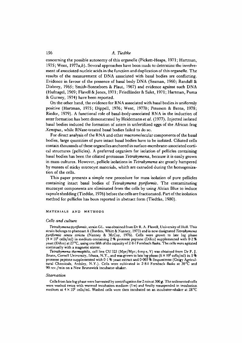
concerning the possible autonomy of this organelle (Pickett-Heaps, 1971; Hartman,1975; Went, 1977a,fe). Several approaches have been made to determine the involve-ment of associated nucleic acids in the function and duplication of this organelle. Theresults of the measurement of DNA associated with basal bodies are conflicting.Evidence in favour of the presence of basal body DNA (Seaman, 1960; Randall &Disbrey, 1965; Smith-Sonneborn & Plaut, 1967) and evidence against such DNA(Hufnagel, 1969; Flavell & Jones, 1971; Friedlander & Salet, 1971; Hartman, Puma& Gurney, 1974) have been reported.
On the other hand, the evidence for RNA associated with basal bodies is uniformlypositive (Hartman, 1975; Dippell, 1976; Went, 19776; Peterson & Berns, 1978;Rieder, 1979). A functional role of basal-body-associated RNA in the induction ofaster formation has been demonstrated by Heidemann et al. (1977). Injected isolatedbasal bodies induced the formation of asters in unfertilized eggs of the African frogXenopus, while RNase-treated basal bodies failed to do so.
For direct analysis of the RNA and other macromolecular components of the basalbodies, large quantities of pure intact basal bodies have to be isolated. Ciliated cellscontain thousands of these organelles anchored in surface-membrane-associated corti-cal structures (pellicles). A preferred organism for isolation of pellicles containingbasal bodies has been the ciliated protozoan Tetrahymena, because it is easily grownin mass cultures. However, pellicle isolations in Tetrahymena are greatly hamperedby masses of sticky mucocyst materials, which are extruded during the homogeniza-tion of the cells.
This paper presents a simple new procedure for mass isolation of pure pelliclescontaining intact basal bodies of Tetrahymena pyriformis. The contaminatingmucocyst components are eliminated from the cells by using Alcian Blue to inducecapsule shedding (Tiedtke, 1976) before the cells are fractionated. Part of the isolationmethod for pellicles has been reported in abstract form (Tiedtke, 1980).
MATERIALS AND METHODS
Cells and cultureTetrahymena pyriformis, strain GL. was obtained from Dr R. A. Flavell, University of Hull. This
strain belongs to phenoset A (Borden, Whitt & Nanney, 1973) and is now designated Tetrahymenapyriformis sensu strictu (Nanney & McCoy, 1976). Cells were grown to late log phase(4 X 10⁵ cells/ml) in medium containing 2 % proteose peptone (Difco) supplemented with 0-2%yeast (Difco) at 27 °C, using one fifth of the capacity of 2-8-1 Fernbach flasks. The cells were agitatedcontinually with a magnetic stirrer.
Tetrahymena thermopm'la, cell line CU325 (Mpr/Mpr; 6mps, V) was obtained from Dr P. J.Bruns, Cornell University, Ithaca, N.Y., and was grown to late log phase (6 X 10s cells/ml) in 1 %proteose peptone supplemented with 0-1 % yeast extract and 0-003 % Sequestrene (Geigy Agricul-tural Chemicals, Ardsley, N.Y.). Cells were cultivated in 2-8-1 Fernbach flasks at 30CC and90 rev./min on a New Brunswick incubator-shaker.
Starvation
Cells from late log phase were harvested by centrifugation for 2 min at 300 g. The sedimented cellswere washed twice with warmed incubation medium (1M) and finally resuspended in incubationmedium at 4 X 10⁵ cells/ml. Washed cells were then incubated on an incubator-shaker at 28 °C
Basal bodies of Tetrahymena 157
(T. pyriformis) and 30°C (T. thermophila), 95 rev./min, using one fifth of the capacity of 2-8-1Fernbach flasks.
The incubation medium (MgPB) used for T. pyriformis was 5 mM-phosphate buffer (pH6-6)containing 1 mM concentrations of CaCk, MgCl2, MgSO4, NaCl and KNO3.
T. thermophila was incubated in a 10-fold dilution of Wagner's solution (pH7-0). The com-position of Wagner's solution is, per litre: NaCl, 2-75g; KC1, l-49mg; MgSCv7H20, 246mg,Na2HPO4, l-37g; KH2PO4, 320mg.
Induction of capsule sheddingCapsule shedding was carried out after appropriate times of incubation as follows: 200-ml samples
of cells were mixed under vigorous shaking with an equal volume of capsule-shedding medium.Capsule-shedding medium was prepared just before use by diluting a 2 % (w/v) stock solution ofAlcian Blue 8GS (Chroma-Gesellschaft, Stuttgart) with distilled water. Immediately after induc-tion of capsule shedding, dissolved egg albumen (Sigma) was added to a final concentration of0-2%.
Separation of capsule-free cellsThe capsule-shedding mixture of T. pyriformis was washed twice (300 g, 2min) in fresh incuba-
tion medium (1M) and finally resuspended in 1 M medium. Cells that had left their capsules within20 min were separated from the empty capsules and from the cells remaining in their capsules bynitration through 10/an mesh polyamide gauze (Vercinigte Seidenwebercien, Krefeld).
Isolation of pelliclesThe homogenization medium (HM) consisted of 10 mM-Tris • HC1 (pH 6-0), supplemented with
lOmM-sodium ethylene diamine tetraacetate (EDTA), and 0-01% Triton X-100. Cells thatescaped from their capsules were homogenized with an Ultra-Turrax rotary homogenizer (Janke &Kunkel, Staufen). Steel sieves with 5 /an sized pores for separation of pellicles were obtained fromVeco-Werke, Solingen.
Electron microscopyPellicles washed with 10 mM-sodium phosphate (pH 7-2) were fixed with glutaraldehyde (1 -25 %)
for 30min at 4°C. The washed sedimented pellicles were postfixed with 1-5 % O8O4 for 30min at20 CC, washed in 70% (v/v) ethanol, dehydrated and embedded in Epon. Thin sections werestained with lead citrate and uranyl acetate, and were examined with a Siemens electron microscopeat 60 or 80 kV.
RESULTS
Elimination of mature mucocysts by capsule shedding
The isolation of pellicles of Tetrahymena is hampered by copious gels, whichenmesh pellicles and other subcellular particles. These voluminous gels originatefrom the contents of secretory vesicles (mucocysts), which are discharged at themoment of homogenization. In order to avoid the annoying difficulties caused bythese gels, capsule shedding induced by Alcian Blue (Tiedtke, 1976) was used toeliminate the contents of mature mucocysts before cell homogenization.
As seen in Fig. 1A, capsule shedding was induced quantitatively by addition ofAlcian Blue. Under the ionic conditions used (see Materials and Methods), AlcianBlue stimulated discharge of mature mucocysts and precipitated their contents in theform of a capsule encaging the cell. The immediate inactivation of unbound Alcian
158 A. Tiedtke
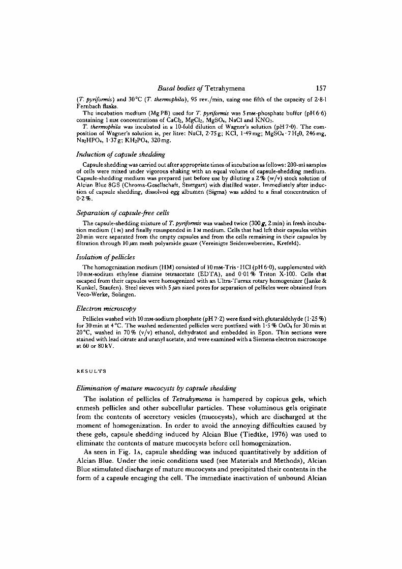
Fig. 1A-B. Capsule shedding induced by treatment with Alcian Blue 8GS. Phase-contrastmicrographs of fixed cells. X 210.A. The fraction of cells capable of mucocyst extrusion and capsule formation approaches100%.B. A sample of the same preparation fixed lOmin later. Cells can leave their capsules(arrows) through holes (arrowheads). About 40 % of the cells leave their capsules withinlOmin.
Blue by ovalbumen guaranteed the survival of these cells. Cells were able to move androtate inside their capsules and to wriggle out of them through small holes (Fig. 1B).The location of the holes suggests that the oral apparatus of the cell might have beenlocated in this position at the moment of capsule shedding.
Basal bodies of Tetrahymena 159
•• :
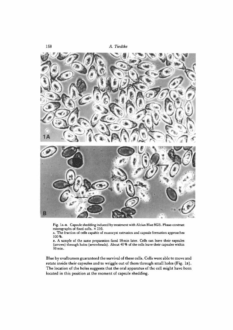
2A , 8Fig. 2A-B . Appearance of cell cortices, loaded with mucocysts (mu) before induction of cap-sule shedding (A), and devoid of mucocysts in slightly compressed living cells (B). X 1350.A. Mucocysts appear as small dark dots and cylinders, mainly in secondary meridians (2°)between the ciliary meridians (1 °).B. Mature mucocysts are not detectable following capsule shedding. Cilia and basal bodiesof ciliary meridians are visible.
1 2 3 4 5 6 7 8 9 10 11 12
Starvation (h)
Fig. 3. Kinetics of capsule shedding. Percentage of cells in capsules is calculated as thequotient of the cells inside capsules plus empty capsules, divided by the sum of all cellscounted. The standard deviation of quadruplicates was less than 5% in all cases.(O—O) T. thermopkila; ( • — • ) T. pyriformis.

160 A. Tiedtke
CULTURE1000 ml Tetrahvmena pvriformis culture.late log phase ~4 x 10* cells/ml. Wash 2xin 1M Resuspend in IM: 4 x 10⁵ cells/ml.Shake for 12 h (Gyrotory shaker 95 rev./min. 28°C)
STARVATION (I)
2x1:::::::::::: SOOmf"
ISOLATION OF PELLICLESCulture: Tetrahvmena pvriformis axenic inproteose peptone medium, late log phase
Incubation medium = IM:5 DIM phosphate bufferpH6-6 +CaCI2, MgCI,. MgSO4. NaCI. K.NO,. all 1 mm
Capsule shedding medium = CM: 0-4 % Alcian Blue8GS (Chroma)
Homogenization medium = HM. 10 mM TrisHCIp H 6 + 10niM EDTA
Centrifuges: Hettich Roto Silentu III. HeraeusChrist UJ II KS, swing-out-rotor
CAPSULESHEDDING (2)
200 mlstarved cells
W/M\ 200 ml CM (0-4% Alcian Blue 8 GS)
sediment (300g. 2rnin). wash 2x in IMresuspend in 200 ml IM ^
SEPARATION OFCAPSULE LEAVING CELLS (3)
HOMOGENIZATION (4)
Buchner funnel, 10/im mesh gazeempty capsules + cells remainingin capsules
capsule leaving cells (devoidof dischargeable mucocysts)
sediment (300g, 2min)resuspend in 4°C HM (107 cells/5 ml)
—M^— Ultra-Turrax 104 rev./minJ 2-5min~0°C
- pellicles + smallcell debris
SEPARATION OF PELLICLES (5)discard
wash 4x withcold HM
steel sieve5 /im mesh
sediment:pure pellicles
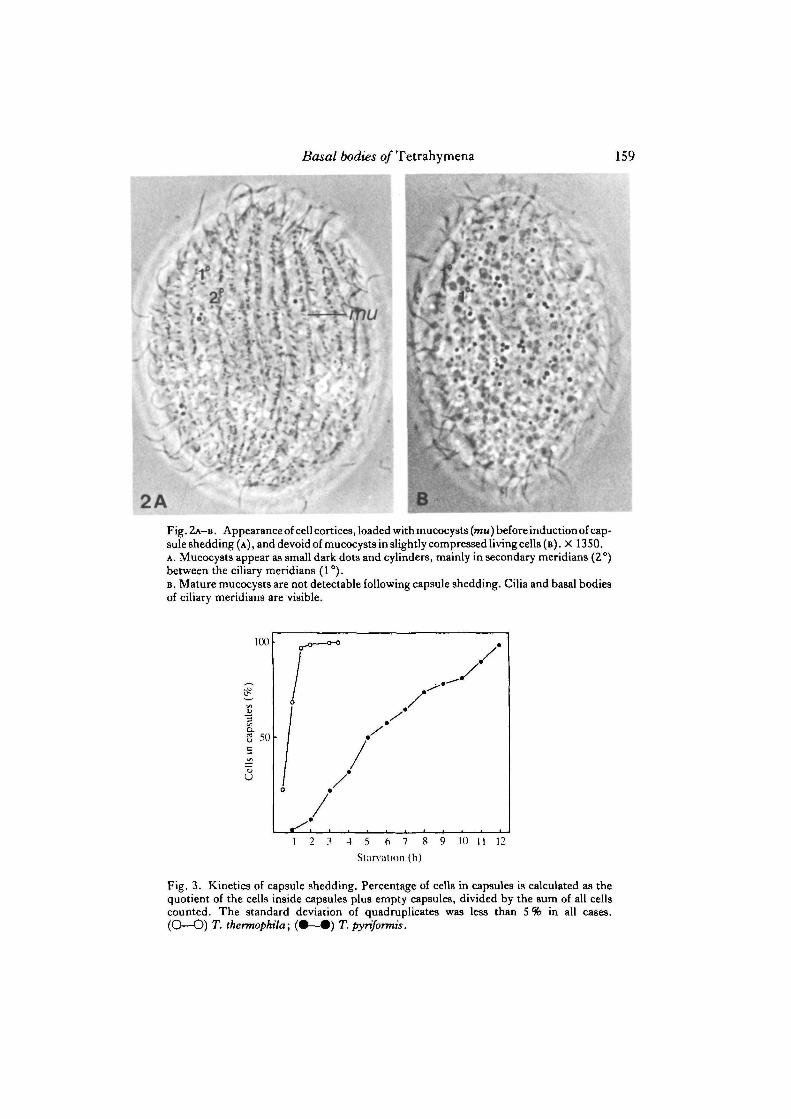
Fig. 4. Illustration of the procedure of pellicle isolation for T. pyriformis.
Basal bodies of Tetrahymena 161
I have shown previously by electron microscopy that the capsules are built up ofextruded mucocysts precipitated by the Alcian Blue. Following capsule shedding, thetreated cells are devoid of mature mucocysts (see pp. 240-241 in the review byHausmann, 1978). For routine controls, the success of capsule shedding was followedby phase-contrast microscopy of living cells. While starved cells were loaded withmucocysts, mainly in rows between the ciliary rows (Fig. 2A), no mature mucocystswere detected in the cortex of cells that escaped their capsules (Fig. 2B). Whenphotographed and counted at different focal planes, untreated cells yield roughestimates of 2000—3000 mucocysts per cell.
In order to determine the minimum time of starvation needed to achieve maximumrates of capsule shedding, both species of Tetrahymena were starved in their respec-tive incubation media (1M) for increasing times. As shown in Fig. 3, the maximumrate of capsule shedding was achieved in T. thermophila after 2h of starvation. T.pyriformis had to be incubated for at least 12 h to stimulate 95 % of the cells to forma capsule.
Isolation of pelliclesCells that escaped from their capsules during the 10—20 min following induction of
capsule shedding (about 40 %) were separated from cells that remained inside theircapsules and from empty capsules. These mucocyst-depleted cells were sedimented(300 g, 2 min) and resuspended in ice-cold homogenization medium (HM) at a con-centration of 107 to 108 cells/5 ml. Cells homogenized at 5000 rev./min and 4°C wereobserved by phase-contrast to be converted quantitatively into whole pure pellicles.Pellicles floated freely in the homogenate; no gels enmeshing the pellicles were detec-ted. The pellicles were separated from cell debris at 4°C by use of steel sieves with5 [im sized pores. Only the pellicles remained on the sieve plate and were washedseveral times with HM without Triton X—100. They were finally flushed off the sieveplates, sedimented and stored at 4°C. The complete procedure for isolation of pell-icles is illustrated in Fig. 4. Although the further experiments were performed onpellicles of T. pyriformis, equal yields of pure pellicles were obtained with T. ther-mophila (Tiedtke, unpublished).
Purified isolated pellicles are shown in Fig. 5. The regularly spaced dark spotsrepresent the basal bodies of the ciliary meridians (see also Fig. 7A). Isolated pellicleswere rigid and, owing to their larger size, easily separated from the cell homogenate,which passed through the pores (5 ̂ m diameter) of the steel sieves.
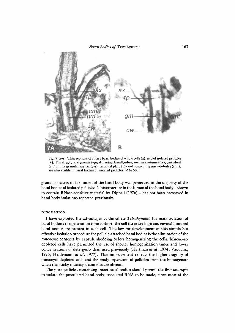
Electron micrographs of thin-sectioned isolated pellicles showed no contaminationwith particulate cell constituents (Fig. 6). Most of the surface membranes wereremoved by the action of 0-01 % Triton X-100, but membrane blebs are occasionallyseen near the basal bodies. The isolated pellicles consisted of typical surface-membrane-associated cytoskeletal elements (Williams, Vaudaux & Skriver, 1979): acontinuous layer of epiplasm, microtubular ribbons above the epiplasm, and basalbodies attached to it. The basal bodies of isolated purified pellicles were well preser-ved (Fig. 7B). Compared with basal bodies of whole cells, those of isolated pelliclescontain essentially all the known structural elements of this organelle. Even the

162 A. Tiedtke
'/,
s.
Fig. 5. Whole pure pellicles. Rows of dark spots represent the ciliary basal bodies. Oralapparatuses (arrows) are visible in some pellicles. Phase-contrast. X 360.
Fig. 6. Electron micrograph of thin-sectioned isolated pellicles. Intact basal bodies(arrows) are anchored in the continuous epiplasmic layer of the pellicle. Cilia are detachedabove the level of the axosome. Microtubular ribbons (arrowheads) are seen above theepiplasmic layer. No contaminations with cell debris is visible. X 7500.
Basal bodies of Tetrahymena
B
Fig. 7. A-B. Thin sections of ciliary basal bodies of whole cells (A) , and of isolated pellicles(B). The structural elements typical of intact basal bodies, such as axosome (ax), cartwheel(cw), inner granular matrix igm), terminal plate (tp) and connecting microtubules (cmt),are also visible in basal bodies of isolated pellicles. X 62500.
granular matrix in the lumen of the basal body was preserved in the majority of thebasal bodies of isolated pellicles. This structure in the lumen of the basal body- shownto contain RNase-sensitive material by Dippell (1976) - has not been preserved inbasal body isolations reported previously.
DISCUSSION
I have exploited the advantages of the ciliate Tetrahymena for mass isolation ofbasal bodies: the generation time is short, the cell titres are high and several hundredbasal bodies are present in each cell. The key for development of this simple buteffective isolation procedure for pellicle-attached basal bodies is the elimination of themucocyst contents by capsule shedding before homogenizing the cells. Mucocyst-depleted cells have permitted the use of shorter homogenization times and lowerconcentrations of detergents than used previously (Hartman et al. 1974; Vaudaux,1976; Heidemann et al. 1977). This improvement reflects the higher fragility ofmucocyst-depleted cells and the ready separation of pellicles from the homogenatewhen the sticky mucocyst contents are absent.
The pure pellicles containing intact basal bodies should permit the first attemptsto isolate the postulated basal-body-associated RNA to be made, since most of the
164 A. Tiedtke
basal bodies of pellicles isolated show preserved central matrices. A rough calculationprovides an estimate of the number of cells needed to obtain microgram quantities ofRNA. On the basis of an estimation of the amount of RNA per basal body(Heidemann et al. 1977) of 5 X 10~16 g, a minimum of 5 X 10¹⁰ intact basal bodies isneeded to obtain 1 /ig of RNA. Assuming that the RNA is extracted quantitativelyfrom the isolated intact basal bodies, then 1 X 10⁸ cells, each containing 5 X 10² basalbodies, is the minimum number needed. Since cell titres of 2 X 10⁶ cells/ml can beobtained, 10 litres of cell culture will provide 1 X 10¹³ basal bodies. Even if a morerealistic yield of only 0-1 % of the input is calculated, microgram quantities of basal-body-associated RNA from pellicle isolates should still be obtained.
For a direct characterization of the other macromolecules of basal bodies these mustbe stripped off the pellicles. Techniques for this purpose, e.g. the grinding of pellicleswith sand (Seaman, 1960), result in basal bodies associated with fragments of thepellicle of various sizes and structures connected to basal bodies. Efforts to separatebasal bodies from pellicles, therefore, were not promising. However, the major pell-icle proteins of Tetrahymena have been identified and their localization has beenshown by using pellicles isolated with high concentrations of detergent (Vaudaux,1976; Williams et al. 1979; Vaudaux & Williams, 1979). These preparations regularlycontained basal bodies without the central matrix material. Comparison of proteinsfrom pellicles isolated by different methods may enable us to identify the basal bodyproteins. Recently, a basal-body-associated protein that is not tubulin has been iden-tified (Conolly & Kalnins, 1978; Turksen, Aubin & Kalnins, 1982). This protein hasbeen immunoprecipitated with antibodies present in some normal rabbit sera, it hasa molecular weight of 50 000 and is a common constituent of basal bodies of speciesas distantly related as Tetrahymena and the chicken.
I thank Dr K. Heckmann for initiating and supporting this study, and for many encouragingdiscussions. I also thank Dr D. L. Nanney for many helpful comments and critically reading themanuscript.
REFERENCESBORDEN, D., WHITT, G. S. & NANNEY, D. L. (1973). Electrophoretic characterization of classical
Tetrahymena pyriformis strains. .7. Pmtozool. 20, 639—700.CONOIXY, J. A. & KALNINS, V. J. (1978). Visualization of centrioles and basal bodies by fluores-
cent staining with nonimmune rabbit sera. J. Cell Biol. 79, 526—532.DIPPELL, R. (1976). Effects of nuclease and protease digestion on the ultrastructure of Paramecium
basal bodies. J . Cell Biol. 69, 622-637.FLAVELL, R. A. & JONES, J. G. (1971). DNA from isolated pellicles of Tetrahymena. J. Cell Sci.
9, 719-726.FRIEDLANDER, M. & SALET, C. (1971). Absence of DNA in giant centrioles of the neuropteran
insect Chrysopa cornea. A laser microbeam irradiation study. Cytobios 4, 171-176.FULTON, C. (1971). Centrioles. In Origin and Continuity of Cell Organelles (ed. J. Reinert & H.
Ursprung), pp. 170-221, Heidelberg: Springer-Verlag.HARTMAN, H., PUMA, J. P. & GURNEY, T. JR (1974). Evidence for the association of RNA with
the ciliary basal bodies of Tetrahymena. J. Cell Sci. 16, 241-259.HARTMAN, H. (1975). The centriole and the cell. .7. theor. Biol. 51, 501-509.HAUSMANN, K. (1978). Extrusive organelles in protists. Int. Rev. Cytol. 52, 197-276.
Basal bodies of Tetrahymena 165
HEIDEMANN, S. R., SANDER, G. & KIRSCHNER, M. W. (1977). Evidence for a functional role ofRNA in centrioles. Cell 10, 337-350.
HUFNAGEL, L. A. (1969). Properties of DNA associated with raffinose-isolated pellicles ofParameciwn aurelia.J. Cell Set. 5, 561-573.
NANNEY, D. L. & MCCOY, J. W. (1976). Characterization of the species of the Tetrahymenapyriformis complex. Trans. Am. Microsc. Soc. 95, 664-682.
PETERSON, S. P. & BERNS, M. W. (1978). Evidence for centriolar region RNA functioning inspindle formation in dividing PTK2 cells. J . Cell Sci. 34, 289-301.
PETERSON, S. P. & BERNS, M. W. (1980). The centriolar complex. Int. Rev. Cytol. 64, 81-106.PICKETT-HEAPS, J. D. (1971). The autonomy of the centriole: fact or fallacy. Cytobios 3, 205-214.RANDALL, J. & DISBREY, C. (1965). Evidence for the presence of DNA at basal body sites in
Tetrahymena pyriformis. Proc. Ry. Soc.. B, 162, 473-491.RIEDER, C. L. (1979). Ribonucleoprotein staining of centrioles and kinetochores in newt lung cell
spindles. J. CellBiol. 80, 1-9.SEAMAN, G. R. (1960). Large-scale isolation of kinetosomes from the ciliated protozoan
Tetrahymena pyriformis. Expl Cell Res. 21, 292-302.SMITH-SONNEBORN, J. & PLAUT, W. (1967). Evidence for the presence of DNA in the pellicle of
Paramecium.J. Cell Sci. 2, 225-234.SOROKIN, S. P. (1968). Reconstructions of centriole formation and ciliogenesis in mammalian
lungs. J . Cell Sci. 3, 207-230.TIEDTKE, A. (1976). Capsule shedding in Tetrahymena. Naturwissenschaften 63, 93.TIEDTKE, A. (1980). A simple method for isolation of pure pellicles in Tetrahymena. Eur.J. Cell
Biol. 22, 598.TURKSEN, K., AUBIN, J. E. & KALNINS, V. J. (1982). Identification of a centriole-associated
protein by antibodies present in normal rabbit sera. Nature, Land. 298, 763-765.VAUDAUX, P. (1976). Isolation and identification of specific cortical proteins in Tetrahymena
pyriformis GL.J. Protozool. 23, 458-464.VAUDAUX, P. E. & WILLIAMS, N. E. (1979). Cytoskeletal proteins of the cell surface in
Tetrahymena. II. Turnover of major proteins. Expl Cell Res. 123, 321-331.WENT, H. A. (1977a). Can a reverse transcriptase be involved in centriole duplication?^, theor.
Biol. 68, 95-100.WENT, H. A. (19776). Centriole duplication in sand dollar eggs. I. The effects of selected reagents.
Expl Cell Res. 108,63-73.WILLIAMS, N. E., VAUDAUX, P. E. & SKJUVER, L. (1979). Cytoskeletal proteins of the cell surface
in Tetrahymena. I. Identification and localization of major proteins. ExplCellRes. 123, 311-320.WOLFE, J. (1972). Basal body fine structure and chemistry. In Advances in Cell and Molecular
Biology (ed. E. J. Du Praw), vol. 2, pp. 151-192. New York: Academic Press.
(Received 14 February 1983 -Accepted, in revised form, 12 March 1985)


![hypnouniversity.com · entered !at stationers' hall.] the bodie book. hypnotism. electricity. mental suggestion. magnetic touch. clairvoyance. telepathy. by walford bodie,](https://img.pdfslide.us/doc/110x75/5f51056c7a48362a307331b7/entered-at-stationers-hall-the-bodie-book-hypnotism-electricity-mental-suggestion.jpg)