Embed Size (px)
Citation preview
International Journal of Agricultural Technology 2018 Vol. 14(7): 1787-1800
Available online http://www.ijat-aatsea.com
ISSN: 2630-0613 (Print) 2630-0192 (Online)
Isolation and characterization of keratinolytic bacteria from soil
samples of poultry waste dumping sites
Reyes, A.1,2 *
, Ambita, I. D.1, Batalon, J. L.
1, Aba, B. L.
1, Cortes, A.
1,
Macabecha, C. G.1 and Montecillo, A.
1
1University of the Philippine Los Baños, College Laguna Philippines;
2College of Fisheries-
Freshwater Aquaculture Center, Central Luzon State University, Science City of Muñoz, Nueva
Ecija, Philippines.
Reyes, A., Ambita, I. D., Batalon, J. L., Aba, B. L., Cortes, A., Macabecha, C. G. and
Montecillo, A. (2018). Isolation and characterization of keratinolytic bacteria from soil samples
of poultry waste dumping sites. International Journal of Agricultural Technology 14(7): 1787-
1800.
Abstract The keratinolytic bacteria isolated from soil samples which containing the degrading
feathers was investigated. Thirteen bacterial isolates were selected and were subjected to
preliminary screening through protease assay using Milk Agar Medium. Six isolates were
detected positive for protease activity and were further characterized via biochemical and
microscopic assays. Isolates were grown in basal medium containing feathers as sole nutrient
source and the degree of feather degradation was monitored. Two isolates exhibited
conspicuous keratinase activity. DNA from these two candidate organisms were isolated and
subjected to PCR using 16S rRNA specific primers. PCR products were sequenced and analysis
revealed that both of them belong to Bacillus cereus species. Isolation of potential keratinolytic
microorganisms could have potential biotechnological used especially in processes which
involved keratin hyrdolysis.
Keywords: Keratinolytic bacteria, soil, biochemical assays, Bacillus cereaus, 16S rRNA
Introduction
Feathers are produced in large amounts as waste by-products of poultry
farms from small-scale to large-scale processing plants. This is tantamount to
the numerous chickens killed for consumption worldwide. Recently, a value-
added use for feathers is its conversion to feather meal, a digestible dietary
protein for animal feed, using physical and chemical treatments (Riffel et al.,
2006). However, in the Philippines, most of these feather wastes are burned,
dumped or stacked in waste areas which cause problems in storage and
emissions control. This is because the bulk of feather produced by farms is
poorly recycled (Jeevana Lakshmi et al., 2013) when left alone in dumping
areas unlike other animal wastes. Also, it has limited use due to the chemically
unreactive nature of keratin. Consequently, with improper handling of
* Coressponding Author: Reyes, A.T.; Email: [email protected]

1788
discarded feathers, it could become a pool for other pathogenic microorganisms
causing mycoplasmosis, chlorosis and fowl cholera (Williams et al., 1991).
Feathers are slowly degraded and are resistant to other soil
microorganism's proteolytic enzymes, due to the complex structure of its β-
keratin filaments. In addition, it has disulfide cross-links which produce a
compact three dimensional network (Bradbury et al., 1973), resulting to
intermolecular disulfide bonds between rod domains and terminal domains of
the constituent molecules (Parry et al., 1998). These molecular configurations
of its amino acids ensure the structural rigidity posing a challenge for its
disposal or utilization as feather feed. Physical and chemical processes used to
increase the digestibility of feather keratin require consumption of large
amounts of energy and destruction of certain amino acids, thus yielding
products of poor digestibility and variable nutrient quality. Dymatic hydrolysis
by microorganisms that possess keratinolytic activity represents an attractive
alternative to improve the nutritional value of feather wastes (Xu et al., 2009).
A number of studies that describe nutritional upgrading of feather meal
through microbial or enzymatic treatments have been published over the years.
Some of these are feather meal fermentation with Streptomyces fradiae
supplemented with methionine (Elmayergi and Smith, 1971) and feather-lysate
from Bacillus licheniformis with amino acid supplementation resulting to a
similar growth rate in chickens fed with soybean meal (Williams et al., 1991).
Also, the crude keratinase enzyme produced by B. licheniformis was found to
significantly increase total amino acid digestibility of raw feathers and
commercial feather meal (Lee et al., 1991).
Moreover, keratinolytic microorganisms and their enzymes could be used
to enhance the digestibility of feathers (Odetallah et al., 2003) which is
primarily a garbage problem for a number of poultry farms and other feathered
livestock raisers in the Philippines. Furthermore, although a number of
keratinolytic microorganisms have already been reported, the full commercial
potential of keratinases is yet to be realized. Molecular approaches targeting the
keratin gene from reported isolates yet needs to be established. The objectives
were to isolate, characterize and identify keratinolytic bacteria from soil
samples containing degrading feathers in the Philippines.
Materials and Methods
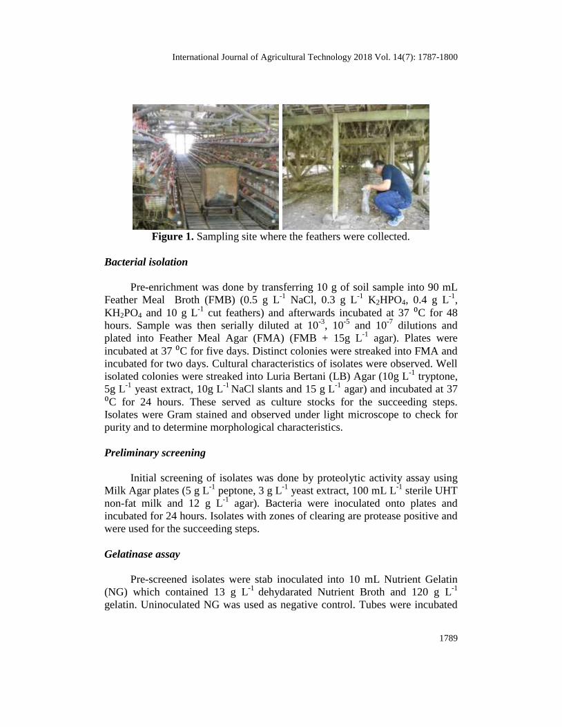
Sample collection
Soil samples were collected from poultry waste dumping sites at ERI
Poultry Farm in Victoria, Laguna, Philippines. Composite sampling was done
using sterile falcon tubes and autoclavable plastic bags (Figure 1).
International Journal of Agricultural Technology 2018 Vol. 14(7): 1787-1800
1789
Figure 1. Sampling site where the feathers were collected.
Bacterial isolation
Pre-enrichment was done by transferring 10 g of soil sample into 90 mL
Feather Meal Broth (FMB) (0.5 g L-1
NaCl, 0.3 g L-1
K2HPO4, 0.4 g L-1
,
KH2PO4 and 10 g L-1
cut feathers) and afterwards incubated at 37 ⁰C for 48
hours. Sample was then serially diluted at 10-3
, 10-5
and 10-7
dilutions and
plated into Feather Meal Agar (FMA) (FMB + 15g L-1
agar). Plates were
incubated at 37 ⁰C for five days. Distinct colonies were streaked into FMA and
incubated for two days. Cultural characteristics of isolates were observed. Well
isolated colonies were streaked into Luria Bertani (LB) Agar (10g L-1
tryptone,
5g L-1
yeast extract, 10g L-1
NaCl slants and 15 g L-1
agar) and incubated at 37
⁰C for 24 hours. These served as culture stocks for the succeeding steps.
Isolates were Gram stained and observed under light microscope to check for
purity and to determine morphological characteristics.
Preliminary screening
Initial screening of isolates was done by proteolytic activity assay using
Milk Agar plates (5 g L-1
peptone, 3 g L-1
yeast extract, 100 mL L-1
sterile UHT
non-fat milk and 12 g L-1
agar). Bacteria were inoculated onto plates and
incubated for 24 hours. Isolates with zones of clearing are protease positive and
were used for the succeeding steps.
Gelatinase assay
Pre-screened isolates were stab inoculated into 10 mL Nutrient Gelatin
(NG) which contained 13 g L-1
dehydarated Nutrient Broth and 120 g L-1
gelatin. Uninoculated NG was used as negative control. Tubes were incubated
1790
at room temperature for 48 hours. Upon incubation, tubes were placed in
freezer for 15 minutes and allowed to solidify. Liquefaction of gelatin indicated
positive for gelatinase enzyme.
Catalase test
Presence of catalase enzyme was tested on pre-screened isolates by
teasing loopfuls of colonies into separate glass slides. Hydrogen peroxide was
then poured unto the cells using Pasteur pipet. Presence of bubbles would be
indicative of catalase enzyme’s presence in the cells.
Spore staining
Isolates were tested for presence of spores. Schaefer-Fulton method of
spore staining was utilized as staining procedure (Schaeffer and Fulton, 1933).
Smears of each isolate were first prepared, air dried and heat fixed. The smears
were then flooded with malachite green. Slides were steamed for 8-10 minutes
and in the process, smears were kept saturated with malachite green. Smears
were then washed thoroughly with water and counterstained with safranin for
one minute. After which, smears were washed and dried. Microcopic
observation was done under oil immersion. Spores were stained blue while
vegetative cells were red.
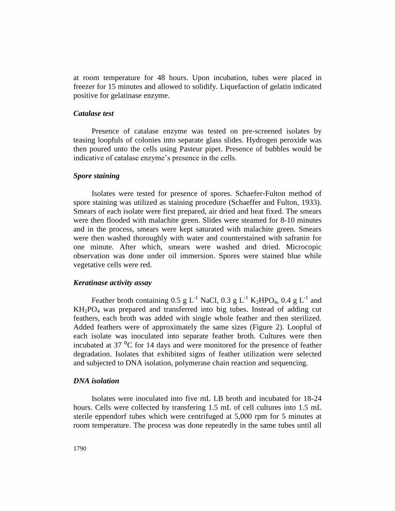
Keratinase activity assay
Feather broth containing 0.5 g L-1
NaCl, 0.3 g L-1
K2HPO4, 0.4 g L-1
and
KH2PO4 was prepared and transferred into big tubes. Instead of adding cut
feathers, each broth was added with single whole feather and then sterilized.
Added feathers were of approximately the same sizes (Figure 2). Loopful of
each isolate was inoculated into separate feather broth. Cultures were then
incubated at 37 ⁰C for 14 days and were monitored for the presence of feather
degradation. Isolates that exhibited signs of feather utilization were selected
and subjected to DNA isolation, polymerase chain reaction and sequencing.
DNA isolation
Isolates were inoculated into five mL LB broth and incubated for 18-24
hours. Cells were collected by transfering 1.5 mL of cell cultures into 1.5 mL
sterile eppendorf tubes which were centrifuged at 5,000 rpm for 5 minutes at
room temperature. The process was done repeatedly in the same tubes until all
International Journal of Agricultural Technology 2018 Vol. 14(7): 1787-1800
1791
cells in the culture broths were collected. Cells were washed twice with sterile
distilled water and recentrifuged at the same conditions. Cells were
resuspended with 200 µL Tris-EDTA (TE) buffer and mixed briefly. Tubes
were first placed into boiling water bath for five minutes and then placed in
freezer for 15 minutes. This freeze-thaw cycle was repeated three times.
Resulting suspension was centrifuged at 5,000 rpm for five minutes.
Supernatant was transferred to new sterile eppendorf tubes and labeled
accordingly.
Figure 2. Feathers used as carbon source for keratinolytic bacteria
Polymerase Chain Reaction (PCR) using 16S rRNA primers
Extracted DNA samples were subjected to PCR using 16S ribosomal
RNA specific primers. Reactions of 20 µL total volume contained 1X PCR
Buffer (10 mM Tris-HCL, 50 mM KCl, 1.5 mM MgCl2, pH 8.3 at 25⁰C), 0.2
mM dNTPs (Invitrogen, USA), 1 µL of each primer, 0.5 unit of Taq
polymerase (New England Biolabs, USA) and 1µL of DNA template. PCR
cylcing profile included an initial denaturation at 95 ⁰C for five minutes
followed by a 25x cycling profile constituted of a denaturation step at 95 ⁰C for
one minute, an annealing temperature of 55 ⁰C for one minute and an extension
step at 72 ⁰C for one minute. A final extension step at 72 ⁰C for 10 minutes was
included after the cycling step. All PCR products were visualized through
agarose gel electrophoresis with 1.0 % agarose gel. PCR products with correct
bands were sent for sequencing.
1792
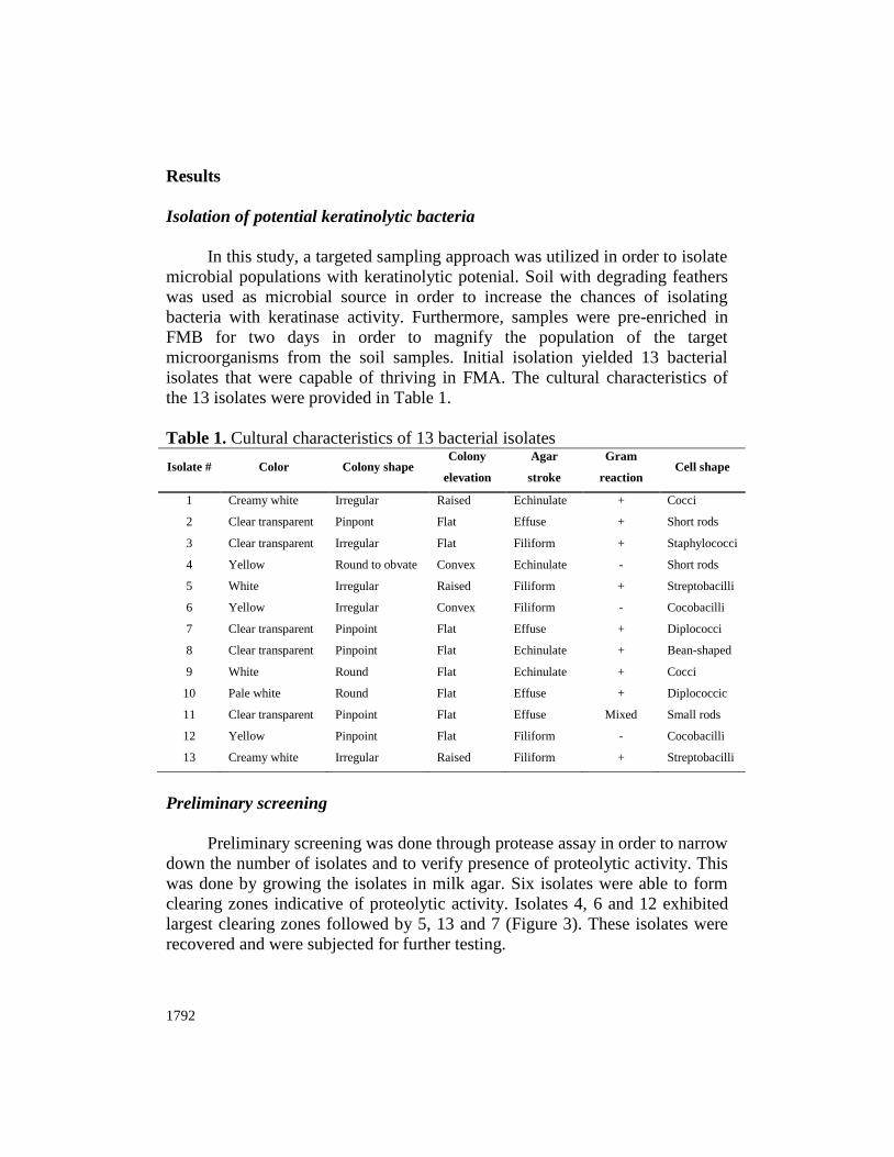
Results
Isolation of potential keratinolytic bacteria
In this study, a targeted sampling approach was utilized in order to isolate
microbial populations with keratinolytic potenial. Soil with degrading feathers
was used as microbial source in order to increase the chances of isolating
bacteria with keratinase activity. Furthermore, samples were pre-enriched in
FMB for two days in order to magnify the population of the target
microorganisms from the soil samples. Initial isolation yielded 13 bacterial
isolates that were capable of thriving in FMA. The cultural characteristics of
the 13 isolates were provided in Table 1.
Table 1. Cultural characteristics of 13 bacterial isolates
Isolate # Color Colony shape Colony
elevation
Agar
stroke
Gram
reaction Cell shape
1 Creamy white Irregular Raised Echinulate + Cocci
2
3
Clear transparent
Clear transparent
Pinpont
Irregular
Flat
Flat
Effuse
Filiform
+
+
Short rods
Staphylococci
4 Yellow Round to obvate Convex Echinulate - Short rods
5
6
White
Yellow
Irregular
Irregular
Raised
Convex
Filiform
Filiform
+
-
Streptobacilli
Cocobacilli
7
8
9
10
11
12
13
Clear transparent
Clear transparent
White
Pale white
Clear transparent
Yellow
Creamy white
Pinpoint
Pinpoint
Round
Round
Pinpoint
Pinpoint
Irregular
Flat
Flat
Flat
Flat
Flat
Flat
Raised
Effuse
Echinulate
Echinulate
Effuse
Effuse
Filiform
Filiform
+
+
+
+
Mixed
-
+
Diplococci
Bean-shaped
Cocci
Diplococcic
Small rods
Cocobacilli
Streptobacilli
Preliminary screening
Preliminary screening was done through protease assay in order to narrow
down the number of isolates and to verify presence of proteolytic activity. This
was done by growing the isolates in milk agar. Six isolates were able to form
clearing zones indicative of proteolytic activity. Isolates 4, 6 and 12 exhibited
largest clearing zones followed by 5, 13 and 7 (Figure 3). These isolates were
recovered and were subjected for further testing.
International Journal of Agricultural Technology 2018 Vol. 14(7): 1787-1800
1793
Figure 3. Protease assay of the 13 isolates using milk agar medium
Biochemical and microscopic assays
Pre-screened isolates were subjected to spore staining and some
biochemical assays such as gelatinase (Figure 4) and catalase (Figure 5). These
tests were done in order to elucidate some characteristics of the isolates which
could be useful in their identification. Results of the mentioned tests are
summarized in Table 2.
Figure 4. Representative for gelatinase activity test. All isolates are positive for
gelatinase activity; left (negative control) and right (unknown isolate)
Figure 5. Catalase test of several isolates positive for keratinolytic activity

1794
Table 2. Biochemical and microscopic assay of the pre-screened isolates Isolate # Gelatinase Catalase Spore stain
4 + - +
5 + + +
6 + - -
7 + + +
12 + + -
13 + + -
Keratinase assay
Isolates with highest keratinase activity were identified by further
interrogating their capacity to degrade feathers. This was done by using feather
broth, a minimal medium with feathers as sole carbon and nitrogen source.
Whole feathers were supplemented in the media in order to visually inspect
feather degradation. Results revealed that isolates 5 and 13 exhibited the most
visible feather degradation activity (Figures 6 to 8). This was further supported
by the observed highest turbidity in these isolates indicative of feather
utilization for growth. These two isolates were recovered and identified.
Figure 6. Keratinolytic activity assay using feather as carbon source at 0 h
incubation
Figure 7. Keratinolytic degradation of several isolates after 7 days incubation
using feathers as source of carbon
International Journal of Agricultural Technology 2018 Vol. 14(7): 1787-1800
1795
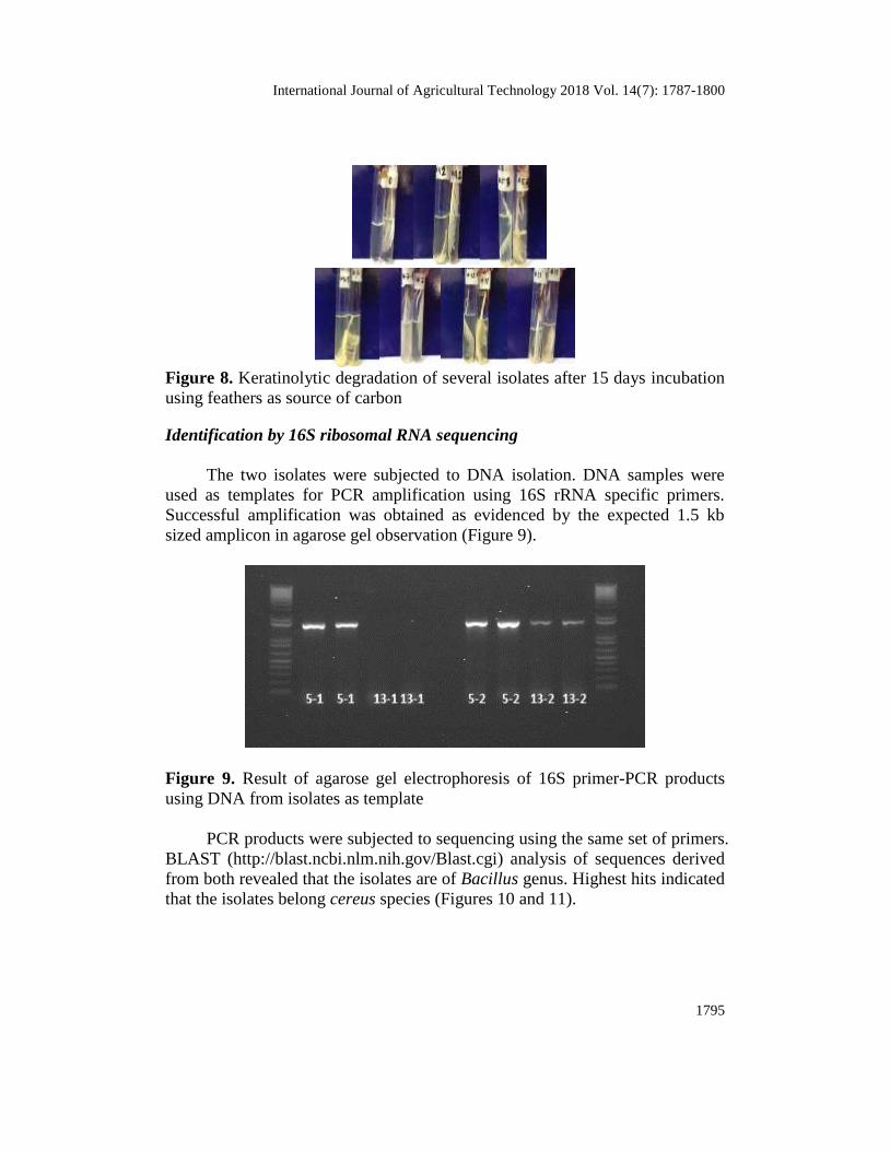
Figure 8. Keratinolytic degradation of several isolates after 15 days incubation
using feathers as source of carbon
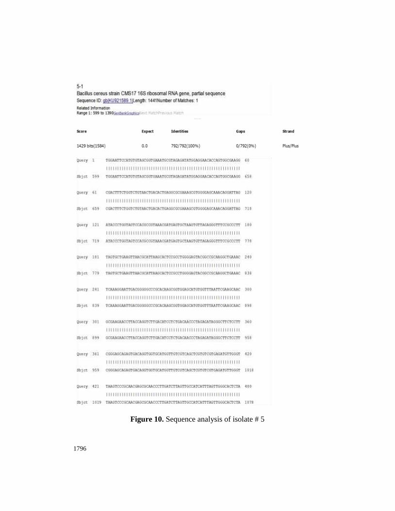
Identification by 16S ribosomal RNA sequencing
The two isolates were subjected to DNA isolation. DNA samples were
used as templates for PCR amplification using 16S rRNA specific primers.
Successful amplification was obtained as evidenced by the expected 1.5 kb
sized amplicon in agarose gel observation (Figure 9).
Figure 9. Result of agarose gel electrophoresis of 16S primer-PCR products
using DNA from isolates as template
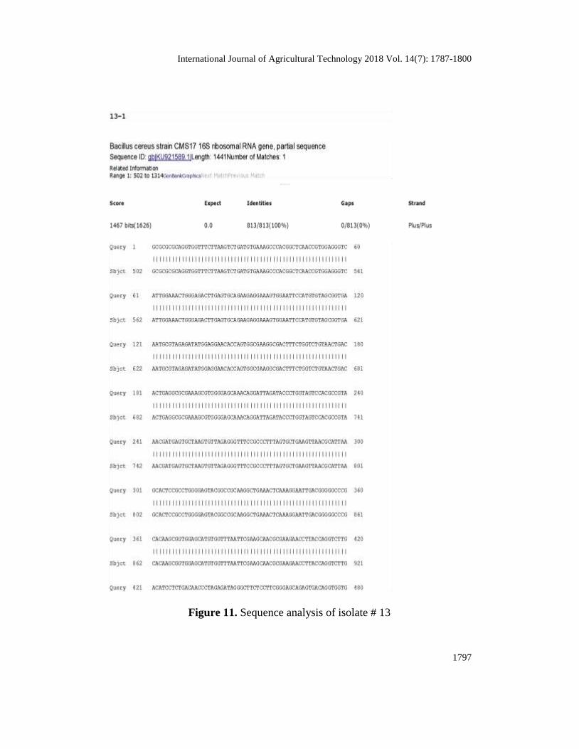
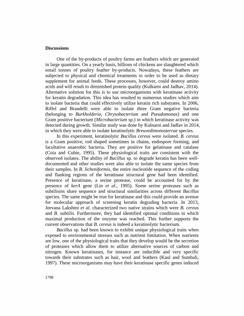
PCR products were subjected to sequencing using the same set of primers.
BLAST (http://blast.ncbi.nlm.nih.gov/Blast.cgi) analysis of sequences derived
from both revealed that the isolates are of Bacillus genus. Highest hits indicated
that the isolates belong cereus species (Figures 10 and 11).
1796
Figure 10. Sequence analysis of isolate # 5
International Journal of Agricultural Technology 2018 Vol. 14(7): 1787-1800
1797
Figure 11. Sequence analysis of isolate # 13
1798
Discussions
One of the by-products of poultry farms are feathers which are generated
in large quantities. On a yearly basis, billions of chickens are slaughtered which
entail tonnes of poultry feather by-products. Nowadays, these feathers are
subjected to physical and chemical treatments in order to be used as dietary
supplement for animal feeds. These processes, however, could destroy amino
acids and will result to diminished protein quality (Kulkarni and Jadhav, 2014).
Alternative solution for this is to use microorganisms with keratinase activity
for keratin degradation. This idea has resulted to numerous studies which aim
to isolate bacteria that could effectively utilize keratin rich substrates. In 2006,
Riffel and Brandelli were able to isolate three Gram negative bacteria
(belonging to Burkholderia, Chrysobacterium and Pseudomonas) and one
Gram positive bacterium (Microbacterium sp.) in which keratinase activity was
detected during growth. Similar study was done by Kulnarni and Jadlav in 2014,
in which they were able to isolate keratinolytic Breuvedimonosterrae species.
In this experiment, keratinolytic Bacillus cereus were isolated. B. cereus
is a Gram positive, rod shaped sometimes in chains, endospore forming, and
facultative anaerobic bacteria. They are positive for gelatinase and catalase
(Coia and Cubie, 1995). These physiological traits are consistent with the
observed isolates. The ability of Bacillus sp. to degrade keratin has been well-
documented and other studies were also able to isolate the same species from
their samples. In B. licheniformis, the entire nucleotide sequence of the coding
and flanking regions of the keratinase structural gene had been identified.
Presence of keratinase, a serine protease, could be accounted for by the
presence of kerA gene (Lin et al., 1995). Some serine proteases such as
subtilisins share sequence and structural similarities across different Bacillus
species. The same might be true for keratinase and this could provide an avenue
for molecular approach of screening keratin degrading bacteria. In 2013,
Jeevana Lakshmi et al. characterized two native strains which were B. cereus
and B. subtilis. Furthermore, they had identified optimal conditions in which
maximal production of the enzyme was reached. This further supports the
current observations that B. cereus is indeed a keratinolytic bacterium.
Bacillus sp. had been known to exhibit unique physiological traits when
exposed to environmental stresses such as nutrient limitation. When nutrients
are low, one of the physiological traits that they develop would be the secretion
of proteases which allow them to utilize alternative sources of carbon and
nitrogen. Known keratinases, for instance are inducible and very specific
towards their substrates such as hair, wool and feathers (Kaul and Sumbali,
1997). These microorganisms may have their keratinase specific genes induced
International Journal of Agricultural Technology 2018 Vol. 14(7): 1787-1800
1799
which made them adapted to utilize substrates such as feathers. Isolation of
such microorganisms could be of biotechnological importance for improving
nutritional qualities of feeds containing treated poultry feather wastes.
Furthermore, this would provide avenues for alternative means of reducing
feather wastes and at the same time preventing generation of pools of
pathogenic microorganisms.
Acknowledgement
The authors would like to exend their appreciation to Prof. Andrew Montecillo for his
guidance in pursuing this study.
References
Bradbury, J. H. (1973). The structure and chemistry of keratin fibers. Advances in Protein
Chemistry. 27:111-211.
Coia, J. and Cubie, H. (1995). Bacillus cereus the immunoassay kit directory. Springer
Netherlands. 1:648-649.
Elmayergi, H. H. and Smith, R. E. (1971). Influence of growth of Streptomyces fradiae on
pepsin-HCl digestibility and methionine content of feather meal. Canadian Journal of
Microbiology. 17:1067-1072.
Jeevana Lakshmi, P., Kumari Chitturi, Ch. M. and Lakshmi, V. V. (2013). Efficient degradation
of feather by keratinase producing Bacillus sp. International Journal of
Microbiology.Article ID 608321. doi:10.1155/2013/608321.
Kaul, S. and Sumbali, G. (1997). Keratinolysis by poultry farm soil fungi. Mycopathologia.
139:137-40.
Kulkarni, S. A. and Jadhav, A. R. (2014). Isolation and characterization of keratinolytic bacteria
from poultry farm soils. International Research Journal of Biological Sciences. 3:29-33.
Lee, C. G., Ferket, P. R. and Shih, J. C. H. (1991). Improvement of feather digestibility by
bacterial keratinase as a feed additive. FASEB Journal. 59:1312.
Lin, X., Kelemen, D. W., Miller, E. S. and Shih, J. C. H. (1995). Nucleotide sequence and
expression of kerA, the gene encoding akeratinolytic protease of Bacillus
licheniformisi. Applied and Environmental Microbiology. 61:1469-1474.
Odetallah, N. H., Wang, J. J., Garlich, J. D. and Shih, J. C. H. (2003). Keratinase in starter diets
improve growth of broiler chicks. Poultry Science. 82:664-670.
Parry, D. A. T. and North, A. C. T. (1998). Hard a-keratin intermediate filament chains: substructure
of the N- and C-terminal domains and the predicted structure and function of the C-terminal
domains of type i and type II chains. Journal of Structural Biology. 122:67-75.
Riffel, A. and Brandelli, A. (2006). Keratinolytic bacteria isolated from feather waste. Brazilian
Journal of Microbiology. 37:395-399.
Schaeffer, A. B. and Fulton, M. D. (1933). A simplified method of staining endospores. Science.
17:194.
1800
Williams, C. M., Lee, C. G., Garlich, J. D. and Shih, J. C. H. (1991). Evaluation of a bacterial
feather fermentation product, feather-lysate, as a feed protein. Poultry Science. 70:85-94.
Xu, B., Zhong, Q., Tang, X., Yang, Y. and Huang, Z. (2009). Isolation and characterization of a
new keratinolytic baterium that exhibits significant feather-degrading capability.
African Journal of Biotechnology. 8:4590-4596.
(Received: 9 September 2018, accepted: 31 October 2018)