Embed Size (px)
Citation preview
DOI:10.21276/sjpm
153
Original Research Article
Saudi Journal of Pathology and Microbiology ISSN 2518-3362 (Print) Scholars Middle East Publishers ISSN 2518-3370 (Online)
Dubai, United Arab Emirates
Website: http://scholarsmepub.com/
Isolation and Identification of Gram-Negative Bacteria Responsible for
Bacteremia in Leukemia Patient and Detection of Procalcitonin Levels in Serum
of Leukemic Patients Naba'a A. Muhammed
1, Muthana B. Farhan
2, Zeyad A. Shabeeb
3
1B.Sc., Biology Division, Collage of Education for Women, University of Anbar, Iraq
2Ph.D., Microbiology, Biology Division, Collage of Education for Women, University of Anbar, Iraq
3Ph.D., Clinical Immunology, Collage of medicine, University of Al-Mustansiriyah, Iraq
*Corresponding Author: Naba'a A. Muhammed
Email: [email protected]
Abstract: Gram negative bacteria (GNB) is the most common causative agent for morbidities and mortalities in
leukemic patient, because of their receiving immunosuprresive chemotherapeutic agents, aggressive devices like
catheters used for giving them those treatments and Hospitalization, in addition to GNB increasing resistance to many of
antibiotics. The study include isolation of GNB from blood samples of leukemia patients and identification of GNB
species isolated from those samples then testing its susceptibility to 15 antibiotics, moreover, calculating Procalcitonin
(PCT) concentration in the samples as immunological marker to detect bacteremia in those patients. The results show
presence of 9 GNB species in blood samples of leukemia patients including: Enterobacter cloacae, E. sakazakii, Serratia
marcescens, S. ficaria, S. liquifaciens, S. rubideae, S. odorefera, Klebsiella oxytoca, and Pseudomonas fluorescens. The
most common species was E. cloacae followed by E. sakazakii. The higher effective antibiotics were Ciprofloxacin,
Gentamicin and Amikacin; also PCT concentrations were ranged between (0.1 and 8.23) ng.ml-1
. The study concluded
that Gram negative bacteremia was common causative agent for infections in Leukemia Patients, Enterobacteriacae was
the the most common GNB causing infection, and the Most effective antibiotic for it were Ciprofloxacin, Gentamicin,
and Amikacin which can be used as prophylactic therapies for those infections.
Keywords: Gram Negative Bacteria, Bacteremia, Leukemia, Procalcitonin, Immunological marker, Antibiotic Resistant.
INTRODUCTION
Leukemia is a worldwide disease, it happened
in all ages and both genders from Females and males
[1]. It's one of the basic reasons for Pathogenicity and
mortality in Iraq according to Cancer registration center
in Iraq [2] since The world health organization report
demonstrate that Leukemia is the second cause for
mortalities in Females and the third in males in Iraq in
2014 [3].
Bacterial infections is one of the most
important reasons for morbidity and mortality in
Leukemia, [4] Including Blood stream infections
particularly bacteremia which is the basic factor for
life-threating infections in leukemic patients receiving
chemotherapy, because of complications resulted from
these infection; in spite of development in Antibiotics
therapy and health care especially in Malignant diseases
including leukemia which still one of the therapeutics'
challenges as a result to immune system defect causing
deficiency in immunity, also because of little using of
prophylactics therapy [5-10]. In addition chemotherapy
related neutropenia is a main cause of immune defects,
because they receive either myelosuppressive or
immunosupperessive drugs. Moreover, the early treated
patients develop infections greater then untreated early.
so that early diagnosis of those infections will be
helpful largely to take decisions related to
Antimicrobial drugs and hospitalization [11,12,5]. So
that the expert physicians consider infections to be the
possible causative factor of fever in any patient
especially leukemic patients [13]. Because of the weak
immune response in those patients, the traditional signs
and symptoms being unclear and the high temperature
considered to be the single diagnostic sign, so that the
blood culture results take the axial diagnostic role for
bacteremia [7,14,11]. So that it's considered to be the
most relevant bacteria in blood particularly in adults in
spite of Gram positive bacteremia is more predominant
in children with association with leukemia [15]. and it
responsible for most frequently infections with high
mortality [16] although developments in antibiotics'
therapy, it stay the basic reason for mortalities in
hospitalized patients especially critically ill patients like
leukemic patients [17].
Mahenaz khan et al.; Saudi J. Pathol. Microbiol.; Vol-2, Iss-5 (May, 2017):153-166
Available Online: http://scholarsmepub.com/sjpm/ 154
Bacteremia is the presence of live bacteria in
bloodstream which can translocate from commensally
bacterial flora or from injection of some bacterial
contaminated materials directly into circulation, which
can be filtrated often from blood in minutes; so that the
bacteremia considered to be silent and transient, but if
the immune system destroyed or in weak performance;
this bacteria will stay in the blood causing pathogenic
symptoms, and the basic symptom is fever [6]. Most of
gram negative bacteria causing these infections arise
from intestinal flora and reach to blood stream by
process called bacterial translocation which induce the
intestine colonization and the increased growth of
transferred bacteria because immunosuppression or by
changes in intestinal mucosa , which all happened in
leukemic patients [16].
The potential inducer for PCT production in
the body were bacterial endotoxin and PCT
concentrations stay high in immunocompromised
patients in infections case also PCT in the serum stay
very stable and its doesn’t analyzed to hormonally
active calcitonin. The pathophysiology rule of PCT was
still under investigation and supposed to be one of acute
phase proteins. Also according to the study, PCT good
maker for infections incidence, moreover; its detect the
intensity of infections also [18]. So many studies
focused on the importance of Procalcitonon (PCT) in
diagnosis of bacteremia, as Giani study show that PCT
concentrations were high in bacteremic patients so that
it was considered to be immunological marker for
detecting the intensity of bacteremia [19,11,20]. AS
PCT concentrations changes during infection were
rapid; and its melcules were constant and easy
detectable, its concentrations can raise to high values in
the blood because of its releasing in the blood during
bacterial infection [21,20,11]. The study aim to detect
the incidence of Gram Negative Bacteremia in leukemia
patients, its susceptibility to Antibiotics, the relation
between Those infections and leukemia types, and
detect PCT values and its relation to Bacteremia
incidence.
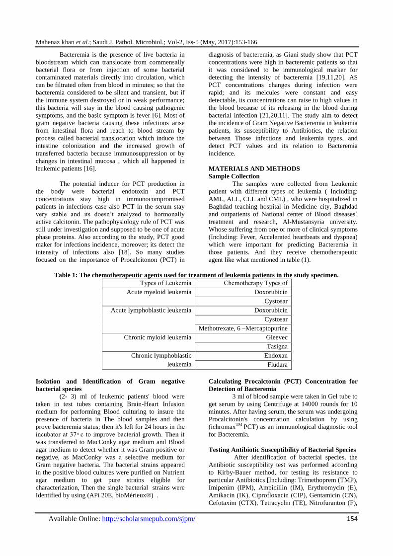
MATERIALS AND METHODS
Sample Collection
The samples were collected from Leukemic
patient with different types of leukemia ( Including:
AML, ALL, CLL and CML) , who were hospitalized in
Baghdad teaching hospital in Medicine city, Baghdad
and outpatients of National center of Blood diseases`
treatment and research, Al-Mustansyria university.
Whose suffering from one or more of clinical symptoms
(Including: Fever, Accelerated heartbeats and dyspnea)
which were important for predicting Bacteremia in
those patients. And they receive chemotherapeutic
agent like what mentioned in table (1).
Table 1: The chemotherapeutic agents used for treatment of leukemia patients in the study specimen.
fyoshpyo ytheehtomehC ehtomehCefo sosyy
Doxorubicin Acute myeloid leukemia
Cystosar
Doxorubicin Acute lymphoblastic leukemia
Cystosar
Methotrexate, 6 –Mercaptopurine
Gleevec Chronic myloid leukemia
Tasigna
Endoxan Chronic lymphoblastic
leukemia Fludara
Isolation and Identification of Gram negative
bacterial species
(2- 3) ml of leukemic patients' blood were
taken in test tubes containing Brain-Heart Infusion
medium for performing Blood culturing to insure the
presence of bacteria in The blood samples and then
prove bacteremia status; then it's left for 24 hours in the
incubator at c to improve bacterial growth hen it
was transferred to MacConky agar medium and Blood
agar medium to detect whether it was Gram positive or
negative, as MacConky was a selective medium for
Gram negative bacteria. The bacterial strains appeared
in the positive blood cultures were purified on Nutrient
agar medium to get pure strains eligible for
characterization, Then the single bacterial strains were
Identified by using (APi 20E, bioMérieux®) .
Calculating Procalctonin (PCT) Concentration for
Detection of Bacteremia
3 ml of blood sample were taken in Gel tube to
get serum by using Centrifuge at 14000 rounds for 10
minutes. After having serum, the serum was undergoing
Procalcitonin's concentration calculation by using
(ichromaxTM
PCT) as an immunological diagnostic tool
for Bacteremia.
Testing Antibiotic Susceptibility of Bacterial Species
After identification of bacterial species, the
Antibiotic susceptibility test was performed according
to Kirby-Bauer method, for testing its resistance to
particular Antibiotics [Including: Trimethoprem (TMP),
Imipenim (IPM), Ampicillin (IM), Erythromycin (E),
Amikacin (IK), Ciprofloxacin (CIP), Gentamicin (CN),
Cefotaxim (CTX), Tetracyclin (TE), Nitrofuranton (F),
Mahenaz khan et al.; Saudi J. Pathol. Microbiol.; Vol-2, Iss-5 (May, 2017):153-166
Available Online: http://scholarsmepub.com/sjpm/ 155
Piperacillin (PRL), Chloramphnicol (C), Cefalothin
(KF), Naldixic acid (NA) and Amoxicillin (AUG)]
purchased from Bioanalyse® by culturing bacteria on
uller- inton agar and using antibiotic s discs for
hours in c in the incubator then calculating the
inhibition zone for all of them according to CLSI [22].
Statistical Analysis
The statistical analysis was performed by using
chi- square test and UNIANOVA test (SPSS program,
version 19).
RESULTS
Patients Sample
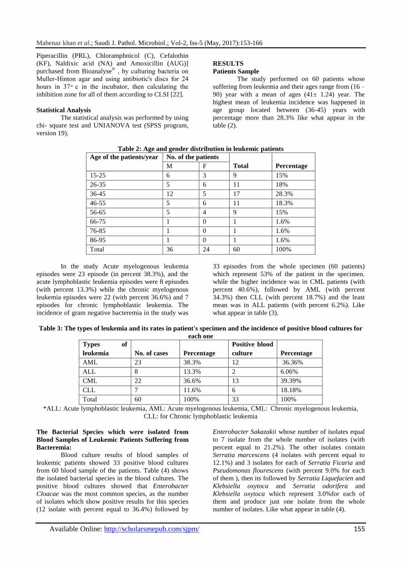
The study performed on 60 patients whose
suffering from leukemia and their ages range from (16 –
90) year with a mean of ages (41± 1.24) year. The
highest mean of leukemia incidence was happened in
age group located between (36-45) years with
percentage more than 28.3% like what appear in the
table (2).
Table 2: Age and gender distribution in leukemic patients
Age of the patients/year
No. of the patients
Total
Percentage M F
15-25 6 3 9 15%
26-35 5 6 11 18%
36-45 12 5 17 28.3%
46-55 5 6 11 18.3%
56-65 5 4 9 15%
66-75 1 0 1 1.6%
76-85 1 0 1 1.6%
86-95 1 0 1 1.6%
Total 36 24 60 100%
In the study Acute myelogenous leukemia
episodes were 23 episode (in percent 38.3%), and the
acute lymphoblastic leukemia episodes were 8 episodes
(with percent 13.3%) while the chronic myelogenous
leukemia episodes were 22 (with percent 36.6%) and 7
episodes for chronic lymphoblastic leukemia. The
incidence of gram negative bacteremia in the study was
33 episodes from the whole specimen (60 patients)
which represent 53% of the patient in the specimen.
while the higher incidence was in CML patients (with
percent 40.6%), followed by AML (with percent
34.3%) then CLL (with percent 18.7%) and the least
mean was in ALL patients (with percent 6.2%). Like
what appear in table (3).
Table 3: The types of leukemia and its rates in patient's specimen and the incidence of positive blood cultures for
each one
Types of
leukemia
No. of cases
Percentage
Positive blood
culture
Percentage
AML 23 38.3% 12 36.36%
ALL 8 13.3% 2 6.06%
CML 22 36.6% 13 39.39%
CLL 7 11.6% 6 18.18%
Total 60 100% 33 100%
*ALL: Acute lymphoblastic leukemia, AML: Acute myelogenous leukemia, CML: Chronic myelogenous leukemia,
CLL: for Chronic lymphoblastic leukemia
The Bacterial Species which were isolated from
Blood Samples of Leukemic Patients Suffering from
Bacteremia: Blood culture results of blood samples of
leukemic patients showed 33 positive blood cultures
from 60 blood sample of the patients. Table (4) shows
the isolated bacterial species in the blood cultures. The
positive blood cultures showed that Enterobacter
Cloacae was the most common species, as the number
of isolates which show positive results for this species
(12 isolate with percent equal to 36.4%) followed by
Enterobacter Sakazakii whose number of isolates equal
to 7 isolate from the whole number of isolates (with
percent equal to 21.2%). The other isolates contain
Serratia marcescens (4 isolates with percent equal to
12.1%) and 3 isolates for each of Serratia Ficaria and
Pseudomonas flourescens (with percent 9.0% for each
of them ), then its followed by Serratia Liquefacien and
Klebsiella oxytoca and Serratia odorifera and
Klebsiella oxytoca which represent 3.0%for each of
them and produce just one isolate from the whole
number of isolates. Like what appear in table (4).
Mahenaz khan et al.; Saudi J. Pathol. Microbiol.; Vol-2, Iss-5 (May, 2017):153-166
Available Online: http://scholarsmepub.com/sjpm/ 156
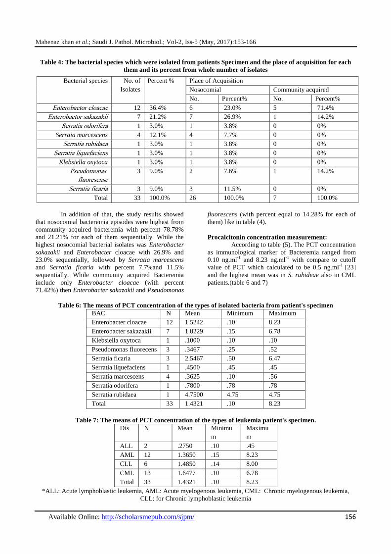
Table 4: The bacterial species which were isolated from patients Specimen and the place of acquisition for each
them and its percent from whole number of isolates
In addition of that, the study results showed
that nosocomial bacteremia episodes were highest from
community acquired bacteremia with percent 78.78%
and 21.21% for each of them sequentially. While the
highest nosocomial bacterial isolates was Enterobacter
sakazakii and Enterobacter cloacae with 26.9% and
23.0% sequentially, followed by Serratia marcescens
and Serratia ficaria with percent 7.7%and 11.5%
sequentially. While community acquired Bacteremia
include only Enterobacter cloacae (with percent
71.42%) then Enterobacter sakazakii and Pseudomonas
fluorescens (with percent equal to 14.28% for each of
them) like in table (4).
Procalcitonin concentration measurement:
According to table (5). The PCT concentration
as immunological marker of Bacteremia ranged from
0.10 ng.ml-1
and 8.23 ng.ml-1
with compare to cutoff
value of PCT which calculated to be 0.5 ng.ml-1
[23]
and the highest mean was in S. rubideae also in CML
patients.(table 6 and 7)
Table 6: The means of PCT concentration of the types of isolated bacteria from patient's specimen
BAC N Mean Minimum Maximum
Enterobacter cloacae 12 1.5242 .10 8.23
Enterobacter sakazakii 7 1.8229 .15 6.78
Klebsiella oxytoca 1 .1000 .10 .10
Pseudomonas fluorecens 3 .3467 .25 .52
Serratia ficaria 3 2.5467 .50 6.47
Serratia liquefaciens 1 .4500 .45 .45
Serratia marcescens 4 .3625 .10 .56
Serratia odorifera 1 .7800 .78 .78
Serratia rubidaea 1 4.7500 4.75 4.75
Total 33 1.4321 .10 8.23
Table 7: The means of PCT concentration of the types of leukemia patient's specimen.
Dis N Mean Minimu
m
Maximu
m
ALL 2 .2750 .10 .45
AML 12 1.3650 .15 8.23
CLL 6 1.4850 .14 8.00
CML 13 1.6477 .10 6.78
Total 33 1.4321 .10 8.23
*ALL: Acute lymphoblastic leukemia, AML: Acute myelogenous leukemia, CML: Chronic myelogenous leukemia,
CLL: for Chronic lymphoblastic leukemia
Place of Acquisition Percent % No. of
Isolates
Bacterial species
Community acquired Nosocomial
Percent% No. Percent% No.
71.4% 5 23.0% 6 36.4% 12 eapo hcytph etohytyo
14.2% 1 26.9% 7 21.2% 7 eapo hcytph emysyrysyy
0% 0 3.8% 1 3.0% 1 ao ypyyehah yCo y
0% 0 7.7% 4 12.1% 4 Serraia marcescens
0% 0 3.8% 1 3.0% 1 ao ypyye cyayoy
0% 0 3.8% 1 3.0% 1 ao ypyyeoyl oCytyoam
0% 0 3.8% 1 3.0% 1 Klebsiella oxytoca
14.2% 1 7.6% 2 9.0% 3 mo ahshayme
Co h omoamo
0% 0 11.5% 3 9.0% 3 ao ypyyeCyty yy
100.0% 7 100.0% 26 100.0% 33 Total
Mahenaz khan et al.; Saudi J. Pathol. Microbiol.; Vol-2, Iss-5 (May, 2017):153-166
Available Online: http://scholarsmepub.com/sjpm/ 157
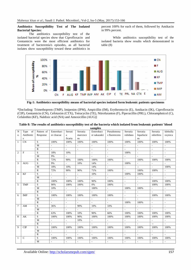
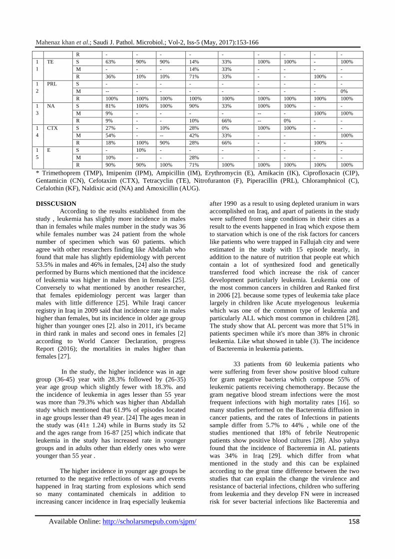
Antibiotics Susceptibility Test of The Isolated
Bacterial Species:
The antibiotics susceptibility test of the
isolated bacterial species show that Ciprofloxacin eand
Gentamicin were the most efficient antibiotics for
treatment of bacteremia's episodes, as all bacterial
isolates show susceptibility toward these antibiotics in
percent 100% for each of them, followed by Amikacin
in 99% percent.
While antibiotics susceptibility test of the
isolated bacteria show results which demonstrated in
table (8)
Fig-1: Antibiotics susceptibility means of bacterial species isolated form leukemic patients specimens
*[Including: Trimethoprem (TMP), Imipenim (IPM), Ampicillin (IM), Erythromycin (E), Amikacin (IK), Ciprofloxacin
(CIP), Gentamicin (CN), Cefotaxim (CTX), Tetracyclin (TE), Nitrofuranton (F), Piperacillin (PRL), Chloramphnicol (C),
Cefalothin (KF), Naldixic acid (NA) and Amoxicillin (AUG)]
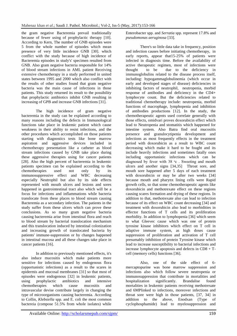
Table 8: The results of antibiotics susceptibility test of the bacteria which isolated from leukemic patients' blood
samples N
o.
Type of
Antibiotic
s
Pattern of
Response
Enterobact
er cloacae
Serrati
a
ficaria
Serratia
marcesce
ns
Enterobact
er sakazakii
Pseudomona
s fluorescens
Serratia
rubidaea
Serratia
liquefacie
ns
Serratia
odorifera
klebsilla
oxytoca
1 CN S 100% 100% 100% 100% 100% 100% 100% 100% 100%
M - - - - - - - - -
R - - - - - - - - -
2 F S 18% 10% - - 100% - - -
M 9% - - - - - - -
R 72% 90% 100% 100% 100% - 100% 100% 100%
3 AUG S 9% - 10% 14% - 100% - - -
M 18% 10% 14% - - -- - 100%
R 72% 90% 90% 71% 100% - 100% 100% -
4 KF S - - - 10% - 100% 100% - -
M - - - - - - - - -
R 100% 100% 100% 90% 100% - - 100% 100%
5 TMP S 90% 100% 100% 0% 100% - - 100% 100%
M 10% - - 100% - 100% 100% - -
R - - - - - - - - -
6 IMP S 100% 100% 100% 100% 100% - - 100% 100%
M - - - - - - - - -
R - - - - - 100% 100% - -
7 AM S 36% - 90% 10% 33% - - - -
M - - - - - - - - -
R 63% 100% 10% 90% 66% 100% 100% 100% 100%
8 AK S 100% 100% 90% 100% 100% 100% 100% 100% 100%
M - - 10% - - - - - -
R - - - - - - - -- -
9 CIP S 100% 100% 100% 100% 100% 100% 100% 100% 100%
M - - - - - - - - -
R - - - - - - - - -
1 C S 100% 100% 100% 100% 100% 100% 100% 100% 100%
M - - - - - - - - -
Mahenaz khan et al.; Saudi J. Pathol. Microbiol.; Vol-2, Iss-5 (May, 2017):153-166
Available Online: http://scholarsmepub.com/sjpm/ 158
R - - - - - - - - -
1
1
TE S 63% 90% 90% 14% 33% 100% 100% - 100%
M - - - 14% 33% - - - -
R 36% 10% 10% 71% 33% - - 100% -
1
2
PRL S - - - - - - - - -
M -- - - - - - - - 0%
R 100% 100% 100% 100% 100% 100% 100% 100% 100%
1
3
NA S 81% 100% 100% 90% 33% 100% 100% - -
M 9% - - - - -- - 100% 100%
R 9% - - 10% 66% -- 0% - -
1
4
CTX S 27% - 10% 28% 0% 100% 100% - -
M 54% - -- 42% 33% - - - 100%
R 18% 100% 90% 28% 66% - - 100% -
1
5
E S - 10% - - - - - - -
M 10% - - 28% - - - - -
R 90% 90% 100% 71% 100% 100% 100% 100% 100%
* Trimethoprem (TMP), Imipenim (IPM), Ampicillin (IM), Erythromycin (E), Amikacin (IK), Ciprofloxacin (CIP),
Gentamicin (CN), Cefotaxim (CTX), Tetracyclin (TE), Nitrofuranton (F), Piperacillin (PRL), Chloramphnicol (C),
Cefalothin (KF), Naldixic acid (NA) and Amoxicillin (AUG).
DISSCUSION According to the results established from the
study , leukemia has slightly more incidence in males
than in females while males number in the study was 36
while females number was 24 patient from the whole
number of specimen which was 60 patients. which
agree with other researchers finding like Abdallah who
found that male has slightly epidemiology with percent
53.5% in males and 46% in females, [24] also the study
performed by Burns which mentioned that the incidence
of leukemia was higher in males then in females [25].
Conversely to what mentioned by another researcher,
that females epidemiology percent was larger than
males with little difference [25]. While Iraqi cancer
registry in Iraq in 2009 said that incidence rate in males
higher than females, but its incidence in older age group
higher than younger ones [2]. also in 2011, it's became
in third rank in males and second ones in females [2]
according to World Cancer Declaration, progress
Report (2016); the mortalities in males higher than
females [27].
In the study, the higher incidence was in age
group (36-45) year with 28.3% followed by (26-35)
year age group which slightly fewer with 18.3%. and
the incidence of leukemia in ages lesser than 55 year
was more than 79.3% which was higher than Abdallah
study which mentioned that 61.9% of episodes located
in age groups lesser than 49 year. [24] The ages mean in
the study was (41± 1.24) while in Burns study its 52
and the ages range from 16-87 [25] which indicate that
leukemia in the study has increased rate in younger
groups and in adults other than elderly ones who were
younger than 55 year .
The higher incidence in younger age groups be
returned to the negative reflections of wars and events
happened in Iraq starting from explosions which send
so many contaminated chemicals in addition to
increasing cancer incidence in Iraq especially leukemia
after 1990 as a result to using depleted uranium in wars
accomplished on Iraq, and apart of patients in the study
were suffered from siege conditions in their cities as a
result to the events happened in Iraq which expose them
to starvation which is one of the risk factors for cancers
like patients who were trapped in Fallujah city and were
estimated in the study with 15 episode nearly, in
addition to the nature of nutrition that people eat which
contain a lot of synthesized food and genetically
transferred food which increase the risk of cancer
development particularly leukemia. Leukemia one of
the most common cancers in children and Ranked first
in 2006 [2]. because some types of leukemia take place
largely in children like Acute myelogenous leukemia
which was one of the common type of leukemia and
particularly ALL which most common in children [28].
The study show that AL percent was more that 51% in
patients specimen while it's more than 38% in chronic
leukemia. Like what showed in table (3). The incidence
of Bacteremia in leukemia patients.
33 patients from 60 leukemia patients who
were suffering from fever show positive blood culture
for gram negative bacteria which compose 55% of
leukemic patients receiving chemotherapy. Because the
gram negative blood stream infections were the most
frequent infections with high mortality rates [16]. so
many studies performed on the Bacteremia diffusion in
cancer patients, and the rates of Infections in patients
sample differ from 5.7% to 44% , while one of the
studies mentioned that 18% of febrile Neutropenic
patients show positive blood cultures [28]. Also yahya
found that the incidence of Bacteremia in AL patients
was 34% in Iraq [29]. which differ from what
mentioned in the study and this can be explained
according to the great time difference between the two
studies that can explain the change the virulence and
resistance of bacterial infections, children who suffering
from leukemia and they develop FN were in increased
risk for sever bacterial infections like Bacteremia and
Mahenaz khan et al.; Saudi J. Pathol. Microbiol.; Vol-2, Iss-5 (May, 2017):153-166
Available Online: http://scholarsmepub.com/sjpm/ 159
the gram negative Bacteremia prevail traditionally
because of fewer using of prophylactic therapy [10].
According to Kern, The number of GNB episodes were
5 from the whole number of episodes which mean
presence of very little incidence GNB [30]. which
conflict with the study because of high incidence of
Bacteremia episodes in study's' specimen resulted from
GNB. Also gram negative bacteria responsible for 14%
of blood stream infections in AML patient Receiving
extensive chemotherapy in a study performed in united
states between 1995 and 2000 which also conflict with
the results of other studies found that gram negative
bacteria was the main cause of infections in those
patients. This study returned its result to the possibility
that prophylactic antibiotics inhibit GNB resulting in
increasing of GPB and increase GNB infections [31].
The high incidence of gram negative
bacteremia in the study can be explained according to
many reasons including the defects in Immunological
functions take place in leukemic patient which cause
weakness in their ability to resist infections, and the
other procedures which accomplished on those patients
starting with diagnostics tests like bone marrow
aspiration and aggressive devices included in
chemotherapy presentation like a catheter as blood
stream infections caused by GNB take place during
these aggressive therapies using for cancer patients
[28]. Also the high percent of bacteremia in leukemic
patients specimen can be explained according to the
chemotherapies used not only by its
immunosuppressive effect and WBC decreasing
especially Neutrophil but also by its side effects
represented with mouth ulcers and lesions and sores
happened in gastrointestinal tract also which will be a
focus for infections and inflammations while bacteria
translocate from these places to blood stream causing
Bacteremia as a secondary infection. The patients in the
study suffer from these ulcers which can prove these
conclusions. As so many gram negative bacteria
causing bacteremia arise from intestinal flora and reach
to blood stream by bacterial translocation mechanism
and this translocation induced by intestinal colonization
and increasing growth of translocated bacteria by
patients' immune-suppression or by changes happened
in intestinal mucosa and all these changes take place in
cancer patients [16].
In addition to previously mentioned effects, it's
also induce mucositis which make patients more
sensitive for infections caused by endogenous flora
(opportunistic infections) as a result to the scares in
epidermis and mucosal membranes [31] so that most of
episodes were endogenous [32] in leukemic patients,
using prophylactic antibiotics and intensive
chemotherapies which cause mucositis and
intravascular devise contribute largely in changing the
type of microorganisms causing bacteremia. According
to Collin, Klebsiella spp. and E. coli the most common
bactereia (compose 51.5% from whole isolates) while
Enterobacter spp. and Serratia spp. represent 17.8% and
pseudomonas aeruginosa [33].
There's so little data take in frequency, position
and infection causes before initiating chemotherapy, in
early reports, appear that15-25% of patients were
infected in diagnosis time. Before the availability of
active therapeutic regimen, most of infections were
thought to be due to the deficiency in
immunoglobulins related to the disease process itself,
including: hypogammaglobulinemia (which occur in
early and developed stages of disease) deficiencies in
inhibiting factors of neutrophil, neutropenia, morbid
response of antibodies and deficiency in the CD4+
lymphocyte count. But the deficiencies related to
traditional chemotherapy include: neutropenia, morbid
functions of macrophage, lymphopenia and inhibition
of antibodies productions [12]. In the study, the
chemotherapeutic agents used correlate generally with
those effects, omidvari proves doxorubicin effect which
lead to Neutropenia and stomatitis which happened for
intestine system. Also Batra find oral mucositis
presence and granulocytipenia development and
infections as most frequently effects in the treatment
period with doxorubicin as a result to WBC count
decreasing which make it hard to be fought and its
include heavily infections and sometimes fatally ones
including opportunistic infections which can be
diagnosed by fever with 8 ᵒc Sweeting and mouth
ulcers and another signs [34,35] Throat sore, and
mouth sore happened after 5 days of each treatment
with doxorubicin or may be after two weeks [34]
because mouth and pharynx lining cells were Rapid
growth cells, so that some chemotherapeutic agents like
doxorubicin and methotrexate effect on these regions
causing scares formation and drying of these regions. In
addition to that, methotrexate also can lead to infection
because of its effect on WBC count decreasing [34] and
treatment with doxorubicin also lead to morbidities in
effector functions of T cells and its proliferation
morbidity. In addition to lymphopenia [36] which seem
to what Gleevec cause which was one of protein
tyrosine kinase inhibitors which effect on T cell in
adaptive immune system, as high doses cause
suppression of proliferation and activation of T cell
presumably inhibition of protein Tyrosine kinase which
lead to increase susceptibility to bacterial infections and
increase lymphocyte apoptosis and defects in CD8 + T-
cell (memory cells) functions [36].
Also, one of the side effect of 6-
mercaptopurine was bone marrow suppression and
infections also which follow severe neutropenia or
immunosuppression that contribute in mortalities and
hospitalization significantly. Brandalise found
mortalities in leukemic patients receiving methotrexate
and 6MPlinked to infections, moreover infections and
throat sore were high in those patients, [37, 34] in
addition to the above, Enodxan (Type of
cyclophosphamide) lead to myelosuppression and
Mahenaz khan et al.; Saudi J. Pathol. Microbiol.; Vol-2, Iss-5 (May, 2017):153-166
Available Online: http://scholarsmepub.com/sjpm/ 160
significant suppression of immune response and cause
leukopenia and neutropenia leading to intensive and
sometimes fatal bacterial infections like sepsis or septic
shock also activation of potential infection and its must
be stopped or its decreased dose in case of severe
infections and can cause sores and bladder necrosis [34]
Treatment with fludarabine (fludara) one type of purine
analogues was high risk factor for side effects in GIT
[12] while the most common side effect of
Anthracyclines (doxorubicin) was myelosuppression
and stomatitis, [38] Tasigna cause many common side
effects including infections like upper respiratory tract
infections and stomatitis and not common to develop
sepsis abnormal hepatic functions and gastrointestinal
ulcer perforation but acute pancreatitis and mouth
ulceration were less common. Also its lead to
myelosuppression and can cause neutropenia, septic
shock and sore throat and low blood count and the latest
effect was so common in case of Tasigna treatment
[39].
By all what previously mentioned, There's
explanation for the high percentage of bacterial isolates
subsidiary to Enterobacteriaceae in the study like
Enterobacter cloacae, Enterobacter sakazakii and
Serratia spp. as most of bacteria in GIT were from
Enterobacteriaceae which can linked to the side effects
of chemotherapies as its can produce primary source of
infections (secondary infections) also wisplinghoff
found that most of secondary BSI resulted from
intravenous catheter and from UTI followed by lower
respiratory tract and GIT but there's no significant
difference noticed between Neutropenia and those who
didn’t suffer from it about infection source [ 0] most of
BSI in immunocompromised patients arise from
previous local infections compared with BSI in
Neutropenia patient which arise largely from
endogenous infections like GIT infections [40,25] The
data taken from extensive studies in cancer research
centers in USA and Europe show that
Enterobacteriaceae represent 65-80% from registered
gram negative infections [28] also Batzar found that E.
cloacae was the most common bacteria in ICU patients
from children [41] in addition to all of that Lio-ten foe
isolate Klebsiella oxytoca and E. cloacae and P.
fluorescens from pharynx, saliva and rectum from ICU
patients [42] in the study. The highest percentage of
positive isolates were in CML with percent higher than
40% from the whole number of specimens followed by
AML with percent higher than 34%, Then CLL (18%)
and the lesser percent was in ALL patients (6.25%)
which differ from what noticed in Abdallah study and
AL -Neemy [24,26] which found higher percent of
Bacteremia in AML with 41% percent in Abdallah
study [24] and mortality rates caused by infections in
AML patients range from (5.5-13)% during treatment.
[43] also it differ from Burns study who find ALL
infections higher than AML ones [25]. according to
study accomplished in Eduard Herriot hospital in
France about the annual mean of Hospital acquired
infections, this study showed that Bacteremia percent in
AML was 12.9% and ALL (13.4%) and gram negative
bacilli bacteremia was (4%) but cocci was 1% from
whole episodes of bacteremia, this study also show
increasing incidence of bacteremia and most of them
were Endogenous [32] as Enterobacter from one of
endogenous flora of human [44] in addition to that E.
cloacae was opportunistic pathogen [54] also
chemotherapeutics regimens for AML induce
Neutropenia which stay for a long periods and expose
them for frequent infections, Basically arise through the
first cycle of chemotherapy [43] in spite of the fact that
statistical analysis from the study show absence of
significant difference in the relationship between
bacteremia and leukemia patient, also between the types
of bacterial isolates species and leukemic types which
agree completely with what mentioned by Abdallah
study [24] in spite of using appropriate number of
patient in specimen which can give statistical results
near from reality also Kern found that incidence of
bacteremia in leukemic patients was 36% [30] so that
its differ from the study that show higher incidence also
Kern found that 4 patients from study specimen died,
[30] while in the study 2 patient were died and they
show positive blood cultures for Enterobacter sakazakii
(PCT 4.60ng.ml-1
) and the other for Serratia
marcescens (PCT 0.45ng.ml-1
) so that mortality rate in
the study 6.25% while in Kern study, 4 patients died.
[30] from the whole number of isolates, Enterobacter
cloacae from the highest percent with 37.5% followed
by Enterobacter sakazakii with 25% which form the
most common species in the study while 12.5% from
the isolates affiliate to Serratia marcescens and Both of
pseudomonas fluorecens and Serratia ficaria was
9.37% and the least percent of isolates was for Serratia
rubidaea, Serratia liquefaciens, Klebsiella oxytoca
which represent 3% of the whole number of isolates,
which agree with the isolated species causing
bacteremia in Kern study whose include Enterobacter
sp. In addition to E. coli. [30] It's important to notice
Abdallah results whose study accomplished in the same
environment of the study, and it's found that the most
common species was Klebsiella pneumonia with
21.42% followed by E. coli and Pseudomonas
aeruginosa with 14.28% then Salmonella typhi with
7.14% and the least percentage was for proteus penneri
with 3.57% [24] all these results differ from what found
in the study, which can be explained according to the
time difference between the two studies and the
possibility of prophylactic treatments difference and the
resistance and virulence differences of bacteria which
were in continuous progress. In his book, Faguet
mentioned that the most common bacterial isolates were
P.aeruginosa, Klebsiella sp. And E.coli [12] which
agrees somewhat with study finding because of
emergence of species affiliate to Klebsiella represented
with Klebsiella oxytoca. inaddition to emergence of
Pseudomonas fluorescens. As in one of studies which
accomplished in Benha hospital, the incidence of
bacteremia was 30.61% and from this percent, GNB
Mahenaz khan et al.; Saudi J. Pathol. Microbiol.; Vol-2, Iss-5 (May, 2017):153-166
Available Online: http://scholarsmepub.com/sjpm/ 161
represent 63.9% and E. coli was the Hugo percent as its
form 46.1% and Klebsiella form 30.8% followed by
15.4% of Pseudomonas then Acinobacter form 7.7%
[54] Body and his collagenous consider FN and specific
pathogens like Pseudomonas aeruginosa and Serratia
marcescens to be challenge for cancer treatment. In this
time, the mortalities followed P. aeruginosa Bacteremia
form nearly 90% in spite of availability of laboratory
active antibiotics [7]. in recent studies, BSI volume
estimated in Neutropenia AML to be ranged between
(34-38)% also in Madani study, who said that GN
bacilli was 12.1%. Composed of Klebsiella pneumonia
(3%) followed by Enterobacter cloacae (1.5%) [43].
The epidemiological observations on community
acquired pneumonia (CAP) show increasing incidence
of severe Bacteremia resulted from Enterobacter as one
study show that En CAP incidence stay severe in
number of cancer episodes including CML [55]. In
India, parabgsh and his collagenous in 2010 found that
the most common causative agents of infections in
leukemic patients was Pseudomonas spp. with 30.37%
percent. [28] while chency study GNB from 60% from
infections in Neutropenic patients with 60% including
E. coli (12%) and Klebsiella pneumonia (10%), [28]
The most common Bacterial pathogen in Santolya study
which composed pediatric cancer patients receiving
chemotherapy and suffering from FN and Bacteremia
was E, coli followed by Klebsiella sp and Pseudomonas
aeruginosa then Enterobacter aerugenes. (221) but
Wisplinghoff mentioned nearly same results to Santolya
that E.coli, The most common isolates followed by
Klebsiella sp. and Pseudomonas sp. Then Enterobacter
sp [40]. These studies differ from the study with
important point, which involve that E.coli the most
common pathogen while in the study, there's not any
isolate of E. coli and Enterobacter spp. Were the most
common isolates which can be explained by the type of
prophylactic antibiotic used which selected to target the
most common pathogens like E. coli and the other types
which appear with little percent in the study. Because
prevention and treatment of infection critical part of
leukemia treatment which can be accomplished with
empirical treatment with antibiotics which cover the
wide range of pathogens [8] also Abdollahi found that
E. coli was the most common isolate from Blood
cultures of leukemia patients with 34%. [28] because of
increasing mortalities in leukemic patients resulted from
E. coli and P. aeruginosa [40] Serratia cause 1.4% of
nosocomial bacteremia because its contain
chromosomally induced Amc β lactamases which give
it the ability to develop rapid resistance to many of β
lactam antibiotic and give it ability to cause Bacteremia
and sever nosocomial infections. In Choi study which
contains leukemic patients Receiving
immunosuppressive agents and chemotherapeutic
agents, Serratia bacteremia diagnosed with higher
percentage in the first group while 96% of them
returned to S. marcescens and 4% for Serratia
liquifaciens [45]. in the study the higher percent of
Bacteremia resulted from nosocomial infections with
81.25% while little percent resulted from community
acquired Bacteremia. Because leukemia patients were
in increased risk for nosocomial BSI because of disease
severity which need intensive chemotherapy or stem
cell or bone marrow transplantation which lead to
severe infections and worsen the cases so that
mortalities of Nosocomial bacteremia were ranged from
10-20% [25, 32].
Apostlopolou and his collagenous calculate
Bacteremia incidence in 102 leukemic patients who
were hospitalized for more than 48 hour and he find that
21.99/1000 patient perday suffering from Bacteremia
especially women in contrast the age doesn’t have any
effect on Bacteremia expectance in spite of that its
considered to be risk factor for severe Neutropenia.
Also Garcia -Suarez found the same incidence of
Bacteremia in leukemic patient from elderly and young
who suffering from FN. there's so little data available
on the effect of comorbidities of leukemia as risk factor
for Bacteremia, [7] also E. cloacae considered to be the
most common causative agent for Nosocomial
infections as its cause sepsis and can be found in GIT,
UT, RT, also its resistant to disinfectants and
Antimicrobial agents from Enterobacteriaceae which
can explain its increasing responsibility of Nosocomial
Infections and its wide spreading [45].
PCT concentration measurement
Some studies suggest that PCT have secondary
mediated rule in immunological pathogenicity in sepsis
cases, and many studies focused on the diagnostic rule
of PCT for diagnosis of Bacteremia and sepsis in
patients who require intensive care, and one study
found sensitivity between 65% to 97% and specfity
between 48% to 94% for PCT in diagnosis of
Bacteremia, Three studies found that PCT was the best
immunological marker for sepsis, and the best marker
for bacterial infections according to Chan study [19]
also according to Leli and his Collageous study, [23] as
patients suffering from severe sepsis have 10 times PCT
concentrations with comparison to sepsis episodes.
Several studies focused on the abilities of PCT for
diagnosing infections ICU [19]. which help physicians
to determinate the primary antibacterial therapy suitable
for those patients while there's correlation between
starting unsuitable therapy with resulted complications
largely [23]. The PCT values in the study range
between (0.1 to 8.23) ng.ml-1
and Eleven sample from
33 sample in the study show PCT concentration higher
than cut–off value which was 0.5 ng.ml-1`
which show
that they were infected with bacteremia, while the other
values were less than 0.5 ng.ml-1
, so that it may indicate
two possibilities; first one that there's no bacteremia in
those patient and the other possibility that its values
decreased as a result to disease itself. As Hepatocytes
considered to be known sources of PCT so that any
damages in the liver resulted from chemotherapy may
be possible source for decreasing PCT levels in cancer
patients [46,11] Like in Giamarellos-Baboulis study,
Mahenaz khan et al.; Saudi J. Pathol. Microbiol.; Vol-2, Iss-5 (May, 2017):153-166
Available Online: http://scholarsmepub.com/sjpm/ 162
who mentioned that PCT concentrations in leukemic
patients before chemotherapy in his study were in 0.16
mean while its 0.05 mean in Febrile neutropenic
patients [47]. which suppose that in case of
neutropenia, PCT values less than non-neutropenic
patients. Which agree with what Schuttrumpf and his
collagenous mentioned when they calculate PCT
concentrations in plasma in for 111 patient with
hematological malignancies conditions and they found
that PCT concentrations higher significantly in patients
with infections than in those without infections also its
higher in patients with infections who didn’t suffer from
leukopenia from those who have leukopenia and PCT
concentrations higher in Bacteremic patients than in
local viral o fungal infections [48]. Engle and his
colleagues found that PCT levels during fever incidence
stay under 0.5 ng.ml-1
also Hambach study found that
PCT levels higher in Bacterial infections with mean
equal to 2.3 ng.ml-1
. Also Pihusch and his collagenous
found increasing in PCT levels after infections
complications but only PCT can distinguish between
infections and related complications to stem cells
transplantation in cancer patients [48].
Also PCT levels in Chan study were equal or
larger than 0.6 ng.ml-1
in 69.5% of patient with
infections and PCT mean was 5.30ng/ml for both
infected and non-infected patients while its mean in
non-infected patients only were 0.5 ng.ml-1
and there's
no relation between PCT concentration and the type of
infected organism. Also PCT level was the best for
predicting bacteremia incidence was 1 ng.ml-1
with 63%
specifity according to Chan study. PCT concentrations
in healthy plasma were under 0.1 ng.ml-1
[18] while in
our study the cut-off value used according to
ichromaxTM
PCT instructions was 0.5 ng.ml-1
But Leli
study found that cutoff value of PCT to distinguish
between Enterobacteriaceae and non fermentive gram
negative was 3.1 ng.ml-1
with 90% sensitivity [23]
While Ugarit study indicate that PCT cutoff value was
0.6 ng.ml-1
with 67.6% sensitivity [18] but So many
studies mention the cutoff values of PCT to distinguish
bacterial infections from other infections which range
between 0.5 ng.ml-1
to 1.3 ng.ml-1
[48] PCT values
ranged from 0.5 to 2 considered to be indicator for life
threatened conditions [46] which agree with what used
in the study. The PCT values larger than 2 ng.ml-1
seem
to be enough to distinguish between sever sepsis and
local infections with 90.9% sensitivity approximately
and 80.9% specifity [47].
The study results statistics indicate that there's
no significant relationship between PCT values and the
types of bacterial species, or with types of leukemia
while SVALDI study found that PCT concentrations in
AML patients were higher than 2 ng.ml-1
with high
percent in comparison with CML followed by ALL
[46]. also one study found that PCT levels were less in
children who suffer from neutropenia and bacterial
infections from those without neutropenia [11]
Hatzitilianon found high significant difference in PCT
levels between bacterial infections and non-bacterial
infections moreover its significantly lower in
Neutropenia who have bacterial infections [11]. PCT
concentration in gram negative bacteremia was higher
than gram positive bacteremia significantly in positive
blood cultures, [23] because GNB induce production of
significantly higher PCT levels than GPB and the LPS
composition of outer membrane was the responsible for
that effect. [46] One study found that GN bacteremia
had largest PCT values then gram positive bacteremia
while another study didn’t find any difference between
them and this study didn’t contain any sepsis episodes
[47]. the higher PCT mean found in S. rubideae (4.75
ng.ml-1
) followed by S. ficaria then in E. cloacae and E.
sakazakii (1.82 and 1.52) ng.ml-1
sequentially and the
least value was for Klebseilla oxytoca (0.1 ng.ml-1
)
moreover Leli study found that PCT concentrations in
BSI resulted from GNB including Enterobacteriaceae
(Enterobacter cloacae, E. coli, Klebsiella sp. Serratia
marcescens, K. oxytoca and proteus mirabilis) were 17
ng.ml-1
which were higher significantly than non
fermentive bacteria including Pseudomonas aeruginosa
(3.5ng.ml-1
) and Serratia marcescens whose value was
14.9 ng.ml-1
and Klebsiella oxytoca (2.3 ng.ml-1
) and
Pseudomonas aeruginosa (6.8 ng.ml-1
) [23]. and PCT
concentrations in bacteremia patients resulted from
GNB in the first day of fever were 12.37 ng.ml-1
mean
ranged from 0.09 to 143.98 and its high particularly in
immunocompromised patients serum who suffer from
sepsis. After incidence of fever, PCT levels say within
normal limits for Both Neutropenic patients who suffer
from cancers and Neutropenic patients without fever
(The values means were 0.29 ng.ml-1
and 0.18 ng.ml-1
sequentially while PCT values mean in the first day of
bacteremia was 8.23 ng.ml-1
also high significant
difference noticed between the mean value of PCT in
the first day of bacteremia and PCT main in the first
day of local bacterial infections whose mean was 0.86
ng.ml-1
[47]. In the study, there's no ability to detect
whether PCT concentrations measured in the first day
or any other day because it wasn't taken in mind during
information's registration.
Antibiotic susceptibility test
Antibiotic administration in the beginning of
disease symptoms may prevent its diffusion and
completely decrease mortality rates and other
complications resulted from cancer [28] while the first
source of infection often stay unknown, so that
choosing of first therapy of antibiotic often being
empirically and targeted primarily toward GNB [43]
because of noticed significant increasing in GN Bacilli
resistance to antibiotics [28] and the changing in the
spectrum of pathogens causing infections in last
decades [43] many studies accomplished on main
microbes which cause infections and its susceptibility to
specific antibiotics in cancer patients, because of high
rates of bacterial infections and difficulty to detect the
patients who were more exposure to severe infections
Mahenaz khan et al.; Saudi J. Pathol. Microbiol.; Vol-2, Iss-5 (May, 2017):153-166
Available Online: http://scholarsmepub.com/sjpm/ 163
risk. so that all neutropenic patients receive wide
spectrum antibiotics until fever excluding and maintain
integrity immune system this strategy was very
successful for decreasing mortalities and secondary
complications of infections. Instead of side effects risk
of severe regimens, resistant bacterial strains, fungal
infections and followed psychological and economic
effects. Because isolated bacteria from clinical and
ecological samples acquired increased resistant to
conventional antibiotics and GNB represent the largest
risk on public health. Not just by its rapid resistance
development then positive bacteria but also by little
availability of new and developed active antibiotics
against it, and its increasing resistance resulted mainly
by mobile genes on plasmids which rapidly transferred
through bacterial groups [49]. According to results
obtained in the study; the most efficient antibiotics were
Ciproflxacin and Gentamicin with percent equal to
100% followed by Amikacin antibiotic (with 99%) so
that these antibiotics can be used effectively, also in
case of prophylactic antibiotic therapy. Since Rahman
study concluded that fluoroqinolone therapy (like
ciprofloxacin) effect on bacterial infections as its delay
fever in FN patients resulted from chemotherapy [28] as
ciprofloxacin used for recent years intensively to
prevent bacterial infections in severe Neutropenia. In
spite of it's decreased GNB infections but its cause
increasing emergence of GPB with Resistance
emergence for many GNB. Also its must be limited for
patients with high risk of developing severe
Neutropenia follow chemotherapy [12] in addition to
that Mohammed study concluded that Amikacin the
most efficient one [50] while Kumarasamy study
showed Resistance of Enterobacteriacea to Imipenem
and Amikacin in each of USA, China and India and
sensitive with 8% to Ciprofloxacin and 3% for
Gentamicin [49] while study results show 78%
sensitivity to Imipenem but Patzer mentioned that
Imipenem, Ciprofloxacin and Gentamicin the most
effective antibiotic against GNB [41] as in E. cloacae
Acr AB efflux pump mechanism play role in its
pathogenicity and resistance [49] as some antibiotics
considered to be good substrates to be excluded by
efflux mechanism which provide internal resistance for
these some antibiotics, as high resistance found in
isolates express this mechanism including
Ciprofloxacin [51] also Perez found that its resistance
depend largely on efflux pump mechanism which
interfere also with its pathogenicity [45]. Burns indicate
that Enterobacter spp. Resistance were uncommon,
because its slightly resistant to Gentamicin without any
significant difference between Neutropenic patients and
others [25] while wisplinghoff noticed that Imipenem
was the most effective antibiotic toward Enterobacter
isolates and 33% of them resistant to Piperecillin and so
little percent of the isolates were resistant to CIP and
Gentamicin [40]. In another study, most of E. cloacae
was sensitive to Ciprofloxacin, TMP, aminoglycosides
and piperacillin but also resistant to Ampicillin and
Amoxicillin β lactmases was the most important
mechanism responsible for β lactam Antibiotics
resistance because its contain all the types of β
lactamaces (Including class A penicillinaces class β
metallo-enzymes and class C cephalosporinases and
class D oxacillinaces) and able for increasing reduction
of Ampc β lactamaces also with removing of depression
from chromosomal genes or by acquisition of Ampc
genes known with Ampc plasmid mediated genes
Which can be distinguished from chromosomal genes.
Also increasing production of chromosomal
cephalosporines with porines changes responsible for
Imipenem resistance [44] also Paterson found that 37%
of infections caused by Enterobacter in ICU include
resistant strains to third generation cephalosporines.
Because of its increasing production of Ampc β
lactamases and some E. cloacae were Amp and ESBL β
lactamaces producers which give it resistance toward
cephalospoius while Quinolone resistance in
Enterobacteriaceae always come from chromosomal
mutations lead to changes in the enzymes. Hospital
acquired Enterobacter sp were Amp β lactamaces
producers so that any exposure to β lactam antibiotics
can induce production of those enzymes also mutations
can lead to its increasing and produce permanent
resistance. And Quinolone resistance in
Enterobacterceae. Result from changes or morbidities in
DNA gyrase and topoisomerase enzymes which happen
either by changes in porine expression or Efflux
mechanisms and both of those mechanisms resulted
from chromosomal mutations [52]. Klebsiella oxytoca
show compeletely sensitivity toward Imipenem; while
In one study, 50% of isolated Klebsiella spp. Show
multi resistance toward antibiotics including Imipenem
[28] also resistance of Klebsiella sp noticed toward
Imipenem with 1% according to Burns study [25].
Abdollahi study agree with the study results in spite of
some differences in Bacterial species isolates, that all
isolated bacterial species from blood cultures of
leukemia patients were sensitive toward CIP [28] but in
spite of CIP using through Neutropenia, GN Bacteremia
study high risk [43] also Abdollahi said that
Pseudomonas and Enterobacter isolates didn’t show
any resistance toward Imipenem [28] which agree with
study results, which indicate that P. flourescens, E.
cloacae and E .sakazakii toward Imipenem with 100%
[28] also Mohammed study show that E. cloacae, E.
sakazakii, K. oxytoca and S .marcescens isolated from
UTI were completely sensitive toward Amikacin,
Gentamicin and Imipenem while its 100% resistant
toward Ampicillin and E.sakazakii sensitive toward
CIP, TMP and Piperacillin in spite of E. cloacae and K.
oxytoca were resistant for them. [50] Collin
demonstrated that (40-45)% of Enterobacter spp. And
Serratia spp. were sensitive toward Piperacillin and
Kllebsiella spp. completely sensitive toward
Piperacillin and Pseudomonas sp. Were sensitive for
many factors including Imipenem [33] also
Wisplinghoff study noticed that 80% from Klebsiella
spp. were sensitive toward 3rd
generation
cephalosporins, Imipenem, Aminoglycoside and
Mahenaz khan et al.; Saudi J. Pathol. Microbiol.; Vol-2, Iss-5 (May, 2017):153-166
Available Online: http://scholarsmepub.com/sjpm/ 164
flouroquinolons while 98% of them and Serratia spp.
were resistance toward Ampicillin and little percentage
were resistant for Piperacillin, Imipenem, CIP and
Gentamicin [40, 45] while in Choi study, most of
isolates were resistant to Ampicillin, Piperacillin,
Gentamicin and Coprofloxacin moreover its sensitive
toward Imipenem which show the least resistance rates
followed by Amikacin, so that Imipenem stay the
important therapeutic choice but its excessive using
lead to later resistance to IMP with 3rd
generation
cephalosporins recent study showed emergence of
significant high resistance rate of Serratia bacteremia
during treatment with wide spectrum cephalosporins
while quinolone therapy didn’t link to any resistance
emergence so that, 3rd
generation cephalosporins were
described to be avoided as bacteremia treatment [45]
also blood culture results from samples of ICU patient
in Lockhart study showed species which include E.
cloacae, S. marcescens and K. oxytoca, since K. oxytoca
show the higher resistance toward β lactam antibiotics
with decreasing in E. cloacae sensitivity toward CIP, at
the same time, he indicated that Amikacin effective
widely toward Pseudomonas sp. and Enterobacteriaceae
[53]. Flouoquinolone work by interfering with Type II
topoisomerases (DNA gyrase and Topoisomerase III),
according to this mechanism, Bacteria work on
developing resistance mechanisms which include
mutations target Gyra / GYRB for DNA Gyrase and
parC/parE for Topoisomerase III or decrease the access
of the target its self by decreasing its permeability or by
Efflux pump mechanism, and most effective antibiotics
were Gentamyicin and Amikacin since isolates stay
sensitive in spite of presence of gene responsible for
Imipenem resistance [44] also most of studies
mentioned that its using doesn't decrease the frequency
of fever incidence and empirical antibacterial therapy
and its should be limited for patients who have great
possibility to produce sever neutropenia follow
chemotherapy [12].
REFERENCES
1. John, A., Haslett, A., Chilvers, C., Edwin, R., &
Nicki, R. C. (2002). DAVIDSON'S Principles and
Practice of Medicine. 19th Edition, British Library
Cataloguing in Publication Data.
2. Iraqi Cancer Registry. (2011, 2009, 2006). Ministry
of Health of Iraq.
3. Al-Jobori, & Shahrazad, S. (2016). Iraqi National
Bone Marrow Registry Project. Journal of Cellular
Cancer, 8 (1), 34-40.
4. Corey, G., & Ralph. (2009). Staphylococcus aureus
Blood Stream Infections: Defentions and
Treatment. Clinical Infectious Diseases, 48, S254-
S259
5. Zembower, & Teresa, R. (2014). Epidemiology and
Infections in Cancer Patients. North Western
University Division of Infections Disease, Feinberg
School of Medicine.
6. Ashour, Hossam, M., & El-Sharif, Amany. (2007).
Microbial Spectrum and Antibiotic Susceptibility
Profile of Gram Positive Aerobic Bacteria Isolated
from Cancer Patients. Journal of Clinical
Oncology. 25 (36), 5763- 5769.
7. Norgaard, & Mette. (2012). Risk of Infections in
Adult Patients with Hematological Malignancies.
The open Infectious Diseases Journal, (6), 46-51.
8. Luciano, & Azevedo. (2012). Sepsis- An Ongoing
and Significant Challenge. InTech.
9. Plummer, Kathleen, & Hope. (2014). Cancer and
Infection" Department of Cell Biology,
Microbiology and Molecular Biology. College of
Arts and Sciences, University of South Florida.
10. Asturias, Ei, J., Corral, J. E., & Quezada, J. (2010).
Evaluation of Six Risk Factors for The
Development of Bacteremia in Children with
Cancer and Febrile Neutropenia. Pediatric
Oncology, 17 (2), 59-63.
11. Maria, H., Aleka, R., Fanni, A., & Dorothea, C.
(2010). Procalcitonin as an Early Marker of
Bacterial Infection in Neutropenic Febrile Children
with Acute Lymphoblastic Leukemia.
Inflammation Research, 59, 339-347.
12. Faguet, & Guy, B. (2004). Chronic Lymphocytic
Leukemia: Molecular Genetics, Biology,
Diagnosis, and Management. Humana Press Inc.
13. René , H., & John, A. M. (2001). Surgical
Treatment: Evidence Based and Problem Oriented.
Zuckschwerdt.
14. Mette, N. (2005). Hematological Malignancies and
Bacteremia: Risk and Prognosis. Department of
Clinical Epidemiology, University of Aarhus.
15. Mohamed, A. M., & Ali, H. M. (2015). New
Biomarkers AS Indicators for Sepsis in Febrile
Leukemic Iraqi Patients. World Journal of
Pharmaceutical Research SJIF, 4 (5), 480-496.
16. Farida, H., Raymond, R., Patrick, S., Christian, B.,
Chantal, L., Michel, T., & Antoine, A. (2000).
Prevalence of Virulence Genes and Clonality in
Escherichia Coli Strains That Cause Bacteremia in
Cancer Patients. Infections and Immunity, 68 (7),
3983-3989.
17. Michael, T. L., & Donald, A. (1974). Bacteremia
Due to Pseudomonas aeruginosa Complicating
Neoplastic Disease: A progress Report. The journal
of Infectious Diseases, 130, S14-S23.
18. Yi-Ling, C., Ching-Ping, T., Pei-Kuei, T., Shy-
Shin, C., & Te-Fa, C. (2004). Procalcitonin as A
marker of Bacterial Infection in The emergency
Department: An Observational Study. Critical
Care, 8 (1), R13-R20.
19. Shanin, G., Ole, K., Pederson, G. C., and Stenvang,
P. S. (2006). Procalcitonin, Lipopolysaccharide
Binding protein, Interleukin-6 and C - reactive
protein in Community Acquired Infections and
Sepsis: A prospective Study. Clinical Care, 10
(R53), 1-10.
20. Nicolas, B., Michael, D., Isaline, C., Jean-Paul, F.,
Stephane, L., Nadir, A., Benoit, S., & Elie, A.
(2011). Diagnostic Accuracy of Procalcitonin in
Mahenaz khan et al.; Saudi J. Pathol. Microbiol.; Vol-2, Iss-5 (May, 2017):153-166
Available Online: http://scholarsmepub.com/sjpm/ 165
Critically Ill Immunocompromised Paients. BMC
Infectious Diseases, 11 (224), 1-6.
21. Rudolf, A., and Rene, S. (2010). Clinical
Biomarkers in Sepsis. Frontiers in Bioscience.
22. Giovanna, B., Silvana, M., Rosanna, I., Agata, S.,
& Giuseppe, N. (2014). Antimicrobial
Susceptibility of Strains of Enterobacteriaceae
Isolated From Bloodstream Infections Using
Current CLSI and EUCAST Breakpoints. Health, 6
(2), 153-157.
23. Christian, L., Marta, F., Amedo, M., Salim, A. Z.,
& Antonella, C. (2015). Procalcitonin Levels In
Gram Positive, Gram egative, and Fungal
Bloodstream Infections. Disease Markers, 2015, 1-
8.
24. Sevan, A. N., Rasool, A. A. D., & Haitheam, B. I.
(2008). Nosocomial Bacteremia in Leukopenic
Patients with Leukemia in Baghdad Teaching
Hospital. The Iraqi Postgraduate Medical Journal,
7 (4), 327_331.
25. Burns, C., Patrick. Armitage, James, O. Frey,
Anthony, L. Dick, Fred, R. Jordan, James, E. &
Woolson, Robbert, F. (1981). Analysis of The
Presenting Features OF Adult Leukemia. Cancer,
47, 2460-2469.
26. Al-Neemy, E. J. (1999). Bacteremia in leukemic
patients during course of cytotoxic therapy. College
of medicine, University of Baghdad.
27. World Cancer Declaration Progress Report.
(2016). International Cancer Control.
28. Abdollahi, A., Hakimi, F., Doomanlou, M., &
Azadegan, A. (2016). Microbial and Antibiotic
Susceptibility Profile among Clinical Samples of
Patients with Acute Leukemia. International
journal of hematology-oncology and stem cell
research, 10(2), 61.
29. Yahya, H. I., Al-Bayati, F. A., and Al-Khoja, M.
(1985). Bacteremia in patients with acute leukemia.
Journal of Facility Medicine of Baghdad, 27, 45-
51.
30. Kern, W. V., Heiss, M., & Steinbach, G. (2001).
Prediction of gram-negative bacteremia in patients
with cancer and febrile neutropenia by means of
interleukin-8 levels in serum: targeting empirical
monotherapy versus combination therapy. Clinical
infectious diseases, 32(5), 832-835.
31. Biswal, S., & Godnaik, C. (2013). Incidence and
management of infections in patients with acute
leukemia following chemotherapy in general
wards. ecancermedicalscience, 7.
32. Huoi, C., Vanhems, P., Nicolle, M. C., Michallet,
M., & Bénet, T. (2013). Incidence of hospital-
acquired pneumonia, bacteraemia and urinary tract
infections in patients with haematological
malignancies, 2004–2010: a surveillance-based
study. PloS one, 8(3), e58121.
33. Collin, B. A., Leather, H. L., Wingard, J. R., &
Ramphal, R. (2001). Evolution, incidence, and
susceptibility of bacterial bloodstream isolates from
519 bone marrow transplant patients. Clinical
infectious diseases, 33(7), 947-953.
34. Terry, P. (2008). Cancer Chemotherapy in Clinical
Practice. Spriger -Verlag London Limited.
35. Omidvari, S., Hosseini, S., Ashouri, Y.,
Tahmasebi, S., Talei, A., Nasrolahi, H., ... &
Mohammadianpanah, M. (2011). Comparison of
docetaxel, doxorubicin and cyclophosphamide
(TAC) with 5-fluorouracil, doxorubicin and
cyclophosphamide (FAC) neoadjuvant
chemotherapy in locally advanced breast cancer: A
phase III clinical trial. Middle East Journal of
Cancer, 2(2), 51-58.
36. Zitvogel, L., Apetoh, L., Ghiringhelli, F., &
Kroemer, G. (2008). Immunological aspects of
cancer chemotherapy. Nature reviews immunology,
8(1), 59-73.
37. Brandalise, S. R., Pinheiro, V. R., Aguiar, S. S.,
Matsuda, E. I., Otubo, R., Yunes, J. A., ... & Lee,
M. L. (2010). Benefits of the intermittent use of 6-
mercaptopurine and methotrexate in maintenance
treatment for low-risk acute lymphoblastic
leukemia in children: randomized trial from the
Brazilian Childhood Cooperative Group—protocol
ALL-99. Journal of Clinical Oncology, 28(11),
1911-1918.
38. Frost, B. M. (2002). Chemotherapy in Childhood
Acute Lymphoblastic Leukemia: In vitro cellular
drug resistance and pharmacokinetics (Doctoral
dissertation, Acta Universitatis Upsaliensis).
39. Hochhaus, A., Le Coutre, P. D., Kantarjian, H. M.,
Baccarani, M., Erben, P., Reiter, A., ... & Giles, F.
J. (2013). Effect of the tyrosine kinase inhibitor
nilotinib in patients with hypereosinophilic
syndrome/chronic eosinophilic leukemia: analysis
of the phase 2, open-label, single-arm A2101 study.
Journal of cancer research and clinical oncology,
139(12), 1985-1993.
40. Wisplinghoff, H., Bischoff, T., Tallent, S. M.,
Seifert, H., Wenzel, R. P., & Edmond, M. B.
(2004). Nosocomial bloodstream infections in US
hospitals: analysis of 24,179 cases from a
prospective nationwide surveillance study. Clinical
infectious diseases, 39(3), 309-317.
41. Patzer J A Dzierżanowska D & Turner, P. J.
(2008). Trends in antimicrobial susceptibility of
Gram-negative isolates from a paediatric intensive
care unit in Warsaw: results from the MYSTIC
programme (1997–2007). Journal of antimicrobial
chemotherapy, 62(2), 369-375.
42. Lo-Ten-Foe, J. R., de Smet, A. M. G., Diederen, B.
M., Kluytmans, J. A., & van Keulen, P. H. (2007).
Comparative evaluation of the VITEK 2, disk
diffusion, Etest, broth microdilution, and agar
dilution susceptibility testing methods for colistin
in clinical isolates, including heteroresistant
Enterobacter cloacae and Acinetobacter baumannii
strains. Antimicrobial agents and chemotherapy,
51(10), 3726-3730.
Mahenaz khan et al.; Saudi J. Pathol. Microbiol.; Vol-2, Iss-5 (May, 2017):153-166
Available Online: http://scholarsmepub.com/sjpm/ 166
43. Sori, H. (2009). Severe Spesis in Neutropenic
Hamatological Patients. University of Kuopio,
Department of Medicine, Finland.
44. Mezzatesta, M. L., Gona, F., & Stefani, S. (2012).
Enterobacter cloacae complex: clinical impact and
emerging antibiotic resistance. Future
microbiology, 7(7), 887-902.
45. Choi, S. H., Kim, Y. S., Chung, J. W., Kim, T. H.,
Choo, E. J., Kim, M. N., ... & Ryu, J. (2002).
Serratia bacteremia in a large university hospital:
trends in antibiotic resistance during 10 years and
implications for antibiotic use. Infection Control &
Hospital Epidemiology, 23(12), 740-747.
46. Svaldi, M., Hirber, J., Lanthaler, A. I., Mayr, O.,
Faes, S., Peer, E., & Mitterer, M. (2001).
Procalcitonin‐reduced sensitivity and specificity in
heavily leucopenic and immunosuppressed
patients. British journal of haematology, 115(1),
53-57.
47. Giamarellos-Bourboulis, E. J., Grecka, P.,
Poulakou, G., Anargyrou, K., Katsilambros, N., &
Giamarellou, H. (2001). Assessment of
procalcitonin as a diagnostic marker of underlying
infection in patients with febrile neutropenia.
Clinical infectious diseases, 32(12), 1718-1725.
48. Sakr, Y., Sponholz, C., Tuche, F., Brunkhorst, F.,
& Reinhart, K. (2008). The role of procalcitonin in
febrile neutropenic patients: review of the
literature. Infection, 36(5), 396-407.
49. Kumarasamy, K. K., Toleman, M. A., Walsh, T.
R., Bagaria, J., Butt, F., Balakrishnan, R., ... &
Krishnan, P. (2010). Emergence of a new antibiotic
resistance mechanism in India, Pakistan, and the
UK: a molecular, biological, and epidemiological
study. The Lancet infectious diseases, 10(9), 597-
602.
50. Mohammed, M. A., Alnour, T. M., Shakurfo, O.
M., & Aburass, M. M. (2016). Prevalence and
antimicrobial resistance pattern of bacterial strains
isolated from patients with urinary tract infection in
Messalata Central Hospital, Libya. Asian Pacific
journal of tropical medicine, 9(8), 771-776.
51. Pérez, A., Poza, M., Fernández, A., del Carmen
Fernández, M., Mallo, S., Merino, M., ... & Bou, G.
(2012). Involvement of the AcrAB-TolC efflux
pump in the resistance, fitness, and virulence of
Enterobacter cloacae. Antimicrobial agents and
chemotherapy, 56(4), 2084-2090.
52. Paterson, D. L. (2006). Resistance in gram-
negative bacteria: Enterobacteriaceae. The
American journal of medicine, 119(6), S20-S28.
53. Lockhart, S. R., Abramson, M. A., Beekmann, S.
E., Gallagher, G., Riedel, S., Diekema, D. J., ... &
Doern, G. V. (2007). Antimicrobial resistance
among Gram-negative bacilli causing infections in
intensive care unit patients in the United States
between 1993 and 2004. Journal of clinical
microbiology, 45(10), 3352-3359.
54. Kamal, A. S. (2010). Risk of Bacteremia and
Fungemia in Children with Acute Lymphoblastic
Leukemia and Febrile Neutropenia. Benha
University.
55. Alexandre, B., Brice, A., Fredenic, V., Ma, Y.,
Sylvie, M., Veronique, D., Cecil, B., Annemarie,
R., & Didier, G. (2011). Severe Community -
Acquird Enterobacter Pneumonia: A plea for
Greater A wariness of the Concept of Health Care
Associated Pneumonia. BMC Infectious Diseases,
11, 1-7