Embed Size (px)
Citation preview
Islet Cell Autoantigen 69 kD (ICA69)Molecular Cloning and Characterization of a Novel ciated Autoantigen
Massimo Pietropaolo, * Luis CastaIto, * Sunanda Babu, * Roland Buelow,t Yu-Ling S. Kuo, Stephan Martin, Andrea Martin,IAlvin C. Powers,11 Michal Prochazka,' Jurgen Naggert,' Edward H. Leiter,' and George S. Eisenbarth** Barbara Davis Center for Childhood Diabetes, University of Colorado Health Sciences Center, Denver, Colorado 80262; and Elliot P.Joslin Research Laboratory, Joslin Diabetes Center, Brigham and Women's Hospital, NewEngland Deaconess Hospital, HarvardMedical School, Boston, Massachusetts 02215 $ImmuLogic Pharmaceutical Corporation, Palo Alto, California 94394; §DiabetesForschungsinstitut, D-4000 Dusseldorf; Germany" IlDivision of Endocrinology, Nashville Veterans Affairs Medical Center,Vanderbilt University, Nashville, Tennessee 37232; and 'The Jackson Laboratory, Bar Harbor, Maine 04609
Abstract
Wehave identified a novel 69-kD peptide autoantigen (ICA69)associated with insulin-dependent diabetes mellitus (IDDM)by screening a human islet Xgtl 1 cDNAexpression library withcytoplasmic islet cell antibody positive sera from relatives ofIDDM patients who progressed to the overt disease. The de-duced open reading frame of the ICA69 cDNApredicts a 483-amino acid protein. ICA69 shows no nucleotide or amino acidsequence relation to any known sequence in GenBank, exceptfor two short regions of similarity with BSA. The ICA69 cDNAprobe hybridizes with a 2-kb mRNAin poly(A+) RNAfromhuman pancreas, brain, heart, thyroid, and kidney, but not withskeletal muscle, placenta, spleen, or ovary. Expression ofICA69 was also detected in cells and cell lines, as well as intumoral tissue of islet cell origin. The native ICA69 moleculemigrates to 69 kD in SDS-PAGEas detected with specific anti-bodies. Serum samples from relatives of IDDMpatients specifi-cally reacted with affinity-purified recombinant ICA69 on
Western blotting. The structural gene for ICA69 was desig-nated ICAI. A homologue in the mouse, designated Ica-] was
mapped to the proximal end of chromosome 6 (within 6 cM ofthe Met protooncogene). ICA69 adds a novel autoantigen tothe family of identified islet target molecules, and by the man-
ner of its identification and characterization large amounts ofantigen are available for development of quantitative, conve-
nient predictive assays for autoantibodies and analysis of therole of this molecule in diabetes autoimmunity, as well as itsphysiologic function. (J. Clin. Invest. 1993. 92:359-371.) Keywords: autoantigens - preclinical diabetes * molecular cloningautoimmunity * ICA69
IntroductionThere is evidence that insulin-dependent diabetes mellitus(IDDM)' is a chronic autoimmune disease in which the pres-
Address correspondence and reprint requests to Dr. George S. Eisen-barth, The Barbara Davis Center for Childhood Diabetes, University ofColorado Health Sciences Center, 4200 East 9th Ave., Box B 140,Denver, CO80262.
Receivedfor publication 6 October 1992 and in revisedform 3 Feb-ruary 1993.
1. Abbreviations used in this paper: APAAP, alkaline phosphatase-an-tialkaline phosphatase; GAD, glutamic acid decarboxylase; ICA, isletcell antibodies; IDDM, insulin-dependent diabetes mellitus; IPTG, iso-propyl-,3-D-thiogalactopyranoside; LB, Luria-Bertani; PAP, peroxidaseantiperoxidase; pfu, plaque-forming units; RFLV, restriction lengthfragment variation.
ence of autoantibodies, such as cytoplasmic islet cell antibodiesor insulin autoantibodies, can be present years before the clini-cal onset of the disease (1). A common feature of type I dia-betes and other autoimmune diseases is a humoral immuneresponse characterized by the appearance of autoantibodiesagainst cellular proteins, including islet peptides (2-4). Al-though all the target antigens in type I diabetes have not beenidentified, several autoantigens associated with the diseasehave been molecularly characterized using different experimen-tal approaches, namely insulin (5), glutamic acid decarboxyl-ase (GAD) (6), carboxypeptidase H (7), as well as the glycolip-ids GT3 (8) and GM2- 1 (9). Recently, cDNAencoding for afragment of carboxypeptidase H (7), a granule-associated en-zyme, has been reported to react with sera from prediabeticpatients and another peptide expressed in a Xgt 11 phage from ahuman islet library appears to be recognized by IDDM sera( 10). Cellular proteins of unknown sequence whose molecularmasses are 38 (11), 52 (12), and 69 kD ( 13), have also beenreported to be recognized by a humoral and/or a cellular im-mune response. It is of interest that almost all patients withtype I diabetes have elevated levels of IgG anti-BSA antibodiesrelated to a 69,000-Mr islet peptide, which may represent atarget antigen for cow milk-induced islet autoimmunity(14, 15).
The present study was undertaken to isolate clones thatcode for some of these or other unidentified autoantigens andcharacterize their molecular structure. Isolation of cDNAclones expressing antigenic determinants has been extensivelyused to identify clones coding for autoantigens in different au-toimmune diseases ( 16). This approach offers the possibility ofidentifying and characterizing novel autoantigens that may beof restricted cellular distribution as well as low cellular expres-sion (17). Such proteins may not be detected by routinescreening tests such as immunofluorescence or immunoprecipi-tation.
Wehave used this approach to immunoscreen a humanislet Xgtl 1 expression library with a pool of sera from predia-betic relatives of IDDMpatients, identify, sequence the clones,and characterize the expressed proteins. In this report, we de-scribe the cloning of a cDNAthat encodes a novel islet autoan-tigen, whose apparent migration is 69 kD on SDS-polyacryl-amide gel chromatography.
MethodsSerum samples. Sera were obtained from first degree relatives of pa-tients with type I diabetes. All of them were at high risk of developingIDDM, and some have already progressed to the overt disease on pro-spective follow-up. Clinical studies were performed with informed con-sent, as well as approval from the Joslin Clinic and University of Colo-rado institutional review boards. All the sera used for the screening ofthe human islet Xgt 1 I library expressed high titer of islet cell antibodies(> 80 Juvenile Diabetes Foundation units). The sera were repeatedlyabsorbed with a protein lysate of a wild Xgtl 1 phage-infected Esche-
ICA69: A Novel Islet Autoantigen 359
J. Clin. Invest.© The American Society for Clinical Investigation, Inc.0021-9738/93/07/359/13 $2.00Volume 92, July 1993, 359-371
richia coli strain Y1090 (18) to remove anti-E. coli antibodies. Ab-sorbed antibodies were stored at -20'C in the presence of 0.05% so-dium azide until used for immunological screening. Originally, a poolof three sera was used to identify positive clones, and subsequently seraof three other relatives were studied for reactivity with the positiveclone. 10 sera of normal individuals were also tested for reactivity withthe positive clone. Sera from additional prediabetic relatives (subjectsfollowed to diabetes onset) (n = 23), autoantibody positive butcurrently nondiabetic relatives (n = 31 ), and normal controls (n = 70)were tested for reactivity to the expressed molecule on Western blots.
XgtJ 1 expression libraries. Two Xgt 11 libraries were used, a humanislet library provided by Dr. Alan Permutt (Washington University, St.Louis, MO) and a human insulinoma library generated by Alvin C.Powers (Vanderbilt University, Nashville, TN). A human Xgtl 1 isletlibrary was constructed from human islet poly(A') mRNAby Clon-tech (Palo Alto, CA), with I x 09 plaque-forming units (pfu)/mland 85% being recombinants. A human insulinoma library was gener-ated from insulinoma poly(A+) mRNAand then cDNAwas producedand packaged into the Xgtl 1 phage ( 19), with - 1.3 x I09 pfu/ ml anda recombinant rate of more than 80%.
Screening of Xgtl I expression libraries with antibody and cDNAprobes. A phage human islet Xgtl 1 expression library was screened witha pool of sera from preclinical IDDM relatives (20). Isolated recombi-nant phages were plated on Luria-Bertani (LB) agar plates ( 150 mmdiameter) with E. coli strain Y1090 at 0.5-1 X 10⁴ pfu/plate. Aftera 3-h incubation at 42°C, a nitrocellulose filter (Schleicher & Schuell,Keene, NH) saturated with 10 mMisopropyl-#-D-thiogalactopyrano-side (IPTG) (BRL, Grand Island, NY) was overlaid on the agar over-night at 37°C to induce the expression of f-galactosidase fusion pro-teins. After that, the filters were blocked with 1%BSA(Sigma Immuno-chemicals, St Louis, MO) in Tris-buffered saline (TBS), incubatedcontaining 0.05% Tween, incubated for 2 h at room temperature), andthen incubated with 1/500 diluted sera overnight at 4°C. After severalwashes with TBS, the bound antibodies were detected by incubationwith anti-human IgG alkaline phosphatase (Cappel Laboratories,Durham, NC) diluted 1/ 100 (2 h at room temperature). A phagehuman islet Xgtl I expression library was initially screened with pooledsera from three prediabetics. The original positive plaque was replatedand rescreened sequentially until all progeny of plaques were recog-nized by the sera. To determine whether prediabetic sera or controlsreacted with the product of the clone and to reduce the possibilities offalse positivity, plaque-purified recombinant bacteriophage was mixed
- 1:1 with a wild-type Xgtl 1 and plated with E. coli Y1090 as forscreening. Pieces of nitrocellulose carrying plaque proteins were thenincubated with individual sera. Reactions were considered positive ifsignificant staining of - 50%of the plaques was observed. Intensity ofstaining was estimated to score reactivity of individual sera on a 0(negative) to 4+ (strongest) scale. The cDNA insert of the originalpositive clone, termed PMI / 1 (Fig. I B), was used as probe to furtherscreen the human islet library and a human insulinoma library byplaque hybridization (21 ) to obtain several longer and overlappingcDNA clones. The probe was labeled with [a32P]dCTP by randompriming (21, 22) using Klenow fragment (Amersham Corp., ArlingtonHeights, IL) and used to rescreen the libraries.
Amplification of XgtlJ cDNA insert and cloning. The Xgtl 1 cDNAinsert from the positive clones was amplified by PCR(23, 24) usingXgtl 1 primers complementary to the fl-galactosidase portion of theXgtl 1 template (primer 1218: 5'-GGTGGCGACGACTCCTGGAGC-CCG-3'; primer 1222: 5'-TTGACACCAGACCAACTGGTAATG-3',NewEngland Biolabs, Beverly, MA). Reaction mixtures for PCR(0.1ml) contained cDNAtemplate, 100 pmol each of the primers, and 2.5U of Taq I DNApolymerase (Perkin-Elmer Cetus Instruments, Nor-walk, CT) in 10 mMTris/HCI, pH 8.3,50 mMKCI, 1.5 mMMgCI2containing dNTPs at 0.2 mMeach and 0.01 1%gelatin. Reactions werecarried out in a thermal cycler (Perkin-Elmer Cetus) for 30 cycles ofdenaturation (92°C, 1 min), annealing (60°C, 1.5 min), and elonga-tion (720C, 1 min). After Eco RI digestion and fractionation on 1%agarose gel stained with ethidium bromide to visualize the PCRprod-ucts, the product of interest was excised, purified, and subcloned into
the Eco RI site of pBluescript II vector. This vector was used to trans-form E. Coli strain XLl Blue, and to sequence the PCRproducts acrossits polylinker arms (Stratagene, La Jolla, CA). cDNAsamples for PCRwere obtained from phage suspension.
DNAsequencing and computer analysis of nucleic acid and proteinsequences. Nucleotide sequences were determined by using the dideoxy-nucleotide chain termination method of Sanger et al. (25), using T7DNApolymerase (Sequenase; United States Biochemical Corp., Cleve-land, OH). To avoid compression in G+ C-rich sequences, additionalsequencing reactions were performed with dITP alternating withdGTP (26).
Sequences were aligned and analyzed using the EUGENE, SAM,PIMA.SH, and PROSITE programs. The GenBank (DNA and AminoAcid Databank) was searched for similarities, and the PLSEARCHprogram analyzed for protein sequence patterns derived from the se-quences of homologous protein families (Molecular Biology Comput-ing Research Resource, Dana Farber Cancer Institute, and HarvardSchool of Public Health, Cambridge, MA). Hydropathy plots from thededuced amino acid sequence were prepared as described by Kyte andDoolittle (27, 28) and Klein et al. (29).
Cell lines. Cells were used at late log phase, when almost all wereviable. RIN 1046-38, derived from a rat insulinoma (kindly providedby Christopher Newgard, Southwestern Medical Center, University ofTexas, Dallas, TX), were cultured in DMEsupplemented with 10%FBS, and 5.6 mMglucose in a humidified atmosphere of 10% C02/90% air at 370C (30). flTC- I and aTC- I were derived from progeny oftransgenic mice expressing SV40 large T-antigen under control of therat insulin II 5'-flanking region or rat preproglucagon 5'-flanking re-gion respectively (31-33). The FTC- I and aTC-6 cell lines were main-tained in DMEsupplemented to a final concentration of 16.5 mMglucose and supplemented with Eagle's MEM, nonessential aminoacids component, 44 mMsodium bicarbonate, 15 mMHepes, 50 sg/liter gentamicin sulphate, and 10% heat-inactivated FBS in a humidi-fied atmosphere of 5%C02/95% air. HIT cells, derived from a hamsterinsulin producing cell line (34), were grown in 5% C02/95% air inRPMI 1640 medium containing 10% FCSand 1 1.1 mMglucose. HeLacells (ATCC CCL 2.2; American Type Culture Collection, Rockville,MD) (35), JEG cells (human choriocarcinoma; ATCCHTB36) (36),and HepG2 cells (human hepatoma; ATCCHB8065) (37) were main-tained in RPMI 1640 supplemented with 10% FCS, 2 mML-glutamineand 5 jig/ml gentamicine sulfate in 10% C02/90% air incubator. Ahuman islet carcinoid cell line designated BON-l (provided by Dr.Cortney Townsend, Department of Surgery, University of Texas Medi-cal Branch, Galveston, TX) was maintained in DMEwith 10%heat-in-activated FCSand 5.6 mMglucose in a humidified atmosphere of 10%C02/90% air.
RNA isolation and Northern analysis. Total RNAsand poly(A+)RNAsfrom various tissues and cell lines were prepared by the guani-dinium isothiocyanate method, enriched for the polyadenylated (poly-A) fraction with an oligo(dT)-cellulose column and analyzed onNorthern blots according to standard procedures (38). The hybridiza-tion was carried out for 18 h at 42°C in the prehybridization buffer(50% formamide, 5X SSPE [lx SSPE consists of 150 mMNaCl, 10mMsodium phosphate, and 1 mMEDTA, pH 7.4]), 5X Denhardt'ssolution, 100 ,ug/ml denaturated salmon sperm DNA, and 0.1% SDS)( 18) containing [a32P]dCTP labeled cDNA purified probe. Theprobes consisted of either a 0.95-kb fragment from the original PM1 / 1positive clone identified, or a 1.78-kb gtl I insert from an overlappingclone; 100 ng of each probe was labeled by the random primingmethod. 100 ng of a 2-kb human #l-actin cDNA was used as controlprobe (39). The fresh hybridization solution contained the denaturedradiolabeled DNAprobes at a concentration of 2-4 x 10⁶ cpm/ml witha specific activity 2 5 X 10⁸ cpm/sg ( 18, 40). The nitrocellulose filterswere washed in three changes of 2x SSC and 0.05% SDS at roomtemperature each time. The final three washes were carried out in 0.1 xSSCand 0.1% SDS from room temperature to 65°C depending uponthe stringency conditions required for each experiment. Filters wereexposed to Kodak film at -80'C with intensifying screens. Ribosomalbands were used as size markers (41, 42).
360 Pietropaolo et al.
Preparation of anti-ICA69 antibodies from synthetic peptides andfrom the purified molecule. Rabbit antibodies were produced using syn-thetic peptides from the deduced amino acid sequence as well as theICA69 recombinant expressed molecule. Rabbits were immunized inorder to generate antibodies against specific domains (28, 43). Tworegions of the molecule, one corresponding to the COOHterminus,residues 471-483: GKTDKEHELLNA,and one to an internal poly-peptide close to the COOH terminus, residues 458-470:ADLDPLSNPDAV,and the serum generated against the whole mole-cule, were used and found to yield antisera which reacted with thenative ICA69 molecule on Western blots (44). The synthetic polypep-tides were coupled to a carrier protein, keyhole limpet hemocyaninlinked to bromoacetyl bromide. Five female NewZealand white rab-bits were immunized with 1 mg of the keyhole limpet hemocyanin-peptide conjugate suspended in 1 ml of complete Freund's adjuvant.Rabbits were boosted three times with 1 mgof the specific polypeptidein incomplete Freund's adjuvant at 30-d intervals and serum sampleswere collected and stored in aliquots at -20'C. An ELISA was used todetect specific antipeptide antibodies.
Indirect ELISA. Indirect ELISA was performed for the detection ofspecific antibodies generated in rabbits against ICA69 polypeptides(45). 1 jig of specific polypeptide was used to coat each well of a micro-titer plate (Immulon; Dymatech Laboratories, Inc., Chantilly, VA)(46), and after blocking residual binding of the plate with a PBS solu-tion containing 1%BSAfor 2 h, appropriate dilutions of rabbit pre- andpostimmune sera were added to each well (1:100-1:32,000) and incu-bated overnight. All dilutions were tested in triplicate. After washingaway unbound antibodies, a solution containing anti-rabbit IgG(whole molecule) peroxidase conjugate (Sigma Immunochemicals) asdeveloping reagent was added to the wells. After 2 h incubation, un-bound conjugate was washed away and a substrate solution (o-phenyl-enediamine dihydrochloride) (Sigma Immunochemicals), was added.A specific hyperimmune serum raised to another polypeptide (PEP-80,of the IRS- 1 molecule kindly provided by Dr. M. White, Joslin Dia-betes Center) (47) and preimmune sera from normal rabbits were usedin each assay as positive and negative controls respectively. The opticaldensity of the solutions in the wells was measured with a spectropho-tometer through a 405-nm filter.
SDS-PAGEand immunoblotting. Cell line extracts and total ho-mogenates of rat brain tissues were prepared as described by Laemmli(48). Cell line extracts and total-homogenate proteins were separatedby SDS-PAGEusing a constant voltage of 180 V for 4 h through stack-ing and the resolving gel. Bromophenol blue was included in the sam-ple buffer to visualize buffer front. A mixture of individually coloredand purified proteins were used as protein standards (Rainbow(IB Pro-tein Molecular Weight Markers, Amersham Corp.): myosin, mol wt200,000, blue; phosphorylase b, mol wt 97,400, brown; BSA, mol wt69,000, red; ovalbumin, mol wt 46,000, yellow; carbonic anhydrase,mol wt 30,000, orange; trypsin inhibitor, mol wt 21,000, green; andlysozyme, mol wt 14,300, magenta. Homogenate protein concentra-tions were determined by Lowry's method (Pierce Chemical Co.,Rockford, IL) and 4-50 jg of proteins per lane (depending on the sizeof the PAGE) were run on a 10% SDS-PAGEunder reducing condi-tions. Proteins were then transferred onto nitrocellulose according toTowbin et al. (49) in transfer buffer (12.5 mMTris, 96 mMglycine,20% methanol) for 1 h on a semi-dry electrophoretic transfer cell at 15V. The nitrocellulose was cut into strips, and incubated for 2 h at 37°Cin 5% (wt/vol) nonfat dried milk diluted in PBS (Blotto buffer) toblock the nonspecific binding sites. The nitrocellulose strips were thenincubated with a 1: 100 dilution of a rabbit anti-ICA69 antiserum andthen washed in 5% (wt/vol) nonfat dried milk diluted in PBSaddingTween 20 to a final concentration of 0.01%. After incubation of thefilters at room temperature for 2 h with '251-Protein A (AmershamCorp.) to detect the rabbit anti-ICA69 antibodies, unbound 1251-Pro-tein A was removed by washing as described above. Blots were exposedto Kodak film at -80°C with intensifying screens for 12-24 h.
Expression of the recombinant ICA69. PM1/3 clone cDNA wasamplified by PCR. The PCRproduct was generated using a primerspanning the PM1/3 start codon and encoding the first eight amino
acids (5'-TCAGGACACAAATGCAGTTATCCC-3'), and a primercontaining the codon sequence for the last seven amino acids, a transla-tional stop codon and a HindIII restriction site ( 5'-TTTAAGCTTTCA-TGCATTGAGCAATTCGTGTTC-3').The pMAL-c vector (50, 51 ),which encodes for maltose binding protein as product of the malEgene, was cut with StuI and HindIII restriction enzymes and ligatedwith the PM1 / 3 PCRproduct. The constructs were then transfectedinto the appropriate E. coli, strain TB I ( 52 ). Ampicillin-resistant colo-nies were grown overnight in 3 ml LB medium containing 100jIg/mlampicillin. 100 jil of TBI pMAL-c-PMI /3 transformants were dilutedin 1 ml LB/ampicillin medium and grown 1 h at 370C followed byinduction with 1 mMIPTG for 2 h. Lysates were prepared by centrifu-gation of 200 il bacterial cultures for 1 min and boiling the cell pelletwith 50 jl SDS sample buffer with 5% mercaptoethanol. After SDS-PAGEof lysates (10 Al) with and without IPTG induction, gels werestained with Coomassie blue. One colony expressing a protein whosemolecular mass migrated - 105 kD was identified, and the correct sizeof the insert was confirmed by restriction analysis.
Another vector system was used to express the purified recombi-nant ICA69 protein without maltose binding protein. The coding re-gion of the PM1/3 cDNA clone, was amplified by PCR using theprimers 5'-GAAGGATCCATGTCAGGACACAAATGCAG-3'and5'-GGTCTCGAGTCATGCATTGAGCAATTCGTG-3'and clonedinto the BamHI and XhoI sites of the expression vectorpTrc99A(His6). This vector was constructed by insertion of a syntheticDNAfragment encoding six histidines ([CAC]6) into the polylinker ofpTrc 99A (53). Recombinant proteins were tagged with six histidineresidues at the NH2terminus. The plasmid construct was transformedinto E. coli-Tgl and protein expression was induced by the addition ofIPTG to the culture medium. After 2 h at 37°C, bacteria were lysed in100 mMTris pH 8.0, 6 MGuHCl, and 10 mMDTT, and insolublematerial was removed by centrifugation at 40,000 g for 30 min. Recom-binant (His)6-ICA69 was purified using Ni-NTA-agarose (Qiagen,Chatsworth, CA) in the presence of 6 MGuHCl, 1 mMDTT buffer.The correct size of the ICA69 cDNA in the vector was confirmed bysequencing.
Lysates containing ICA69 and maltose binding protein fusion pro-tein, as well as the purified recombinant ICA69, have been used assource for performing Western blots with control sera such as rabbitanti-ICA69 sera (pre- and postimmune), human control sera, and pre-diabetic sera at a dilution 1:100. Optical density of the bands corre-sponding to the ICA69 fusion protein and the affinity-purified ICA69has been evaluated to quantitate the reactivity of the serum samples toICA69 using a video densitometer (Bio Rad Laboratories, Hercules,CA), and the results were expressed as relative densitometric units.
Immunohistochemistry. Immunohistochemistry has been per-formed in formalin fixed rat pancreas paraffin embedded sections (4Amthickness). A double immunoenzymatic labeling of rat islet cellu-lar constituents has been performed (54, 55) using as detection systemhorseradish peroxidase antiperoxidase (PAP) and alkaline phospha-tase-antialkaline phosphatase (APAAP). The PAP immune complexserved for the identification of ICA69, whereas the APAAPcomplexfor the identification of insulin, glucagon and somatostatin. After re-moval of paraffin and rehydratation of tissue, the pancreas sectionswere first treated with an hydrogen peroxidase solution to suppresspossible endogenous peroxidase activity. This was followed by an incu-bation with normal serum to quench nonspecific protein binding tocertain tissue elements, and then the sections were incubated with aprimary antibody mixture (rabbit anti-ICA69 antibody generated tothe whole molecule, and a mouse mAbgenerated insulin [ HPI-005] toglucagon [GLU 001], or anti-ICA69 and a mouse mAbgenerated tosomatostatin [SOM 018] [Novo Nordisk, Denmark]) for 30 min atroom temperature. Unbound antibodies were washed with TBS. Anti-body to target antigens (primary antibody), antibody to the primaryantibody (link antibody: swine anti-rabbit for ICA69, or goat anti-mouse for antiinsulin, antiglucagon or antisomatostatin mAbs),APAAPand PAP reagents (Dako Corp., Santa Barbara, CA) were ap-plied sequentially for simultaneous double staining. The color develop-ment was stopped by washing the slides thoroughly in deionized water.
ICA69: A Novel Islet Autoantigen 361
CGGGCGGGGGATACCCCAGGAGATGGGGGTCGAGGAGAGACCCCGGGGAGTAGAGAGAGAGAAACTCACTCCCCGAGTCCCCGACCCTCCCCAAGCAAGGTTATAATATAACTTATCCTCTCATGCTTTTTTCCTGCCCCTTCTCCCCAAATCATCAACAATAGAAGAAGAAGAAAACATG TCA GGA CAC
Met Ser Gly His
AAA TGC AGT TAT CCC TGG GAC TTA CAG GAT CGA TAT GCT CAA GAT AAG TCA GTT GTA AAT AAG ATG CAA CAG AGALys Cys Ser Tyr Pro Trp Asp Leu Gln Asp Arg Tyr Ala Gln Asp Lys Ser Val Val Asn Lys Met Gln Gln Arg
AMD CK2TAT TGG GAG ACG AAG CAG GCC TTT ATT AAA GCC ACA GGGAAG AAG GAA GAT GAACAT GTT GTT GCC TCTGAC GCGTyr Trp Glu Thr Lys Gin Ala Phe Ile Lys Ala Thr Gly Lys Lys Glu Asp Glu His Val Val Ala Ser Asp Ala
GAC CTG GAT GCC AAG CTA GAG CTG TTT CAT TCA ATT CAG AGA ACC TGT CTG GACTTA TCG AAA GCA ATT GTA CTCAsp Leu Asp Ala Lys Leu Glu Leu Phe His Ser Ile Gln Arg Thr Cys Leu Asp Leu Ser Lys Ala Ile Val Leu
CK2TAT CAA AAG AGG ATA TGT TTC TTG TCT CAA GAA GAA AAC GAA CTG GGA AAA TTT CTT CGA TCC CAAGGT TTC CAATyr Gln Lys Arg Ile Cys Phe Leu Ser Gln Glu Glu Asn Glu Leu Gly Lys Phe Leu Arg Ser Gln Gly Phe Gln
PKCGAT AAA ACC AGA GCA GGA AAG ATG ATG CAA GCG ACA GGA AAG GCC CTC TGC TTT TCT TCC CAG CAA AGG TTG GCCAsp Lys Thr Arg Ala Gly Lys Met Met Gln Ala Thr Gly Lys Ala Leu Cys Phe Ser Ser Gln Gln Arg Leu Ala
PKCTTA CGA AAT CCT TTG TGT CGA TTT CAC CAA GAA GTG GAG ACT TTT CGG CAT CGG GCC ATC TCA GAT ACT TGG CTGLeu Arg Asn Pro Leu Cys Arg Phe His Gln Glu Val Glu Thr Phe Arg His Arg Ala Ile Ser Asp Thr Trp Leu
ACG GTG AAC CGC ATG GAA CAG TGC AGG ACG GAA TAT AGA GGA GCA CTA TTA TGG ATG AAG GAC GTGTCT CAG GAGThr Val Asn Arg Met Glu Gln Cys Arg Thr Glu Tyr Arg Gly Ala Leu Leu Trp Met Lys Asp Val Ser Gln Glu
CTT GAT CCA GAC CTC TAC AAG CAA ATG GAG AAG TTC AGG AAG GTG CAA ACA CAA GTG CGC CTT GCA AAA AAA AACLeu Asp Pro Asp Leu Tyr Lys Gln Met Glu Lys Phe Arg Lys Val Gln Thr Gln Val Arg Leu Ala Lys Lys Asn
TTT GAC AAA TTG AAG ATG GAT GTG TGT CAA AAA GTG GAT CTT CTT GGA GCG AGC AGA TGC AAT CTC TTG TCT CACPhe Asp Lys Leu Lys Met Asp Val Cys Gln Lys Val Asp Leu Leu Gly Ala Ser Arg Cys Asn Leu Leu Ser His
ATG CTA GCA ACA TAC CAG ACC ACT CTG CTT CAT TTT TGG GAG AAA ACT TCT CAC ACT ATG GCA GCCATC CAT GAGMet Leu Ala Thr Tyr Gln Thr Thr Leu Leu His Phe Trp Glu Lys Thr Ser His Thr Met Ala Ala Ile His GluPKC PKC CK2AGT TTC AAA GGT TAT CAA CCA TAT GAA TTT ACT ACT TTA AAG AGC TTA CAA GACCCT ATG AAA AAA TTA GTT GAGSer Phe Lys Gly Tyr Gln Pro Tyr Glu Phe Thr Thr Leu Lys Ser Leu Gln Asp Pro Met Lys Lys Leu Val Glu
CK2AAA GAA GAG AAG AAG AAA ATC AAC CAG CAG GAA AGT ACA GAT GCA GCC GTGCAG GAG CCG AGC CAATTA ATT TCALys Glu Glu Lys Lys Lys Ile Asn Gln Gln Glu Ser Thr Asp Ala Ala Val Gln Glu Pro Ser Gln Leu Ile Ser
AMP PKC CK2TTA GAG GAA GAA AAC CAG CGC AAG GAA TCCTCT AGT TTT AAG ACT GAA GAT GGA AAA AGT ATT TTA TCT GCC TTA
Leu Glu Glu Glu Asn Gln Arg Lys Glu Ser Ser Ser Phe Lys Thr Glu Asp Gly Lys Ser Ile Leu Ser Ala Leu
GAC AAA GGC TCT ACA CAT ACT GCA TGC TCAGGA CCC ATA GATAsp Lys Gly Ser Thr His Thr Ala Cys Ser Gly Pro Ile Asp
TGC CTG GGA GTG GCA GGG ACCCCG GAA CCT GAA GGTGCTPCysLeu Gly ro Val Ala Gly Thr Pro Glu Pro Glu Gly Ala
* CK2TTC AAT GCT TCC TCC TTG GAA GAG GGC GAG TTC AGC AAA GAGPhe Asn Ala Ser Ser Leu Glu Glu Gly Glu Phe Ser Lys Glu
GAG CCA GTG CCC ACT ATG GCC CTG GGA GAG CCA GAC CCC AAGGlu Pro Val Pro Thr Met Ala Leu Gly Glu Pro Asp Pro Lys
CK2CTT TTA GAC CAA AAT ATG AAA GAC TTA CAG GCC TCG CTA CAALeu Leu Asp Gln Asn Met Lys Asp Leu Gln Ala Ser Leu Gln
CK2TGG TTC AGC CTC TTC GCT GAC CTC GAC CCA CTC TCA AAT CCTTrp Phe Ser Leu Phe Ala Asp Leu Asp Pro Leu Ser Asn Pro
GAA CTAGlu Leu
GAC AAAAsp Lys
TGG GCCTrp Ala
GCC CAGAla Gln
GAA CCTGlu Pro
GAT GCTAsp Ala
TTA GAC ATG AAA TCTLeu Asp Met LYS Ser
GAT GAC CTG CTG CTGAsp Asp Leu Leu Leu
GCT GTG TTT GGA GACAla Val Phe Gly Asp
ACA GGC TCA GGT TTCThr Gly Ser Gly Phe
GCT AAG GCT GCC TCAAla Lys Ala Ala Ser
PKCGTT GGGAAA ACC GATVal Gly Lys Thr Asp
GAG GAAGlu Glu
TTG AGTLeu Ser
GGC CAAGly Gln
CTT CCTLeu Pro
GAC CTGAsp Leu
AAA GAALys Glu
GGT1 G-1Gly Ala
GAG ATCGlu Ile
GTG AAGVal Lys
TCG CAGSer Gln
ACT GCCThr Ala
CAC GAAHis Glu
TTG CTC AAT GCA TGA ATCTGTACCCTTCGGAGGGCACTCACATGCCGCCCCCAGCAGCTCCCCTGGGGGCTAGCAGAAGTATAAAGTGATCAGTLeu Leu Asn Ala END
ATGCTGTT'AAT'AAT'AT UAT1-'1T1TAATAAAATbAAAGGTCIlAAu'iCGC1TTAAAAAAAAAAAAAAAAAA
-17812
4
8729
16254
23779
312104
387129
462154
537179
612204
687229
762254
837279
912304
987329
1062354
1137379
1212404
1287429
1362454
1437479
1531483
1607
b 0.5Kb 1.0Kb 1.5Kb
OKb 1.8KbA HM N B
AAAA PM1/3
PM1/1
PM1/2
Figure 1. (a) Complete nucleotide and deduced amino acid sequence of the ICA69 molecule. Underlined are the first upstream in frame stopcodon (TAA) at nucleotide -75 and the polyadenylation signal 29 bp upstream of the poly(A) tail. Homologous subunits with BSA are inboxes. A potential N-linked glycosylation site is indicated by an asterisk. Potential phosphorylation sites are as follows: PKC(protein kinase C),CK2 (casein kinase II), and AMP(cAMP/cGMP-dependent kinase). A potential amidation site is indicated as AMD. (b) Strategy for se-quencing cDNA encoding ICA69 polypeptide and partial restriction map. Sequencing was performed with synthetic oligonucleotide primers,and the direction of sequencing is indicated by arrows. Restriction sites are: A, AccII; B, Bgl II; H, HgiAI; M, MaeII; and N, NdeI. The poly(A)tail (AAAA) was found in the PM1 /3 clone. The coding region is indicated by solid bars and 5' and 3' untranslated regions are represented byempty bars. These sequence data are available from GenBank under accession number LO1100.
a
The sections were then counterstained with Mayer's hematoxylin. Cov-erslips were mounted with an aqueous mounting medium without alco-hol (Glicergel; Dako Corp.).
Mapping of the mouse homologue of Ica-]. Presence of a homolo-gous locus in the mouse genome (Ica-J) was established by analysis ofSouthern blots of kidney DNA from the NOD(nonobese diabetic)mouse digested with a variety of restriction endonucleases and probedwith the PM1/1 cDNA insert according to the procedure described indetail elsewhere (56). An XbaI restriction fragment length variation(RFLV) distinguishing the Ica-] locus in the diabetes-susceptibleNOD/Lt (- 8 kb fragment) from the related, but diabetes-resistantNON/ Lt strain ( - 11 kb fragment), was used both to assign a chromo-somal location and to assess whether a gene conferring susceptibility toIDDMwas closely linked to the NOD/Lt allele of Ica-L. Segregation ofthis Ica-I RFLVwas studied in a panel of kidney DNAsprepared from19 first backcross diabetic mice from an NOD/Lt X NON/Lt outcrosspreviously typed for other DNAmarkers (57), plus kidney DNAsfrom40 diabetic F2 mice produced in an outcross between NOD/Lt and adiabetes-resistant NON/Lt stock congenic for the diabetogenic H-2g7haplotype of NOD(NON.NOD-H-2g7) (58). A HindIII RFLVdistin-guished NOD/Lt ( - 8.5 kb fragment) from both NON/Lt and NON-NOD-H-2 7 ( 7.8 kb fragment). Comparison of the Ica-J segregationpattern with previously typed markers indicated that this locus waslinked to the Met protooncogene on proximal chromosome 6. To con-firm this putative linkage, segregation of D6Rck2, D6Mitl, andD6MitJ6 was assessed by PCRusing the oligonucleotide primer se-quences described by Dietrich et al. (59). The recombination frequen-cies reported represent a weighted average using the information func-tion (60).
Statistical analysis. Differences between groups of relatives andcontrols were analyzed by Wilcoxon rank sum test.
Results
Isolation of cDNA clones encoding the ICA69 molecule. A hu-man islet Xgt 11 expression library was immunoscreened with apool of sera from three prediabetic relatives of IDDM patients,which contained a high titer of cytoplasmic islet cell antibodies.Approximately 0.4 X 10⁶ plaques were screened and a single,consistently positive 0.95 kb clone, designated PM1/1, wasidentified. Fusion protein from the purified clone induced byIPTG, reacted with three out of six ICA positive prediabeticsubjects' sera (individually tested at 1:500 dilutions of thesera), whereas no reaction was obtained with 10 control indi-vidual sera. A labeled cDNA probe derived from the PM1 / 1clone was used to screen both a human Xgt 11 islet library and ahuman insulinoma Xgtl 1 library to obtain the full-lengthcDNA. Screening - 6.5 x 10⁴ pfu, two additional hybridizingand overlapping clones were identified from the human isletXgt 11 expression library, both of which retained specificityafter secondary and tertiary screening to 100% purity. DNAsequence analysis (see below) confirmed that the clones con-tained fragments of the same gene.
DNA sequence. After PCRamplification and subcloninginto pBluescript II vector, partial sequence indicates that thesmallest overlapping clone (PM 1/2), whose size is 0.6 kb, re-veals sequence totally contained within the original sequencedPM1 / 1 insert (Fig. 1 B). The results of sequencing both cDNAstrands of the longest clone (PM 1/3), whose size is 1.78 kb,indicates complete identity in the region of the molecule over-lapping with the two clones and sequence not contained withinthe previous clones.
Analysis of the nucleotide sequence 1,785 bp cDNArevealsa 1 ,449-bp open reading frame coding for 483 amino acids andending in a poly (A) tail 29 bases downstream of the canonical
polyadenylation signal (AATAAA). Translation of the ICA69message putatively initiates from the first in frame ATGac-cording to the criteria defined by Kozak (61 ). Upstream fromthe first ATG, there is an in frame stop codon (TAA) at -75 bp(Fig. 1 A). The predicted open reading frame from the deducedATGstart codon, codes for a protein with an estimated linearrelative molecular mass of 54,600, which contains one poten-tial N-linked glycosylation site.
Using computer programs to search databases of knownnucleic acid or amino acid sequences, no significant similari-ties were found except for minimal homology with BSA, im-plying that our sequence was new and unique. Two short re-gions of bovine but not human serum albumin precursor ap-pear to have similarities with the ICA69 molecule (Fig. 2).
A hydrophobicity plot generated from the ICA69 inferredamino acid sequence reveals a number of slightly hydrophobicregions, alternating with several very hydrophilic segments,which suggests that the molecule does not contain any mem-brane spanning domain, according to the criteria defined byboth Kyte and Doolittle (27, 28) and Klein et al. (29). Thesegments of hydrophobicity appear not to be long enough to bepotential transmembrane-spanning regions. The molecule ispredominantly hydrophilic, with 27% of its amino acid resi-dues positively or negatively charged.
Analysis of ICA69 transcripts in normal and tumor cells.The cDNA derived from two clones (PM 1/1, PM1 / 3) wasused to probe for transcripts in human and animal tissues andin several cell lines by Northern analysis (Figs. 3 and 4). Probesconsisting of 0.95 kb (PM 1/ 1 ) and 1.78 kb (PM 1/3 ) hybrid-ized with a 2-kb mRNAband in islet-derived cell lines, and insome tissues, with a 2.7-kb band. Fig. 3 shows a Northern blotof ICA69 transcripts in human tissues. A 2-kb poly(A+) RNAband was detected in abundant amounts in both human pan-creas and heart, and then in brain, and in small amounts inlung, liver and kidney, but not in placenta and skeletal muscle.In brain and heart, an additional 2.7-kb band is visible. A 2-kbpoly( A') RNAband was also detected in human thyroid, butnot in ovary and spleen (not shown). The /3-actin controlprobe hybridized with a 2-kb band in all tissues with differentintensity, and with a smaller band ( 1.6 kb) in both heart and
IC 69 N FDKLK MDVC 214
im 373 V FDKLK HL V D: 382
HSA 398 V F DEF P LV E407
ICA 69 351SE:E GACLGP V 3
SM 172ED:K GACL L P K8
HSA 197AD AACLLPK2
Figure 2. Regions of similarities between ICA69 molecule, BSA, andhuman serum albumin precursor (HSA). Solid line encloses identicalamino acid residues. Dashed line encloses amino acid residues withsimilar charge. Numbers correspond to the amino acid residue num-bers.
ICA69: A Novel Islet Autoantigen 363
4) Figure 3. Northern blotanalysis of poly(A+)
EXu - X RNAfrom different hu-
co . 0 C<,, @ 4) man tissues hybridizinga.mji 3 * cn S X with the 0.95-kb cDNA
insert of the PM1/1clone. The poly(A+)RNAwas obtained fromClontech. 2 ,g of purepoly(A') RNAfrom
2 Kb X each tissue was appliedand resolved on dena-turating gel as describedunder Methods. Lane1, human heart; lane 2,
B-Actin - _ human brain; lane 3,W _ human placenta; lane 4,
human liver; lane 5,I 2 .5 4 C w1 8 human lung; lane 6, hu-
man skeletal muscle;lane 7, human kidney; and lane 8, human pancreas. The same blotwas rehybridized with a human fl-actin cDNAcontrol probe to showthat the mRNAwas not degraded. Hybridization and washing wereperformed with identical conditions in the two different experiments.The blot was exposed for 4 h (j3-actin cDNAprobe) and 24 h (PM /
1 cDNAprobe) at -80°C with intensifying screen.
skeletal muscle. This is because ,B-actin is not expressed inequal amount in all tissues and because there are two forms off3-actin mRNAin both heart and skeletal muscle (2 and 1.6 kb)(40). The labeled cDNAPM1 / 1 insert hybridizes with a 2-kbmRNAband in total RNAfrom rat pancreas, brain, and cere-bellum and kidney (in the last three tissues, also with a 5-kbband). In contrast, no ICA69 transcripts were detectable in ratspleen, thymus, bowel, lymph nodes, and salivary gland (notshown). The heterogeneity of mRNAsize among tissues maybe caused by an alternative splicing of the ICAI gene. As shownin Fig. 4, the 2-kb ICAI transcript was detected in total RNAfrom human insulinoma and from a variety of endocrine celllines, such as a human islet carcinoid cell line (BON- 1), ahamster insulin-producing cell line (HIT), and three rodentislet cell lines, namely the rat RIN 1046-38 insulinoma cellline, the mouse f3TC- 1 (producing primarily insulin, but alsosome glucagon, and which shows a transcript after longer expo-sure than shown in Fig. 4), and the mouse aTC-6 (glucagon-
Crc Figure 4. Northern blot% v4 N *.+, ^O analysis of ICA69 ex-
t'& 0 pression in human in-sulinoma, and rat, ham-ster, and mouse trans-
E formed cell lines. A 2-kbmRNAis detected in
2Kb -W - total RNAfrom human(H) insulinoma, a hu-
U man islet carcinoid cell* line (BON-J), a ham-
ster insulin-producingcell line (HIT), RIN
1046-38, and the mouse islet lines OTC-I (which is visible after longerexposure), and aTC-6 (a clonal line producing glucagon). No de-tectable mRNAwas found in total RNAfrom three human nonisletcell lines: HepG2-hepatoma, HeLa-fibroblast, and JEG-choriocarci-noma. Autoradiograph exposure time was 2-7 d.
producing clonal line). No ICAI transcripts were detected intotal RNA from three human nonislet cell lines, namelyHepG2-hepatoma, HeLa-cells, and JEG-choriocarcinoma(Fig. 4).
Immunoblotting. Western blots of cell line extracts (RIN1046-38, BON-l ) and brain tissue homogenate revealed a spe-cific band of 69 kD after incubation with rabbit antibodiesraised to the COOHterminus of ICA69 and an internal poly-peptide. Fig. 5 illustrates that rabbit antiserum (rabbit number1) raised against the COOHterminus of the molecule specifi-cally reacted. with a protein of 69 kD in RIN 1046-38 andBON-1 (visible after longer exposure than shown in Fig. 5)total cell homogenate, but not with HeLa cell line homogenate.The specific 69-kD band disappears after absorption with thepolypeptide against which specific antibodies were produced.The control serum from the same rabbit before the polypeptideimmunization does not show any 69-kD reactivity. The samespecific 69-kD reactivity was also detectable in rat brain totalhomogenate (not shown), and also using hyperimmune seragenerated to an internal polypeptide as well as antiserum pro-duced against the whole ICA69 molecule (see Methods). Sincethe deduced amino acid sequence of ICA69 is 483 residues withan estimated relative molecular mass of 54,600, the differencebetween the Western blot size of the protein fractionated in theSDS-PAGE and the estimated size based on the deducedamino acid sequence is likely caused by an aberrant migrationof the RIN and the brain tissue proteins in SDS-PAGEas aresult of detergent solubilization, as previously observed for abrain protein of approximately the same sequence deducedmolecular mass and a similarly discrepant migration on SDS-PAGE(62, 63).
Reactivity of recombinant ICA69 with serum of first degreerelatives of IDDMpatients on Western blotting. To determinewhether sera from prediabetic subjects reacted with the recom-binant ICA69 fusion protein, and then with the purified ICA69recombinant molecule, further testing of Western blots was
GoCD
0
I C _
x co a:
coCtD
0
-i za) 0 ZI c c
69 kD -
CDC.,co
0
I co cc
so
Pre-immune Post-immune Post-absorbed
Figure 5. Rabbit antiserum raised against a COOH-terminal ICA69peptide sequence specifically reacted with a protein of 69 kD in RIN1046-38 and BON-l (visible after longer exposure) cell total homog-enate but not with homogenate from HeLa cells. The specific 69-kDband disappears after absorption with the polypeptide against whichspecific antibodies were produced. The serum from the same rabbit(rabbit number 1) before the polypeptide immunization does notreact with the 69-kD band.
364 Pietropaolo et al.
_ I- C 'tUC) DflO co 0) °0 LC0.C 0. 0 0 0 00 0 0 00 0
_ N CO) t LO (t -_ C _ II<I
M M + + M M 2 _- M _a D -_ a a a _ a aCL X + + 0. X 0. + 0 CL CL 0
105 kD ON
97.4 kD lo-
69 kD w0. a
also
-4--105 kD.....
.- 97.4 kD
&- 69 kD.
-4-. 46 kD46 kD -
1 2 3 4 5 6 7 8 9 10 11 12
CO
+ + 2+ + 0.
CM IX 7 VI Le) VI' If,I
I
0 0 00 000
1 2 3 4 5 6 7 8 9 10 11 12
0, C')"
2 + 20 - aCL 0
0
co I.+ 2 0
(. L- U
co Ca0
0 0 00 0
d
1 2 3 4 5 6 7 8 9 10 11 12 1 2 3 4 5 6 7 8 9 10 11 12
Figure 6. Anti-ICA69 sera from relatives of IDDM patients recognize a maltose binding protein-ICA69 fusion protein (a and b) andaffinity-purified recombinant ICA69 without fusion protein (c and d) on Western blots. Fluorograms of 10% SDS-PAGEshowing reactivity ofnormal control (C), prediabetic relatives' sera (PDM) from preclinical IDDM subjects (at dilution 1:1 00) and reactivity of serum samples fromautoantibody positive relatives (citoplasmic islet cell antibody/insulin autoantibody +/+, +/-) who have not yet developed diabetes. Bands at
- 105 kD (arrows) represent the PMI /3 clone fusion protein as indicated by reactivity with rabbit-anti ICA69 peptide antibody. The 105kD bands correspond to the SDS-PAGEmigration of the whole fusion protein. Lanes 1-2 (a), samples from the same rabbit (rabbit number 1)before (Pre-J) and after (Post-i) the immunization with the deduced COOHterminus polypeptide of ICA69 (see Methods). Lanes 3-12, serum
samples from 10 healthy individuals. In b, sera from seven prediabetics (PDM-l to 7), from two cytoplasmic islet cell antibody+/insulin au-
toantibody+ relatives who have not developed diabetes (+ / +), from one cytoplasmic islet cell antibody+ /insulin #utoantibody negative (+ / -)and two additional human control sera (C- I 1, C- 12 ). Immunoblots showing reactivity of preclinical IDDM relatives cytoplasmic islet cell anti-body-positive serum samples (PDM) with Ni-NTA-agarose purified recombinant (His)6-ICA69 (c and d). The purified ICA69 was separatedby a 10% SDS-PAGE, and probed with sera from relatives of IDDM patients, and from controls (at 1:100 dilution). Lanes 1, 2, and 5 (c) andlanes 3 and 6 (d) show reactivity of sera from relatives of IDDM patients followed to the overt disease (PDM) with the 69-kD band. Cytoplasmicislet cell antibody/insulin autoantibodies (+/ + and -/+), serum samples from relatives of IDDMpatients that have not developed the disease,show also reactivity with ICA69. Note the absence of detectable reactivity with control sera (lanes 6-12 in c; lanes 4 and 7-12 in d). All thesera from both prediabetics and controls used in the preliminary MBP/PM1/3 clone fusion protein Western blot assay gave consistent resultsusing the affinity-purified recombinant ICA69 as source of antigen for performing Western blots (some of the same serum samples, used in theimmunoblots in a and b, are indicated with the same label in the two sets of immunoblots [Fig. 6, a, b, c and d]). Rabbit hyperimmune sera
produced against the whole ICA69 and the COOHterminus of the molecule specifically reacted also with the Ni-NTA-agarose purified (His)6-ICA69 (not shown). Positions of molecular mass markers (Mr X 10-'), and - 105 kD bands are indicated at right and left edges. The gels were
exposed for 6-12 h.
performed. Wefirst assessed whether antiserum raised againstthe COOHterminus and the whole ICA69 molecule, as well asthe three sera from prediabetics previously reacting with theXgtl IPTG-induced PM1 / 1 insert, could react on a Westernblot with the PM1 /3 clone expressed as a fusion protein withmaltose binding protein. The rabbit hyperimmune serum and6 out of 10 sera from relatives with IDDM specifically reactedwith a - 105 kD band (Fig. 6, a and b) which corresponds tothe SDS-PAGEmigration of the whole fusion protein, whereasnone of the 12 control sera reacted significantly (quantitating
the sera reactivity to the - 105 kD band by scanning densitom-etry).
Sera from first degree relatives from patients with insulin-dependent diabetes mellitus demonstrated specific binding tothe affinity purified recombinant ICA69 on Western blotting(Fig. 6, c and d, and Fig. 7). Serum samples from 23 relatives ofIDDMpatients who were initially found to be ICA positive andthen followed to the clinical onset of the disease (Pre-DM; Fig.7), 70 healthy volunteers, serum samples from 13 ICA+/IAA+ (cytoplasmic islet cell antibody+/insulin autoanti-
ICA69: A Novel Islet Autoantigen 365
a b
C
0 00.0.
200 kD 1-b
97.4 kD -
69 kD - wa46 kD o
- 200 kD
-o 97.4 kO
*-69 kD
--o-46 kD
0
ICA-AAM+ CONTROLS
Figure 7. Quantitative comparison of serum binding to recombinantaffinity-purified ICA69. Results of Western blots for individual serum
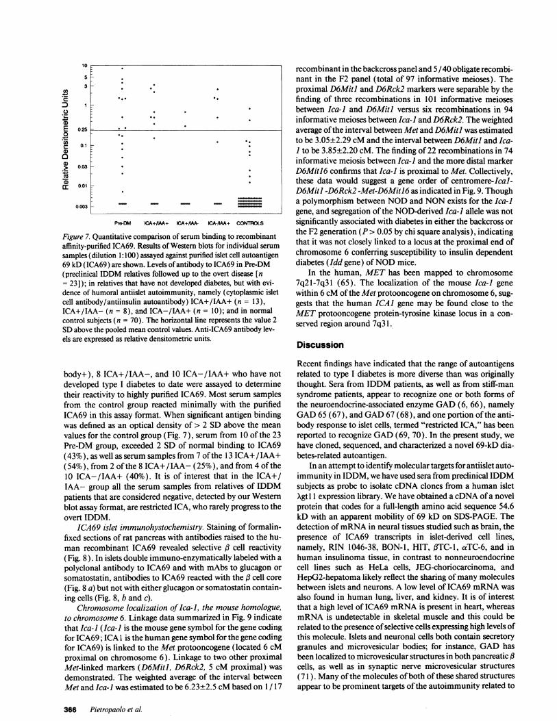
samples (dilution 1:100) assayed against purified islet cell autoantigen69 kD (ICA69) are shown. Levels of antibody to ICA69 in Pre-DM(preclinical IDDM relatives followed up to the overt disease [n= 23]); in relatives that have not developed diabetes, but with evi-dence of humoral antiislet autoimmunity, namely (cytoplasmic isletcell antibody/antiinsulin autoantibody) ICA+/IAA+ (n = 13),ICA+/IAA- (n = 8), and ICA-/IAA+ (n = 10); and in normalcontrol subjects (n = 70). The horizontal line represents the value 2SDabove the pooled mean control values. Anti-ICA69 antibody lev-els are expressed as relative densitometric units.
body+), 8 ICA+/IAA-, and 10 ICA-/IAA+ who have notdeveloped type I diabetes to date were assayed to determinetheir reactivity to highly purified ICA69. Most serum samplesfrom the control group reacted minimally with the purifiedICA69 in this assay format. When significant antigen bindingwas defined as an optical density of > 2 SD above the mean
values for the control group (Fig. 7), serum from 10 of the 23Pre-DM group, exceeded 2 SD of normal binding to ICA69(43%), as well as serum samples from 7 of the 13 ICA+/IAA+(54%), from 2 of the 8 ICA+/IAA- (25%), and from 4 of the10 ICA-/IAA+ (40%). It is of interest that in the ICA+/IAA- group all the serum samples from relatives of IDDMpatients that are considered negative, detected by our Westernblot assay format, are restricted ICA, who rarely progress to theovert IDDM.
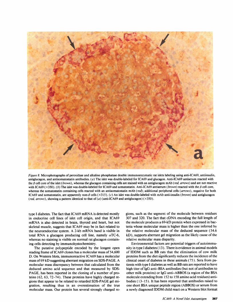
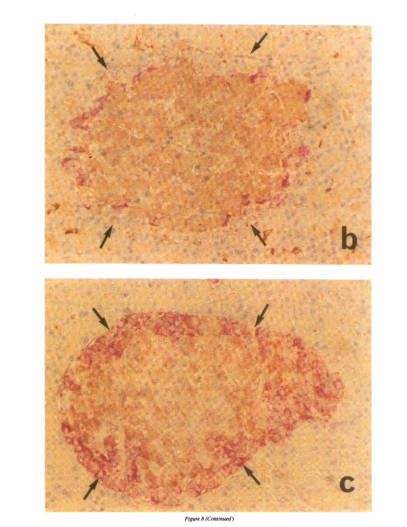
ICA69 islet immunohystochemistry. Staining of formalin-fixed sections of rat pancreas with antibodies raised to the hu-man recombinant ICA69 revealed selective cell reactivity(Fig. 8). In islets double immuno-enzymatically labeled with a
polyclonal antibody to ICA69 and with mAbs to glucagon or
somatostatin, antibodies to ICA69 reacted with the cell core
(Fig. 8 a) but not with either glucagon or somatostatin contain-ing cells (Fig. 8, b and c).
Chromosome localization of Ica-i, the mouse homologue,to chromosome 6. Linkage data summarized in Fig. 9 indicatethat Ica-I (Ica-I is the mouse gene symbol for the gene codingfor ICA69; ICA 1 is the human gene symbol for the gene codingfor ICA69) is linked to the Met protooncogene (located 6 cMproximal on chromosome 6). Linkage to two other proximalMet-linked markers (D6Miti, D6Rck2, 5 cM proximal) was
demonstrated. The weighted average of the interval betweenMet and Ica-i was estimated to be 6.23±2.5 cMbased on 1/17
recombinant in the backcross panel and 5 / 40 obligate recombi-nant in the F2 panel (total of 97 informative meioses). Theproximal D6MitJ and D6Rck2 markers were separable by thefinding of three recombinations in 101 informative meiosesbetween Ica-] and D6MitJ versus six recombinations in 94informative meioses between Ica-I and D6Rck2. The weightedaverage of the interval between Met and D6Mitl was estimatedto be 3.05±2.29 cM and the interval between D6Miti and Ica-I to be 3.85±2.20 cM. The finding of 22 recombinations in 74informative meiosis between Ica-] and the more distal markerD6Miti6 confirms that Ica-] is proximal to Met. Collectively,these data would suggest a gene order of centromere-Ical-D6Mitl -D6Rck2 -Met-D6MitJ6 as indicated in Fig. 9. Thougha polymorphism between NODand NONexists for the Ica-Igene, and segregation of the NOD-derived Ica-i allele was notsignificantly associated with diabetes in either the backcross orthe F2 generation (P > 0.05 by chi square analysis), indicatingthat it was not closely linked to a locus at the proximal end ofchromosome 6 conferring susceptibility to insulin dependentdiabetes (Idd gene) of NODmice.
In the human, METhas been mapped to chromosome7q2l-7q3 1 (65). The localization of the mouse Ica-] genewithin 6 cM of the Met protooncogene on chromosome 6, sug-gests that the human ICAI gene may be found close to theMETprotooncogene protein-tyrosine kinase locus in a con-served region around 7q3 1.
Discussion
Recent findings have indicated that the range of autoantigensrelated to type I diabetes is more diverse than was originallythought. Sera from IDDM patients, as well as from stiff-mansyndrome patients, appear to recognize one or both forms ofthe neuroendocrine-associated enzyme GAD(6, 66), namelyGAD65 (67), and GAD67 (68), and one portion of the anti-body response to islet cells, termed "restricted ICA," has beenreported to recognize GAD(69, 70). In the present study, wehave cloned, sequenced, and characterized a novel 69-kD dia-betes-related autoantigen.
In an attempt to identify molecular targets for antiislet auto-immunity in IDDM, we have used sera from preclinical IDDMsubjects as probe to isolate cDNA clones from a human isletXgtl 1 expression library. Wehave obtained a cDNAof a novelprotein that codes for a full-length amino acid sequence 54.6kD with an apparent mobility of 69 kD on SDS-PAGE. Thedetection of mRNAin neural tissues studied such as brain, thepresence of ICA69 transcripts in islet-derived cell lines,namely, RIN 1046-38, BON-1, HIT, f3TC-1, aTC-6, and inhuman insulinoma tissue, in contrast to nonneuroendocrinecell lines such as HeLa cells, JEG-choriocarcinoma, andHepG2-hepatoma likely reflect the sharing of many moleculesbetween islets and neurons. A low level of ICA69 mRNAwasalso found in human lung, liver, and kidney. It is of interestthat a high level of ICA69 mRNAis present in heart, whereasmRNAis undetectable in skeletal muscle and this could berelated to the presence of selective cells expressing high levels ofthis molecule. Islets and neuronal cells both contain secretorygranules and microvesicular bodies; for instance, GADhasbeen localized to microvesicular structures in both pancreatic (cells, as well as in synaptic nerve microvesicular structures(71 ). Many of the molecules of both of these shared structuresappear to be prominent targets of the autoimmunity related to
366 Pietropaolo et al.
10
5
3
0.25
0.1
0.03
0.01
Voc
._0
tr
E0
00
CU
0.003
Pre-DM KA+/IM+ CA+/IM-
0
0 00
. . 0 0 1
0 a 00
00 .
0
. I
6
0
I
j6p
aFigure 8. Microphotographs of peroxidase and alkaline phosphatase double immunoenzymatic rat islets labeling using anti-ICA69, antiinsulin,antiglucagon, and antisomatostatin antibodies. (a) The islet was double-labeled for ICA69 and glucagon. Anti-ICA69 antiserum reacted withthe f3 cell core of the islet (brown), whereas the glucagon containing cells are stained with an antiglucagon mAb(red, arrows) and are not reactivewith ICA69 (X350). (b) The islet was double-labeled for ICA69 and somatostatin. Anti-ICA69 antiserum (brown) reacted with the d3 cell core,whereas the somatostatin containing cells reacted with an antisomatostatin mAb (red); additional peripheral cells (arrows), negative for bothICA69 and somatostatin, are apparently non-g cells (X315). (c) An islet was double-labeled with mAbanti-insulin (brown) and antiglucagon(red, arrows), showing a pattern identical to that of (a) (anti-ICA69 and antiglucagon) (X350).
type I diabetes. The fact that ICA69 mRNAis detected mostlyin endocrine cell lines of islet cell origin, and that ICA69mRNAis also detected in brain, thyroid and heart, but notskeletal muscle, suggests that ICA69 may be in fact related tothe neuroendocrine system. A 2-kb mRNAband is visible intotal RNA a glucagon producing cell line, namely aTC-6,whereas no staining is visible on normal rat glucagon contain-ing cells detecting by immunohystochemistry.
The putative polypeptide encoded by the longest openreading frame of ICA69 clones has a molecular mass of 54,600D. On Western blots, immunoreactive ICA69 has a molecularmass of 69 kD suggesting aberrant migration on SDS-PAGE. Amolecular mass discrepancy between that calculated from thededuced amino acid sequence and that measured by SDS-PAGE, has been reported in the cloning of a number of pro-teins (62, 63, 72-74). These proteins have highly charged re-gions that appear to be related to retarded SDS-PAGEgel mi-gration, resulting thus in an overestimation of the truemolecular mass. Our protein has several strongly charged re-
gions, such as the segment of the molecule between residues307 and 320. The fact that cDNAencoding the full length ofthe molecule produces a 69-kD protein when expressed in bac-teria whose molecular mass is higher than the one inferred bythe relative molecular mass of the deduced sequence (54.6kD), supports aberrant gel migration as the likely cause of therelative molecular mass disparity.
Environmental factors are potential triggers of autoimmu-nity in type I diabetes ( 13). There is evidence in animal modelsof IDDM such as BB rats that the elimination of cow milkproteins from the diet significantly reduces the incidence of theclinical onset of diabetes in these animals (75). Sera from pa-tients with type I diabetes as well as BBrats are reported to havehigh titer of IgG anti-BSA antibodies (but not of antibodies toother milk proteins) or IgG anti-ABBOS (a region of the BSAmolecule extending from 152 to 158 amino acid residues) anti-bodies ( 13-15). It has been reported that antibodies raised toone short BSAunique peptide region (ABBOS) or serum froma newly diagnosed IDDMchild react on a Western blot format
ICA69: A Novel Islet Autoantigen 367
x
V4:.
A.
er.r
I~f
4k
/
.A~~~~~~~.
RI. N
a.J
LS:~~
Xi
4.k
Figure 8 (Continued)
t-i
-h'
4p
I
rv
fl- II a, %j
.6
A'V
:d` -.0iv;, " .:
i..;
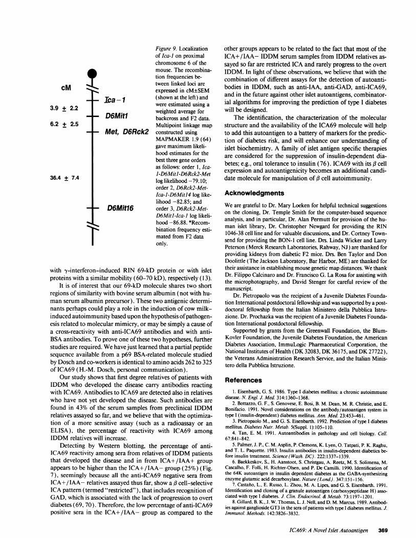
Figure 9. Localizationof Ica-] on proximalchromosome 6 of themouse. The recombina-tion frequencies be-tween linked loci areexpressed in cM±SEM(shown at the left) andwere estimated using aweighted average forbackcross and F2 data.Multipoint linkage mapconstructed usingMAPMAKER1.9 (64)gave maximum likeli-hood estimates for thebest three gene ordersas follows: order 1, Ica-I-D6MitJ-D6Rck2-Metlog likelihood -79.10;order 2, D6Rek2-Met-Ica-i-D6MitJ4 log like-lihood -82.85; andorder 3, D6Rck2-Met-D6Mitilca-l log likeli-hood -86.88. *Recom-bination frequency esti-mated from F2 dataonly.
with y-interferon-induced RIN 69-kD protein or with isletproteins with a similar mobility (60-70 kD), respectively (13).
It is of interest that our 69-kD molecule shares two shortregions of similarity with bovine serum albumin (not with hu-man serum albumin precursor). These two antigenic determi-nants perhaps could play a role in the induction of cow milk-induced autoimmunity based upon the hypothesis of pathogen-esis related to molecular mimicry, or may be simply a cause ofa cross-reactivity with anti-ICA69 antibodies and with anti-BSAantibodies. To prove one of these two hypotheses, furtherstudies are required. Wehave just learned that a partial peptidesequence available from a p69 BSA-related molecule studiedby Dosch and co-workers is identical to amino acids 262 to 325of ICA69 (H.-M. Dosch, personal communication).
Our study shows that first degree relatives of patients withIDDM who developed the disease carry antibodies reactingwith ICA69. Antibodies to ICA69 are detected also in relativeswho have not yet developed the disease. Such antibodies arefound in 43% of the serum samples from preclinical IDDMrelatives assayed so far, and we believe that with the optimiza-tion of a more sensitive assay (such as a radioassay or anELISA), the percentage of reactivity with ICA69 amongIDDM relatives will increase.
Detecting by Western blotting, the percentage of anti-ICA69 reactivity among sera from relatives of IDDM patientsthat developed the disease and in from ICA+/IAA+ groupappears to be higher than the ICA+/IAA- group (25%) (Fig.7), seemingly because all the anti-ICA69 negative sera fromICA+/IAA- relatives assayed thus far, show a /3 cell-selectiveICA pattern (termed "restricted"), that includes recognition ofGAD, which is associated with the lack of progression to overtdiabetes (69, 70). Therefore, the low percentage of anti-ICA69positive sera in the ICA+/IAA- group as compared to the
other groups appears to be related to the fact that most of theICA+/IAA- IDDM serum samples from IDDM relatives as-sayed so far are restricted ICA and rarely progress to the overtIDDM. In light of these observations, we believe that with thecombination of different assays for the detection of autoanti-bodies in IDDM, such as anti-IAA, anti-GAD, anti-ICA69,and in the future against other islet autoantigens, combinator-ial algorithms for improving the prediction of type I diabeteswill be designed.
The identification, the characterization of the molecularstructure and the availability of the ICA69 molecule will helpto add this autoantigen to a battery of markers for the predic-tion of diabetes risk, and will enhance our understanding ofislet biochemistry. A family of islet antigen specific therapiesare considered for the suppression of insulin-dependent dia-betes; e.g., oral tolerance to insulin (76). ICA69 with its fi cellexpression and autoantigenicity becomes an additional candi-date molecule for manipulation of f cell autoimmunity.
AcknowledgmentsWeare grateful to Dr. Mary Loeken for helpful technical suggestionson the cloning, Dr. Temple Smith for the computer-based sequenceanalysis, and in particular, Dr. Alan Permutt for provision of the hu-man islet library, Dr. Christopher Newgard for providing the RIN1046-38 cell line and for valuable discussions, and Dr. Cortney Town-send for providing the BON-l cell line. Drs. Linda Wicker and LarryPeterson (Merck Research Laboratories, Rahway, NJ) are thanked forproviding kidneys from diabetic F2 mice. Drs. Ben Taylor and DonDoolittle (The Jackson Laboratory, Bar Harbor, ME) are thanked fortheir assistance in establishing mouse genetic mapdistances. WethankDr. Filippo Calcinaro and Dr. Francisco G. La Rosa for assisting withthe microphotography, and David Stenger for careful review of themanuscript.
Dr. Pietropaolo was the recipient of a Juvenile Diabetes Founda-tion International postdoctoral fellowship and was supported by a post-doctoral fellowship from the Italian Ministero della Pubblica Istru-zione. Dr. Prochazka was the recipient of a Juvenile Diabetes Founda-tion International postdoctoral fellowship.
Supported by grants from the Greenwall Foundation, the Blum-Kovler Foundation, the Juvenile Diabetes Foundation, the AmericanDiabetes Association, ImmuLogic Pharmaceutical Corporation, theNational Institutes of Health (DK 32083, DK36175, and DK27722),the Veterans Administration Research Service, and the Italian Minis-tero della Pubblica Istruzione.
References1. Eisenbarth, G. S. 1986. Type I diabetes mellitus: a chronic autoimmune
disease. N. Engl. J. Med. 314:1360-1368.2. Bottazzo, G. F., S. Genovese, E. Bosi, B. M. Dean, M. R. Christie, and E.
Bonifacio. 1991. Novel considerations on the antibody/autoantigen system intype I (insulin-dependent) diabetes mellitus. Ann. Med. 23:453-461.
3. Pietropaolo M., and G. S. Eisenbarth. 1992. Prediction of type I diabetesmellitus. Diabetes Nutr. Metab. 5(Suppl. 1): 105-1 10.
4. Tan, E. M. 1991. Autoantibodies in pathology and cell biology. Cell.67:841-842.
5. Palmer, J. P., C. M. Asplin, P. Clemons, K. Lyen, 0. Tatpati, P. K. Raghu,and T. L. Paquette. 1983. Insulin antibodies in insulin-dependent diabetics be-fore insulin treatment. Science (Wash. DC). 222:1337-1339.
6. Baekkeskov, S., H. Aanstoot, S. Christgau, A. Reetz, M. S. Solimena, M.Cascalho, F. Folli, H. Richter-Olsen, and P. De Camilli. 1990. Identification ofthe 64K autoantigen in insulin dependent diabetes as the GABA-synthesizingenzyme glutamic acid decarboxylase. Nature (Lond.). 347:151-156.
7. Castano, L., E. Russo, L. Zhou, M. A. Lipes, and G. S. Eisenbarth. 1991.Identification and cloning of a granule autoantigen (carboxypeptidase H) asso-ciated with type I diabetes. J. Clin. Endocrinol. & Metab. 73:1197-1201.
8. Gillard, B. K., J. W. Thomas, L. J. Nell, and D. M. Marcus. 1989. Antibod-ies against ganglioside GT3 in the sera of patients with type I diabetes mellitus. J.Immunol. Methods. 142:3826-3832.
ICA69: A Novel Islet Autoantigen 369
cM'r :ca -1
4- D6Mit1
+I- Met, D6Rck2
3.9 + 2.2
6.2 + 2.5
36.4 + 7.4
D6Mit6
9. Dotta, F., P. G. Colman, D. Lombardi, D. W. Scharp, D. Andreani, G. M.Pontieri, U. Di Mario, L. Lenti, G. S. Eisenbarth, and R. C. Nayak. 1989. Ganglio-side expression in human pancreatic islets. Diabetes. 38:1478-1483.
10. Rabin, D. U., S. Pleasic, R. Palmer-Crocker, and J. A. Shapiro. 1992.Cloning and expression of IDDM-specific human autoantigens. Diabetes.41:183-186.
11. Roep, B. O., A. A. Kallan, W. L. W. Hazenbos, G. J. Bruining, E. M.Bailyes, S. Arden, J. C. Hutton, and R. R. P. DeVries. 1991. T-cell reactivity to 38kD insulin-secretory-granule protein in patients with recent-onset type 1 diabetes.Lancet. 337:1439-1441.
12. Karounos, D. G., and J. W. Thomas. 1990. Recognition of common isletantigen by autoantibodies from NODmice and humans with IDDM. Diabetes.39: 1085-1090.
13. Martin, J. M., B. Trink, D. Daneman, H.-M. Dosch, and B. Robinson.1991. Milk proteins in the etiology of insulin-dependent diabetes mellitus(IDDM). Ann. Med. 23:447-452.
14. Kadjalainen, J., J. M. Martin, M. Knip, J. Ilonen, B. H. Robinson, E.Savilahti, H. K. Akerblom, and H.-M. Dosch. 1992. A bovine albumin peptide asa possible trigger of insulin-dependent diabetes mellitus. N. Engl. J. Med.327:302-307.
15. Kardalainen, J., T. Saukkonen, E. Savilahti, and H.-M. Dosch. 1992.Disease-associated anti-bovine serum albumin antibodies in type 1 (insulin-de-pendent) diabetes mellitus are detected by particle concentration fluoroimmu-noassay and not by enzyme linked immunoassay. Diabetologia. 35:985-990.
16. Coppel, R. L., M. E. Gershwin, and A. D. Sturgess. 1989. Cloned autoanti-gens in the study and diagnosis of autoimmune diseases. Mol. Biol. Med. 6:27-34.
17. Dropcho, E. J., Y.-T. Chen, J. B. Posner, and L. J. Old. 1987. Cloning of abrain protein identified by autoantibodies from a patient with a paraneoplasticcerebellar degeneration. Proc. Nail. Acad. Sci. USA. 84:4552-4556.
18. Sambrook, J., E. F. Fritsch, and T. Maniatis. 1989. Molecular Cloning: ALaboratory Manual. Cold Spring Harbor Laboratory Press, Cold Spring Harbor,NY.
19. Huynh, T. V., R. A. Young, and R. W. Davis. 1985. In Constructing andScreening cDNA libraries XgtlO and Xgtl 1. D. M. Glover, editor. JRL Press,Oxford. 49-78.
20. Young, R. A., and R. W. Davis. 1984. Yeast RNApolymerase II genes:isolation with antibody probes. Science (Wash. DC). 222:778-782.
21. Feinberg, A. P., and B. Vogelstein. 1983. A technique for radiolabelingDNArestriction endonuclease fragments to high specific activity. Anal. Biochem.132:6-13.
22. Wallace, R. B., M. J. Johnson, T. Hirose, T. Miyake, E. H. Kawashima,and K. Itakura. 1981. The use of synthetic oligonucleotides as hybridizationprobes. Hybridization of oligonucleotides of mixed sequence to rabbit fl-globinDNA. Nucleic Acids Res. 9:879-894.
23. Friedman, K. D., N. L. Rosen, P. J. Newman, and R. R. Montgomery.1988. Enzymatic amplification of specific cDNA from Xgtl 1 libraries. NucleicAcids Res. 16:8718.
24. Innis, M., D. Gelfand, J. Sninsky, and T. White. 1990. PCRProtocols: aGuide to Methods and Applications. Academic Press, Inc., San Diego, CA253-258.
25. Sanger, F., S. Nickel, and A. R. Consoln. 1977. DNAsequencing withchain terminating inhibitors. Proc. Natl. Acad. Sci. USA. 74:5463-5467.
26. Tabor, S., and C. C. Richardson. 1975. DNAsequence analysis with amodified bacteriophage T7 DNA polymerase. Proc. Natl. Acad. Sci. USA.84:4767-4771.
27. Kyte, J., and R. F. Doolittle. 1982. A simple method for displaying hydro-phobic character of a protein. J. Mol. Biol. 157:105-132.
28. Doolittle, R. F. 1987. Of URSFand ORFS. A Primer on Howto AnalyzeDerived Amino Acid Sequences. University Sciences Books, Mill Valley, CA.
29. Klein, P., M. Kanehisa, and C. DeLisi. 1985. The detection and classifica-tion of membrane-spanning proteins. Biochim. Biophys. Acta. 815:468-476.
30. Clark, S. A., B. Burnham, and W. L. Chick. 1990. Modulation of glucose-induced insulin secretion from a rat clonal beta-cell line. Endocrinology. 127,6:2779-2788.
31. Hamaguchi, K., and E. H. Leiter. 1990. Comparison of cytokine effects onmouse pancreatic alpha cell and beta cell lines. Viability, secretory function, andMHCantigen expression. Diabetes. 39:415-425.
32. Powers, A. C., S. Efrat, S. Mojsov, D. Spector, J. F. Habener, and D.Hanahan. 1990. Proglucagon processing similar to normal islets in pancreaticalpha-like cell line derived from transgenic mouse tumor. Diabetes. 39:406-414.
33. Breant, B., C. Lavergne, and G. Rosselin. 1990. Cell cycle and gene expres-sion in the insulin producing pancreatic cell line #TC-1. Diabetologia. 33:586-592.
34. Purrello, F., M. Buscema, M. Vetri, C. Vinci, C. Gatta, F. Forte, A. M.Rabuazzo, and R. Vigneri. 1991. Glucose regulates both glucose transport andthe glucose transporter gene expression in a hamster-derived pancreatic beta-cellline (HIT). Diabetologia. 21:366-369.
35. Contreras, G., D. F. Summers, J. Maizel, and E. Ehrenfeld. 1973. HeLacell nucleolar RNAsynthesis after poliovirus infection. Virology. 53:120-129.
36. Kohler, P. O., W. E. Bridson, J. M. Hammond, B. Weintraub, M. A.
Kirschner, and D. M. Van Thiel. 1971. Clonal lines of human choriocarcinomacells in culture. Acta Endocrinol. 153(Suppl.):137-150.
37. Broze, G. J., and J. P. Miletich. 1987. Isolation of the tissue factor inhibi-tor produced by HepG2 hepatoma cells. Proc. Nall. Acad. Sci. USA. 84:1886-1890.
38. Thomas, P. S. 1980. Hybridization of denatured RNAand small DNAfragments transferred to nitrocellulose. Proc. Natl. Acad. Sci. USA. 77:5201-5205.
39. Cleveland, D. W., M. A. Lopata, R. J. MacDonald, N. J. Cowan, W. J.Rutter, and M. W. Kirschner. 1980. Number and evolutionary conservation ofalpha- and beta-tubulin and cytoplasmic beta- and gamma-actin genes usingspecific cloned cDNA probes. Cell. 20:95-105.
40. Pari, G., K. Jardine, and M. W. McBurney. 1991. Multiple CArG boxes inthe human cardiac actine gene promoter required for expression in embryoniccardiac muscle cells developing in vitro from embryonal carcinoma cells. Mol.Cell. Biol. 11:4796-4803.
41. Hassouna, N., B. Michot, and J.-P. Bachellerie. 1984. The complete nu-cleotide sequence of mouse 28 S RNAgene. Implications for the process of sizeincrease of the large subunit rRNA in higher eukaryotes. Nucleic Acids Res.12:3563.
42. Raynal, F., B. Michot, and J.-P. Bachellerie. 1984. Complete nucleotidesequence of mouse 18 S rRNA gene: comparisons with other available homologs.FEBS (Fed. Eur. Biochem. Soc.) Lett. 167:263.
43. Van Regenmortel, M. H. V., J. P. Briand, S. Muller, and S. Plaue. 1988.Syntetic polypeptides antigens. In Laboratory Techniques in Biochemistry andMolecular Biology. R. H. Burdon and P. H. von Knippenberg, editors. ElsevierScience Publishing Co., Inc., New York.
44. Walter, G. 1986. Production and use of antibodies against synthetic pep-tides. J. Immunol. Methods. 88:149-161.
45. Kurstak, E. 1986. Enzyme Immunodiagnostic. Academic Press, SanDiego, CA.
46. Jitsukawa, T., S. Nakajima, I. Sugawara, and H. Watanabe. 1989. In-creased coating efficiency of antigens and preservation of original antigenic struc-ture after coating in ELISA. J. Immunol. Methods. 116:251-257.
47. Sun, X. J., P. Rothenberg, C. R. Kahn, J. M. Backer, E. Araki, P. A.Wilden, D. A. Cahill, B. J. Goldstein, and M. F. White. 1991. Structure of theinsulin receptor substrate IRS-1 defines a unique signal transduction protein.Nature (Lond.). 352:73-77.
48. Laemmli, U. K. 1970. Cleavage of structural proteins during the assemblyof the head of bacteriophage T4. Nature (Lond.). 227:680-685.
49. Towbin, H., T. Staehelin, and J. Gordon. 1979. Electrophoretic transfer ofproteins from polyacrylamide gels to nitrocellulose sheets: procedure and someapplications. Proc. Natl. Acad. Sci. USA. 76:4350.
50. Maina, C. V., P. D. Riggs, A. G. Grandea III, B. E. Slatko, L. S. Moran,J. A. Tagliamonte, L. A. McReynolds, and C. di Guan. 1988. An Escherichia colivector to express and purify foreign proteins by fusion to and separation frommaltose-binding protein. Gene (Amst.). 74:365-373.
51. di Guan, C., P. Li, P. D. Riggs, and H. Inouye. 1988. Vectors that facilitatethe expression and purification of foreign peptides in Escherichia coli by fusion tomaltose-binding protein. Gene (Amst.). 67:21-20.
52. Johnston, T. C., R. B. Thompson, and T. 0. Baldman. 1986. Nucleotidesequence of the luxB gene of vibrio harveyi and the complete amino acid se-quence of the beta subunit of bacterial luciferase. J. Biol. Chem. 261:4805-481 1.
53. Aman, E. B., B. Oclis, and K. J. Abel. 1988. Tightly regulated tac pro-moter useful for the expression of unfused and fused proteins in Escherichia coli.Gene (Amst.). 69:301-315.
54. Mason, D., and Y. R. Sammons. 1978. Alkaline phosphatase and peroxi-dase for double immunoenzimatic labelling of cellular constituents. J. Clin.Pathol. 31:454.
55. Mason, D. Y., B. F. Abdulaziz, and H. Stein. 1983. Double immunoenzi-matic labelling. In Immunocytochemestry. Practical Application in Pathologyand Biology. J. M. Polak and S. Van Noorden, editors. Wright PSG, Boston.
56. Prochazka, M., D. V. Serreze, S. M. Worthen, and E. H. Leiter. 1989.Genetic control of diabetogenesis in NOD/Lt mice: development and analysis ofcongenic stocks. Diabetes. 38:1446-1455.
57. Prochazka, M., E. H. Leiter, D. V. Serreze, and D. L. Coleman. 1987.Three recessive loci required for insulin-dependent diabetes in NODmice.Science (Wash. DC) . 237:286-289.
58. Leiter, E. H., and D. V. Serreze 1992. Antigen presenting cells and theimmunogenetics of autoimmune diabetes in NODmice. Reg. Immunol. 4:263-273.
59. Dietrich, W., H. Katz, S. Lincoln, H.-S. Shin, J. Friedman, N. Dracopoli,and E. S. Lander. 1992. A genetic mapof the mouse suitable fortyping intraspeci-fic crosses. Genetics. 131:423447.
60. Green, E. L. 1985. Tables and a computer program for analyzing linkagedata. Mouse News Let. 73:20-21.
61. Kozak, M. 1987. An analysis of 5'-noncoding sequences from 699 verte-brate messenger RNAs. Nucleic Acids Res. 20:8125-8132.
62. Krumar, K. N., N. Tilakaratne, P. S. Johnson, A. E. Allen, and E. K.
370 Pietropaolo et al.
Michaelis. 1991. Cloning of cDNA for the glutamate-binding subunit of theNMDAreceptor complex. Nature (Lond.). 354:70-73.
63. Krumar, K. N., K. T. Eggman, J. L. Adams, and E. K. Michaelis. 1991.Hydrodynamic proterties of the purified glutamate-binding protein subunit ofthe N-methyl-D-aspartate receptor. J. Bio. Chem. 23:14947-14952.
64. Lander, E. S., P. Green, J. Abrahamson, A. Barlow, M. J., Daly, S. E. Daly,and L. Newberg. 1987. MAPMAKER:an interactive computer package for con-structing primary genetic linkage maps for experimental and natural populations.Genomics. 1:174-181.
65. Dean, M., M. Park, M. M. LeBeau, T. S. Robins, M. 0. Diaz, J. D.Rowley, D. G. Blair, and G. F. Vande Woude. 1985. The human met oncogene isrelated to the tyrosine kinase oncogenes. Nature (Lond.). 318:385-388.
66. Thivolet, C. H., M. Tappaz, A. Durand, J. Petersen, A. Stefanutti, P.Chaelain, B. Bialettes, W. Scherbaum, and J. Orgiazzi. 1992. Glutamic acid decar-boxylase (GAD) autoantibodies are additional predictive markers of type I (in-sulin-dependent) diabetes mellitus in high risk individuals. Diabetologia.35:570-576.
67. Hagopian W. A., B. Michelsen, A. E. Karlsen, F. Larsen, A. Moody, C. E.Grubin, R. Rowe, J. Petersen, R. McEvoy, and A. Lernmark. 1993. Autoantibod-ies in IDDM primarily recognize the 65,000-M, rather than 67,000-M, isoform ofglutamic acid decarboxylase. Diabetes. 42:631-636.
68. Kaufman, D. L., M. G. Erlander, M. Clare-Salzer, M. A. Atkinson, N. K.Maclaren, and A. J. Tobin. 1992. Autoimmunity to two forms of glutamatedecarboxylase in insulin-dependent diabetes mellitus. J. Clin. Invest. 89:283-292.
69. Gianani, R., A. Pugliese, S. Bonner-Weir, A. J. Shiffrin, J. S. Soeldner, H.Erlich, Z. L. Awdeh, C., A. Alper, R., A. Jackson, and G. S. Eisenbarth. 1992.
Prognostically significant heterogeneity of cytoplasmic islet cell antibodies inrelatives of patients with type I diabetes. Diabetes. 41:347-353.
70. Genovese, S., E. Bonifacio, J. M. McNally, B. M. Dean, R. Wagner, E.Bosi, E. A. M. Gale, and G. F. Bottazzo. 1992. Distinct cytoplasmic islet cellantibodies with different risks for type I (insulin-dependent) diabetes mellitus.Diabetologia. 35:385-388.
71. Christgau, S., H. J. Aanstoot, H. Schierbeck, K. Begley, T. Tullin, K.Hejnaes, and S. Baekkeskov. 1992. Membrane anchoring of the autoantigenGAD65to microvesicles in pancreatic beta-cells by palmitoylation in the NH2-ter-minal domain. J. Cell Biol. 11 8(2):309-320.
72. McCauliffe, D. P., F. A. Lux, T.-S. Lieu, I. Sanz, J. Hanke, M. M. New-kirk, L. L. Bachinski, Y. Itoh, M. J. Siciliano, M. Reichlin, R. D. Sontheimer, andJ. D. Capra. 1990. Molecular cloning, expression, and chromosome 19 localiza-tion of a human Ro/SS-A autoantigen. J. Clin. Invest. 85:1379-1391.
73. Benedum, U. M., D. S. Baeuerle, D. S. Konecki, R. Frank, J. Powell, J.Mallet, and W. B. Huttner. 1986. The primary structure of bovine chromograninA: a representative of a class of acidic secretory proteins commonto a variety ofpeptidergic cells. EMBO(Eur. Mol. Biol. Organ.) J. 5:1495-1502.
74. Spritz, R. A., K. Strunk, C. S. Surowy, S. 0. Hoch, D. E. Barton, and U.Francke. 1987. The human U1-70K snRNP protein: cDNA cloning, chromo-somal localization, expression, alternative splicing and RNA-binding. NucleicAcids Res. 15:10373-10391.
75. Elliot, R. B., and J. M. Martin. Dietary protein: a trigger of insulin-depen-dent diabetes in rat? 1984. Diabetologia. 26:297-299.
76. Zhang, Z. J., L. Davidson, G. S. Eisenbarth, and H. L. Weiner. 1991.Suppression of diabetes in NODmice by oral administration of porcine insulin.Proc. Natl. Acad. Sci. USA. 88:10252-10256.
ICA69: A Novel Islet Autoantigen 371