Embed Size (px)
Citation preview
ISCHEMIC HAERT DISEASES
BYDR\ RAMY A. SAMY
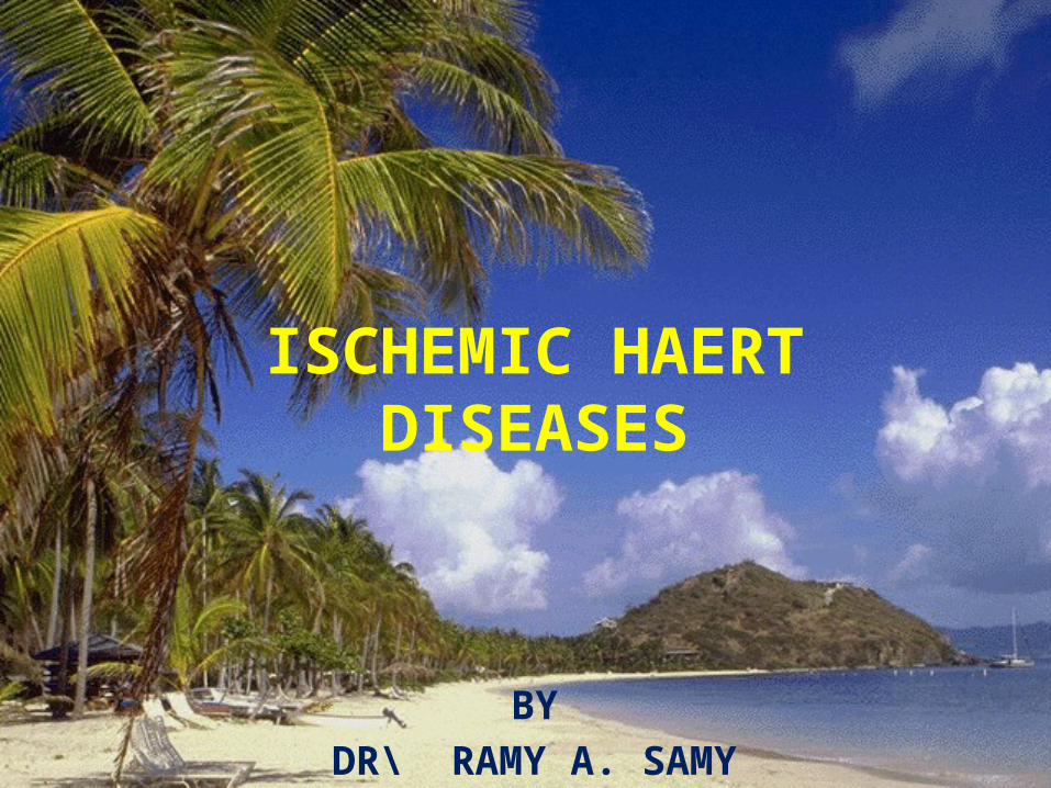
Ischemic Heart Disease Hypoxemia (diminished transport of oxygen by
the blood) less deleterious than ischemia Also called coronary artery disease (CAD) or
coronary heart disease IHD =Syndromes
◦ late manifestations of coronary atherosclerosis Cause => 90% of cases, coronary
atherosclerotic arterial obstruction

Ischemic Heart Disease Classification = mainly 4 types
◦ Myocardial infarction (MI)◦ Sudden cardiac death◦ Angina pectoris◦ Chronic IHD with heart failure
Acute Coronary syndromes◦ important predisposing factor -Plaque disruption or
Acute plaque change Acute myocardial infarction Unstable angina Sudden cardiac death
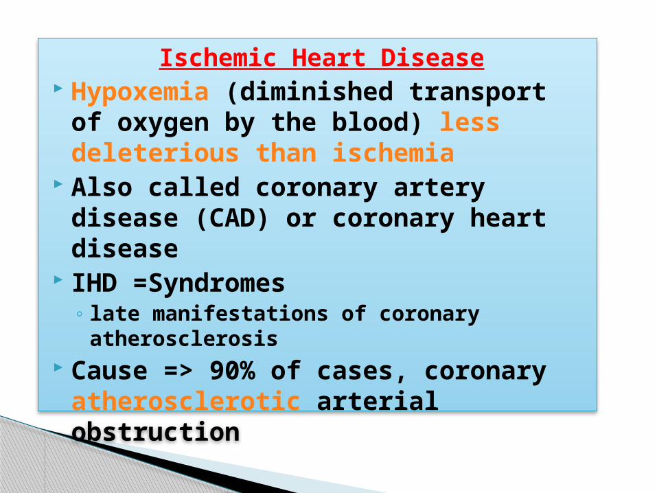
Ischemic Heart Disease 75% stenosis = symptomatic ischemia induced by
exercise 90% stenosis = symptomatic even at rest Pathogenesis
◦ ↓ coronary perfusion relative to myocardial demand◦ Role of Acute Plaque Change
(Erosion/ulceration, Hemorrhage into the atheroma, Rupture/fissuring, Thrombosis)
◦ Role of Inflammation T cell, Macrophages (MMPs), CRP
◦ Role of Coronary Thrombus The most dreaded complication
◦ Role of Vasoconstriction (VC) Platelet & Endothelial factors, VC substances
Results when there is an imbalance between myocardial oxygen supply and demand
Most occurs because of atherosclerotic plaque with in one or more coronary arteries
Limits normal rise in coronary blood flow in response to increase in myocardial oxygen demand
Myocardial Ischemia
Imbalance between Myocardial oxygen supply and demand = Myocardial hypoxia and accumulation of waste metabolites due to atherosclerotic disease of coronary arteries
Ischemic Heart Disease

Angina Pectoris: uncomfortable sensation in the chest or neighboring anatomic structures produced by myocardial ischemia
Angina Pectoris
Stable Angina: chronic pattern of transient angina pectoris precipitated by physical activity or emotional upset, relieved by rest with in few minutes
Temporary depression of ST segment with no permanent myocardial damage
Stable Angina
Typical anginal discomfort usually at rest Develops due to coronary artery spasm
rather than increase myocardial oxygen demand
Transient shifts of ST segment – ST elevation
Variant Angina
Increased frequency and duration of Angina episodes, produced by less exertion or at rest = high frequency of myocardial infarction if not treated
Unstable Angina
Asymptomatic episodes of myocardial ischemia
Detected by electrocardiogram and laboratory studies
Silent Ischemia
Arcus senilis, xanthomas, funduscopic exam: AV nicking, exudates
Signs and symptoms: hyperthyroidism with increased myocardial oxygen demand, hypertension, palpitations
Auscultate carotid and peripheral arteries and abdomen: aortic aneurysm
Cardiac: S4 common in CAD, increased heart rate, increased blood pressure
Physical Examination
Myocardial ischemia may result in papillary muscle regurgitation
Ischemic induced left ventricular wall motion abnormalities may be detected as an abnormal precordial bulge on chest palpation
A transient S3 gallop and pulmonary rales = ischemic induced left ventricular dysfunction
Ischemia
Blood tests include serum lipids, fasting blood sugar, Hematocrit, thyroid (anemias and hyperthyroidism can exacerbate myocardial ischemia
Resting Electrocardiogram: CAD patients have normal baseline ECGs◦ pathologic Q waves = previous infarction◦ minor ST and T waves abnormalities not specific
for CAD
Diagnostic Tests
Electrocardiogram: is useful in diagnosis during cc: chest pain
When ischemia results in transient horizontal or downsloping ST segments or T wave inversions which normalize after pain resolution
ST elevation suggest severe transmural ischemia or coronary artery spasm which is less often
Electrocardiogram

Used to confirm diagnosis of angina Terminate if hypotension, high grade
ventricular disrrhythmias, 3 mm ST segment depression develop
(+): reproduction of chest pain, ST depression
Severe: chest pain, ST changes in 1st 3 minutes, >3 mm ST depression, persistent > 5 minutes after exercise stopped
Low systolic BP, multifocal ventricular ectopy or V- tach, ST changes, poor duration of exercise (<2 minutes) due to cardiopulmonary limitations
Exercise Stress Test
Radionuclide studies Exercise radionuclide ventriculography Echocardiography Ambulatory ECG monitoring Coronary arteriography
Other Diagnostic Tests
Prevent complications – myocardial infarction, and to prolong life
No smoking, lower weight, control hypertension and diabetes
Patients with CAD – LDL cholesterol should achieve lower levels (<100)
HMG-COA reductase inhibitors are effective
Management Goals to reduce Anginal Symptoms
Therapy is aimed in restoring balance between myocardial oxygen supply and demand
Useful Agents: nitrates, beta-blockers and calcium channel blockers
Pharmacologic Therapy
Reduce myocardial oxygen demand Relax vascular smooth muscle Reduces venous return to heart Arteriolar dilators decrease resistance
against- which left ventricle contracts and reduces wall tension and oxygen demand
Nitrates
Dilate coronary arteries with augmentation of coronary blood flow
Side effects: generalized warmth, transient throbbing headache, or lightheadedness, hypotension
ER if no relief after X2 nitro's: unstable angina or MI
Nitrates: cont

Drug tolerance Continued administration of drug will
decrease effectiveness Prevented by allowing 8 – 10 hours nitrate
free interval each day. Elderly/inactive patients: long acting
nitrates for chronic antianginal therapy is recommended
Physical active patients: additional drugs are required
Problems with Nitrates
Prevent effort induced angina Decrease mortality after myocardial
infarction Reduce Myocardial oxygen demand by
slowing heart rate, force of ventricular contraction and decrease blood pressure
Beta Blockers

Block myocardial receptors with less effect on bronchial and vascular smooth muscle- patients with asthma, intermittent claudication
Beta -1
With partial B-agonist activity: Intrinsic sympathomimetic activity (ISA)
have mild direct stimulation of the beta receptor while blocking receptor against circulating catecholamines
Agents with ISA are less desirable in patients with angina because higher heart rates during their use may exacerbate angina
not reduce mortality after AMI
Beta-Agonist blockers
Short acting administered intravenously Can be used to test tolerability of beta-
blockage Used for tachydysrhythmias and unstable
angina Primary prevention trials: beta blockers
decrease incidence of first MIs with hypertensive patients
Esmolol
Symptomatic CHF, history of bronchospasm, bradycardia or AV block, peripheral vascular disease with s/s of claudication
Contraindications

Bronchospasm (RAD), CHF, depression, sexual dysfunction, AV block, exacerbation of claudication, potential masking of hypoglycemia in IDDM patients
Side Effects
Tachycardia, angina or MI Inhibit vasodilatory beta 2 receptors Should be avoided in patients with
predominant coronary artery vasospasm
Abrupt Cessation
Serum lipids: decrease of HDL cholesterol and increased triglycerides
Effects do not occur with beta-blockers with B-agonist activity or alpha-blocking properties
Beta-Blockers: Long Term effects
Anti-anginal agents prevent angina Helpful: episodes of coronary vasospasm Decreases myocardial oxygen requirements
and increase myocardial oxygen supply Potent arterial vasodilators: decrease
systemic vascular resistance, blood pressure, left ventricular wall stress with decrease myocardial oxygen consumption
Calcium Channel Blockers
Fall in blood pressure, trigger increase heart rate
Undesirable effect associated with increased frequency of myocardial infarction and mortality
Nifedipine and other dihydropyridine calcium channel blockers
Secondary agents in management of stable angina
Are prescribed only after beta blockers and nitrate therapy has been considered
Potential to adversely decrease left ventricular contractility
Used cautiously in patients with left ventricular dysfunction
Calcium Channel Blockers

Are newer CCB Decrease (-) inotropic effects Amlodipine is tolerated in patients with
advanced heart failure without causing increase mortality when added with ace inhibitor, diuretic, and digoxin
amlodipine and felodipine

Undergoes oxidation in proximity of arterial wall = prone to atherosclerotic process
Vitamin E 200 – 400 IU daily may lower coronary death rates
LDL
Chronic Stable Angina: beta blocker and long acting nitrate or calcium channel blocker (not verapamil: bradycardia) or both.
If contraindication to BB a CCB is recommended (bronchospasm, IDDM, or claudication) any of CCB approved for angina are appropriate.
Drug Selection
Verapamil and Cardizem is preferred because of effect on slowing heart rate
Patients with resting bradycardia or AV block, a dihydropyridine calcium blocker is better choice
Patients with CHF: nitrates preferred amlodipine should be added if additional therapy is needed
Drugs
Primary coronary vasospasm: no treatment with beta blockers, it could increase coronary constriction
Nitrates and CCB are preferred Concomitant hypertension: BB or CCB are
useful in treatment Ischemic Heart Disease & Atrial Fibrillation:
treatment with BB, verapamil or Cardizem can slow ventricular rate
Drugs
If patients do not respond to initial antianginal therapy – a drug dosage increase is recommended unless side effects occur.
Combination therapy: successful use of lower dosages of each agent while minimizing individual drug side effects
Combination Therapy
Nitrate and beta blocker Nitrate and verapamil or cardizem for
similar reasons Long acting dihydropyridine calcium
channel blocker and beta blocker A dihydropyridine and nitrate is often
not tolerated without concomitant beta blockade because of marked vasodilatation with resultant head ache and increased heart rate
Combination Therapy Include:
Beta blockers should be combined only very cautiously with verapamil or cardizem because of potential of excessive bradycardia or CHF in patients with left ventricular dysfunction
Combinations
Patients with 1 – 2 vessel disease with normal left ventricular function are referred for catheter based procedures
Patients with 2 and 3 vessel disease with widespread ischemia, left ventricular dysfunction or DM and those with lesions are not amendable to catherization based procedures and are referred for CABG
Other methods

THANK YOU