Embed Size (px)
Citation preview

April, 1996
NEXT MEETING:
GUEST SPEAKER:
DATE:
TIME:
LOCATION:
i s C A M l l N e w s l e t t e r Vol. 14, N0.12
Eumida and related genera
Danny Eibye-Jacobsen
May 13 -14, 1996
9:30am-3:30pm
Worm Lab, Natural History Museum of Los Angeles County 900 Exposition Blvd., Los Angeles, CA
MAY 13 - 14 MEETING
The May meeting will be held over two days at the Worm Lab of the Natural History Museum and hosted by Dr. Danny Eibye-Jacobsen from the Zoological Museum, University of Copenhagen. The meeting will be a discussion of phyllodocid polychaetes, especially Eumida species. Members should bring any problem specimens for examination by Danny along with any questions on this group of polychaetes.
Eumida longicornuta (from Eibye-Jacobsen 1991)
FUNDS FOR THIS PUBUCATION PROVIDED, IN PART, BY THE ARCO FOUNDATION, CHEVRON USA, AND TEXACO INC.
SCAMIT Newsletter is not deemed to be a valid publication for formal taxonomic purposes.

February, 1996 SCAMIT Newsletter Vol. 14, No. 10
MEETINGS, MEETINGS, MEETINGS ANOTHER INTRODUCED SPECIES
There are several important meetings that may be of interest to members that will be occurring in the next few months.
May 3 - 4 So. Calif. Academy of Sciences at Loyola Marymount University. One of the Friday symposia is on regional marine monitoring in the southern California Bight.
June 23 - 27 Western Society of Malacologists in San Diego at the Handlery Hotel and Country Club. Refer to March newsletter Vol. 14(11) for details.
July 14 -18 The Crustacean Society Meeting and the 3rd International Large Branch-iopod Symposium (held jointly) at the University of San Diego. Refer to Feb. newsletter Vol. 14(10) for details.
August 5-9 9th International Echinoderm Conference, San Francisco. Seven Hills Conference Center, San Francisco State University. Contact Rich Mooi at the California Academy of Sciences @ (415) 750-7086 or [email protected] for further information.
At the April meeting John Ljubenkov (MEC) informed members of an introduced species of anemone that has large stinging cells on its tentacles and lives on eelgrass. This anemone has been recently reported in Mission Bay by scientific divers. It is an apparently undescribed species in the genus Bunodeopsis which also has been taken in the Gulf of California according to John.
The City of San Diego has also recently seen another species of heart urchin Nacospatangus depressus in their trawls, which was previously unreported in local monitoring samples. This species was listed and illustrated in Charwat and Word 1975 as Gonimaretia laevis. They indicated it had been taken at 18m depths off San Clemente Island. It is more oval in shape than Lovenia cordiformis, but generally resembles that species although it lacks both long dorsal spines and an anterior ambulacrum. It is actually more closely related to Spatangus catifornicus and is in the family Spatangidae, not the Loveniidae. Both Nacospatangus and Lovenia were noted as occurring in the same trawl off Pt. Loma, so we should all critically examine our "Lovenia" to be sure they don't hide a few Nacospatangus as well.
"SCAM IT" NEWSLETTER
The April 1st edition, Volume 14(13) seems to have been well received by the membership. Those of you who let your newsletter pile up before reading it or don't read it all the way thru missed out on a good chuckle last month. The newsletter staff is planning another edition for next year and will certainly welcome articles from other "twits" and "fools". Please submit articles as soon as Thalia (the Muse of Comedy -for the classically impaired) smites you with the whoopie cushion of inspiration. All submissions are welcome, but, with a year to prepare, we will attempt more subtlety in Volume 15(13).
According to Maluf (1988), the synonymy of Gonimaretia laevis with Nacospatangus depressus is provisional, and may be reversed once a definitive study of these animals is done. She lists it as having a depth range of 5-302m. The record from San Clemente Island represents the recorded northern range limit for the species, which ranges south to Isla Espiritu Santu.
BUBBLE SHELL NAMES
In Edition 2 of the SCAMIT Taxonomic Listing bubbleshells of the genus Haminaea were indicated as being in the family Atyidae. This placement is no longer accurate. For some time
2

February, 1996 SCAMIT Newsletter Vol. 14, No. 10
a problem in nomenclature at the family level has been noticed by some workers. Dr. Myra Keen, in a letter to Jim McLean in the early 70's pointed out the problem, and suggested that the solution rested with the ICZN.
The problem was one of homonymy at the family level. In Mollusca there is a family Atyidae based on the type genus Atys de Montfort 1810. In Crustacea there is a family Atyidae based on the type genus Atya Leach, 1816. This later taxon was a replacement name for Atys Leach, 1815 [in Crustacea] non Atys de Montfort 1810 [in Mollusca]. In either case the family name derived from the generic name is the same, Atyidae. The family name was introduced in Crustacea by De Haan 1849, while it did not find usage in Mollusca until Thiele 1926. The next available family name is Haminaidae Pilsbry 1895. This name has been variously spelled as Haminaidae, Haminoeidae, and Haminaeidae based on the transliteration of the diphthong ae. There is currently before the ICZN a proposed use of the plenary powers for conservation of the name Haminaea Leach 1820, and for fixation of the correct spelling of the family name as Haminaeidae (Giannuzzi-Savelli & Gentry 1990). While the commission has not as yet formally acted, the evidence that the proposed approach is the correct one is persuasive (at least to your editor). Accordingly, the next edition of the Taxonomic Listing will replace the family Atyidae in mollusks with the family Haminaeidae [the generic change from Haminoea to Haminaea has already been made]. - Don Cadien
NEW LITERATURE
Volume 9 of the MMS Taxonomic Atlas, The Mollusca, Part 2, Gastropoda with sections by Jim McLean and Terry Gosliner is published and subscribers should be receiving it soon.
A Listing of Living Mollusca by Yoshihiro Goto and Guido T. Poppe has been recently published. It is in two parts and four volumes and includes
41,861 species. Its cost is $US 185 plus postage for all four volumes. The weight of all 4 volumes together is almost 10 kg. Orders for this set may be sent to:
Mostra Mondiale Malacologia Via Adriatica Nord, 240 63012 Cupra Marittima (AP - Italy) tel: 39(0)735 777550 fax: 39 (0) 735 777232
Also, recendy published by Y. Goto and P. Anseeuw is The Living Pleurotomariidae. This is a comprehensive synopsis of recent Pleurotomariidae which includes 24 species and 2 subspecies. It not only includes many text illustrations, color plates and distribution maps, but SEM images of radula as well. It costs $US 210 and it is hardbound with a cloth cover and slipcase.
It may be ordered from:
Naturama C.P. 28 - Succ.26 90146 Palermo Italy e-mail: [email protected] fax:+(91) 6713568
Payment should be made with your order by international postal money order or with a VISA card (for a 5% charge) by fax not e-mail for obvious reasons.
Also from the same publisher. Seashells of Eastern Arabia by D. Bosch, P. Dance, R. Moolenbeek and G. Oliver. It is hardbound and includes more than 1000 species from Oman and the Arabian Gulf illustrated with color plates and SEM images. It costs $US 80.
Shell-bearing Gastropods of the Arctic by A.N. Golikov is also available from Naturama. Its a softbound monograph with black and white plates only. It costs $US 45.
3

February, 1996 SCAMIT Newsletter Vol. 14, No. 10
The latest number of the Proceedings of the Biological Society of Washington contains a paper by member Dr. Mary Wicksten on the taxonomy of local Neocrangon species. It reports the results of her reexamination of the validity of Neocrangon zacae, and concludes it is a synonym of N. resima. This is another in the series of papers on shrimp and other decapods in preparation for the upcoming large scale revision of the decapods of California. Mary has recently indicated that the first installment of this is already accepted and in press at the California Academy of Sciences.
In the same issue the thalassinid genus Calocarides is reviewed (Kensley 1996), and our local Acanthaxius spinulicaudus transferred into it in the process. This is now properly known as Calocarides spinulicauda (Rathbun 1902),
MINUTES OF APRIL 22 MEETING
The meeting began with the guest speakers Don Cadien (CSDLAC) and John Ljubenkov (MEC) giving members some background information on the taxonomy, biology, and anatomy of cephalaspid mollusks. This general information was distributed thru handouts, which have been included in this newsletter.
Most cephalaspid mollusks are predatory animals. They have radula and gizzard plates that they use to catch, consume and process their prey. Species of the genus Bulla are mostly vegetarian, while most other cephalaspid groups are carnivorous. Generally, cephalaspids live on the bottom of the ocean and burrow into the sediments. Most have glands that secrete a mucous sheet on or within which they crawl, so that the fine particulate matter of the sediment does not clog up their respiratory system.
The shape of the gizzard plates gives the taxonomist an idea of what the animals are eating. Species with large robust plates generally consume animals with strong shelly protection.
Those with more gracile plates use them in crushing relatively fragile prey such as foraminifers, or use them only to hold the prey in position in the gut for gradual digestion.
Some have 3 equal or similar shaped plates while others have 2 equal or "paired" plates and 1 unequal or "unpaired" plate. The shape of the gizzard plates should allow the taxonomist to differentiate these animals to the generic level. While most of the gizzard plates of Philine are diamond shaped there seems to be some variation in die size and proportion among different species.
Before we broke for lunch Kelvin Barwick (CSDMWWD) showed members a videotape of cephalaspids from Pt. Loma. These included Acteocina, Bullomorpha, Parvaplustrum, Volvulella, Philine sp. A and Philine californxca.
After lunch we examined specimens. First we examined Philine auriformis, the size of the animal making gizzard plate dissection a breeze. Megan Lilly (CSDMWWD) dissected the gizzard mass out with a ventral incision thru the foot using forceps and then cut the gizzard sheath with a small scalpel to separate the three plates. (Refer to the section on locating gizzard plates in the attached handouts.) We then compared these plates to Philine sp. A, which had 3 equal plates that were long and slender with a ventral rib that runs the entire length of the plates.
The P. auriformis plates were as illustrated by Gosliner (1995), flat laterally with a strengthening longitudinal rib, and prominently humped medially (the portion pointing into the lumen of the digestive tract) at midlength, although, not having a distinct rib. The lateral flat face was excavated near its center so the strengthening rib was almost free-standing near the middle of the plate.
We next compared shell specimens of Cylichna diegensis, C. attonsa, and C. alba. The specimens of C. attonsa came from Catalina Island, the Aleutian Islands, and Oregon. There
4

February, 1996 SCAM1T Newsletter Vol. 14, No. 10
seemed to be quite a difference in opinion amongst members as to whether these shells were more straight sided or rounded at the shoulder and base. While we were unable to see much difference between the shells of C. diegensis and C. attonsa, the shell of C. alba was very different. It was thicker and had a closely adherent brown periostracum. The differences between C. attonsa and C. diegensis need to be further investigated by examination of the types.
We then looked at a very different cephalaspid shell, Diniatys dentifera, which had a small, but prominent tooth at the base of the columella. The specimens we examined were from Hawaii, but the species is also known from the eastern tropical Pacific. The small Micraenigma oxystoma Berry 1953 belongs in Diniatys (see Burn 1978) and Berry's species oxystoma may be a synonym of the nearly circumtropical D. dentifera. The holotype of D. oxystoma came from off the Coronados Islands just south of San Diego and, although not reported since, this species may occur in our samples.
The next specimens examined were Acteocina inculta from shallow water in outer Los Angeles Harbor. We dissected out the gizzard plates to see the two paired plates and the one unpaired, cordiform or heart-shaped plate, which is generally larger or equal in size to the paired plates. We also examined the suture of the whorls to see if these indeed belonged to Acteocina and not Tornastra, which is now distinguished by its deep groove or channel in the whorls {as well as by gizzard plate shape as in Marcus 1977). These specimens of Acteocina did not have a deep groove such as that expected in Tornastra. However, Acteocina culcitella does have this groove so it should be referred to Tornastra culcitella. Two other local species also appear to belong in Tornastra, Acteocina infrequens, and Acteocina cerealis.
We examined specimens from the mollusk collections of the Natural History Museum of Los Angeles County identified as all three of these species of Tornastra, but were only able to
clearly separate T. infrequens on conchological grounds. This predominantly southern species was differentiated from the others by presence of three spiral color bands on the shell. These faint purple-black bands were separated by slightly less than their width, and stood out prominently against the white base color of the shell.
Bullomorpha sp A, which is listed in the SCAMIT Taxonomic Listing Ed2 as family uncertain, has been further examined anatomically in efforts to place it within one of the existing families. This effort was not successful. The radula of Bullomorpha sp A was examined and found to have the formula 6-7.1.0.1.6-7. The lateral tooth was about twice the size of the largest marginal tooth, and there was no central tooth. All teeth lacked denticles, and had a robust base and a long hooked cusp. Gizzard plates were lacking in the animal. The only family known to occur in the eastern Pacific which lacks gizzard plates and has a radular formula like that of the examined animal is the Gastropteridae. Bullomorpha is clearly not a gastropterid so we are must assume this animal requires a new family to contain it, and cannot be allocated to any of the existing families of cephalaspids.
Another change in local southern California cephalaspid taxonomy is Meloscaphander sp. A which will now be called Parvaplustrum sp. B due to successful removal, mounting and examination of the radula (FINALLY!!!) of both Parvaplustrum sp. A and Meloscaphander sp A. In both species the radula is very small relative to the size of the animal, and has a formula of 1.0.1 with numerous rows of flattened leaf-like teeth closely packed in series. The bases of these teeth were very small, as shown for the generotype of Parvaplustrum by Marcus and Marcus 1969. This radular configuration is very unusual, and differs from that of Meloscaphander (illustrated by Bouchet 1975 for M. imperceptus) which has differently shaped teeth, and a radular formula of 1.1.1. The two species are clearly congeneric based on their radulae, and appear properly placed in or near Parvaplustrum. In
5
February, 1996 SCAMIT Newsletter Vol. 14, No. 10
both species gizzard plates are absent. Despite the similarity in shell to Meloscaphander, it's anatomy proves our species does not belong in that genus.
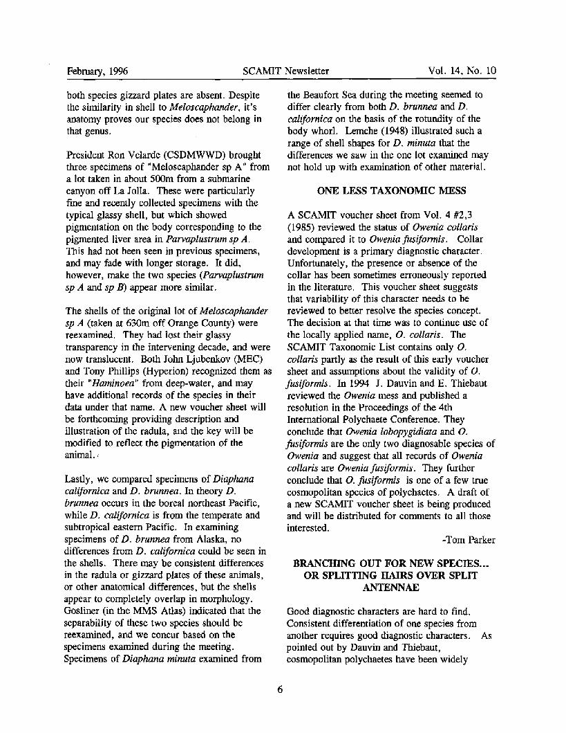
President Ron Velarde (CSDMWWD) brought three specimens of "Meloscaphander sp A" from a lot taken in about 500m from a submarine canyon off La Jolla. These were particularly fine and recently collected specimens with the typical glassy shell, but which showed pigmentation on the body corresponding to the pigmented liver area in Parvaplustrum sp A, This had not been seen in previous specimens, and may fade with longer storage. It did, however, make the two species (Parvaplustrum sp A and sp B) appear more similar.
The shells of the original lot of Meloscaphander sp A (taken at 630m off Orange County) were reexamined. They had lost their glassy transparency in the intervening decade, and were now translucent. Both John Ljubenkov (MEC) and Tony Phillips (Hyperion) recognized them as their "Haminoea" from deep-water, and may have additional records of the species in their data under that name. A new voucher sheet will be forthcoming providing description and illustration of the radula, and the key will be modified to reflect the pigmentation of the animal.
Lastly, we compared specimens of Diaphana californica and D. brunnea. In theory D. brunnea occurs in the boreal northeast Pacific, while D. californica is from the temperate and subtropical eastern Pacific. In examining specimens of D. brunnea from Alaska, no differences from D. californica could be seen in the shells. There may be consistent differences in the radula or gizzard plates of these animals, or other anatomical differences, but the shells appear to completely overlap in morphology. Gosliner (in the MMS Atlas) indicated that the separability of these two species should be reexamined, and we concur based on the specimens examined during the meeting. Specimens of Diaphana minuta examined from
the Beaufort Sea during the meeting seemed to differ clearly from both D. brunnea and D. californica on the basis of the rotundity of the body whorl. Lemche (1948) illustrated such a range of shell shapes for D. minuta that the differences we saw in the one lot examined may not hold up with examination of other material.
ONE LESS TAXONOMIC MESS
A SCAMIT voucher sheet from Vol. 4 #2,3 (1985) reviewed the status of Owenia collaris and compared it to Owenia fusiformis. Collar development is a primary diagnostic character. Unfortunately, the presence or absence of the collar has been sometimes erroneously reported in the literature. This voucher sheet suggests that variability of this character needs to be reviewed to better resolve the species concept. The decision at that time was to continue use of the locally applied name, O. collaris. The SCAMIT Taxonomic List contains only O. collaris partly as the result of this early voucher sheet and assumptions about the validity of O. fusiformis. In 1994 J. Dauvin and E. Thiebaut reviewed the Owenia mess and published a resolution in the Proceedings of the 4th International Polychaete Conference. They conclude that Owenia lobopygidiata and O. fusiformis are the only two diagnosable species of Owenia and suggest that all records of Owenia collaris are Owenia fusiformis. They further conclude that O. fusiformis is one of a few true cosmopolitan species of polychaetes. A draft of a new SCAMIT voucher sheet is being produced and will be distributed for comments to all those interested.
-Tom Parker
BRANCHING OUT FOR NEW SPECIES... OR SPLITTING HAIRS OVER SPLIT
ANTENNAE
Good diagnostic characters are hard to find. Consistent differentiation of one species from another requires good diagnostic characters. As pointed out by Dauvin and Thiebaut, cosmopolitan polychaetes have been widely
6
February, 1996 SCAMIT Newsletter Vol. 14, No. 10
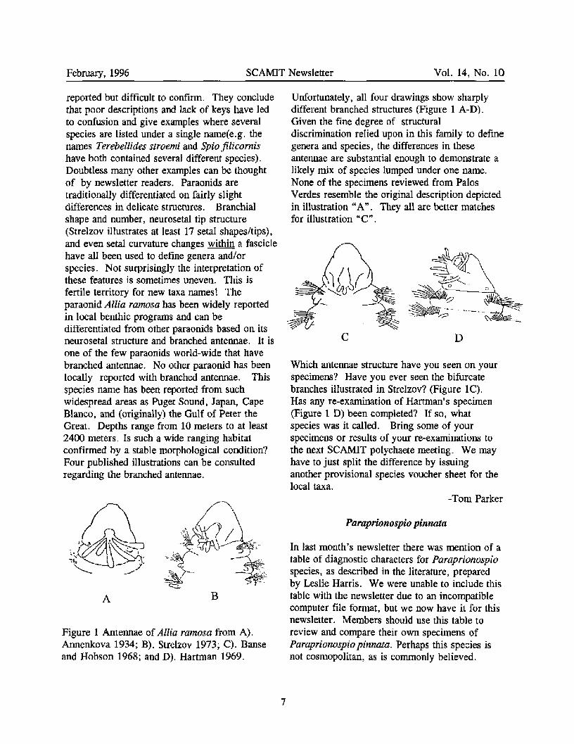
reported but difficult to confirm. They conclude that poor descriptions and lack of keys have led to confusion and give examples where several species are listed under a single name(e.g. the names Terebellides stroemi and Spio filicornis have both contained several different species). Doubtless many other examples can be thought of by newsletter readers. Paraonids are traditionally differentiated on fairly slight differences in delicate structures. Branchial shape and number, neurosetal tip structure (Strelzov illustrates at least 17 setal shapes/tips), and even setal curvature changes within a fascicle have all been used to define genera and/or species. Not surprisingly the interpretation of these features is sometimes uneven. This is fertile territory for new taxa names! The paraonid Allia ramosa has been widely reported in local benthic programs and can be differentiated from other paraonids based on its neurosetal structure and branched antennae. It is one of the few paraonids world-wide that have branched antennae. No other paraonid has been locally reported with branched antennae. This species name has been reported from such widespread areas as Puget Sound, Japan, Cape Blanco, and (originally) the Gulf of Peter the Great. Depths range from 10 meters to at least 2400 meters. Is such a wide ranging habitat confirmed by a stable morphological condition? Four published illustrations can be consulted regarding the branched antennae.
Figure 1 Antennae of Allia ramosa from A). Annenkova 1934; B). Strelzov 1973; C). Banse and Hobson 1968; and D). Hartman 1969.
Unfortunately, all four drawings show sharply different branched structures (Figure 1 A-D), Given the fine degree of structural discrimination relied upon in this family to define genera and species, the differences in these antennae are substantial enough to demonstrate a likely mix of species lumped under one name. None of the specimens reviewed from Palos Verdes resemble the original description depicted in illustration "A". They all are better matches for illustration "C".
C D
Which antennae structure have you seen on your specimens? Have you ever seen the bifurcate branches illustrated in Strelzov? (Figure 1C). Has any re-examination of Hartman's specimen (Figure 1 D) been completed? If so, what species was it called. Bring some of your specimens or results of your re-examinations to the next SCAMIT polychaete meeting. We may have to just split the difference by issuing another provisional species voucher sheet for the local taxa.
-Tom Parker
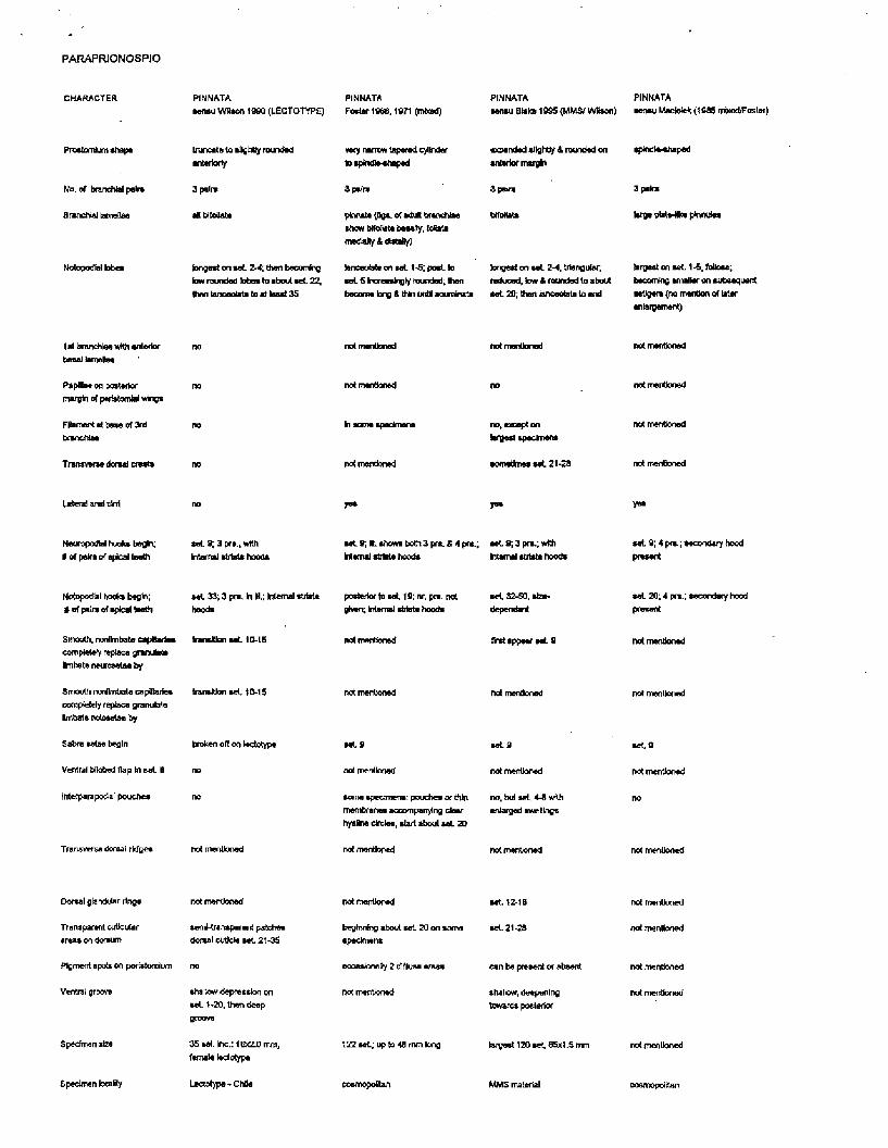
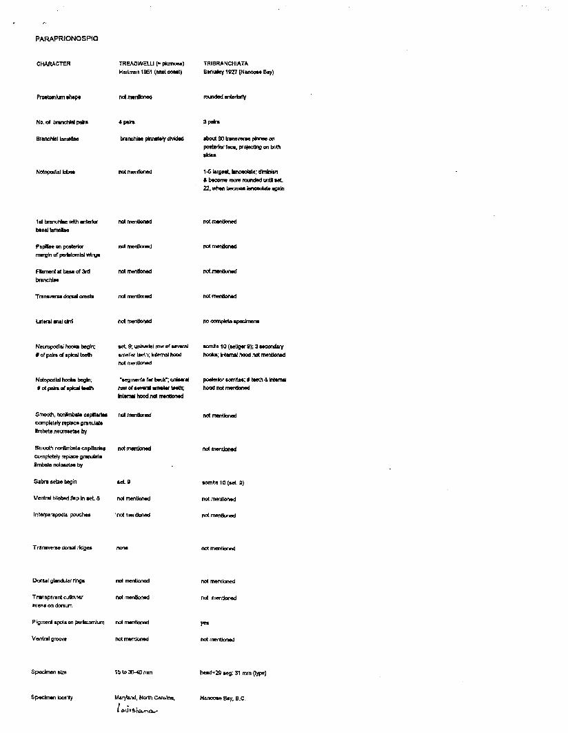
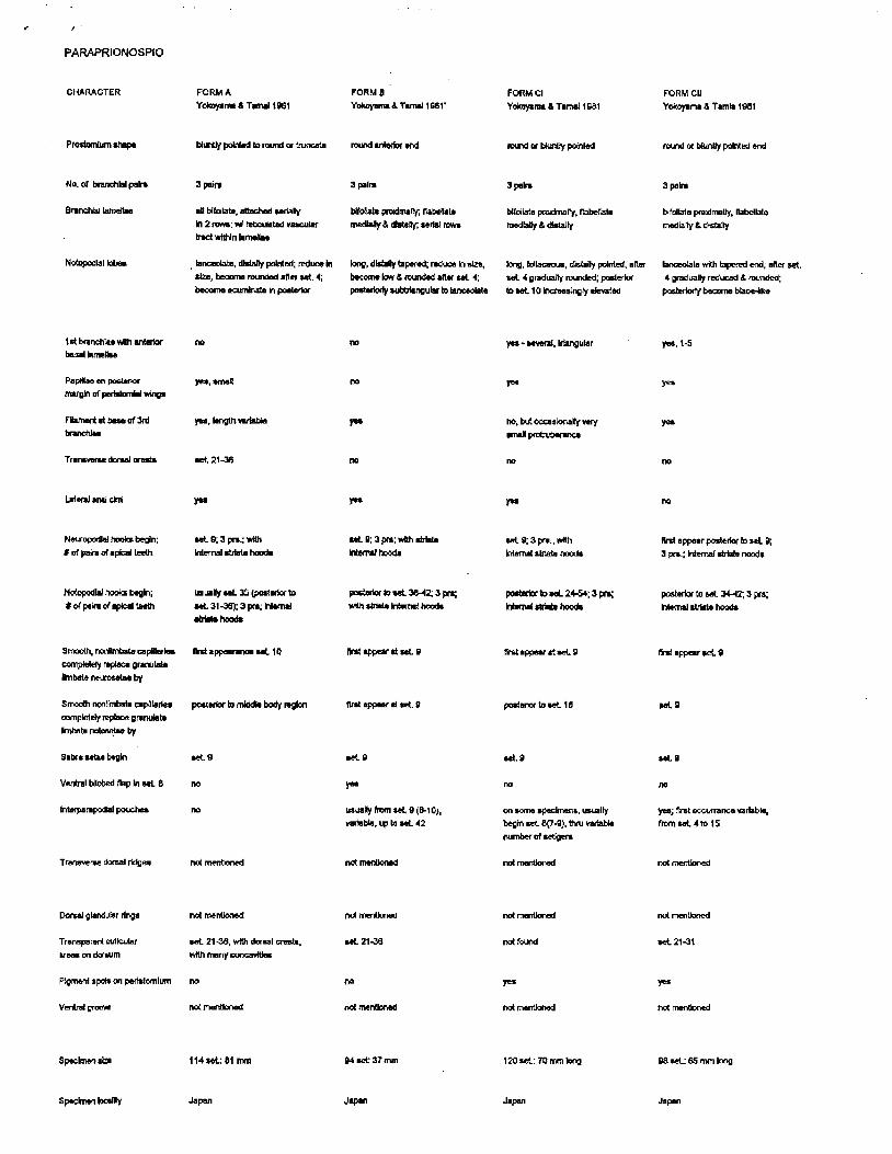
Paraprionospio pinnata
In last month's newsletter there was mention of a table of diagnostic characters for Paraprionospio species, as described in the literature, prepared by Leslie Harris. We were unable to include this table with the newsletter due to an incompatible computer file format, but we now have it for this newsletter. Members should use this table to review and compare their own specimens of Paraprionospio pinnata. Perhaps this species is not cosmopolitan, as is commonly believed.
7
February, 1996 SCAMIT Newsletter Vol. 14, No. 10
A RAPID TECHNIQUE FOR STAINING CIRRATULID POLYCHAETES
Local workers now use methyl green as the stain for species level identification of cirratulid polychaetes. Specimens of Aphelochaeta and Monticellina are abundant in many survey samples, with single benthic Van Veen grabs containing several hundred cirratulid specimens. These specimens require a great deal of time to handle and manipulate during staining, rinsing, destaining and identification. Problems with the current methods include slow stain uptake, long destaining process times, variable periods of staining and destaining, and messy liquid stain transfer procedures.
In Vol. 14, No. 6 of the SCAMIT Newsletter it was reported that formulation for methyl green stain varied between taxonomists, but that most people used 70% ETOH to dissolve the stain powder. The new modified technique reported here produces a much faster acting stain. The time necessary to process these specimens is greatly reduced. The new formula is:
3.0 grams methyl green powder 30 ml absolute ETOH 100 ml DI water O.Olgms KOH
This solution must be stirred to completely dissolve the powder.
New Staining materials: Flat bottomed watch glass. 20-30 ml methyl green stain solution. Gooch-style ceramic crucible (Coors 60151) with perforated bottom (5 cm tall). Small disc of nitex screen cut to fit inside bottom of crucible. Deep sided 300 ml pyrex dish for rinsing specimens in crucible. 70% ETOH for destaining.
New Staining Method 1. Set-up on stain resistant surface near sink
with bulk 70% ETOH supply. 2. Pour or eyedropper stain solution into
watch glass. 3. Place nitex screen in bottom of crucible. 4. With forceps, place a large number (50-
100) of specimens inside crucible on top of nitex screen.
5. Immerse perforated bottom of crucible into solution in watch glass.
6. Allow solution to completely cover specimens, add drops of additional stain over specimens if necessary.
7. Wait approximately 1-2 minutes while specimens soak in stain.
8. Remove crucible and specimens to deep sided dish about half full of 70% ETOH.
9. Gently raise and lower crucible within this dish to rinse out the excess stain. Drain and replace with fresh ETOH until most of excess stain is gone.
10. Remove specimens from nitex screen with forceps and place in dish for examination with microscope. With a large number of specimens, an additional step of destaining may be necessary while in the watch glass.
Aphelochaeta marioni specimens will stain in less than two minutes and reveal their characteristic barring pattern after 2-3 brief rinses; while Aphelochaeta sp. C stains with it's general non-barred pattern. There does not appear to be any great change in the reported stain patterns with this formulation. Advantages of this new method:
oa Rapid stain uptake by specimens greatly reduces identification time. «s=Use of crucible to stain and destain assures uniform treatment to all specimens. «afRapid destaining of specimens in crucible. ispRemoves stain handling from the microscope area. ^Produces same stain patterns reported in other technique.
8

February, 1996 SCAMIT Newsletter Vol. 14, No. 10
Please send any comments you may have about this technique to the newsletter editor.
-Tom Parker
BIBLIOGRAPHY
ANNENKOVA, N. P. 1934. Paraonidae of the Far-Eastern Seas of the USSR. [English translation from Russian], Comptes Rendus d'Academy des Sciences, U.S.S.R 1934:656-661.
BANSE, KARL, and Katharine D. Hobson. 1968. Benthic Polychaetes from Puget Sound Washington, with Remarks on Four Other Species. Proceedings of the United States National Museum 125:1-52.
BERRY, S. STILLMAN. 1953. Notices of New West American Marine MoIIusca. Transactions of the San Diego Society of Natural History XI(16):405-428.
BOUCHET, PHILIPPE. 1975. Opisthobranches de profondeur de l'Ocean Atlantique. 1. - Cephalaspidea. Cahiers de Biologie Marine 16(3): 317-365.
BURN, ROBERT F. 1978. A review of Australian species of Austrocylichna, Nipponatys. Cyltchnatys and Diniatvs (MoIIusca: Gastropoda: Haminoeidae). Journal of the Malacological Society of Australia 4(l/2):93-112.
CHARWAT, DANUTA K, and Jack Q Word. 1975. Key to irregular urchins. Pp.119-132. IN:: Word, Jack Q., and Danuta K. Charwat (eds.), Invertebrates of Southern California Coastal Waters I. Select Groups of Annelids, Arthropods, Echinoderms, and Molluscs. Southern California Coastal Water Research Project: El Segundo, California, U.S.A. 164pp.
DAUVIN, JEAN-CLAUDE, and Eric Thiebaut. 1994. Is Owenia fusiformis Delle Chiaje a cosmopolitan species? Pp.383-404. IN:: Dauvin, J. C, L. Laubier, and D. J. Reish (eds.), Actes de la 4eme Conference internationale des Polychetes. Memoires du Museum National d'Histoire Naturelle, Paris. 642pp.
EIBYE-JACOBSEN, DANNY. 1991. A Revision of Eumida Malmgren, 1865 (Polychaeta: Phyllodocidae). Steenstrupia 17(3):81-140.
G1ANNUZZI-SAVELLI, RICCARDO, and Anthea Gentry. 1990. Haminaea Leach, [1820](Mollusca, Gastropoda): proposed conservation. Bulletin of Zoological Nomenclature 47(4): 263-269.
GOSLINER, TERRENCE M. 1995. Introduction and spread of Philine auriformis (Gastropoda: Opisthobranchia) from New Zealand to San Francisco Bay and Bodega Harbor. Marine Biology 122(2):249-255.
HARTMAN, OLGA. 1969. Atlas of the Sedentariate Polychaetous Annelids from California. Allan Hancock Foundation, University Of Southern California: Los Angeles, Ca.
KENSLEY, BRIAN. 1996. Systematics and distribution of the genus Calocarides (Crustacea: Decapoda: Axiidae). Proceedings of the Biological Society of Washington 109(l):53-69.
LEMCHE, HENNING. 1948. Northern and Arctic tectibranch gastropods. 2. A revision of the cephalaspid species. Det Kongelige Danske Videnskabernes Selskab, Biologiske Skrifter 5(3):32-136.
MALUF, LINDA YVONNE. 1988. Composition and Distribution of the Central Eastern Pacific Echinoderms. Technical Report Number 2, Allen Press, Inc.: Lawrence, Kansas.
MARCUS, EVELINE DU BOIS-REYMOND. 1977. On the genus Tomatina and related forms. Journal of Molluscan Studies 2(2-35)
MARCUS, EVELINE DUBOIS-REYMOND, and Ernst Marcus. 1969. Opisthobranchian and lamellarian gastropods collected by the'Vema'. American Museum Novitates (2368): 1-33.
RATHBUN, MARY J. 1902. Descriptions of new decapod crustaceans from the west coast of North America. Proceedings of the United States National Museum 24(1272): 885-905.
STRELZOV, V. E. 1973. Polychaete Worms of the Family Paraonidae Cemiti, 1909 (Polychaeta, Sedentaria)[1979 translation]. Amerind Publishing Co. Pvt. Ltd.: New Delhi.
9

February, 1996 SCAMIT Newsletter Vol. 14, No. 10
WICKSTEN, MARY K. 1996. Neocraneon zacae (Chace, 1937) synonymized with N. resima (Rathbun, 1902), and compared with N. communis (Rathbun, 1899)(Decapoda: Caridea: Crangonidae). Proceedings of the Biological Society of Washington 109(l):39-43.
Ogyrides orientalis (Stimpson, 1860) from Holthuis, L. B. 1993. Recent genera of caridean and stenopodidean shrimps. Nationaal Natuurhistorisch Museum, Leiden.
SCAMIT OFFICERS: If you need any other information concerning SCAMIT please feel free to contact any of the officers.
e-mail address President Ron Velarde (619)692-4903 [email protected] Vice-President Don Cadien (310)830-2400 ext. 403 [email protected] Secretary Cheryl Brantley (310)830-2400 ext. 403 [email protected] Treasurer Ann Dalkey (310)648-5611 cam@san,ci.la.ca.us Back issues of the newsletter are available. Prices are as follows:
Volumes 1 -4 (compilation) $ 30.00 Volumes 5 - 7 (compilation) $ 15.00 Volumes 8 - 13 $ 20.00/vol.
Single back issues are also available at cost.
10
SCAMIT TREASURY SUMMARY. 1995-96
During the past fiscal year, April 1995 though March 1996, expenses were twice as great as income reflecting SCAMIT's increased activity in producing newsletters and publications. Costs for producing the newsletter (including printing, postage, and supplies) increased from $2163.60 for last fiscal year to $3399.61 due to the increased content of the newsletter. The second edition of the Taxonomic Listing was published ($779.40) and SCAMIT Grant #96-1 was issued to Larry Lovell for a Nephytid publication ($584.34). SCAMIT's primary source of income, $1570.00, came from membership dues which covered half the costs for producing the newsletter. Grants and workshops will continue to be funded from the money collected for creating the Taxonomic Listing for SCCWRP during the 1994-95 fiscal year. The following is a summary of the expenses and income:
Expenses Newsletter Publications (Taxonomic Listing, Grants Miscellaneous Total
Income Dues Interest T-Shirts Donations Miscellaneous Total
Account balances (March 31, Checking Savings Total
1996)
$3399.61 2nd ed) 779.40
584.34 183.72
$5100.77
$1570.00 385.76
0.00 10.00
510.25 $2476.01
$ 999.49 17068.11
$18067.60
REVIEW OF THE CEPHALASPIDS OF CALIFORNIA Don Cadien (CSDLAC) and John Ljubenkov (MEC)
SCAMIT Meeting 22 APRIL 1996
Mollusks of the Order Cephalaspidea (s.l.) are well represented in the waters of the Northeast Pacific. They are frequently encountered in environmental sampling, and nearly always identified to at least generic level by local taxonomists. There are, however, many problems associated with the taxonomy of the group which should be further resolved. This is especially true at this time, when two separate treatments of the group which embody differing authoritative views are being introduced into the literature {the sections on Opisthobranch Mollusks by Gosliner and Gastropod Mollusks by McLean in the Taxonomic Atlas of the Santa Maria Basin and Western Santa Barbara Channel).
This group of opisthobranchs is transitional in that some are externally shelled, and some virtually lack shells. They have been treated both by workers who deal exclusively with shelled mollusks, and those who deal primarily with shell-less mollusks. Shelled mollusks have a fossil record, and many groups were established on the basis of fossils, or on the basis of the shell without reference to the animal which created it. This is a very different approach from that of the general run of opisthobranch workers who usually base their taxonomy on soft anatomy rather than hard parts of the animals they study. In both "camps" the radula is considered an important source of characters for differentiating species and/or higher taxa, but in groups which have been established based on fossils, radular evidence is lacking for the types. Nomenclaturai schisms have developed, particularly over genera such as Acteocina and Sulcoretusa where the generotype is either a fossil or described on shell characters alone.
Characterization and Phytogeny of the group
Until fairly recently the cephalaspids were easily identified by the synapomorphy of possession of a cephalic shield. Recent cladistic analyses have drawn this into question by demonstrating that some groups with cephalic shields belong to other groups. Groups traditionally considered within the confines of the cephalaspids have been excluded on the basis of such cladistic reassessments.
There have been several cladistic analyses of the group, the most complete and recent being that of Mikkelsen 1996. She was very thorough in laying the groundwork for her analysis, testing and discarding many traditionally used characters as inappropriate to the purpose (Mikkelsen 1993). As in the analysis of Gosliner 1981, which was not based on a full application of cladistic methods, characters of the nervous, digestive, and reproductive systems were viewed as of importance in establishing the phylogeny of the group.
In Mikkelsen's analysis the family Acteonidae is removed from the Opisthobranchia, and combined with a few disparate groups at the base of the Heterobranchia. She terms this stem group "unresolved", but there is little doubt that it is very primitive. Other groups traditionally grouped with the Acteonidae in the Acteonoidea were retained in the Opisthobranchia, but outside the Cephalaspidea in the analysis. Representatives of the Hydatinidae and Ringiculidae were joined into another unresolved primitive group the "Architectibranchia"
With the Acteonoidea thus fragmented and dispersed, only three superfamilies remain in the Cephalaspidea; the Bulloidea, the Philinoidea, and the Runcinoidea (which was not included in Mikkelsen's analysis). The first two were united by four synapomorphies in the analysis: flexed ciliated strips in the mantle cavity, three gizzard plates, a secondarily prepharyngeal nerve ring, and
1

the genital ganglion on the visceral nerve loop (Mikkelsen, 1996). The earlier "synapomorphic" character of posession of a cephalic shield is, based on Mikkelsen's analysis, now not even particular to Opisthobranchia, and within the opisthobranchs is found both in cephalaspids and sacoglossans.
Taxonomy of the group
The taxonomy of the group above the species level is presented in the attached appendix; taxa represented in the northeast Pacific are indicated with asterisks. There are still a number of unresolved problems in the higher classification of the cephalaspids, and the taxonomy as presented here is of necessity subject to immediate or future modification based on new evidence. That presented is derived from traditional sources, and includes several groups now excluded from the cephalaspids on the basis of cladistic analyses.
Californian cephalaspid fauna
Although cephalaspids were treated comprehensively as part of several general mollusk monographs (ie. Pilsbry 1895-96, Oldroyd 1927) they were first specifically addressed for the northeast Pacific by Steinberg (1963). Her treatment is little more than a list of the described species organized into family and higher categories, but it provides a convenient summary of the taxa, and a matrix on which to build. The current list of the California cephalaspids (Table ) is smaller, with a number of species relegated to synonymy. There are also several undescribed taxa not included in Steinberg's list, some of which are familiar members of our soft-bottom fauna.
Biology of cephalaspids
Cephalaspids are predatory gastropods, living as hunters pursuing prey within the oxic surface layers of the bottom sediments. There are, of course, exceptions to this generalization. First among these are the bullids, which are herbivorous or omnivorous species (Rudman 1971b).
Regardless of diet, all members of the group are well adapted for movement on and through soft sediments. They bear a foot, generally broad, which is well supplied with mucous glands and has a ciliated surface for gliding locomotion. On soft sediments the mucous envelope secreted by the animal allows movement through sediments fine enough to clog respiratory surfaces, and fill the pallia! cavity. This is prevented by secretion of a "sheath'1 within which the animal moves forward. This can be on the sediment surface, or below it. Much the same strategy and methods are used by other gastropod groups including the olivids, and the naticids.
The structure of the body also lends itself to rapid and effective burrowing in soft sediments. The shell is either reduced and internal, or external but covered by extensions of the mantle. This provides a smooth flexible surface over which the mucous envelope slides. Once again structure is homologous with the olivids and naticids, which also share the same life style.
Equipped by the above morphology cephalaspids can rapidly glide about in search of prey. Reported predatory strategy usually involves whole consumption of small prey items, but some members of the Acteonidae use a different strategy. These animals, including the local Rictaxis punctocaelatus, harvest exposed palps, cirri, or brancnia of polychaete worms (Marcus 1972). This behavior, akin to siphon nipping by fishes, allows the prey to escape and regenerate the lost structures
2

TABLE 1
CALIFORNIAN CEPHALASPID MOLLUSKS
Superfamily A c t e o n a c e a
Family Acteon idae
Acteon traskii
Microglyphis brevicula
Microglyphis estuarina
Riclaxis painei
Rictaxis punclocaelatus
Family Hydatintdae
Parvaplustrum sp A
Superfamily Diaphanacea
Family Oiaphanidae
Diaphana California*
Woodbridgea williamsi
Superfamily Bullacea
Family Butfidae
Bulla gouldiana
Family Haminaeidae
Atys castas
Atys nonscriptus
Diniatys oxystoma
Micraenigma oxystoma
Haminaea vesicula
Haminaea virescens
Haminoea olgae
Family Retusidae
Sulcoretusa montereyensis
Sulcoretusa xystrum
Volvulella californica
Volvulella calharia
Volvulella cytindrica
Volutella tenuissima
Volvulella panamica
Superfamily Runcinacea
Family Runcinidae
Runcina macfarlandi
Runcinida sp A
superfamily uncertain
family uncertain
Bullomorpha sp A
Stearns 1898
{Dail 1902)
(Dall 1908)
Dal] 1903
{Carpenter 1864)
[SCAMIT 1995]
Dall 1919
Berry 1953
Pilsbry 1895
Carpenter 1864
(A. Adams 1850)
(Berry 1953)
Berry 1953
Gould 1855
(Sowerby 1833)
Dall 1919
(A. G. Smith & Gordon 1948)
{Dall 1919)
Dall 1919
Dall 1919
{Carpenter 1864)
Willed 1944
Dall 1919
Gosliner 1991
[Cadien]
[Ljubenkov 1994]
Superfamily Philinacea
Family Scaphandridae
Acteocina harpa
Acteocina oidroydi
Acteocina mculta
Acteocina carinaia
Acteocina intermedia
Acteocina smirna
Tornastra culcitella
Tornastra rolleri
Tornastra cerealis
Acteocina eximia
Acteocina planata
Tornastra infrequens
Acteocina magdalenensis
Meloscaphander sp A
Family Cytichnidae
Cylichna attonsa
Cylichna diegensis
Family Aglajidae
Agtaja ocelligera
Doridium adellae
Chelidomira phocae
Melanochtamys diomedea
Aglaja nana
Aglaja sp A
Navanax inermis
Doridium purpureum
Agtaja bakeri
Family Philinidae
Philine alba
Phitine bakeri
Philine bakeri
Philine auriformis
Philine bakeri
Philine californica
Philine polystrigma
Broctonia polystrigma
Woodbridgea polystrigma
Philine cfquadrata
Philine sp A
Philine "no radula/gizzard"
Philine "tubular"
Family GasUopteridae
Gastropteron pacificum
(Dail 1871)
Dall 1925
(Gould 1855)
(Carpenter 1857)
Willed !928
Dall 1919
(Gould 1853}
Ev. Marcus 1977
(Gould 1853)
(Baird 1863)
Dall 1919
C, B. Adams 1852
{Dall 1919)
[SCAMIT 1995]
(Carpenter 1865)
(Dall 1919)
(Bergh 1894)
Dall 1894
Marcus 1961
(Bergh 1894)
Steinberg & Jones 1960
[Cadien]
(J. G. Cooper 1863)
Bergh 1893
MacFarland 1924
Mattox 1958
Abbott 1974 (non Dall 1919)
Bern-ens 1993 (non Dall 1919)
Suter 1909
Dall 1919
Willett 1944
(Dall 1908)
Dall 1908
(Dall 1908)
[MMS] (non Wood 1839)
[SCAMIT 1988]
[Gosliner]
[Gosliner]
Bergh 1894
- thus providing a renewable food source. Other acteonids apparently take the entire animal. Hurst (1965), for instance, records finding the polychaete Owenia fusiformis in the gut of Acteon tornatilis.
More typical predatory behavior is shown by Tornastra cuicitella and Cylichna attonsa, which are selective feeders on benthic foraminifers (Shonman and Nybakken 1978). Similar feeding behavior is exhibited by Retusa ckrysoma (Bum and Bell 1974) and Retichna murdochi (Rudman 1971a). Burn and Bell (op. cit.) report R. chrysoma also selectively consumes a small gastropod, Salinator fragilis. Consumption of small gastropods is also reported for the local Acteocina harpa (Beeman and Williams 1980).
Other members of the Philinoidea are their own worst enemies. Members of the Aglajidae are specialized opisthobranch predators, feeding on nudibranchs, other cephalaspids, and in many cases other individuals of their own species. The behavior of these hunters is best known through studies of our local Navanax inermis (Paine 1963, 1965). Hunting in these animals is strongly based on chemosensitivity to chemical cues left in the mucous trails of their opisthobranch prey. Once a trail is crossed by a Navanax, it is turned onto and followed to it's end, either in a meal or not, depending on which direction the predator adopts initially (to or away from the prey). If the prey is encountered it is attacked and swallowed whole by muscular contraction of the Navanax buccal bulb, No crushing or mastication of the prey occurs after swallowing, so that even very delicate shells of shelled prey are undamaged during passage through the Navanax digestive tract. Savvy shell collectors have long used this method of collection for the fragile shells of Haminaea, Philine and other cephalaspids. Not-so-sawy field collectors have found to their chagrin that a bucket full of opisthobranchs + one hungry Navanax — one contented Navanax, and nothing else.
Much more catholic tastes are exhibited by the introduced Philine auriformis. Food appears to be taken in proportion to availability in the environment, with food broadly defined. These hearty eaters have had guts filled with ophiuroid arm fragments, the crushed tests of the foraminifer Rhabdamina, other cephalaspids, Pectinaria, small Parvilucina and a variety of less identifiable fragments of benthic invertebrates. In their native New Zealand they are more specialized on strongly shelled small bivalves (Rudman 1970). Although predation by these animals has not been directly observed, it is assumed that capture is by snagging with the radula, followed by swallowing aided by the buccal pump. Once consumed the food is passed to the gizzard, where it is crushed by the action of the triad of robust gizzard plates and the muscular contractions of the gizzard. For those with sufficient interest in the subject, a wealth of information is available on feeding and digestion in cephalaspids (see for instance Rudman 1971a, b; 1972a, b, c, d).
All known members of the group are hermaphroditic, as are other opisthobranchs. Each animal contains both male and female reproductive organs, although these may not be simultaneously functional. Reproduction involves an exchange of sperm, internal fertilization, and deposition of fertilized eggs m an egg mass. Development is either direct, with larval stages passed within the egg and hatching as a metamorphosed juvenile; or indirect, with hatching of larvae from the egg. In the latter case the larval form may be short lived, metamorphosing rapidly into a benthic juvenile, or planktonic, with the larva (veliger) feeding for some period in the water column prior to metamorphosis.
Literature Cited
BEEMAN, ROBERT D., and G.C.Williams. 1980. Chapter 14. Opisthobranchia and Pulraonata: The Sea Slugs and Allies, pp. 308-354 IN: Morris, Robert R, D. P. Abbott, and E. C. Haderlie. Intertidal Invertebrates of California, Stanford University Press, Stanford. 690pp.
3

BURN, ROBERT F., and K. N. Bell. 1974. Description of Retusa chrysoma Bum sp. nov. (Opisthobranchia) and its food resources from Comer Inlet, Victoria. Memoirs of the National Museum of Victoria 35:115-119.
GOSLINER, TERRENCE M. 1981. Origins and relationships of primitive members of the Opisthobranchia (Mollusca: Gastropoda). Biological Journal of the Linnean Society 16:197-225.
HURST, ANNE. 1965. Studies on the structure and function of the feeding apparatus of Philine aperta with comparative consideration of some other opisthobranchs. Malacologia 2(3):281- 347.
MARCUS, EVELINE DUBOIS-REYMOND. 1972. On some Acteonidae (Gastropoda, Opisthobranchia). Papeis Avulsos deZoologia, Sao Paulo 25(19): 167-188.
MIKKELSEN, PAULA M. 1993. Monophyly versus the Cephalaspidea (Gastropoda, Opisthobranchia) with an analysis of traditional cephalaspid characters. Bolletino Malacologico 29:115-138.
MIKKELSEN, PAULA M. 1996. The evolutionary relationships of Cehalaspidea s. 1. (Gastropoda: Opisthobranchia): a phylogenetic analysis. Malacologia 37(2): 3 75-442.
OLDROYD, IDA SHEPARD. 1927. The Marine Shellsof the West CoastofNorthAmenca. Volume 2 Part 1. Stanford Univerisry Press, Stanford. 297pp. [1978 Reprint]
PAINE, ROBERT T. 1963. Food recognition and predation on opisthobranchs by Navanax inermis (Gastropoda: Opisthobranchia). Veliger 6( 1): 1-9.
—. 1965. Natural history, limiting factors, and energetics of the opisthobranch Navanax inermis. Ecology 46(5):603-619.
PILSBRY, HENRY A. 1895-96. Manual of conchology: structural and systematic 15(59): 133-180; I5(60):181-436; 16(61):l-48; 16(62):49-112; 16(63): 113-160; 16(64):161-262.
RUDMAN, WILLIAM B. 1970. A revision of the genus Philine in New Zealand with descriptions of two new species (Gastropoda, Opisthobranchia). Journal of the Malacological Society of Australia 2(l):23-34.
—. 1971a. On a new genus for 'Tomatina' murdochi. Journal of the Malacological Society of Australia 2(2):I87-193.
—. 1971b. Structure and functioning of the gut in the Bullomorpha (Opisthobranchia). Part 1. Herbivores. Journal of Natural History 5:647-675.
—. 1972a. The anatomy of the opisthobranch genus Hvdatina and the functioning of the mantle cavity and alimentary canal. Zoological Journal of the Linnean Society 51(2): 121-139.
—. 1972b. Structure and functioning of the gut in the Bullomorpha (Opisthobranchia), Part 2. Acteonidae. Journal of Natural History 6:311-324.
—. 1972c. Structure and functioning of the gut in the Bullomorpha (Opisthobranchia). Part 3. Philinidae. Journal of Natural History 6(4):459-474.
—. 1972d. Structure and functioning of the gut in the Bullomorpha (Opisthobranchia). Part 4. Aglajidae. Journal of Natural History 6(5):547-560.
SHONMAN, DAVID, and James W. Nybakken. 1978. Food preferences, food availability and food resource partitioning in two sympatric species of cephalaspidean opisthobranchs. Veliger 21(1):120-126.
STEINBERG, JOAN E. 1963. Notes on the opisthobranchs of the west coast of North America. II. The order Cephalaspidea from San Diego to Vancouver Island. Veliger 5(3): 114-117.
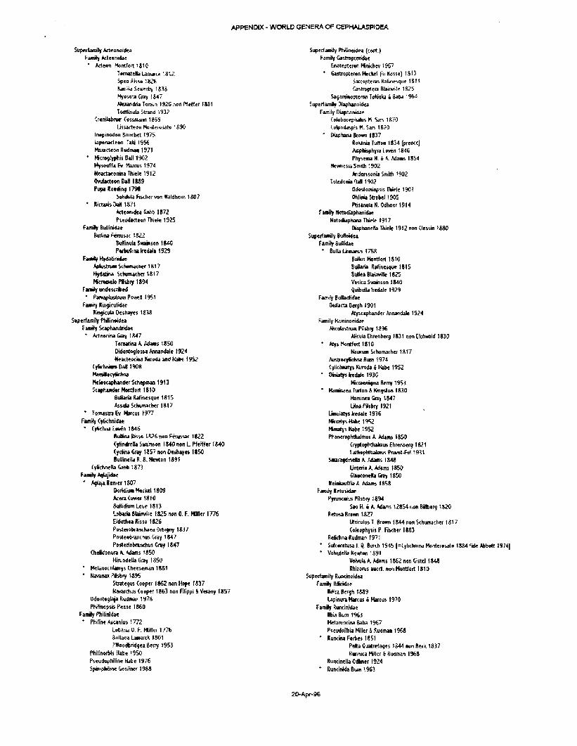
A P P E N D I X - W O R L D G E N E R A O F C E P H A L A S P I D E A
5uperTaintty Acteortordea
Fanny Acttonidae
• Atleoti Montfort 1810
Tomatella Lamarck 18 (2
SpeoKisso 1826
Kanffla Sowerby 1328
Myosota Cray 1847
Alexandria Tomiiri 1926 non f f t f le f 1881
Twnlinula Strand 1332
(rendabnon (ossmann 1869
Lissartean Honterosatc. 1890
Inopinodon Bouchct 1975
laponacteon Tiki 1956
Maxactewi Rudman 1971
' Mcrogfyphis Dall 1902
fyswrffabi. Marcus 1974
NeactaMmha Thide 1912
OvutarteMi Halt 1889
Pupafloeding 1738
SoBAda Fischer vort Waldhcim 1807
' Rktajis Dad 1871
Arteonidea tafib 1872 Pwudacteon Thide 1925
Family BtiHinidae Buifinl Femisac 1822
Buflinula 5wain*on 1840
Pertuina he dale 1929
FaoBfy Hydabnidae
Aplustruai Schumacher 1817
Hydatina Schumacher 1817
Mknwtelo PHsbry 1894
Family undescribed
* Pajwplustrum Powell 1951
Family Ravgit-jlidac
RmgKiia Deshayej 1838
Super! amity fhjlinoidea
Family Scaphaaidridae
* Meer ina Gray 1847
Tematina A. Adams 1850
Didonioglossa Annandale 1924
ttearteocina Kuroda and Habe 1952
(ytktmiim Da* 1908
Mamiltcyikhn]
MekHcaphanderSchepaan 1913
Scaphander Monttort 1S10
Buttaria Rafincigue 1815
As tula Schumacher 1817
* TefnastfiEv. Marcus 1977
Family (ylkhnidae
* [yfcehna Lmrfn 1846
BuDina R K W 1S26 non Fevussat 1822
Cyhndrefla Snamson 1840 non L Pfefffer 1840
(ycltna Gray 1857 non Deshayes 1850
BultinellaR. 8. Newton 1S91
CvfchneRaGahb1873
Family Agtajidae
* AgtajallenierlSO?
Doridi™ Meckel 1809
AceraCuwei 1810
BuHWium Leue 1813
Lobaria Blainviile 1825 non 0. F. MfHIer 1776
Eidothea Risso 1826
Posterohranchaea Orbigny 1837
Posteobfanchu* Gray 1847
Posteriobranchus Gray 1847
Chelidonuta A. Adams 1850
HirundeUa Cray ! 850
* Melanoc hlamys (heeseman 18S1
* Havanax Pusbry 1895
Strategus Cooper 1862 non Hope 1837
Kavafchus Cooper 1863 non Ftfippi 4 v>rany 1857
Odwtoglaja Rudman 1978
Phtfmopsis Pease 1860
Family Philinidae 4 Philine Ascanius 1772
LobariaO.f1. Wilier 1776
Bullaea Lamarck 1801
?n'eodbridgea Berry 1953
Philinorfiis Hate 1950
Pseudophilin* Habe 1976
Spiniphilme Gosiiner 1988
Superfamity Phffinoidea (coiit.)
Family Gastropteridae
Enotepteron Mrachev 1967
* Gastropteran Metluit (in Kesse) 1813
Sarcoptenrs Raiinesque 1814
Gastroplcra Biairwille 1825
Saganunoptergn Tokioka a Baea 1964
SupErfamily Diaphanoidea
Family Eliaphanidae
(olobotepnalus K. Sars 1870
Colpodaspis M. San 1870
" Diapharta 8r°wn 1837
Roxania Turton 1834 [preocc]
Ajsphisphyra Lcven 1846
Physema H. A A. Adams 1854
Newnesia Smith 1902
Andemonia Smith 1902
Totedoriia Dall 1902
Odostomiepsis TWIe 1903
OhliniaStrebeJ1905
ftiS4nulaN.0dhner1914
F unify HotodUphanidae
Notodiaphana Thirie 1917
DiaphafieHa Ihiele 1912 non (kssirt 1880
Superfamily ftuflwdca
Family SuDidae
* Bulla Linnaeus 1758
BuHus Mgntfurt 1810 Suftaria Rafinesqu* 1815
Bufea Blainviile 1S25 Vesica Swainson 1840
Qu&utta trtdale 1929
Family Bcdlactirfae
Buflacta Bergh 1901
Atyscaphander Annandale 1924
Family Haminoeidae
Anculastrun FSsbry 1 8 %
AKcula fhreflbtca 1821 ncm Ektmald 1830
* AtysCtontfort 1810
Naucum Schumacher 1817
AusffwyUchfU Sum 1974
Cylichnatys Kuroda s Kabe 1952
* Dirwitys Iredate 1936
Kccaefligma Berry 1953
* Kaminaea Turton A Kingston 1830
Haminea Gray 1847 LHoa Pilsbry 1921
Limulatys IredaJe 1936
MirratysHabe 1952
Hmatys Habe 1952
Pharterophrhalmus A Adams 1850
{ryptophthabaus E hrenberg 1821
Lattephthatmus Pruvot-Ftri 1931 Smiragdmtlla A. Adams IS48
Linteria A. Adams 1850
GbwcentJla Gray 1 £50
Heinkacrtfia A. Adams 1858
Family Retusidae
PyrutKdus Pilsbry 1894
Sao H. a K Uams 128S4 non Bilberg 1820
Retusa Brawn 1S27
ytritulusT. 8ro*n 1844 ncm Sthuraacher 1817
Uleophysis P. Fischer 18S3
Relkhna Rudman 1971
* Sulcoretusa J, Q. Butch 1945 [=(ylichmru Mcnterosato 1884 Tide Abbott 1974]
* VoWeila Nenton 1891
Votmla A. Adams 1862 non Gistel 1S4S
Rhizorus aucrt. mnMontfort 1810
Superfamity Runcinoidea
Family Itdicidae
UA'ta Bergh 1889
Uptnura Marcus 4 Marcus 1970
Family ftuncinidae
tibia Bum 1963
Metanincina Baia 1%7
PseudoilbiaMilieriftudman 1968
* Runcina Forbes 1851
Pefca Quatre<ages 1844 non fleck 1S37
Runnica Miller i Rudman 1968
8uncinella0dhner1924
" Runcinida Bum 196s
2 0 - A p r - 9 6

ANATOMY OF NEW CEPHALASPIDEA
While most molluscan taxonomists ignore the soft.bodies of their specimens, it is often the case that fleshy parts have many features which aid greatly in identification. These characters allow for the correct identication of damaged specimens with incomplete shells (often the case in screened material).
Paraplustrum sp. A The head and its appendages are quite unique in comparison to other
opisthobranchs. The center frontal portion of the head has a pair of palps(?) with swollen bases and digitiform distal ends. Posterolateral to each palp is a pair of cephalic tentacles whose bases are next to each other, and in one instance the bases were united.. The anterior foot margin is extended on both sides into propodial lobes (which may be hidden by contraction). I have been unable to find eyes, but it would not be surprizing if they possessed them.. The metapodium or posterior end of the foot is extended into a long thread-like structure, also often hidden through contraction, but its exact position in life is unknown.
Bullomorpha sp. A
The foot is broadly cuneate and tapering posteriorly; it possesses two parapodial lobes which broadly flare out at the sides of the body. The cephalic lobe or disc is dorsally attached to the anterior end of the shell and usually protrudes even in contracted specimens. The eyes are positioned on the body just posterior to the base of the cephalic lobe and on either side of the mid-line of the dorsum. The mantle has a thickened edge anteriorly and dextrolaterally (mainly following the lip of the shell. At the rear the mantle flares out into a posterior mantle lobe; of which the central portion is elongated into a pseudo-metapodium that coils into the involute spire. There is always a black, heavily pigmented spot on the mantle on the right side of the animal just forward of the posterior lobe. There is a spermatothecal groove on the dorsal surface of the right-hand parapodial lobe.
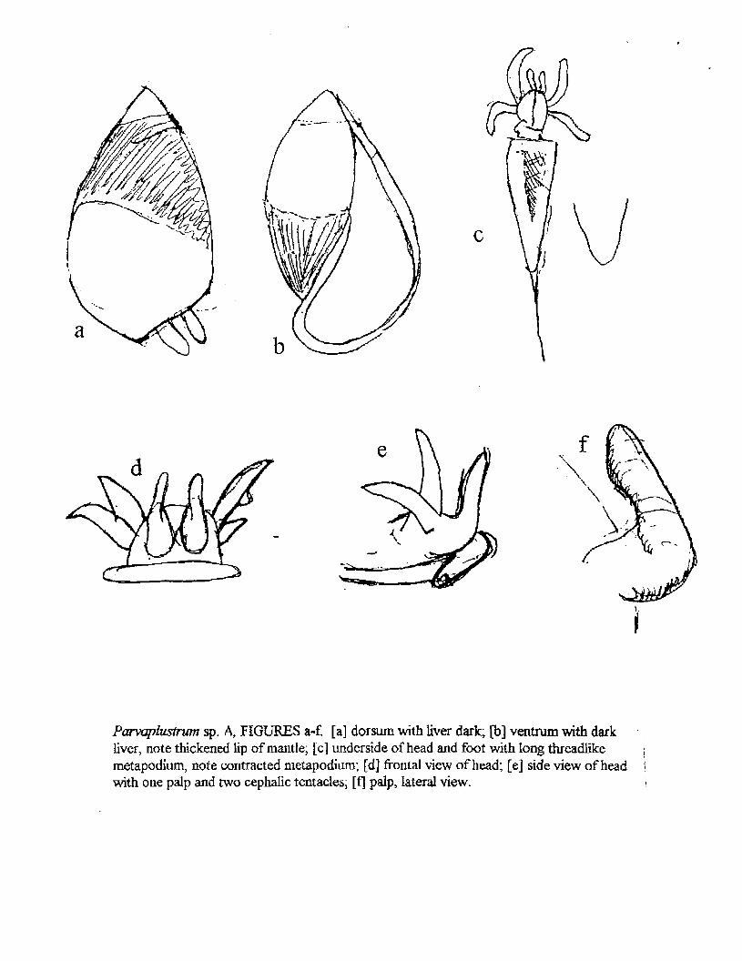
Parvaplustrum sp. A, FIGURES a-f. [a] dorsum with liver dark, [b] ventrum with dark liver, note thickened lip of mantle, [c] underside of head and foot with long threadlike \ metapodium, note contracted metapodium; [d] frontal view of head; [e] side view of head • with one palp and two cephalic tentacles; [f] palp, lateral view. i
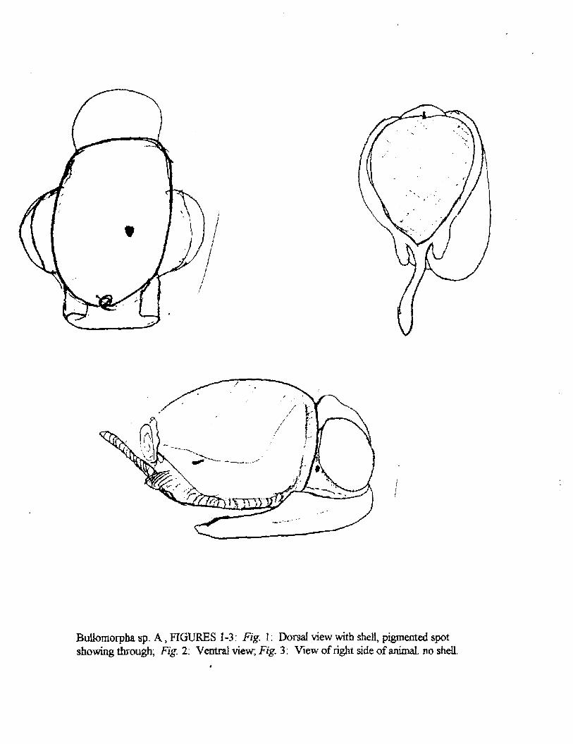
BuUomorpha sp. A, FIGURES 1-3: Fig. 1: Dorsal view with shell, pigmented spot showing through; Fig. 2; Ventral view; Fig. 3: View of right side of animal, no shell.
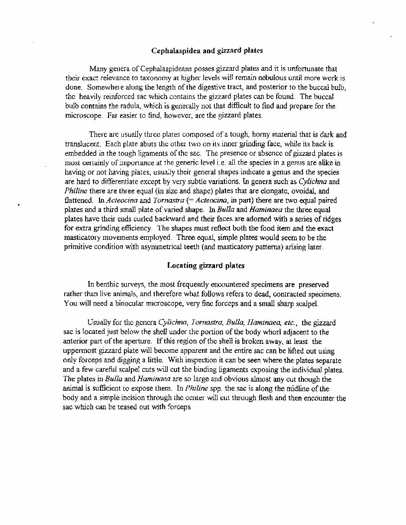
Cephalaspidea and gizzard plates
Many genera of Cephalaspideans posses gizzard plates and it is unfortunate that their exact relevance to taxonomy at higher levels will remain nebulous until more work is done. Somewhere along the length of the digestive tract, and posterior to the buccal bulb, the heavily reinforced sac which contains the gizzard plates can be found. The buccal bulb contains the radula, which is generally not that difficult to find and prepare for the microscope. Far easier to find, however, are the gizzard plates.
There are usually three plates composed of a tough, horny material that is dark and translucent. Each plate abuts the other two on its inner grinding face, while its back is embedded in the tough ligaments of the sac. The presence or absence of gizzard plates is most certainly of importance at the generic level i.e. all the species in a genus are alike in having or not having plates; usually their general shapes indicate a genus and the species are hard to differentiate except by very subtle variations. In genera such as Cylichna and Philine there are three equal (in size and shape) plates that are elongate, ovoidal, and flattened. In Acteocina and Tornastra (= Acteocina, in part) there are two equal paired plates and a third small plate of varied shape. In Bulla and Haminaea the three equal plates have their ends curled backward and their faces are adorned with a series of ridges for extra grinding efficiency. The shapes must reflect both the food item and the exact masticatory movements employed. Three equal, simple plates would seem to be the primitive condition with asymmetrical teeth (and masticatory patterns) arising later.
Locating gizzard plates
In benthic surveys, the most frequently encountered specimens are preserved rather than live animals, and therefore what follows refers to dead, contracted specimens. You will need a binocular microscope, very fine forceps and a small sharp scalpel.
Usually for the genera Cylichna, Tornastra, Bulla, Haminaea, etc., the gizzard sac is located just below the shell under the portion of the body whorl adjacent to the anterior part of the aperture. If this region of the shell is broken away, at least the uppermost gizzard plate will become apparent and the entire sac can be lifted out using only forceps and digging a little. With inspection it can be seen where the plates separate and a few careful scalpel cuts will cut the binding ligaments exposing the individual plates. The plates in Bulla and Haminaea are so large and obvious almost any cut though the animal is sufficient to expose them. In Philine spp. the sac is along the midline of the body and a simple incision through the center will cut through flesh and then encounter the sac which can be teased out with forceps.

FIGURES 1-5: Fig 1: Acteocina inculta, [a] dark area shows general location of gizzard plates; [b] unpaired gizzard plate; Fig. 2: Tornastra cf. cerealis, ala Marcus via MacLean, size 0.4-0.8 mm in length; Fig. 3: Acteocina harpa, size of longest plate about 0,6 mm; Fig. 4: Tornastra cerealis/culcitella/eximia, according to McLean, size unknown; Fig. 5: Sulcoretusa xystrum, size about 0.16 x 0.32 mm.

FIGURES 1-5. Fig. \: Cylichna diegensis, size 0.9 x 0.4 mm, grinding face; Fig. 2: Philine sp. A, size 0.86 x 0.24 mm, [a] grinding face, [b] cross section; Fig. 3; Bulla gouldiana, size 5 ram in length, grinding face with central callous; Fig. 4: Haminaea spp. (generalized), size 1.8 x 1.3 nun, [a] grinding face with transverse ridges, [b] side view of plate with translucent, cartilaginous "backing plate".; Fig. 5: Philine auriformis, size 3.1 mm in length, [a] gringing face, [b] cross section, [c] side view.

PARAPRIONOSRO
CHARACTER AFHICANA ALATA Auyener 1918 Moore 1923 (PL Phot)
COORA Wltson1990
LAMELUBRANCKIA Kertman 1B74
Prwtomturn shape
No. of branchial pah
Branchial tametaa
clearly pointed interior blunt, sightly rounded rounded
anterior shaped
3 paint
double row of p*nn*e on caudal basatty
fee* ftsbeftata
anteriorly, sptndle- ln front
3p*lr*
btfoflate, each hmeOae
penetrated by 3 vascular bops
NotopodW lobe*
1st branchiae with anterior
Papillae on posterior
margin of penstomlaJ wfngs
Filament at base of 3rd branchiae
Transverse dorsal crests
1 at rudmentary; 2-5 then
palatte-shaped, wKh broed free
Bfids covering the back; alter 5
graduafty reducing, by set 24
onry amol. flattened bbe
not mentioned
not mentioned
not mentioned
not menUoned
elongata triangutaf lobe* longest set 1 -4; then reducing hi •&•
1-5 large, triangular, heresslnp,
in ab* thru set. 2-3 ft then
but remaining doraaty acuminata; oVnHshlng thru aeveral segments
efcrngata subutste by set 20, until rounded tobe*
then clntform by set 35
several, trisnguav, on anterior surface*
yes.smal
rrf mentioned
not menUoned
tat eral anal cirri Incomplete ye*, may be «4ramety
Neufopodlal hoofcs begtn;
I of pairs of apical teeth aornile20 act, 0; 2 pre., wtfh
Internal striata hoods
set ft; 2 pre; titemal hood not
menUoned or Duatrated
Motopodlal hooka begtn;
I of pairs of apfcaf teeth
Smooth, nonffmbate capBhriea Eompletftly repfac* granulate Imbate neurosetae by
Smooth nonllmbatecapJRarie*
compWety replace granulate
flrnbata rwtoeetae by
Sabrt setae begin
Ventral Wk>6*d flap Jn set 8
Interparapodttu pouches
Transvwxe dorsal ridges
Dorsal glandular rings
Transparent cutJaJaf
area* on doreum
pigment spots on pertstomfum
Ventral groove
not mentioned; up to somite 50 set, 38-41; 2 pre, shown In I ; wtm only capUarie* Internal striate hoods
appear at set D. replace tmoott]
by set 13 not mentioned
not mentioned
not mentioned
not mentioned
not menUoned
not menUoned
rrf mentioned
not mentioned
not mentioned
nrt mentioned
replaced by set 24
begin set 6« to 13TH (0.9 mm wide), up to
20/21(1.6 mm wide)
set, 13-18; shout 13-15
Ighter colored, 2-3 per
setlger
not mentioned
can be present or absent
not mentioned
past poetmedlan psnipodb; 2 pre;
no mention of Hamal hood
not mentioned
not mentioned
present, but not ipeclfled
not mentioned
present In larger worms, from set
10-1 to 10-20, be*t developed at
aet Ifr-ie
not mentioned
not mentioned
not mentioned
not mentioned
not mentioned
Specimen stza
Specimen locally
n*ed+53aeg, hie: 29x1 .Br
PLPJnom, CA
46 set com.: &Or4mm
37 set inc^ l&i.fimm
New South Wales;
141*2,0 rnm for __ set
Moombtque Channel £ northern

PARAPRIONOSPiO
CHARACTER
Prostomkim ihape
PINNATA PINMATA
tenHJOay ienH iF*uv* l1S32 ( lnd t i )
not mentioned
PINNATA (Iranian Gulf) PINNATA
• m m Weaenbera-Umg 1949 INAEQUIB RANCH IA
C u i f l a y t 9 1 4
nxayted In front
PLUMOSA
Treadwel 1S31 (Chesapeake)
rounded anterior
No. of branchtaftpafc
Branohtal lamelt&e
3*4 p n , moat wfth 4
btfofebi ptnnata, nearly ftabeUlfoim
Spain
ptnnata wfth 2 rowi of pkmJea
• thou ldbe^rpa ln
two row* of f Laments, develop
ment ™ t a Me
NotopodW k>bn not mentioned l ; M i w g * p l a n c * Q W a flattened ciTus-Uke lobv, largest
on se t 1-3, then gradual decrease
so they practically disappear
by se t M O
1st branchiae with anterior
basal temeNae
Papillae cm posterior
margin of perfstomJaJ vrtngs
FRament at baaa of 3rd
branchiae
Tranawnu dorsal crests
Lateral anal cirri
not mentioned
not mentioned
y»e t often very smal
no* mentioned
not mentioned
ye*, Ml. snows 1 ftoment on
left side A 2 on right side
. 2 1 - 0 0 1 female w / e g g * rudefes t * wNUsncfect4 (eg, 11-17.cn
on 21st-2Sth tome sexuaEty m a b n worm*
not mentioned, median onJy not mentioned, onrjr median
not mentioned
not mentioned
not mentioned
not mentioned
Jncompleta specimens
NeuropodfaJ hooka begirt;
f of pair* of apical teeth
NotopodW hooka begin;
I of pairs of apical teeth
Smooth; nonHmbat* capl
ccmpletery replace grsnufata
Bmtnta neurotetae by
Smooth rionJtmbatte cs pittance
complctery replace granulate
Imbate ntfosetae by
Sabre setae begin
VentraJ globed flap In set. B
lnterpsrapodlst pouch** belong
not mentioned
hot mentioned
not mentioned
not mentioned
not mentioned
not mentioned
not menlJorwd
se t 9; teeth 4 Internal nood not 4p
mentioned
not mentioned
not mentioned
not mentioned
not mentioned
not mentioned
not mentioned
s e t 9; tow of 3 smaller teeth above
large sAibtHntilnal tooth; double hood
present
posterior; resemble those of anterior
neunpodta
not mentioned
not mentioned
not mentioned
not mentioned
not mentioned
Transversa dorsal ridges not mentioned not mentioned not mentioned
Dorsal glandular rings
Transparent aMcutaf
areas on dorsum
Pigment spots on pertstomlum
Ventral groovs
not rnentloned
not mentioned
not mentioned
not mentioned
not mentioned
not mentioned
rtot mentioned
not mentioned
not mentioned
not mentioned
not mentioned
net mentioned
Specimen *fc» not mentioned net mentioned ISrt.Gmm incomplete
Specimen beamy lndlahBurma IrsntartGurf Chesapeake Bay
PARAFRJONOSPIO
CHARACTER PINNATA PINNATA aenau Wtaon 1390 (LECTOTYPE) Foaler l«e,1S71 (mined]
PINNATA PINNATA aenau Blalo)1995(NUWS/W>aon} ««wu M«tofeK(t«3S mOaxiFostM)
Preetartum elwpa
No. of branchial pain
Branchial tameflM
truncate ta
enteriony
3 pain
«K biftHUt*
Notopodbl lobaa
vary narrow tapered cylinder
to spaidle-eriaped
pinnate (tiga. of adult branchiae
•how btfotata besaHy, (otata
medJafty&dbtaBy)
Songeat on lot. 2-4; then becoming hncsolate on aet 1-5; {Mat to
tow rounded fobs* to about H L 22, aet 5 hcnaalngty rounded; then
than tancaolata lo i t )n*t 35 become long I thai urtfl acuminata
Spain
brfoMe
expanded eUghby & rounded on apWIa-eliBped
Spain
large ptata-Wta ptnnute*
longest on set 2-4, triangular; targeet on n t . 1-S. foDoaa;
reduced.tewiroundedloabout becoming emaliar
tat 20; then arceofele to and seflgen (no mention of
tat oranchlsa with anterior
banal lanwfoa
p»p«l»on posterior
margin of peristomtat winga
FBamentetti»s*t»f3fd
branchiae
Transversa donal crests
not manUonad
not mentioned
not mentioned
not mentioned •omatimss set 21 -2a
not manUonad
not mentioned
not mentioned
not mentioned
Lateral anal cirri yu
NeuropodU nooks begin;
f of palratf spied teeth
•at 9; 3 pn>„ with Infernal striate hood*
set 9; I . shows both 3 prt, & 4 prs.; Ht. 8; 3 pre.; wtth
LntsfnsJ sti lata hoods
set 9,4 pi*,; secondary hood
present
Notopotfal hooks begin;
# of pain of aptest tenth
set 33; 3 prs, Irt H.; Internal striate posterior to sat tfl; rv, pes, not gbtn; IrtomaJ striate hoods
set 32-50. sb»> set 20; 4 pnr; secondary hood
present
Smooth, rwnfrnbate capMsrtes IrvwUon set 10-15 completely replace gnnultte Irnbate neuroseta-a by
Smooth rwntlmbalfl capttteries transition set ttV15 completely replace granulate Imbate nolosetae by
Sabre setae begin
Ventral bfJobed flap In set S
jntefparapodkl pouches
Transversa dorsal ridge*
broken oh" on tectotyp*
not! mentioned
not mentioned
sets
not mentioned
first sppesf set 9
not mentioned
set9
not mentioned
some specimens: pouches or thin no. but set 4-8 wtth membranes accompanying d u r enlarged sweHlngt hyaHna circles, start about set 20
hot mentioned rut mentioned not mentioned
not mentioned
not menUoned
sets
not menUoned
no
not mentioned
Dorsal glandular rings
Transparent articular areas on dorsum
not mentioned
sertiUnnsparent patches dorml cuticle set 21-35
Pigment spots on perfstomlum no
Ventral groove
Specimen sbe
Specimen locality
shallow depression on
set 1*20, then deep
groove
35 set. Inc.: 16x2.0 mm,
female ledotypa
Ledotjpe * Chile
not mentioned
beglnnEng about set 20 on some specimens
occaaJonflHy 2 diffuse «nw*
not mentioned
122 set; up to 46 mm long
cosmopoliun
set. 12-16
set 21-38
can be present or absent
shallow, deepening towards posterior
tsryest 120 set, €5x1.5 mm
HAMS material
not mentioned
not menUoned
iwt mentioned
not mentioned
not mentioned
cosmopolitan
PARAPRIONOSPIO
CHARACTER TREAOWELLI [" (*imo») TRIBRANCHSATA Hartman1951 (sssleeastj Benteley 1BZ7 (Nanooee Bay)
Prostomkim stop* not mentioned rounded anteriorly
No. of I
Branchial brmfaa
4p*Jr»
branchiae pkinatoV dMded •bout GO transverse pinnae on
posterior face, prolactins on both
Notopodial lobe* not mentioned 1-6 largest, lanceolats; oTmWsfi £ become moos rounded until set. 22, when beams* tanwotrtt again
1 <t branchiae with anterior
basal larneltoe
Papillae on posterior
margin of pertstomlal wring*
Fsemeril at baa* of 3rd
branchiae
Tranewsa dorsal crests
not mentioned
not mentioned
not mentioned
not mentioned
not mentioned
not mentioned
not mentioned
not (masoned
Lateral anal dm* not mentioned no complete specimens
NeuropodlaJ hooka begin; set. 9; unlsarfal row of several somlta 10(seUjerB);3secondaiy
* oT pain of apical teeth entailer teeth: Internal hood hooks; Internal hood not mentioned
not mentioned
Notopodial hooka begin; "segmenta far beck"; unlseral posterior aomltea; f teeth A Internal
f of pain of apical t**th ton ofaevent amlar tseth; hood not mentioned enamel hood not mentioned
Smooth, nonfcnbate capnaoaa not mentioned ccmpleleiy replace granulate irritate neurosetae by
Smooth nonflmbateeaplRane* not mentioned
completely replace granuiat*
imbale notoaelae by
Sabre aetae begin set 9
Ventral bllobad flap In eel 8 not mentioned
Interparapodlal pouches ' not mentioned
not mentioned
not mentioned
somite 10 (set SJ
not mentioned
net mentioned
Transverse dorsal ridges none not mentioned
Dorsal glandular rings
Transparent cutlcuiar areas on dorsum
not mentioned
not mentioned
Pigment epots on partatomhjrm not mentioned
Ventral groove not mentioned
not mentioned
net mentioned
not mentioned
Specimen size 15to3tM0mm head+29 aeg: 31 mm (type)
Specimen locality Maryland, North Carolina, Nsnoos* Bay, B.C.
PARAPRIONOSPIO
CHARACTER FORMA
Yokoy*m**Tafriali9S1 FORMS Yotoyama t Tamil 188T
FORM CI
Yokoyaroa £ Tumi 1BB1 FORM Cli
Yotoyama*Tamla19B1
Prcstomtum shape UunUf pointed to round or truncate round anterior end round or bluntly pointed round or Kunlly pointed end
No. of branchial palm
Branchial lamettae
Notopodlal lobe*
3 pain
at trifoliate, attached eariaty
In 2 rows; W reticulated vascular
beet wtthln lamellae
3 pain
bifoliate pnndmalty; nabeftale
medlajtyi dkttally; aerial rows
Spain
bifoliate proidmalry, RabeTtats
medially SdlstaUy
Spain
bifoliate proidmalry, dabellata
medially & dtitally
i lanceolate, dlslaJly pointed; reduce In long, dWaMy tapered; reduce In size, long, follaceous, dkOalry pointed; after lanceotale with tapered end; after set.
•Ire, become rounded after set 4; become low S, rounded after set 4; set 4 gradually rounded; posterior 4 gradually reduced S rounded;
become acuminate In poatortor posteriony subotsngular to lanceolate toset 10 Increasingly elevsled posteriory become btode-JIfce
let branchiae with anterior no yes * several, triangular yes,l-S
PapMae en posterior ye*, ems)
margin of peristomal wings
Flament at base of 3rd yi
Transverse dorsal crests set 21-38
ye*
no, but occasionally very
emal pratrubersnee
y «
teteral anal cirri yes
Neuropodlal hook* begin;
# of pairs of apical teeth
set 8; 3 pn.; with
Internal striate hoods aeLB; 3 or*; »«h striata
Internal hoods set. 8:3 pre., with
Internal striate hood* first appear posterior to set 9; 3 prs.; Internal striate hoods
Notopodlal hooka begin;
# of pain of apical teeth usually set 35 (posterior to set. 31-361; 3 pre; Hemal
posterior to set 38-12; 3 pre;
with striate Internet hood* posterior to set 24-54; 3 prs; Warn* striate hoods
posterior to set 34-42; 3 prs;
Smooth, nonlmbate capNariea Ant appearance •
completer/ replec* granulate
tmbate neurosetae by
.10 Drst appear at set 9
Smooth nonlfmbsts capillaries posterior to middle body region Drst appear el set a oomplelely replace granulate fcnbata nototetae by
Sabre setae begin set. 0 set 0
Ventnl bllobed flap In set 8 no yea
Interpsnpodtil pouchea no usually from set 9 (8-10},
variable, up to sat 41
Transverse dorsal ridges not mentioned not mentioned
first appear i t set 9
posterior to set 16
set. 9
on some specimens, usually begin set 8(7-9). ttvu variable rtumber of setlgers
not mentioned
drst appear set 9
yea; first occurrence variable, from set 4 to 15
not mentioned
Dorsal glandule/ rings
Transparent cuHcular tree* on dorsum
not mentioned
set 21-36, wKh dorsal crests,
with many concavWe*
Pigment spots on peristomlum no
Ventral groove not mentioned
not mentioned
set 21-39
not mentioned
not mentioned
not found
yes
not mentioned
not mentioned
set 21-31
hot mentioned
Specimen stza
Specimen locality
114 set: 81 mm
Japan
94aet 37 mm
Japan
120set:T0mrnt9na
Japan
S3 set: 65 mm long
Japan