Embed Size (px)
Citation preview

Injury. 2009 Dec;40 Suppl 3:S21-6.
Is there a role for bone morphogenetic proteins in osteoporotic fractures?
Kanakaris NK, Petsatodis G, Tagil M, Giannoudis PV.
Academic Department of Trauma and Orthopaedics, University of Leeds, UK.
The central role of bone morphogenetic proteins (BMPs) in the remodelling process of the human skeleton has been
identified in numerous experimental and clinical studies. BMPs appear to be key agents in the osteoblastic
differentiation of mesenchymal stem cells, and more recent evidence implicates them with the cells of the osteoclastic
lineage. BMP-2, BMP-4, BMP-6 and BMP-7 have been studied in the context of osteoporosis and have been
associated with its pathophysiological pathways. The theoretical advantages of local or systemic treatment of
osteoporotic fractures with BMPs include the potential of inducing a rapid increase in bone strength locally at the
fractured area and systemically in the entire skeleton, as well as accelerating the bone-healing period. Animal models
of osteoporotic fractures suggested that the induction of new bone by local or systemic use of BMP-7 should be
investigated as potential bone augmentation therapy to improve bone quality in symptomatic spinal osteoporosis. As
our knowledge expands, new innovations may provide clinicians with advanced biologically-based therapies for the
successful treatment of osteoporotic fractures. Copyright 2009 Elsevier Ltd. All rights reserved.
_____________________________________________________________________________________________
Biomed Sci Instrum. 2009;45:36-41.
Use of demineralized bone matrix protein in osteoporotic rats: a histological evaluation - biomed 2009.
Aneja A, Krantz C, Tucci M, Benghuzzi HA.
University of Mississippi Medical Center, Jackson, MS.
Osteoporosis is a disease characterized by structural deterioration of bone tissue, leading to fragile bone with an
increased risk for fractures. Bone morphogenetic proteins (BMPs) are supplemental bone graft materials that have
osteoconductive properties of serving as a scaffold for bone to grow on and osteoinductive capability of stimulating

the patient's own stem cells and growth factors to grow new bone. Osteoblast cells in osteoporotic bone have the
ability to produce BMPs and other factors needed for adequate bone formation when activated, demonstrating that
there are factors that can serve as stimulus for fracture repair in osteoporosis. The objective of this study was to
deliver a cascade of growth factors from demineralized bone (DBM), a rich composite of BMP-2, BMP-4, and BMP-7,
to the fracture defect site in an effort to enhance osteoporotic fracture healing. 72 female ovariectomized (OVX) rats
were divided into six treatment groups: intact control, OVX control, intact + drill defect (sham), OVX + drill defect
(sham), intact + drill defect + DBM, and OVX + drill defect + DBM. Ovariectomy induced osteoporosis. DBM was
delivered in a sustained manner via a novel local drug delivery device, tricalcium phosphate combined with lysine
(TCPL). At 2 and 4 weeks post implantation, animals in each group were sacrificed, the femurs were retrieved and
underwent histological analysis. Other surrounding and vital organs were also harvested and analyzed to study the
systemic effects of DBM. The results suggest that DBM was effective in increasing osteocyte number en route to
restoring periosteal and endosteal area in both intact and OVX animal populations.
2006 Nov;5(6):847-66.
Safety and efficacy of use of demineralised bone matrix in orthopaedic and trauma surgery.
Dinopoulos HT, Giannoudis PV.
University of Leeds, Academic Department of Trauma & Orthopaedics, School of Medicine, St. James's University
Hospital, Beckett Street, Leeds, West Yorkshire, LS9 7TF, UK.
Demineralised bone matrix (DBM) acts as an osteoconductive, and possibly as an osteoinductive, material. It is
widely used in orthopaedic, neurosurgical, plastic and dental areas. More than 500,000 bone grafting procedures with
DBM are performed annually in the US. It does not offer structural support, but it is well suited for filling bone defects
and cavities. The osteoinductive nature of DBM is presumably attributed to the presence of matrix-associated bone
morphogenetic proteins (BMPs) and growth factors, which are made available to the host environment by the
demineralisation process. Clinical results have not been uniformly favourable; however, a variable clinical response is
attributed partly to nonuniform processing methods found among numerous bone banks and commercial suppliers.

DBMs remain reasonably safe and effective products. The ultimate safe bone-graft substitute, one that is
osteoconductive, osteoinductive, osteogenic and mechanically strong, remains elusive.
J N Z Soc Periodontol. 2000;(85):10-4.
Tissue engineering in periodontics using rhBMP-2.
Danesh-Meyer MJ.
Institute of Dental Implants & Periodontics, 196 Broadway, Newmarket, Auckland 1001, New Zealand.
The results of these studies show that rhBMP-2 clearly enhances regeneration in periodontal defects (Figures 2&3).
The extent of regeneration appears to be significantly influenced by the nature of the carrier material used to deliver
the rhBMP-2 to the periodontal wound. While the positive effects of rhBMP-2 on osteogenesis are well established,
less is known about the way in which rhBMP-2 effects cementogenesis, or its role in the formation of a new
periodontal ligament. From the studies reviewed, it would appear that rhBMP-2 facilitate in the formation of cellular
cementum on previously denuded root surfaces. This newly formed cementum has also been shown to support an
organised periodontal ligament attachment. Mechanisms related to the possible role of rhBMP-2 in ankylosis are
presently unclear and will require further investigation as such sequelle may complicate the clinical utility of rhBMP-2
in periodontal regeneration. Root resorption has also been reported in the above mentioned studies and appears to
be related to the concentration of rhBMP-2. Further research directed at understanding how different carriers
influence the way in which the rhBMP-2 is released during wound healing should assist researchers with how to best
apply these bioengineered proteins to ultimately achieve a more predictable regeneration of the periodontal
attachment apparatus. Moreover, additional research into the differing biologic effects of other members of the BMP
family of proteins may also hold further promise in the application of this technology to periodontal regeneration.
J Orthop Res. 2009 May;27(5):602-11.
BMP-7 protects against progression of cartilage degeneration after impact injury.

Hurtig M, Chubinskaya S, Dickey J, Rueger D.
Department of Clinical Studies, University of Guelph, 50 McGilvray Lane, Guelph, ON, Canada, N1G 2W1.
In vivo studies were used to characterize a model of cartilage injury leading to osteoarthritis progression in the medial
femorotibial joint of sheep. In three subsequent studies, bilateral impact injuries were created and one joint received
intraarticular injections of 340 microg of rhBMP-7 protein in a collagen particle carrier while the contralateral knee
received the vehicle alone. Sheep were allocated to three groups that received intraarticular injections on day 0
(group A), 21 (group B), or 90 (group C) after experimental knee injury. In each group the, joints were evaluated for
signs of osteoarthritis progression 90 days after the last treatment using India ink stained area, OARSI histological
scoring, cartilage sGAG content, immunostaining for apoptosis (TUNEL), caspase-3, collagen degradation (Col 2
3/4C short collagen epitope), and the endogenous (pro-) form of BMP-7 protein. Knee joints that received rhBMP-7
immediately after injury had small focal lesions at the injury site that did not progress into the surrounding cartilage.
Joints that received BMP-7 3 weeks after injury were improved and had limited progression compared to controls, but
joints that received the protein 12 weeks after injury had no statistically significant improvement. These studies
suggest that BMP-7 may be chondroprotective after traumatic injury in patients if it is administered within 3 to 4
weeks of the index injury. The mechanism of protection after sublethal injury appeared to be an increased survival of
chondrocytes that are able to participate in the repair process. Copyright 2009 Orthopaedic Research Society
Rheum Dis Clin North Am. 2008 Aug;34(3):581-603.
The contribution of genes to osteoarthritis.
Valdes AM, Spector TD.
Twin Research & Genetic Epidemiology Unit, St. Thomas' Hospital Campus, Kings College London School of
Medicine, London SE1 7EH, UK. [email protected]

Osteoarthritis (OA) is the most prevalent form of arthritis in the elderly. A large body of evidence, including familial
aggregation and classic twin studies, indicates that primary OA has a strong hereditary component that is likely
polygenic in nature. Furthermore, traits related to OA, such as longitudinal changes in cartilage volume and
progression of radiographic features, are also under genetic control. In recent years, several linkage analysis and
candidate gene studies have been performed and have unveiled some of the specific genes involved in disease risk,
such as FRZB and GDF5. The authors discuss the impact that future genome-wide association scans can have on
our understanding of the pathogenesis of OA and on identifying individuals at high risk for developing severe OA.
Hum Mol Genet. 2008 May 15;17(10):1497-504. Epub 2008 Feb 24.
A meta-analysis of European and Asian cohorts reveals a global role of a functional SNP in the 5' UTR of GDF5 with osteoarthritis susceptibility.
Chapman K, Takahashi A, Meulenbelt I, Watson C, Rodriguez-Lopez J, Egli R, Tsezou A, Malizos KN, Kloppenburg
M, Shi D, Southam L, van der Breggen R, Donn R, Qin J, Doherty M, Slagboom PE, Wallis G, Kamatani N, Jiang Q,
Gonzalez A, Loughlin J, Ikegawa S.
Institute of Musculoskeletal Sciences, Botnar Research Centre, Nuffield Orthopaedic Centre, University of Oxford,
Oxford, UK.
We have performed a meta-analysis combining data for more than 11,000 individuals. It provides compelling
evidence for a positive association between a functional single-nucleotide polymorphism (SNP) in the 5'-UTR of
GDF5 (+104T/C; rs143383) and osteoarthritis (OA) in European and Asian populations. This SNP has recently been
reported to be associated with OA in Japanese and Han Chinese populations. Attempts to replicate this association in
European samples have been inconclusive, as no association was found in the case-control cohorts from the UK,
Spain and Greece when studied individually. However, the pooled data of UK and Spain found an association of the
T-allele with an odds ratio (OR) of 1.10. Although the European studies had adequate power to replicate the original
findings from the Japanese cohort (OR = 1.79), these results suggest that the role of the GDF5 polymorphism may
not be as strong in Europeans. To clarify whether the European studies were hampered by insufficient power, we
combined new data from the UK and the Netherlands with the three published studies of Europe and Asia. The

results provide strong evidence of a positive association of the GDF5 SNP with knee OA for Europeans as well as for
Asians. The combined association for both ethnic groups is highly significant for the allele frequency model (P =
0.0004, OR = 1.21) and the dominant model (P < 0.0001, OR = 1.48). These findings represent the first highly
significant evidence for a risk factor for the development of OA which affects two highly diverse ethnic groups.
Chin Med J (Engl). 2010 Jan;123(1):84-8.
Effect on cochlea function of guinea pig after controlled release recombinant human bone morphogenetic protein 2.
Li XS, Sun JJ, Jiang W, Liu X.
Center of Otorhinolaryngology of Chinese People's Liberation Army, Naval General Hospital, Beijing 100037, China.
BACKGROUND: The recombinant human bone morphogenetic protein 2 (rhBMP-2) has been used to induce
osteogenesis in animals' middle ear and this technique is possible to be used to reconstruct the defects of ossicles.
The side effects of the rhBMP-2 in middle ear should be observed before using in clinic. Thus we prepared the
controlled release rhBMP-2 and implanted it into the acoustic bulla of guinea pigs. The effect on the cochlea was
observed. METHODS: We prepared the acellular cancellous bone, accompanied with rhBMP-2. The material
accompanied with rhBMP-2 was implanted into one acoustic bulla of the animal and the opposite side of the acoustic
bulla was implanted with acellular cancellous bone without rhBMP-2. Totally 20 guinea pigs were undergone this
procedure. After the operation, the auditory brainstem response (ABR) of the animals was tested according to the
time sequence. Three months after the operation, the animals were sacrificed. The osteogenesis induced by rhBMP-
2, the acoustic bulla and cochlea affected by rhBMP-2 were observed. The structures of hair cells were observed
after silver nitrate staining. RESULTS: The animals were recovered soon after surgery. The hearing thresholds of the
animals were declined slightly just after the surgery and come back completely after 3 months. Also, the bulla and
cochlea were normal in shape. The osteogenesis occurred in the pore of the acellular cancellous bone with rhBMP-2.
There was not any abnormal hyperplasia of bone in the bulla and cochlea. The articulation between the stapes and
oval window was not merged. The shapes of the hair cells were normal and there was no obvious deletion of the hair
cells compared with control group. CONCLUSIONS: The controlled release rhBMP-2 transplanted into the middle ear
could induce osteogenesis in the bulla of the animals. It did not affect the shape of the bulla and the hearing threshold
of the animal, and did not induce the abnormal hyperplasia of bone in the bulla and might be used to reconstruct the
defects of ossicles.
Zhongguo Xiu Fu Chong Jian Wai Ke Za Zhi. 2007 Nov;21(11):1233-7.
[Repair of articular cartilage defect with poly-lactide-co-glycolide loaded with recombinant human bone morphogenetic protein in rabbits]
[Article in Chinese]
Cui Y, Wu J, Hu Y.
Department of Orthopaedics, the Airforce General Hospital, Beijing, 100036, PR China. [email protected]
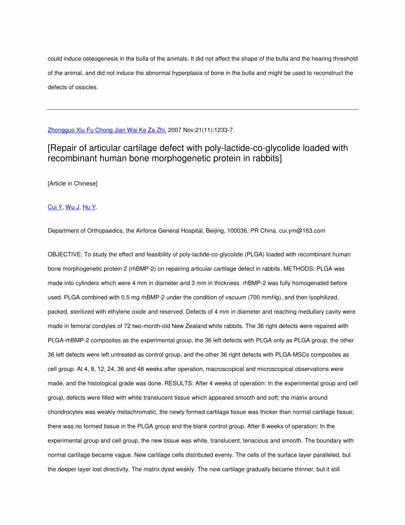
OBJECTIVE: To study the effect and feasibility of poly-lactide-co-glycolide (PLGA) loaded with recombinant human
bone morphogenetic protein 2 (rhBMP-2) on repairing articular cartilage defect in rabbits. METHODS: PLGA was
made into cylinders which were 4 mm in diameter and 3 mm in thickness. rhBMP-2 was fully homogenated before
used. PLGA combined with 0.5 mg rhBMP-2 under the condition of vacuum (700 mmHg), and then lyophilized,
packed, sterilized with ethylene oxide and reserved. Defects of 4 mm in diameter and reaching medullary cavity were
made in femoral condyles of 72 two-month-old New Zealand white rabbits. The 36 right defects were repaired with
PLGA-rhBMP-2 composites as the experimental group, the 36 left defects with PLGA only as PLGA group, the other
36 left defects were left untreated as control group, and the other 36 right defects with PLGA-MSCs composites as
cell group. At 4, 8, 12, 24, 36 and 48 weeks after operation, macroscopical and microscopical observations were
made, and the histological grade was done. RESULTS: After 4 weeks of operation: In the experimental group and cell
group, defects were filled with white translucent tissue which appeared smooth and soft; the matrix around
chondrocytes was weakly metachromatic, the newly formed cartilage tissue was thicker than normal cartilage tissue;
there was no formed tissue in the PLGA group and the blank control group. After 8 weeks of operation: In the
experimental group and cell group, the new tissue was white, translucent, tenacious and smooth. The boundary with
normal cartilage became vague. New cartilage cells distributed evenly. The cells of the surface layer paralleled, but
the deeper layer lost directivity. The matrix dyed weakly. The new cartilage gradually became thinner, but it still

thicker than the normal cartilage ones. The PLGA degraded besides some drops. In the blank control group and
PLGA group, a little white membrane formed at the bottom of the defect. After 12-24 weeks of operation: In the
experimental group and cell group, defects were filled with new tissues which were white, translucent, tenacious and
smooth. The boundary disappeared. The thickness of the new cartilage was similar to that of the normal ones. The
cells of the surface layer paralleled to each other,but the cells of the deeper layer tended to arrange vertically. The
matrix around chondrocytes was metachromatic, but the color was lighter than that of the normal cartilage. Bone
under the cartilage and the tide mark recovered. The new cartilage linked with nomal cartilage finely. In the blank
control group and PLGA group, there was a little fibrous tissue at the bottom of the defect withe obvious boundary.
After 36 weeks and 48 weeks of operation: in the experimental group and the cell group, the new cartilage was
slightly white, continuous and less smooth. The boundary disappeared. There was no proliferated synovial
membrane. The thickenss of the new cartilage was thinner than that of the normal ones. The matrix around
chondrocytes was weakly metachromatic. In the blank control group and PLGA group, the defect still existed, but
became smaller. At the bottom of the defect, fibrous tissues formed. Some cartilage denudated and became less
smooth. Some bone under cartilage exposed,and the synovial membrane became thick. The histologic grade of the
repair tissue at 12 weeks and 24 weeks of operation in experimental group and cell group was significantly different
from that at 4, 8 and 48 weeks of operation (P<0.01). There was also significant difference in the experimental group
and cell group compared with the blank control group and PLGA group at each time after operation (P<0.01). But
there was no significant difference between the experimental group and the cell group. CONCLUSION: In the course
of degradation, PLGA-rhBMP-2 composites releas rhBMP-2, which then act an effect on MSCs around defect and
induced them to differentiate for chondrocytes, and finally the defect is repaired. This simple and easy method may
be used clinically in the future.
Spine (Phila Pa 1976).Spine (Phila Pa 1976).Spine (Phila Pa 1976).Spine (Phila Pa 1976). 2002 Aug 15;27(16 Suppl 1):S40 2002 Aug 15;27(16 Suppl 1):S40 2002 Aug 15;27(16 Suppl 1):S40 2002 Aug 15;27(16 Suppl 1):S40----8.8.8.8.
Safety profile for the clinical use of bone morphogenetic proteins in the spine.
Poynton AR, Lane JM.
Department of Metabolic Bone Diseases, Hospital for Special Surgery, New York, New York 10021, USA.

STUDY DESIGN: A review was conducted. OBJECTIVE: To determine the safety profiles of human recombinant
bone morphogenetic protein-2 (rhBMP-2) and osteogenic protein-1 (OP-1) used clinically in spine applications.
SUMMARY OF BACKGROUND DATA: Safety issues associated with the use of bone morphogenetic proteins in
spine applications include the possibility of bony overgrowth, interaction with exposed dura, cancer risk, systemic
toxicity, reproductive toxicity, immunogenicity, local toxicity, osteoclastic activation, and effects on distal organs.
These issues have been given detailed examination in both human and animal studies, and safety data are available
for both rhBMP-2 and OP-1. The safety data available for OP-1 are less detailed. METHODS: The study involved
reviews of published reports and the safety data submitted to the Food and Drug Administration (rhBMP-2 and OP-1)
and to the European Agency for the Evaluation of Medicinal Products (OP-1), as well as personal communication with
the manufacturers of rhBMP-2 (Medtronic Sofamore Danek, Memphis, TN) and OP-1 (Stryker Biotech, Hopkinton,
MA). RESULTS: Application of either rhBMP-2 or OP-1 to raw decorticated bony surfaces leads to new bone
formation, which is desirable in the intertransverse or interbody regions. However, new bone formation also may
occur if rhBMP-2 or OP-1 comes in contact with laminectomy sites or decompressed neuroforamina, and may lead to
restenosis. Inadvertent placement of either rhBMP-2 or OP-1 in the spinal canal leads to formation of bone. Leakage
of rhBMP-2 or OP-1 outside the fusion area may lead to adjacent-level fusion. Accurate placement of these factors
and adequate retention by their carrier are highly important factors in minimizing these problems. Subdural bone
formation occurs if OP-1 is implanted directly beneath the dura. Osteoclastic overstimulation does not appear to be a
significant problem with rhBMP-2. However, bone resorption has been associated with OP-1 used in the setting of
thoracolumbar fractures. Findings show that RhBMP-2 has an antiproliferative effect on many cancer cells, and no
evidence exists that it is carcinogenic. It is unlikely that OP-1 has carcinogenic potential, although fewer data are
available. Systemic and local toxicity, significant adverse effects, and harmful effects on distant organs have not been
observed in either human or animal studies on rhBMP-2 and OP-1. The benign safety profile of rhBMP-2 may result
from its rapid systemic clearance, which results in very little systemic exposure. Systemic exposure to OP-1 also is
low. No reproductive toxicity has been observed with either rhBMP-2 or OP-1. However, there is no human safety
data. Subclinical immune responses in human subjects to collagen carriers have been reported. Antibody responses
to rhBMP-2 have been detected in less than 1% of spine patients. Low titer immune responses have been observed
in 38% of patients treated with OP-1. There were no associated clinical adverse effects. CONCLUSIONS: Given the
available data, both rhBMP-2 and OP-1 appear to be safe provided they are used appropriately, placed accurately,

not allowed to come into contact with decompressed areas, and contained in the region of fusion. They must be used
with caution in the presence of dural defects.
Eur Spine J. 2003 Oct;12(5):495-500. Epub 2003 Aug 8.
A pilot safety and efficacy study of OP-1 putty (rhBMP-7) as an adjunct to iliac crest autograft in posterolateral lumbar fusions.
Vaccaro AR, Patel T, Fischgrund J, Anderson DG, Truumees E, Herkowitz H, Phillips F, Hilibrand A, Albert TJ.
Department of Orthopaedic Surgery, Thomas Jefferson University, The Rothman Institute, 925 Chestnut St, 5th Floor,
Philadelphia, PA 19107, USA. [email protected]
The ability of bone morphogenetic proteins (BMPs) to induce bone formation has led to an increasing interest in the
potential for their use in fusion surgery. The purpose of this multi-center clinical pilot study was to evaluate the safety
of one such BMP-osteogenic protein 1, in the form of OP-1 putty-combined with autograft for intertransverse process
fusion of the lumbar spine in patients with symptomatic spinal stenosis and degenerative spondylolisthesis following
spinal decompression. Twelve patients with spinal stenosis and degenerative lumbar spondylolisthesis underwent
laminectomy and partial or complete medial facetectomy as required for decompression of the neural elements
followed by intertransverse process fusion by placing iliac crest autograft and OP-1 putty between the decorticated
transverse processes. No instrumentation was used. Patients were followed clinically using the Oswestry scale and
radiographically using static and dynamic radiographs to assess their fusion status. Independent and blinded
radiologists assessed the films for the presence of bridging bone between the transverse processes and measured
translation and angulation on dynamic films using digital calipers. In addition to bridging bone, less than or equal to 5
degrees of angular motion and less than or equal to 2 mm of translation were required to classify the patients as
successfully fused, as per the definition of successful fusion provided by the FDA for use in clinical trials involving
investigational devices to attain spinal fusion. Radiographic outcome was compared to a historical control (autograft
alone fusion without instrumentation for the treatment of degenerative spondylolisthesis). All adverse events were
recorded prospectively. The results showed 9 of the 12 patients (75%) obtained at least a 20% improvement in their
preoperative Oswestry score, while 6 of 11 patients (55%) with radiographic follow-up achieved a solid fusion by the

criteria used in this study. Bridging bone on the anteroposterior film was observed in 10 of the 11 patients (91%). No
systemic toxicity, ectopic bone formation, recurrent stenosis or other adverse events related to the OP-1 putty implant
were observed. A successful fusion was observed in slightly over half the patients in this study, using stringent criteria
without adjunctive spinal instrumentation. This study did not demonstrate the superiority of OP-1 combined with
autograft over an autograft alone historical control, in which the fusion rate was approximately 45%. The lack of
adverse events related to the OP-1 putty implant in this study is in agreement with other studies supporting the safety
of bone morphogenetic proteins in spinal surgery.
PMID: 12908103 [PubMed - indexed for MEDLINE]
Eur Spine J. 2005 Sep;14(7):623-9. Epub 2005 Jan 26.
A 2-year follow-up pilot study evaluating the safety and efficacy of op-1 putty (rhbmp-7) as an adjunct to iliac crest autograft in posterolateral lumbar fusions.
Vaccaro AR, Patel T, Fischgrund J, Anderson DG, Truumees E, Herkowitz H, Phillips F, Hilibrand A, Albert TJ.
Orthopaedic Surgery, Thomas Jefferson University and the Rothman Institute, Philadelphia, PA, USA.
Comment in:
• Eur Spine J. 2006 Jan;15(1):8-15.
The ability of bone morphogenetic proteins (BMPs) to induce bone formation has led to a multitude of investigations
into their use as bone graft substitutes in spinal surgery. The purpose of this multi-center clinical pilot study was to
evaluate the safety and efficacy of BMP-7 (osteogenic protein 1, OP-1), in the form of a putty, combined with
autograft for intertransverse process fusion of the lumbar spine in patients with symptomatic spinal stenosis and
degenerative spondylolisthesis following spinal decompression. Twelve patients with spinal stenosis and
degenerative lumbar spondylolisthesis underwent a laminectomy and partial or complete medial facetectomy as
required for decompression of the neural elements, followed by an intertransverse process fusion by placing iliac

crest autograft and OP-1 putty between the decorticated transverse processes. No instrumentation was used.
Patients were followed clinically using the Oswestry scale and SF-36 outcome forms, and radiographically using
static and dynamic radiographs to assess their fusion status over a 2-year period. Independent and blinded
radiologists assessed the films for the presence of bridging bone between the transverse processes and measured
translation and angulation on dynamic films using digital calipers. Radiographic outcome was compared to a historical
control (autograft alone fusion without instrumentation for the treatment of degenerative spondylolisthesis). All
adverse events were recorded prospectively. The results showed eight of the nine evaluable patients (89%) obtained
at least a 20% improvement in their preoperative Oswestry score, while five of ten patients (50%) with radiographic
follow-up achieved a solid fusion by the criteria used in this study. Bridging bone on the anteroposterior film was
observed in seven of the ten patients (70%). No systemic toxicity, ectopic bone formation, recurrent stenosis or other
adverse events related to the OP-1 putty implant were observed. A successful fusion was observed in slightly over
half the patients in this study, using stringent criteria without adjunctive spinal instrumentation. This study did not
demonstrate the statistical superiority of OP-1 combined with autograft over an autograft alone historical control, in
which the fusion rate was 45%. There were no adverse events related to the OP-1 putty implant in this study, which
supports findings in other studies suggesting the safety of bone morphogenetic proteins in spinal surgery.
Spine J. 2008 May-Jun;8(3):457-65. Epub 2007 May 25.
The safety and efficacy of OP-1 (rhBMP-7) as a replacement for iliac crest autograft for posterolateral lumbar arthrodesis: minimum 4-year follow-up of a pilot study.
Vaccaro AR, Whang PG, Patel T, Phillips FM, Anderson DG, Albert TJ, Hilibrand AS, Brower RS, Kurd MF,
Appannagari A, Patel M, Fischgrund JS.
Department of Orthopaedic Surgery, Thomas Jefferson University and The Rothman Institute, 925 Chestnut Street,
5(th) Floor, Philadelphia, Pennsylvania, PA 19107, USA.
BACKGROUND CONTEXT: Although autogenous bone is still considered to be the gold standard graft material for
promoting spinal fusion, other bone graft substitutes have been developed in an attempt to improve arthrodesis rates
and avoid the complications associated with the procurement of autograft. The bone morphogenetic proteins (BMPs)
represent a family of osteoinductive growth factors that are known to stimulate the osteoblastic differentiation of stem
cells. Osteogenic protein-1 (OP-1) Putty is a commercially available BMP preparation that is already approved for use
in humans. Previous clinical studies involving patients with degenerative spondylolisthesis have reported that the
efficacy and safety of OP-1 Putty is comparable to that of autograft at both 1- and 2-year follow-up. PURPOSE: The
purpose of this study was to evaluate the intermediate-term efficacy and safety of OP-1 Putty as an alternative to
autogenous bone by comparing the 4-year radiographic, clinical, and safety data of these same patients who
underwent decompression and uninstrumented fusion with either OP-1 Putty or iliac crest autograft. STUDY
DESIGN/SETTING: A prospective, randomized, controlled, multicenter clinical pilot study. PATIENT SAMPLE: Thirty-
six patients undergoing decompressive laminectomy and single-level uninstrumented fusion for degenerative
spondylolisthesis and symptomatic spinal stenosis were randomized in a 2:1 fashion to receive either OP-1 Putty (24
patients) or autogenous iliac crest bone graft (12 patients). OUTCOME MEASURES: Patient-reported outcome
measures consisting of Oswestry Disability Index and Medical Outcomes Study 36-Item Short Form Health Survey
(SF-36) scores were used to evaluate clinical efficacy. Perioperative data including operative time, estimated blood
loss, and duration of hospital stay were also recorded for each surgery. Postoperatively, a neurological examination
and an assessment of donor-site pain (if applicable) were performed at every follow-up visit. Radiographic fusion
success was defined as the presence of continuous bridging bone formation between the transverse processes at the
level of the spondylolisthesis with minimal motion evident on dynamic lateral x-ray films. The primary efficacy
endpoint was the overall success rate, a composite measure derived from both radiographic and clinical parameters.
The safety of OP-1 Putty was confirmed by comparing the nature and frequency of all adverse events and
complications that were prospectively observed in either of the groups. METHODS: Thirty-six patients with
degenerative spondylolisthesis and symptoms of neurogenic claudication underwent decompressive laminectomy
and single-level uninstrumented fusion with either OP-1 Putty or autograft. All patients were evaluated at 6 weeks and
3, 6, 9, 12, and 24 months, after which time they were instructed to return on a yearly basis. Multiple
neuroradiologists blinded to the assigned treatment reviewed static and dynamic X-ray films with digital calipers to
assess fusion status according to the presence of continuous bridging bone across the transverse processes as well
as the amount of residual motion evident at the level of interest. Oswestry Disability Index surveys and SF-36
questionnaires were used to assess clinical outcomes. RESULTS: At the 48-month time point, complete radiographic
and clinical data were available for 22 of 36 patients (16 OP-1 Putty and 6 autograft) and 25 of 36 patients (18 OP-1

Putty and 7 autograft), respectively. Radiographic evidence of a solid arthrodesis was present in 11 of 16 OP-1 Putty
patients (68.8%) and 3 of 6 autograft patients (50%). Clinically successful outcomes defined as at least a 20%
improvement in preoperative Oswestry scores were experienced by 14 of 19 OP-1 Putty patients (73.7%) and 4 of 7
autograft patients (57.1%); these clinical findings were corroborated by similar increases in SF-36 scores. The
respective overall success rates of the OP-1 Putty and autograft group were 62.5% and 33.3%. In this study, there
were no incidents of local or systemic toxicity, ectopic bone production, or other adverse events directly related to the
use of OP-1 Putty. CONCLUSION: Despite the challenges associated with obtaining a solid uninstrumented fusion in
patients with degenerative spondylolisthesis, the rates of radiographic fusion, clinical improvement, and overall
success associated with the use of OP-1 Putty were at least comparable to that of the autograft controls for at least
48 months after surgery. These results appear to validate the short-term results previously reported for OP-1 Putty
and suggest that this material may potentially represent a viable bone graft substitute for certain fusion applications.
Growth Factors. 2004 Dec;22(4):233-41.
Bone morphogenetic proteins.
Chen D, Zhao M, Mundy GR.
School of Medicine and Dentistry, Department of Orthopaedics, University of Rochester, Rochester, NY 14642, USA.
Bone morphogenetic proteins (BMPs) are multi-functional growth factors that belong to the transforming growth factor
beta (TGFbeta) superfamily. The roles of BMPs in embryonic development and cellular functions in postnatal and
adult animals have been extensively studied in recent years. Signal transduction studies have revealed that Smad1, 5
and 8 are the immediate downstream molecules of BMP receptors and play a central role in BMP signal transduction.
Studies from transgenic and knockout mice and from animals and humans with naturally occurring mutations in BMPs
and related genes have shown that BMP signaling plays critical roles in heart, neural and cartilage development.
BMPs also play an important role in postnatal bone formation. BMP activities are regulated at different molecular
levels. Preclinical and clinical studies have shown that BMP-2 can be utilized in various therapeutic interventions
such as bone defects, non-union fractures, spinal fusion, osteoporosis and root canal surgery. Tissue-specific

knockout of a specific BMP ligand, a subtype of BMP receptors or a specific signaling molecule is required to further
determine the specific role of a BMP ligand, receptor or signaling molecule in a particular tissue. BMPs are members
of the TGFbeta superfamily. The activity of BMPs was first identified in the 1960s (Urist, M.R. (1965) "Bone formation
by autoinduction", Science 150, 893-899), but the proteins responsible for bone induction remained unknown until the
purification and sequence of bovine BMP-3 (osteogenin) and cloning of human BMP-2 and 4 in the late 1980s
(Wozney, J.M. et al. (1988) "Novel regulators of bone formation: molecular clones and activities", Science 242, 1528-
1534; Luyten, F.P. et al. (1989) "Purification and partial amino acid sequence of osteogenin, a protein initiating bone
differentiation", J. Biol. Chem. 264, 13377-13380; Wozney, J.M. (1992) "The bone morphogenetic protein family and
osteogenesis", Mol. Reprod. Dev. 32, 160-167). To date, around 20 BMP family members have been identified and
characterized. BMPs signal through serine/threonine kinase receptors, composed of type I and II subtypes. Three
type I receptors have been shown to bind BMP ligands, type IA and IB BMP receptors (BMPR-IA or ALK-3 and
BMPR-IB or ALK-6) and type IA activin receptor (ActR-IA or ALK-2) (Koenig, B.B. et al. (1994) "Characterization and
cloning of a receptor for BMP-2 and BMP-4 from NIH 3T3 cells", Mol. Cell. Biol. 14, 5961-5974; ten Dijke, P. et al.
(1994) "Identification of type I receptors for osteogenic protein-1 and bone morphogenetic protein-4", J. Biol. Chem.
269, 16985-16988; Macias-Silva, M. et al. (1998) "Specific activation of Smad1 signaling pathways by the BMP7 type
I receptor, ALK2", J. Biol. Chem. 273, 25628-25636). Three type II receptors for BMPs have also been identified and
they are type II BMP receptor (BMPR-II) and type II and IIB activin receptors (ActR-II and ActR-IIB) (Yamashita, H. et
al. (1995) "Osteogenic protein-1 binds to activin type II receptors and induces certain activin-like effects", J. Cell. Biol.
130, 217-226; Rosenzweig, B.L. et al. (1995) "Cloning and characterization of a human type II receptor for bone
morphogenetic proteins", Proc. Natl Acad. Sci. USA 92, 7632-7636; Kawabata, M. et al. (1995) "Cloning of a novel
type II serine/threonine kinase receptor through interaction with the type I transforming growth factor-beta receptor",
J. Biol. Chem. 270, 5625-5630). Whereas BMPR-IA, IB and II are specific to BMPs, ActR-IA, II and IIB are also
signaling receptors for activins. These receptors are expressed differentially in various tissues. Type I and II BMP
receptors are both indispensable for signal transduction. After ligand binding they form a heterotetrameric-activated
receptor complex consisting of two pairs of a type I and II receptor complex (Moustakas, A. and C.H. Heldi (2002)
"From mono- to oligo-Smads: the heart of the matter in TGFbeta signal transduction" Genes Dev. 16, 67-871). The
type I BMP receptor substrates include a protein family, the Smad proteins, that play a central role in relaying the
BMP signal from the receptor to target genes in the nucleus. Smad1, 5 and 8 are phosphorylated by BMP receptors
in a ligand-dependent manner (Hoodless, P.A. et al. (1996) "MADR1, a MAD-related protein that functions in BMP2

signaling pathways", Cell 85, 489-500; Chen Y. et al. (1997) "Smad8 mediates the signaling of the receptor serine
kinase", Proc. Natl Acad. Sci. USA 94, 12938-12943; Nishimura R. et al. (1998) "Smad5 and DPC4 are key
molecules in mediating BMP-2-induced osteoblastic differentiation of the pluripotent mesenchymal precursor cell line
C2C12", J. Biol. Chem. 273, 1872-1879). After release from the receptor, the phosphorylated Smad proteins
associate with the related protein Smad4, which acts as a shared partner. This complex translocates into the nucleus
and participates in gene transcription with other transcription factors (). A significant advancement about the
understanding of in vivo functions of BMP ligands, receptors and signaling molecules has been achieved in recent
years. <figgrp> <title>Figure 1 BMP signaling and its regulation. BMP signals are mediated by type I and II BMP
receptors and their downstream molecules Smad1, 5 and 8. Phosphorylated Smad1, 5 and 8 proteins form a complex
with Smad4 and then are translocated into the nucleus where they interact with other transcription factors, such as
Runx2 in osteoblasts. BMP signaling is regulated at different molecular levels: (1) Noggin and other cystine knot-
containing BMP antagonists bind with BMP-2, 4 and 7 and block BMP signaling. Over-expression of noggin in mature
osteoblasts causes osteoporosis in mice (<citeref rid="bib9">Devlin et al., 2003</citeref>; <citeref rid="bib65">Wu et
al., 2003</citeref>). (2) Smad6 binds type I BMP receptor and prevents Smad1, 5 and 8 to be activated (<citeref
rid="bib22">Imamura et al., 1997</citeref>). Over-expression of Smad6 in chondrocytes causes delays in
chondrocyte differentiation and maturation (<citeref rid="bib21">Horiki et al., 2004</citeref>). (3) Tob interacts
specifically with BMP activated Smad proteins and inhibits BMP signaling. In Tob null mutant mice, BMP signaling is
enhanced and bone formation is increased (<citeref rid="bib71">Yoshida et al., 2000</citeref>). (4) Smurf1 is a Hect
domain E3 ubiquitin ligase. It interacts with Smad1 and 5 and mediates the degradation of these Smad proteins
(<citeref rid="bib76">Zhu et al., 1999</citeref>). (5) Smurf1 also recognizes bone-specific transcription factor Runx2
and mediates Runx2 degradation (<citeref rid="bib74">Zhao et al., 2003</citeref>). (6) Smurf1 also forms a complex
with Smad6, is exported from the nucleus and targeted to the type I BMP receptors for their degradation (<citeref
rid="bib40">Murakami et al., 2003</citeref>). Over-expression of Smurf1 in osteoblasts inhibits postnatal bone
formation in mice (<citeref rid="bib75">Zhao et al., 2004</citeref>).</title> <fig id="fig1"
name="GGRF0233fig001"></fig> </figgrp>
Med Oral Patol Oral Cir Bucal. 2009 Dec 29. [Epub ahead of print]

A comparative study of platelet-rich plasma, hydroxyapatite, demineralized bone matrix and autologous bone to promote bone regeneration after mandibular impacted third molar extraction.
Arenaz-Búa J, Luaces-Rey R, Sironvalle-Soliva S, Patiño-Seijas B, García-Rozado A, Martín-Sastre R, Ferreras-
Granados J, Lorenzo-Franco F, Vázquez-Mahía I, Lopez-Cedrun JL.
Travesía de Cordelería N 1 4 Izq, 15003 La Coruña, La Coruña, Spain, [email protected].
OBJECTIVES: 1) to compare mandibular bone regeneration by applying autologous bone, platelet-rich plasma and
two biomaterials (synthetic calcium hydroxyapatite, and demineralized bone matrix), and thus establish the potential
benefits of these biomaterials in the regeneration of postextraction alveolar bone, 2)to identify wich of them
accelerates more bone regeneration and 3)to determine whether there are differences in the postoperative period
(pain, swelling, trismus, infection) depending on the material used. STUDY DESIGN: It consists in a prospective,
controlled (with a split- mouth design) and double blinded study. We use as a model an easily reproducible non-
critical bone defect: the defect that remains after extraction of mandibular impacted third molar. The study design is
based on the extraction of two mandibular impacted third molars in a patient during the same surgical procedure by
the same surgeon. We assessed postoperative clinical data, and short, medium and long term neoformation of
alveolar bone after extraction. We compared the two sockets (right and left), which had been grafted in a different
way with the various elements mentioned above. In addition, we compared the postoperative inflammatory symptoms
between groups. RESULTS: The highest acceleration in bone formation was observed in groups in which we used
autologous bone and demineralized bone matrix. There were no statistically significant differences between groups
regarding pain, swelling, trismus and infection throughout the postoperative period. CONCLUSIONS: According to the
results of our study, autologous bone persists as the gold standard material for bone regeneration. Among the
assessed biomaterials, demineralized bone matrix has yielded the best results obtained. No significant differences in
the postoperative (pain, swelling, trismus and infectious events) were observed, depending on the type of material
used as a graft.
J Orthop Res. 2009 Nov 13. [Epub ahead of print]

Influence of bone-derived matrices on generation of bone in an ectopic rat model.
Bahar H, Yaffe A, Boskey A, Binderman I.
Department of Oral Biology, The Maurice and Gabriela Goldschleger, School of Dental Medicine, Tel Aviv University,
Tel Aviv, Israel.
Most bone regeneration experimental models that test bone-derived matrices take place in conjunction with the native
bone. Here, we compared the relative effectiveness of bone matrix components on bone-marrow-directed
osteogenesis in an ectopic model. Cortical bone cylinders consisted of diaphysis of DA rat femurs. They were either
demineralized (DBM), deproteinized (HABM), or nontreated (MBM). Fresh bone marrow was placed into cylinders
and implanted at subcutaneous thoracic sites of 2-month-old DA rats. At designated times the cylinders were
surgically removed from the animals. Microradiographs of DBM and histology of DBM and MBM cylinders
demonstrated progressive increase in mineralized bone volume and its trabecular configuration. Bone filled the inner
volume of DBM and MBM cylinders within 4 weeks, while in HABM cylinders mostly granulation tissue developed. In
the DBM cylinders cartilage deposited within 10 days, while in the MBM cylinders bone was directly deposited. As
early as day 3 after marrow transplantation, marrow cells interacting with DBM increased significantly the genes that
express the cartilage and the bone phenotype. In conclusion, organic components of bone are needed for marrow-
directed osteogenesis. (c) 2009 Orthopaedic Research Society. Published by Wiley Periodicals, Inc. J Orthop Res.
Eur J Orthod. 2010 Jan 11. [Epub ahead of print]
No influence of alimentary zinc on the healing of calvarial defects filled with osteopromotive substances in rats.
Jones L, Thomsen JS, Barlach J, Mosekilde L, Melsen B.
Department of Orthodontics, University of Aarhus, Denmark.

Zinc has been demonstrated to play an important role in bone metabolism and is required for normal growth.
However, no studies have investigated the influence of zinc on calvarial bone healing in aged or adult rats. The aim of
the study was to evaluate whether alimentary zinc supplementation and depletion affect bone healing of calvarial
defects implanted with osteopromotive substances in adult rats. Two 5 mm full thickness critical size bone defects
were trephined in the central part of each parietal bone of 60 six-month-old male Wistar rats. The bone defects were
filled with demineralized bone matrix (DBM), autogenous bone chips, or were left as unfilled controls. The rats were
divided into three groups of 20 rats each and received a semi-synthetic diet containing 20, 60, or 120 mg zinc/kg.
After 4 months, the biomechanical integrity of the healing defects was evaluated by a punch out test and the healed
defects were examined with histomorphometry. Statistical analysis of the data was carried out by two-way analysis of
variance and Wilcoxon's non-parametric signed rank test. Biomechanical testing revealed that the maximum load was
significantly higher in DBM-filled defects than in those filled with autogenous bone, and that the defects filled with
autogenous bone were stronger than the unfilled controls. The biomechanical findings indicated that the alimentary
zinc content did not influence the healing of calvarial defects. No significant difference in maximum load could be
established between the three diet groups for any of the filling materials, whereas the highest zinc supplement
resulted in an increase in the relative extension on mineralizing surfaces in the control group. Thus, healing of adult
rat calvarial defects is not influenced by alimentary zinc supplementation or depletion. Defects filled with DBM were
significantly stronger and exhibited significantly more new bone formation than defects filled with autogenous bone or
unfilled controls.
J Oral Sci. 2009 Sep;51(3):451-6.
Effects of demineralized bone matrix and a 'Ricinus communis' polymer on bone regeneration: a histological study in rabbit calvaria.
Laureano Filho JR, Andrade ES, Albergaria-Barbosa JR, Camargo IB, Garcia RR.
Oral and Maxillofacial Surgery, University of Pernambuco (UPE), Recife, Brazil. [email protected]
The aim of the present study was to histologically analyze the effects of bovine and human demineralized bone
matrix and a Ricinus communis polymer on the bone regeneration process. Two surgical bone defects were created

in rabbit calvaria, one on the right and the other on the left side of the parietal suture. Eighteen rabbits were divided
into three groups. In Group I, the experimental defect was treated with bovine demineralized bone matrix, Group II
with human demineralized bone matrix, and in Group III, the experimental cavity was treated with polyurethane resin
derived from Ricinus communis oil. The control defects were filled with the animals' own blood. The animals were
sacrificed after 7 and 15 weeks. Histological analysis revealed that in all groups (control and experimental), bone
regeneration increased with time. The least time required for bone regeneration was noted in the control group, with a
substantial decrease in the thickness of the defect. All materials proved to be biologically compatible, but
polyurethane resorbed more slowly and demonstrated considerably better results than the demineralized bone
matrices.
Cytokine. 2000 Nov;12(11):1630-8.
Stimulatory effects of cartilage-derived morphogenetic proteins 1 and 2 on osteogenic differentiation of bone marrow stromal cells.
Gruber R, Mayer C, Schulz W, Graninger W, Peterlik M, Watzek G, Luyten FP, Erlacher L.
Department of Rheumatology, Clinic of Internal Medicine III, Austria.
Cartilage-derived morphogenetic proteins 1 and 2 (CDMP-1 and CDMP-2) are members of the bone morphogenetic
protein (BMP) family which play an important role in embryonic skeletal development. Throughout adult life, bone
marrow-derived precursor cells maintain their ability to differentiate into osteoblasts in response to local growth
factors. This study examines the osteogenic potential of CDMP-1, CDMP-2, BMP-6 and osteogenic protein 1 (OP-1)
in bone marrow stromal cells (BMSC) and investigates the endogenous expression of CDMPs/BMPs and their
respective activin receptor-like kinase (ALK) receptors. A 4-day exposure of BMSC to CDMP-1, CDMP-2, BMP-6,
and OP-1 under serum-free conditions stimulated the progression of the osteogenic lineage in a dose-dependent
manner as evaluated by alkaline phosphatase activity and osteocalcin synthesis. In contrast to the BMPs, CDMP-1
and especially CDMP-2 were significantly less osteogenic, as confirmed by Northern blot analysis. Moreover, BMSC
were shown to express endogenously CDMP-2, BMP-2 to -6 and ALK-1, -2, -3, -5 and -6. Phenotypic
characterization of BMSC by RT-PCR showed transcripts of the fat marker adipsin and the prechondrocytic marker
procollagen type IIA; however, we were unable to detect the mature cartilage markers, procollagen type IIB and
aggrecan, even after growth factor treatment. Our data indicate that CDMP-1, CDMP-2, BMP-6 and OP-1 enhance
the osteogenic phenotype in BMSC, with CDMPs being clearly less osteogenic than BMPs. The endogenous
expression of a variety of CDMPs/BMPs and their respective ALK receptors, suggests a possible involvement of
these growth factors in the osteogenic differentiation of bone marrow progenitor cells. Copyright 2000 Academic
Press.
Spine (Phila Pa 1976). 1995 Dec 15;20(24):2633-44.
1995 Volvo Award in basic sciences. The use of an osteoinductive growth factor for lumbar spinal fusion. Part II: Study of dose, carrier, and species.
Boden SD, Schimandle JH, Hutton WC.
Department of Orthopaedics, Emory University School of Medicine, Decatur, Georgia, USA.
STUDY DESIGN: Efficacy of a bovine-derived osteoinductive growth factor was studied in a rabbit model and in a
nonhuman primate model of posterolateral lumbar spinal fusion. OBJECTIVES: To determine the minimum effective
dose of growth factor and the influence of different carrier material on the outcome of intertransverse process lumbar
fusion. SUMMARY OF BACKGROUND DATA: Bone morphogenetic proteins and related growth factors are
becoming increasingly available in purified extract or genetically engineered forms and are capable of inducing new
bone formation in vivo. Osteoinductive growth factors to enhance lumbar spinal infusion have not been well studied in
models of posterolateral intertransverse process fusion. Because of the diminished potential of bone regeneration in
primates (including humans) compared with phylogenetically lower animals, extrapolations regarding dose and
efficacy cannot be made directly from results obtained in experiments performed on phylogenetically lower animals.
Experiments on non-human primates are a critical step before attempting to use these growth factors on humans.
METHODS. One hundred fifteen adult New Zealand white rabbits and 10 adult rhesus macaques underwent single
level posterolateral intertransverse process lumbar spinal arthrodesis to evaluate different doses and carrier materials
for a bovine-derived osteoinductive bone protein extract. Rabbit fusion masses were evaluated 5 weeks after

arthrodesis by manual palpation, radiography, biomechanical testing, and light microscopy. Monkey fusion masses
were evaluated 12 weeks after arthrodesis by radiography and light microscopy. RESULTS: Successful posterolateral
intertransverse process spinal fusions were achieved in the rabbit models using an osteoinductive growth factor with
three different carriers (autogenous iliac bone, demineralized allogeneic bone matrix, and natural coral). There was a
dose-dependent response to the osteoinductive growth factor in the rabbit model, indicating that a threshold must be
overcome before bone formation is induced. The methodology for biologic enhancement of spinal fusion developed in
the rabbit model transferred successfully to the rhesus monkey, where the use of the osteoinductive growth factor
with a demineralized bone matrix carrier resulted in spinal fusion in 12 weeks. CONCLUSION: These experiments
provide an essential building block in the understanding of the biology of spinal fusion and the use of osteoinductive
growth factors to enhance a posterolateral intertransverse process spinal fusion. The achievement of posterolateral
spinal fusion in the rhesus monkey using an osteoinductive growth factor is a significant step toward the biologic
enhancement of spinal fusion in humans.
J Biomed Mater Res B Appl Biomater. 2009 Jan;88(1):115-22.
Tendon bone healing can be enhanced by demineralized bone matrix: a functional and histological study.
Sundar S, Pendegrass CJ, Blunn GW.
Centre for Biomedical Engineering, Institute of Orthopaedics and Musculoskeletal Science, University College
London, Brockley Hill, Stanmore, Middlesex, UK.
Rotator cuff repair surgery has high failure rates, with tendon reattachment to bone remaining a challenging clinical
problem. Increasing the integrity of the healing tendon-bone interface has been attempted by adopting a number of
different augmentation strategies. Because of chondrogenic and osteogenic properties we hypothesise that
demineralized bone matrix (DBM) augmentation of a healing tendon-bone interface will result in improved function,
and a morphology that more closely resembles that of a normal enthesis, compared with nonaugmented controls in
an ovine patellar tendon model. The right patellar tendon was detached from its insertion and reattached to an
osteotomized bone bed using suture anchors. Two groups were analyzed, the control group (without augmentation)
and the DBM group (DBM interposed between the tendon and bone). Animals were sacrificed at 12 weeks. Force
plate, mechanical, and histomorphometric analyses were performed. Tendon repairs failed at a rate of 33 and 0% for
the control and DBM groups, respectively. DBM augmentation resulted in significantly improved functional weight
bearing and increased amounts of fibrocartilage and mineralized fibrocartilage. This study shows that DBM enhances
tendon-bone healing and may reduce the high failure rates associated with rotator cuff repair clinically. (c)
J Bone Joint Surg Am. 2008 Oct;90(10):2206-19.
rhBMP-12 accelerates healing of rotator cuff repairs in a sheep model.
Seeherman HJ, Archambault JM, Rodeo SA, Turner AS, Zekas L, D'Augusta D, Li XJ, Smith E, Wozney JM.
Women's Health and Musculoskeletal Biology, Wyeth Discovery Research, Cambridge, MA 02140, USA.
BACKGROUND: The success rate of rotator cuff repairs is variable. This study was performed to evaluate the ability
of recombinant human bone morphogenetic protein-12 (rhBMP-12), administered in several carriers, to accelerate
healing in a sheep model of rotator cuff repair. METHODS: Local retention of tracer amounts of radiolabeled rhBMP-
12, added to non-radiolabeled rhBMP-12 delivered in buffer, hyaluronan paste or sponges, or Type-I or Type-I/III
collagen sponges was first evaluated with use of gamma scintigraphy in a pilot study of a rat intramuscular implant
model. The rhBMP-12/paste and sponge combinations were then evaluated in eight sheep each with unilateral
complete detachment and subsequent double-row reattachment of the infraspinatus tendon to the proximal part of the
humerus. Contralateral, normal shoulders from sixteen sheep and shoulders in which a repair had been done without
administration of rhBMP-12 in fourteen sheep were also evaluated. The rhBMP-12/Type-I and Type-I/III collagen
sponge combinations were each evaluated in eight additional sheep on the basis of superior efficacy. The Type-I/III
collagen sponge alone was evaluated in ten sheep to examine the effect of a collagen carrier. Ultrasound imaging
was performed at four and eight weeks. Radiographic evaluation, mechanical testing, and biochemical evaluation
were performed at eight weeks. Histological evaluation was performed on specimens from the sites of selected
repairs following mechanical testing. RESULTS: The sponge carriers had longer local retention of rhBMP-12 than did

the buffer or paste carriers in the rat models. All of the sheep shoulder-repair groups demonstrated ultrasound
evidence of a gap between the tendon and the humeral insertion. The gap length and the cross-sectional area of the
repair tissue decreased with time. The mechanical properties of the repairs treated with rhBMP-12 and hyaluronan
paste were similar to those of the untreated repairs. The maximum loads for the rhBMP-12/hyaluronan sponge and
rhBMP-12/collagen sponge-treated repairs were 2.1 and 2.7 times greater, respectively, than the loads for the
untreated repairs and were 33% and 42% of the value for the normal tendon at eight weeks. The maximum loads for
the repairs treated with rhBMP-12 and a Type-I or Type-I/III collagen sponge were 2.1 times greater than those for
the repairs treated with the Type-I/III collagen sponge alone. Changes in maximum stiffness followed a similar
pattern. Histological evaluation demonstrated accelerated healing of the rhBMP-12-treated repairs compared with the
untreated repairs. Bone formation was observed in all repairs, and biochemical measurements were not equivalent to
those of normal tendon at eight weeks. CONCLUSIONS: Delivery of rhBMP-12 in a collagen or hyaluronan sponge
resulted in accelerated healing of acute full-thickness rotator cuff repairs in a sheep model. CLINICAL RELEVANCE:
Delivery of rhBMP-12 in several sponge carriers has the potential to accelerate healing of rotator cuff repairs.
Accelerated repair may allow shorter rehabilitation and an earlier return to occupational and recreational activities.
J Shoulder Elbow Surg. 2007 Mar-Apr;16(2):251-4. Epub 2006 Nov 16.
The effect of cartilage-derived morphogenetic protein 2 on initial healing of a rotator cuff defect in a rat model.
Murray DH, Kubiak EN, Jazrawi LM, Araghi A, Kummer F, Loebenberg MI, Zuckerman JD.
NYU Hospital for Joint Diseases, Department of Orthopaedics, New York, NY 10003, USA.
This animal study evaluated the healing of supraspinatus tendon tears by use of a cartilage-derived morphogenetic
protein 2 growth factor (CDMP-2) delivered to the repair. Forty-eight rats had bilateral, surgically created complete
tears repaired by sutures with the growth factor introduced on one side. They were killed at 2, 3, 4, and 6 weeks, and
the strength of the repairs was determined and histologic analysis performed. At 4 and 6 weeks, the CDMP-2-treated
repairs were significantly stronger than the untreated repairs and histologic analysis showed more organized healing.

The use of growth factors introduced at the time of rotator cuff repair might promote more rapid healing and
subsequent, rapid patient rehabilitation.
J Neurosci. 2010 Jan 27;30(4):1502-11.
BAMBI (bone morphogenetic protein and activin membrane-bound inhibitor) reveals the involvement of the transforming growth factor-beta family in pain modulation.
Tramullas M, Lantero A, Díaz A, Morchón N, Merino D, Villar A, Buscher D, Merino R, Hurlé JM, Izpisúa-Belmonte
JC, Hurlé MA.
Facultad de Medicina, Universidad de Cantabria, 39011 Santander, Spain, Instituto de Formación e Investigación
Marqués de Valdecilla, 39008 Santander, Spain.
Transforming growth factors-beta (TGF-betas) signal through type I and type II serine-threonine kinase receptor
complexes. During ligand binding, type II receptors recruit and phosphorylate type I receptors, triggering downstream
signaling. BAMBI [bone morphogenetic protein (BMP) and activin membrane-bound inhibitor] is a transmembrane
pseudoreceptor structurally similar to type I receptors but lacks the intracellular kinase domain. BAMBI modulates
negatively pan-TGF-beta family signaling; therefore, it can be used as an instrument for unraveling the roles of these
cytokines in the adult CNS. BAMBI is expressed in regions of the CNS involved in pain transmission and modulation.
The lack of BAMBI in mutant mice resulted in increased levels of TGF-beta signaling activity, which was associated
with attenuation of acute pain behaviors, regardless of the modality of the stimuli (thermal, mechanical,
chemical/inflammatory). The nociceptive hyposensitivity exhibited by BAMBI(-/-) mice was reversed by the opioid
antagonist naloxone. Moreover, in a model of chronic neuropathic pain, the allodynic responses of BAMBI(-/-) mice
also appeared attenuated through a mechanism involving delta-opioid receptor signaling. Basal mRNA and protein
levels of precursor proteins of the endogenous opioid peptides proopiomelanocortin (POMC) and proenkephalin
(PENK) appeared increased in the spinal cords of BAMBI(-/-). Transcript levels of TGF-betas and their intracellular
effectors correlated directly with genes encoding opioid peptides, whereas BAMBI correlated inversely. Furthermore,
incubation of spinal cord explants with activin A or BMP-7 increased POMC and/or PENK mRNA levels. Our findings

identify TGF-beta family members as modulators of acute and chronic pain perception through the transcriptional
regulation of genes encoding the endogenous opioids.
BMC Med Genet. 2009 Dec 19;10:141.
Association of a functional microsatellite within intron 1 of the BMP5 gene with susceptibility to osteoarthritis.
Wilkins JM, Southam L, Mustafa Z, Chapman K, Loughlin J.
University of Oxford, Institute of Musculoskeletal Sciences, Botnar Research Centre, Nuffield Orthopaedic Centre,
Oxford, OX3 7LD, UK. [email protected]
BACKGROUND: In a previous study carried out by our group, the genotyping of 36 microsatellite markers from within
a narrow interval of chromosome 6p12.3-q13 generated evidence for linkage and for association to female hip
osteoarthritis (OA), with the most compelling association found for a marker within intron 1 of the bone morphogenetic
protein 5 gene (BMP5). In this study, we aimed to further categorize the association of variants within intron 1 of
BMP5 with OA through an expanded genetic association study of the intron and subsequent functional analysis of
associated polymorphisms. METHODS: We genotyped 18 common polymorphisms including 8 microsatellites and 9
single nucleotide polymorphisms (SNPs) and 1 insertion/deletion (INDEL) from within highly conserved regions
between human and mouse within intron 1 of BMP5. These markers were then tested for association to OA by a two-
stage approach in which the polymorphisms were initially genotyped in a case-control cohort comprising 361
individuals with associated polymorphisms (P < or = 0.05) then genotyped in a second case-control cohort comprising
1185 individuals. RESULTS: Two BMP5 intron 1 polymorphisms demonstrated association in the combined case-
control cohort of 1546 individuals (765 cases and 781 controls): microsatellite D6S1276 (P = 0.018) and SNP
rs921126 (P = 0.013). Functional analyses in osteoblastic, chondrocytic, and adipocytic cell lines indicated that allelic
variants of D6S1276 have significant effects on the transcriptional activity of the BMP5 promoter in vitro.
CONCLUSION: Variability in gene expression of BMP5 may be an important contributor to OA genetic susceptibility.
J Rheumatol. 2010 Feb;37(2):246-56. Epub 2009 Dec 15.
Peripheral blood expression profiles of bone morphogenetic proteins, tumor necrosis factor-superfamily molecules, and transcription factor Runx2 could be used as markers of the form of arthritis, disease activity, and therapeutic responsiveness.
Grcevic D, Jajic Z, Kovacic N, Lukic IK, Velagic V, Grubisic F, Ivcevic S, Marusic A.
Department of Physiology and Immunology, University of Zagreb School of Medicine, Zagreb, Croatia.
OBJECTIVE: To assess whether different forms of arthritis and disease activity could be distinguished by peripheral
blood expression profiles of bone-regulatory factors including tumor necrosis factor (TNF)-superfamily [TNF-related
apoptosis-inducing ligand (TRAIL), the Fas ligand (FasL), and the ligand for herpesvirus entry mediator (LIGHT)] and
bone morphogenetic protein (BMP)-family members (BMP-2, BMP-4, BMP-6) as well as osteoblast differentiation
gene Runx2. METHODS: Blood cells from healthy controls (n = 25) and patients at different disease stages with
rheumatoid arthritis (RA; n = 49), osteoarthritis (OA; n = 17), or spondyloarthritis, including ankylosing spondylitis (AS;
n = 27) or psoriatic arthritis (PsA; n = 23), were processed for quantitative polymerase chain reaction. Gene
expression was assessed in comparison with control samples, correlated with clinical data of different forms of
arthritis, and analyzed for discriminative efficacy between groups by receiver-operation characteristic (ROC) curves.
Results were confirmed on diagnostic RA (n = 5) and AS (n = 8) samples. RESULTS: BMP-4, BMP-6, and Runx2
expressions were significantly decreased in patients with RA and OA versus controls. Patients with RA also had
decreased FasL and LIGHT expression, while patients with AS had increased Runx2 expression. Negative
correlation with disease activity was found for BMP-4, FasL, and Runx2 in RA and for Runx2 in PsA, while positive
correlation was found for BMP-4 in PsA. Gene expression was higher in the therapy-resistant form of AS (for BMP-4,
LIGHT, and Runx2) and in methotrexate-treated patients in RA (for BMP-2 and LIGHT). ROC curve analysis
confirmed discrimination between groups, particularly decreased LIGHT and Runx2 for RA and increased Runx2 for
AS. CONCLUSION: Our study identified BMP and Runx2 as possible biomarkers of bone metabolism in several
forms of arthritis, while lower FasL and LIGHT were associated with RA. Correlation between gene expression and
disease activity may be clinically useful in assessing therapeutic effectiveness and disease monitoring.

Pak J Biol Sci. 2009 Sep 1;12(17):1194-9.
Effect of bone morphogenetic protein-2 on normal and osteoarthritic human articular chondrocytes.
Orazizadeh M, Hashemitabar M, Fakoor M, Moghadam MT.
Department of Anatomical Sciences, Medical School of Ahvaz Jondishapour University, Ahvaz, Iran.
In this study, we investigated whether Bone Morphogenetic Protein-2 (BMP-2) could modulate dedifferentiation,
apoptosis and proliferation capacity in the normal and OA cultured chondrocytes. The articular chondrocytes from
normal (n = 4) and OA (n = 4) cartilages were harvested separately. The chondrocytes were cultured in monolayer in
the presence of 100 ng mL(-1) BMP-2 and 1% FBS as a test group and 1% FBS alone as a control group. Then, the
chondrocytes were harvested and assessed for morphology with invert microscopy, proliferation by using MTT-assay
and apoptosis with caspase-3 immunocytochemistry. The results indicated that the normal and the most OA
chondrocytes showed round and polygonal appearance with chondrocyte-like morphology in BMP-2 treated groups
after 6 days. The MTT proliferation test didn't show significant difference between test and control groups. The OA
cells showed proliferation rate higher than the normal cells and significant difference in the presence of BMP-2 was
observed (p<0.05). Positive immunostaining of caspase-3 in test and control groups was 1 and 20% in normal and 30
and 43% in OA groups, respectively. The percentage of apoptosis was reduced in the presence of BMP-2. In
conclusion, it appears that BMP-2 involves in suppression of dedifferentiation and apoptosis processes of cultured
human chondrocytes.
J Orthop Res. 2009 Aug;27(8):1088-92.
Periodic knee injections of BMP-7 delay cartilage degeneration induced by excessive running in rats.
Sekiya I, Tang T, Hayashi M, Morito T, Ju YJ, Mochizuki T, Muneta T.
Section of Cartilage Regeneration, Graduate School, Tokyo Medical and Dental University, Tokyo, Japan.

Strenuous running of rats enhances mechanical stress on the knee, thereby inducing degeneration of articular
cartilage. Bone morphogenetic protein-7 (BMP-7) has an inhibitory effect on cartilage degeneration, suggesting its
usefulness for human osteoarthritis patients. However, its mode of administration should be investigated. We
examined whether weekly knee injections of BMP-7 delayed the progression of cartilage degeneration. Wistar rats
were forced to run 30 km in 6 weeks on a rodent treadmill, and BMP-7 was injected weekly into the knee.
Macroscopically and histologically, this strenuous running regimen induced cartilage degeneration. Weekly injections
of 250 ng BMP-7 delayed the progression of cartilage degeneration. Immunohistochemically, in the control knee, type
II collagen expression decreased, while BMP-7 expression in chondrocytes slightly increased. Interestingly, weekly
injection of BMP-7 increased BMP-7 expression even 9 days after the final injection. Disulfate disaccharide keratan
sulfate in serum transiently increased in the control group, while it remained at a low level in the BMP-7 group.
Weekly BMP-7 injection increased BMP-7 expression in chondrocytes and its effect seemed to last more than 7 days.
The effect of BMP-7 could be monitored by serum keratan sulfate concentration. Periodical injections of BMP-7
delayed progression of cartilage degeneration induced by excessive running in rats.
Coll Antropol. 2008 Oct;32 Suppl 2:83-7.
Expression of bone morphogenetic proteins, cartilage-derived morphogenetic proteins and related receptors in normal and osteoarthritic human articular cartilage.
Bobinac D, Spanjol J, Marinović M, Zoricić Cvek S, Marić I, Cicvarić T, Fuckar D, Markić D, Vojniković B.
Department of Anatomy, School of Medicine, University of Rijeka, Rijeka, Croatia.
Newborn and adult articular cartilage expresses bone (BMPs) and cartilage derived morphogenetic proteins
(CDMPs). These morphogenetic proteins act over membrane receptors (BMPRs). We examined the expression
pattern of BMP-7, BMP-3, CDMP-1, CDMP-2 and their receptors in adult normal and osteoarthritic, articular, knee
cartilage. Immunostaining was carried out using polyclonal antibodies. The expression of BMP-7,-3, CDMP-1,-2 was
detected in all layers of normal articular cartilage with the strongest expression in chondrocytes of the transitional
layer. BMP-7 and CDMPs expression decreased in osteoarthritic articular cartilage whereas BMP-3 expression was
absent. BMPR-IA and BMPR-II were strongly expressed in both normal and osteoarthritic articular cartilage. BMPR-

IB was not expressed in osteoarthritic (OA) cartilage. BMPs and CDMPs with intact signalling play an important role
in articular cartilage homeostasis, preventing cartilage degeneration
Clin Orthop Relat Res. 2009 Dec;467(12):3221-9. Epub 2008 Oct 22.
Use of bone morphogenic protein-7 as a treatment for osteoarthritis.
Badlani N, Oshima Y, Healey R, Coutts R, Amiel D.
Department of Orthopaedic Surgery, University of California, San Diego, 9500 Gilman Drive, Mail Code 0630, La
Jolla, CA 92093-0630, USA.
Osteoarthritis is a degenerative disorder resulting from breakdown of articular cartilage. Previous work has shown
bone morphogenic protein-7 has a potential protective effect on cartilage during the development of osteoarthritis.
The purpose of this study was to determine whether bone morphogenic protein-7 could decrease the amount of
cartilage degradation in preexisting osteoarthritis. The rabbit ACLT model was used as a model of osteoarthritis.
Bone morphogenic protein-7 was delivered via Alzet osmotic pump to the joint 4 weeks after anterior cruciate
ligament transection; thus cartilage injury was preexisting. The experimental group showed less cartilage degradation
than the controls, with an average Outerbridge score of 1.9 versus 2.6 for the controls. Histomorphometry showed a
trend toward less cartilage degradation in the bone morphogenic protein-7 group when compared with controls.
Semiquantitative real-time polymerase chain reaction showed a considerably greater expression of aggrecan in the
bone morphogenic protein-7-treated cartilage when compared with controls and less expression of matrix
metalloproteinase-3 and matrix metalloproteinase-13, important catabolic mediators. The synovial tissue of the
experimental group also showed considerably less expression of matrix metalloproteinase-3, matrix
metalloproteinase-13, and aggrecanase. These results indicate bone morphogenic protein-7 may reduce degradation
of articular cartilage in osteoarthritis.
Arthritis Res Ther. 2008;10(5):R115. Epub 2008 Sep 24.

Dynamic activation of bone morphogenetic protein signaling in collagen-induced arthritis supports their role in joint homeostasis and disease.
Daans M, Lories RJ, Luyten FP.
Laboratory for Skeletal Development and Joint Disorders, Division of Rheumatology, Department of Musculoskeletal
Sciences, Katholieke Universiteit Leuven, Herestraat 49 box 813, Leuven 3000, Belgium.
INTRODUCTION: Rheumatoid arthritis is a chronic systemic autoimmune disease affecting peripheral joints and
leading to loss of joint function. The severity and outcome of disease are dependent on the balance between
inflammatory/destructive and homeostatic or repair pathways. Increasing evidence suggests a role for bone
morphogenetic protein (BMP) signaling in joint homeostasis and disease. METHODS: Activation of BMP signaling in
collagen-induced arthritis as a model of rheumatoid arthritis was studied by immunohistochemistry and Western blot
for phosphorylated SMAD1/5 at different time points. Expression of different BMP ligands and noggin, a BMP
antagonist, was determined on synovium and cartilage extracts of arthritic knees, at different time points, with
quantitative polymerase chain reaction. At the protein level, BMP2 and BMP7 were studied with
immunohistochemistry. Finally, the effect of anti-tumor necrosis factor-alpha (TNFalpha) treatment on the expression
of BMP2, BMP7, and growth and differentiation factor-5 (GDF5) in synovium and cartilage of arthritic knees was
investigated. RESULTS: A time-dependent activation of the BMP signaling pathway in collagen-induced arthritis was
demonstrated with a dynamic and characteristic expression pattern of different BMP subfamily members in synovium
and cartilage of arthritic knees. As severity increases, the activation of BMP signaling becomes more prominent in the
invasive pannus tissue. BMP2 is present in cartilage and the hyperplastic lining layer. BMP7 is found in the sublining
zone and inflammatory infiltrate. Treatment with etanercept slowed down progression of disease, but no change in
expression of GDF5, BMP2, and BMP7 in synovium was found; in the cartilage, however, blocking of TNFalpha
increased the expression of BMP7. CONCLUSIONS: BMP signaling is dynamically activated in collagen-induced
arthritis and is partly TNFalpha-independent. TNFalpha blocking increased the expression of BMP7 in the articular
cartilage, possibly enhancing anabolic mechanisms. Different types of source and target cells are recognized. These
data further support a role for BMP signaling in arthritis.

Nat Genet. 2009 Apr;41(4):478-81. Epub 2009 Mar 1.
Lack of the bone morphogenetic protein BMP6 induces massive iron overload.
Meynard D, Kautz L, Darnaud V, Canonne-Hergaux F, Coppin H, Roth MP.
Inserm, U563, Toulouse, France.
Comment in:
• Nat Genet. 2009 Apr;41(4):386-8.
• Hepatology. 2009 Jul;50(1):316-9.
• Clin Genet. 2009 Oct;76(4):341-3.
Expression of hepcidin, a key regulator of intestinal iron absorption, can be induced in vitro by several bone
morphogenetic proteins (BMPs), including BMP2, BMP4 and BMP9 (refs. 1,2). However, in contrast to BMP6,
expression of other BMPs is not regulated at the mRNA level by iron in vivo, and their relevance to iron homeostasis
is unclear. We show here that targeted disruption of Bmp6 in mice causes a rapid and massive accumulation of iron
in the liver, the acinar cells of the exocrine pancreas, the heart and the renal convoluted tubules. Despite their severe
iron overload, the livers of Bmp6-deficient mice have low levels of phosphorylated Smad1, Smad5 and Smad8, and
these Smads are not significantly translocated to the nucleus. In addition, hepcidin synthesis is markedly reduced.
This indicates that Bmp6 is critical for iron homeostasis and that it is functionally nonredundant with other members of
the Bmp subfamily. Notably, Bmp6-deficient mice retain their capacity to induce hepcidin in response to inflammation.
The iron burden in Bmp6 mutant mice is significantly greater than that in mice deficient in the gene associated with
classical hemochromatosis (Hfe), suggesting that mutations in BMP6 might cause iron overload in humans with
severe juvenile hemochromatosis for which the genetic basis has not yet been characterized.


![Research Paper COL4A2 in the tissue -specific …binds bone morphogenetic proteins (such as osteogenin and osteogenic protein-1) involved in angiogenesis and osteogenesis [26,27]](https://img.pdfslide.us/doc/110x75/5f0629687e708231d4169a0c/research-paper-col4a2-in-the-tissue-specific-binds-bone-morphogenetic-proteins.jpg)