Embed Size (px)
Citation preview
/ . EmbryoL exp. Morph. Vol. 24, 3, pp. 467-478, 1970 4 6 7Printed in Great Britain
Is the presumptive notochord responsible forsomite genesis in the chick?
By G. NICOLET1
From the Institut d'Histologie et cTEmbryologie,Laboratoire d'Embryologie experiment ale, Ecole de Medecine
de VUniversite de Geneve, Switzerland
SUMMARYFragments of Hensen's node or head process were implanted into the primitive streak
0-8 mm behind the node. The posterior region was isolated from all influences emanatingfrom the anterior part of the blastoderm, either by cutting off a large rectangular piece just infront of the implant or by explanting the region combined with the notochord implant intothe area opaca. As controls, corresponding regions of the primitive streak were isolated orexplanted.
In controls, somite formation never took place, while the implantation of presumptivenotochord into this posterior region was followed by the formation of somites in most cases.
In these experiments, the action of the presumptive notochord in its new situation wassimilar to that of the chorda-factor in amphibians.
INTRODUCTION
Several authors have suggested that a role may be ascribed to Hensen's nodein somite formation (Peebles, 1898; Wetzel, 1929; Fraser, 1954). In the presentwork, we have attempted to analyse how far the presumptive notochord, whichis one of the major components of the node, may be implicated in this processby implanting fragments of presumptive notochord into the posterior parts ofthe primitive streak, as performed in slightly different conditions by Bellairs(1963). Adequate controls were carried out to show that the grafts of presump-tive notochord were not contaminated by presumptive somitic cells and thatthe posterior parts used were unable to form somites spontaneously.
MATERIALS AND METHODS
Chick blastoderms cultured in vitro (Gallera & Nicolet, 1961) were employed.The normal stages of development were defined according to the tables ofHamburger & Hamilton (1951). As mentioned in Table 1, our research isdivided into two experimental systems. In the experimental series, we sometimesused labelled grafts to recognize accurately the structures provided by the1 Author's address: Institut d'Histologie, Ecole de Medecine, 1211, Geneve 4, Switzerland.
468 G. NICOLET
implants and to be sure that they did not contain any presumptive somitic cells.In experimental series as well as in controls, the operation was performed atstage 5, when the length of the head process was about 0-5 mm.
Table 1. Design of experiments
Experimental system 1 Control 1: 8 casesExperiment 1: 8 (5)* casesExperiment 2:18 (6) ""cases
Experimental system 2 Control 2: 8 casesControl 3: 16 casesExperiment 3: 5 (4)* casesExperiment 4: 9 (4)* cases
* The numbers in parentheses represent the cases in which labelled grafts were implanted.
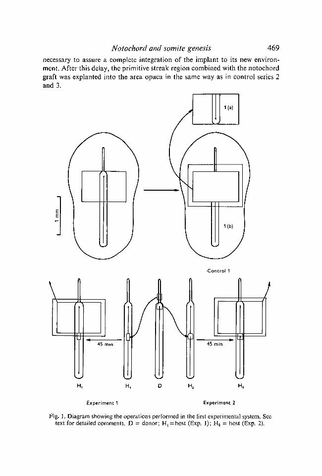
In each series, the surgical operation required several steps as indicated byFig. 1 for the first experimental system. The control series 1 consisted of extir-pating a rectangular area containing the anterior part of the primitive streak.After its antero-posterior axis had been rotated through 180°, it was trans-planted into the area opaca. The germ-wall at the site of explantation was care-fully eliminated before we applied the ventral side of the explant to the ventralsurface of the peripheral ectoblast. The injury made in the blastoderm wascovered by a piece of a thick millipore filter (150 fi) to prevent its enlargement.As two regions were useful for our purposes, they are designated as controlseries \{d) and (b). In Exps 1 and 2, a small square of primitive streak was cutoff 0-8 mm behind the node and was exchanged for a graft of similar size comingeither from the young head process (Exp. 1) or from Hensen's node (Exp. 2).After 45 min, the implant was sufficiently attached to the neighbouring tissuesto make it possible to extirpate the anterior part of the primitive streak andcover the hole by a piece of millipore filter.
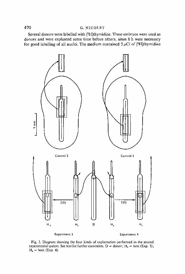
In the second experimental system, various regions of the primitive streakwere explanted on the area opaca (Fig. 2). They were mainly composed ofinvaginating mesoblastic cells but also contained the few endoblastic cells whichcover the ventral side of the primitive streak. On the other hand, some part ofthe neural tissue surrounding the node was involuntarily cut off with the graftin a few cases. Later, this neural tissue was found on sections as a small masstightly attached to the notochord. As in control series l(a), the ventral side ofexplants was applied to the ectoblast and their cephalocaudal axis rotatedthrough 180°. A piece of millipore filter was put on the hole, so that it waspossible to use only one host embryo for all the experimental process. InExps 3 and 4, the explantation was preceded by the implantation of a notochordpiece of two different origins (Fig. 2). The implant was inserted 0-8 mm behindthe node. The host Hensen's node was also excised to slow down the regressionmovement during the time required for the healing. A lapse of time of 2 h was
Notochord and somite genesis 469
necessary to assure a complete integration of the implant to its new environ-ment. After this delay, the primitive streak region combined with the notochordgraft was explanted into the area opaca in the same way as in control series 2and 3.
Control 1
H, H,
Experiment 1 Experiment 2
Fig. 1. Diagram showing the operations performed in the first experimental system. Seetext for detailed comments. D = donor; H^hos t (Exp. 1); H2 = host (Exp. 2).
470 G. NICOLET
Several donors were labelled with [3H]thymidine. These embryos were used asdonors and were explanted some time before others, since 8 h were necessaryfor good labelling of all nuclei. The medium contained 5 /*Ci of [3H]thymidine
Control 2
us 2(h)
H4
Experiment 3 Experiment 4
Fig. 2. Diagram showing the four kinds of explantation performed in the secondexperimental system. See text for further comments. D = donor; H3 = host (Exp. 3);H4 = host (Exp. 4).
Notochord and somite genesis 471
(Radiochemical Centre, Amersham, England). For the auto radiography, thetechnique described by Ficq (1959) was used and Ilford emulsion K2. A correctexposure of the autoradiographic plates required about 30 days.
All the host embryos were incubated for about 36 h (including the incubationtime in ovo). They were fixed with Carnoy, drawn in toto in the camera lucida andoften photographed. The histological study has been made on serial sections of8 /( stained with kernechtrot.
RESULTS
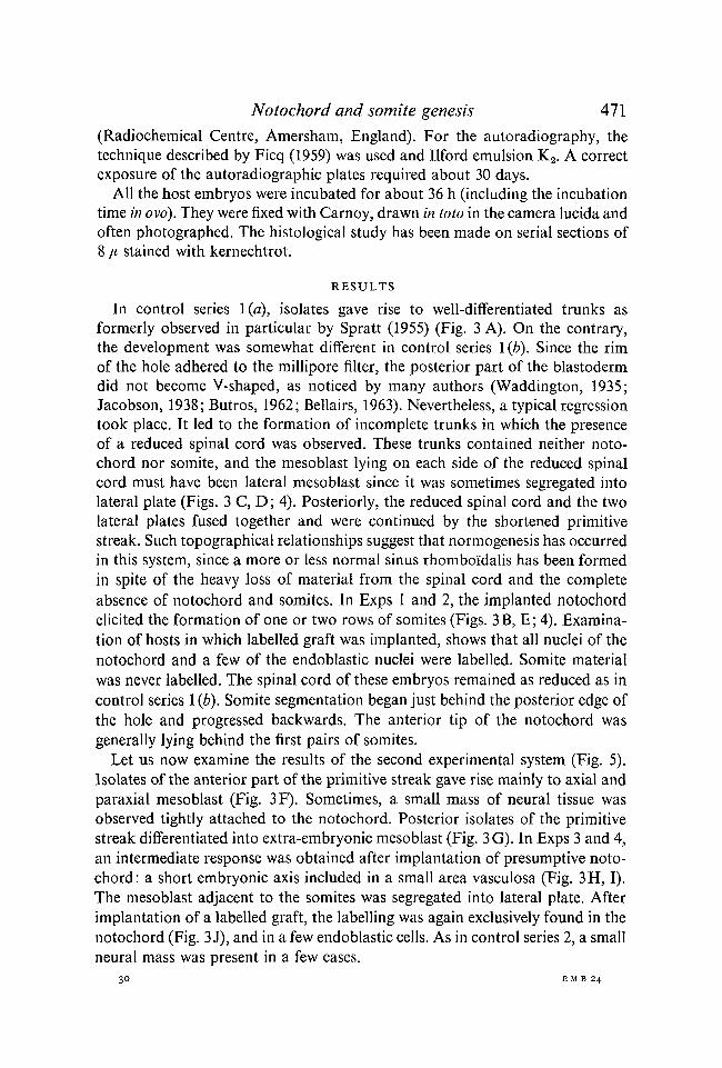
In control series I (a), isolates gave rise to well-differentiated trunks asformerly observed in particular by Spratt (1955) (Fig. 3 A). On the contrary,the development was somewhat different in control series 1 (b). Since the rimof the hole adhered to the millipore filter, the posterior part of the blastodermdid not become V-shaped, as noticed by many authors (Waddington, 1935;Jacobson, 1938; Butros, 1962; Bellairs, 1963). Nevertheless, a typical regressiontook place. It led to the formation of incomplete trunks in which the presenceof a reduced spinal cord was observed. These trunks contained neither noto-chord nor somite, and the mesoblast lying on each side of the reduced spinalcord must have been lateral mesoblast since it was sometimes segregated intolateral plate (Figs. 3 C, D; 4). Posteriorly, the reduced spinal cord and the twolateral plates fused together and were continued by the shortened primitivestreak. Such topographical relationships suggest that normogenesis has occurredin this system, since a more or less normal sinus rhomboiidalis has been formedin spite of the heavy loss of material from the spinal cord and the completeabsence of notochord and somites. In Exps 1 and 2, the implanted notochordelicited the formation of one or two rows of somites (Figs. 3B, E; 4). Examina-tion of hosts in which labelled graft was implanted, shows that all nuclei of thenotochord and a few of the endoblastic nuclei were labelled. Somite materialwas never labelled. The spinal cord of these embryos remained as reduced as incontrol series 1 (6). Somite segmentation began just behind the posterior edge ofthe hole and progressed backwards. The anterior tip of the notochord wasgenerally lying behind the first pairs of somites.
Let us now examine the results of the second experimental system (Fig. 5).Isolates of the anterior part of the primitive streak gave rise mainly to axial andparaxial mesoblast (Fig. 3F). Sometimes, a small mass of neural tissue wasobserved tightly attached to the notochord. Posterior isolates of the primitivestreak differentiated into extra-embryonic mesoblast (Fig. 3G). In Exps 3 and 4,an intermediate response was obtained after implantation of presumptive noto-chord: a short embryonic axis included in a small area vasculosa (Fig. 3H, I).The mesoblast adjacent to the somites was segregated into lateral plate. Afterimplantation of a labelled graft, the labelling was again exclusively found in thenotochord (Fig. 3 J), and in a few endoblastic cells. As in control series 2, a smallneural mass was present in a few cases.
30 E M B 24
472 G. NICOLET
88
18
1659
06
18
049
075
100
080
100
Notochord and somite genesis 473
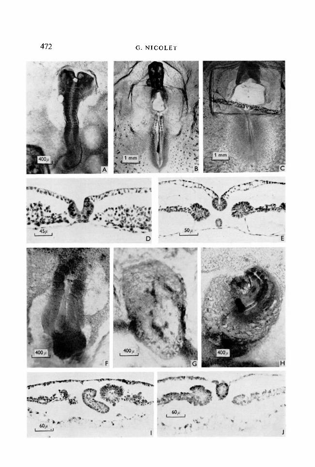
The results are expressed in a semi-quantitative way in two tables (Tables 2, 3).Table 2 shows that no somite formation took place in the posterior region (seecontrol series 1 (b) and 3) unless it was combined with a notochord fragment,but somite formation was observed whenever Hensen's node was implanted.On the other hand, the young head process did not succeed in provoking somitesegmentation in all cases.
Examination of Table 3 shows how far the somitic response was linked eitherto the presence of notochord or to that of neural tissue. The notochord was
Table 2. Frequency of somite formation as observed in experimentalseries and their respective controls
Number Presenceof of Frequency
Series cases somites (%)
Control 1 (b)Experiment IExperiment 2Control 3Experiment 3Experiment 4
Fig. 3 (A). This isolate explanted in the control series l(a) differentiated into atypical trunk. A well-differentiated, widely open neural tube is lying over themesoblast. x 25.(B). This case concerns Exp. 2. Segmented somites are formed along the implantednotochord. x 10.(C). In control series 1(6), the posterior region gives rise only to an incompletetrunk devoid of notochord and somites, x 10.(D). Section across the incomplete trunk seen in (C). The floor of the reduced spinalcord lies directly on the endoblast. On each side, the mesoblast is not well organizedinto lateral plate, x 220.(E). Section through the embryo shown in (B). The topographical relationshipsbetween the various Anlage are almost normal, except that the notochord is notattached to the neural tube, x 200.(F). This explant coming from the anterior part of the primitive streak differentiatesmainly into somites and notochord. x 25.(G). In control series 3, the explant has a posterior origin and yields only extra-embryonic mesoblast. x 25.(H). In Exp. 4, the cells, which are in close contact with the notochord implant,differentiate into somites. The mesoblast contiguous with the somites is convertedinto lateral plate, whereas the peripheral mesoblast gives rise to a small areavasculosa. x25.(I). Section through a differentiated explant in Exp. 3. The segmented somite isclosely associated with the fragment of the implanted head process, x 165.(J). In Exp. 4, almost all the labelling is condensed in the notochord, after implanta-tion of a labelled graft. In the present case, typical lateral plates are adjacent to thesomites, x 165.
30-2
474 G. NICOLET
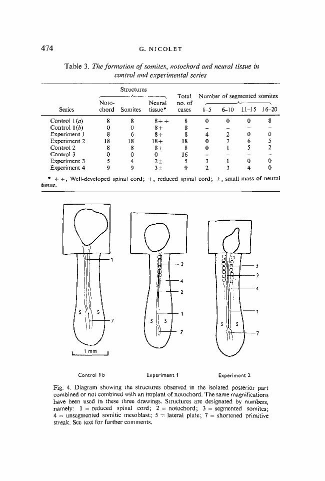
Table 3. The formation of somites, notochord and neural tissue incontrol and experimental series
Series
Control 1 (a)Control 1 (b)Experiment 1Experiment 2Control 2Control 3Experiment 3Experiment 4
(TslntnIN UlU
chord
808
188059
StructuresK
Somites
806
188049
* + + , Well-developed spinal cord;tissue.
\rNvUid.1
tissue*
8+ +8 +8 +
18 +8 +02±3±
Totalo f
cases
888
188
1659
+ , reduced spinal
Number
1-5
0-400_32
cord; ±
of segmented
6-10
0-271-13
, smal
11-15
0-065-04
somites
16-20
8-052-00
1 mass of neural
1 mm
Control 1 b Experiment 1 Experiment 2
Fig. 4. Diagram showing the structures observed in the isolated posterior partcombined or not combined with an implant of notochord. The same magnificationshave been used in these three drawings. Structures are designated by numbers,namely: 1 = reduced spinal cord; 2 = notochord; 3 = segmented somites;4 = unsegmented somitic mesoblast; 5 = lateral plate; 7 = shortened primitivestreak. See text for further comments.
Notochord and somite genesis 475
always present when somite formation occurred, whereas the presence orabsence of neural tissue had apparently no effect upon it. In the right part ofTable 3, the number of segmented somites counted in each case have been distri-buted into four categories. The reliability of this method is limited, since thesize of somites is not constant and their number depends on the stage attainedby the embryo at the time of fixation. In spite of these restrictions, this quantita-tive expression of the somitic response was in agreement with the morphologicalobservations. The impression was gained that, in both experimental systems,
Control 2 Control 3 Experiment 3 Experiment 4
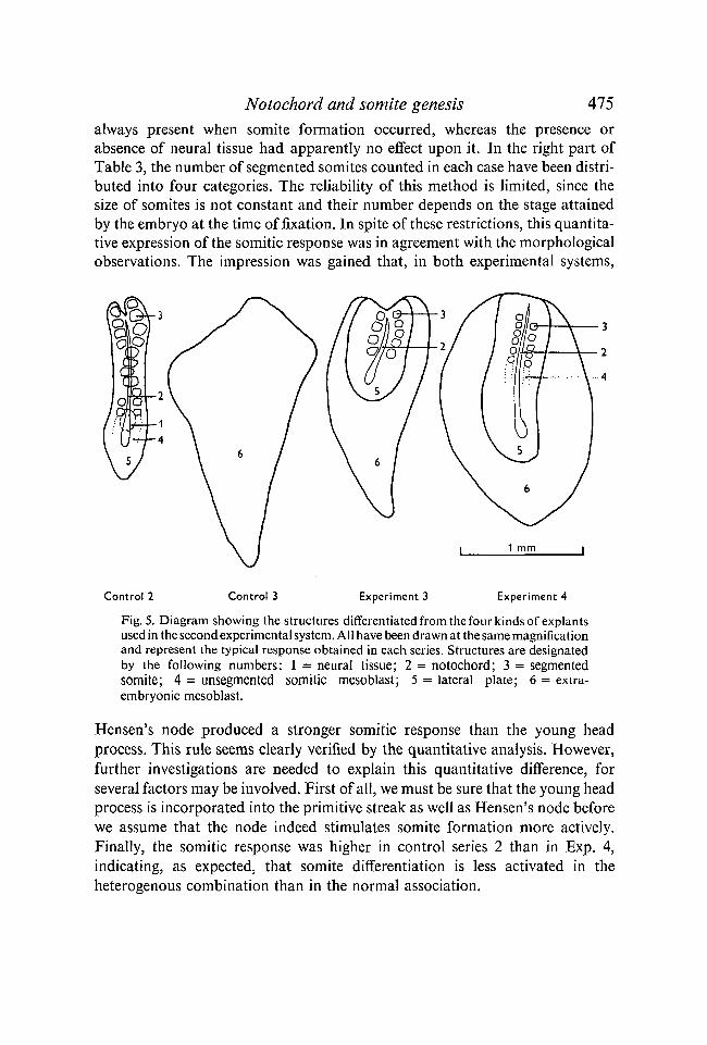
Fig. 5. Diagram showing the structures differentiated from the four kinds of explantsused in the second experimental system. All have been drawn at the same magnificationand represent the typical response obtained in each series. Structures are designatedby the following numbers: 1 = neural tissue; 2 = notochord; 3 = segmentedsomite; 4 = unsegmented somitic mesoblast; 5 = lateral plate; 6 = extra-embryonic mesoblast.
Hensen's node produced a stronger somitic response than the young headprocess. This rule seems clearly verified by the quantitative analysis. However,further investigations are needed to explain this quantitative difference, forseveral factors may be involved. First of all, we must be sure that the young headprocess is incorporated into the primitive streak as well as Hensen's node beforewe assume that the node indeed stimulates somite formation more actively.Finally, the somitic response was higher in control series 2 than in Exp. 4,indicating, as expected, that somite differentiation is less activated in theheterogenous combination than in the normal association.
476 G. NICOLET
DISCUSSION
Whenever labelled grafts were used it was observed that they were notcontaminated by presumptive somitic cells, so that it is very likely that thiscondition was also fulfilled when implants of similar size came from unlabelleddonors. The differentiated notochords found in posterior isolates were entirelycomposed of labelled nuclei, but not all labelled nuclei were contained in themsince a few endoblastic cells and sometimes presumptive neural cells were cutoff with the grafts.
In the first experimental system, a typical pattern of regression was observedin controls as well as in experimental series. Therefore, as already assumed byWaddington (1935, 1952) and Butros (1962, 1967), an autonomous movementof regression occurs in posterior isolates. In fact, this result was expected, sinceseveral authors have shown that backward movements were observed at alllevels of the primitive streak (Spratt, 1947; Vakaet, 1960; Nicolet, 1970).
The posterior isolates used could not differentiate into somites by themselves.In the first experimental system, posterior remnants of the primitive streak gaverise to lateral plate according to their normal prospective significance (Wolff,1936; Nicolet, 1970). On the other hand, after explantation into the area opaca(see control 3), they were no longer able to differentiate into lateral plate, butonly into extra-embryonic mesoblast. The implantation of presumptive notochordin these posterior isolates elicited somite formation in most cases, in agreementwith the results obtained by Bellairs (1963). Hence, it is concluded that thepresumptive notochord has modified the prospective significance of some ofthese cells. The chorda bulb as well as the head process acted in a similar way,though the latter was less efficient as activator.
In our opinion, the major interest of the present experiments is in showingclearly that the action of the presumptive notochord of the chick in this newsituation is similar to that of the chorda-factor discovered in amphibians(Yamada, 1940; Niazi, 1969). At first sight, it seems unlikely that the presump-tive notochord has the same action in the normal development of birds becauseit has been demonstrated that its presence is not required for somite differentia-tion after stage 5 (Waddington, 1932; Spratt, 1955). However, the possibilitycannot be excluded that it may play a role in the earlier steps of somitic deter-mination, since the presumptive notochordal cells start to congregate inHensen's node as soon as stage 3 (Gallera & Nicolet, 1969; Nicolet, 1970).Hence, this accumulation of the presumptive notochord in the node beginsearlier than the invagination of the presumptive somitic cells through theprimitive streak.
Notochord and somite genesis All
RESUME
La chorde presomptive intervient-elle directement dans la differentiationdes somites chez le poulet ?
Les experiences consistent a implanter un fragment de noeud de Hensen ou de prolonge-ment cephalique au sein de la ligne primitive a 0-8 mm derriere Je noeud de Hensen. Pourisoler la region posterieure de toutes influences emanant de la region anterieure du blasto-derme, nous procedons soit a l'excision d'un vaste territoire quadrangulaire situe juste devant1'implantat de chorde presomptive, soit a l'explantation de la region combinee avecPimplantat dans 1'aire opaque. Comme controles, des regions correspondantes de la ligneprimitive ont ete isolees ou explantees.
Chez les controles, la formation des somites n'a jamais lieu, alors que l'implantation demateriel chordal dans le territoire posterieur est presque toujours suivie par l'apparition desomites.
Dans ces experiences, la chorde presomptive agit sur son nouvel environnement comme leferait le facteur chordal qui a ete decouvert chez les Amphibiens.
This work was generously supported by the Fonds national suisse de la Recherchescientifique, Berne, Switzerland.
REFERENCESBELLAIRS. R. (1963). The development of somites in the chick embryo. / . Embryol. exp.
Morph. 11, 697-714.BUTROS, J. (1962). Studies on the inductive action of the early chick axis on isolated post-
nodal fragment. / . exp. Zool. 149, 1-20.BUTROS, J. (1967). Limited axial structures in nodeless chick blastoderms. / . Embryol. exp.
Morph. 17, 119-130.FICQ, A. (1959). Autoradiography. In The Cell, vol. 1 (ed. J. Brachet & A. Mirsky), pp. 67-90.
New York: Academic Press.FRASER, R. C. (1954). Studies on the hypoblast of the young chick embryo. / . exp. Zool. 126,
349-400.GALLERA, J. & NICOLET, G. (1961). Quelques commentaires sur les methodes de cultures
in vitro de jeunes blastodermes de Poule. Experientia 17, 134-137.GALLERA, J. & NICOLET, G. (1969). Le pouvoir inducteur de l'endoblaste presomptif contenu
dans la ligne primitive jeune de Poulet. /. Embryol. exp. Morph. 21, 105-118.HAMBURGER, V. & HAMILTON, H. L. (1951). A series of normal stages in the development of
the chick embryo. /. Morph. 88, 49-92.JACOBSON, W. (1938). The early development of the avian embryo. II. Mesoderm formation
and distribution of presumptive embryonic material. J. Morph. 62, 445-501.NIAZI, I. A. (1969). Differentiation capacities of prospective tail somite region of the neural
plate in the embryos of Ambystoma mexicanum. J. Embryol. exp. Morph. 22, 1-14.NICOLET, G. (1970). Analyse autoradiographique de la localisation des differentes ebauches
presomptives dans la ligne primitive de l'embryon de Poulet. /. Embryol. exp. Morph. 23,79-108.
PEEBLES, F. (1898). Some experiments on the primitive streak of the chick. Arch. EntwMech.Org. 7, 405-429.
SPRATT, N. T. (1947). Regression and shortening of the primitive streak in the explantedchick blastoderm. /. exp. Zool. 104, 69-99.
SPRATT, N. T. (1955). Analysis of the organizer centre in the early chick embryo. I. Localiza-tion of the prospective notochord and somite cells. /. exp. Zool. 128, 121-164.
VAKAET, L. (1960). A propos du raccourcissement de la ligne primitive du blastoderme dePoulet. / . Embryol. exp. Morph. 8, 6-19.
WADDINGTON, C. H. (1932). Experiments on the development of chick and duck embryos,cultivated in vitro. Phil. Trans. R. Soc. Ser. B 221, 179-230.
478 G. NICOLET
WADDINGTON, C. H. (1935). The development of isolated parts of the chick blastoderm./. exp. Zool. 71, 273-291.
WADDINGTON, C. H. (1952). The Epigenetics of Birds. Cambridge University Press.WETZEL, R. (1929). Untersuchungen am Hiihnchen. Die Entwicklung des Keims wahrend der
ersten beiden Bruttage. Wilhelm Roux Arch. EntwMech. Org. 119, 188-321.WOLFF, ET. (1936). Les bases de la teratogenese experimentale des Vertebres amniotes,
d'apres les resultats de methodes directes. Archs Anat. Histol. Embryol. 22, 1-375.YAMADA, T. (1940). Beeinflussung der DifTerenzierungsleistung des isolierten Mesoderms
von Molch-Keimen durch zugefugtes Chorda- und Neuralmaterial. Okajimas Folia anat.jap. 19, 131-197.
{Manuscript received 20 January 1970)