Embed Size (px)
Citation preview

RESEARCH ARTICLE
Is serum level of CC chemokine ligand 18 a
biomarker for the prediction of radiation
induced lung toxicity (RILT)?
Eleni Gkika1*, Werner Vach2, Sonja Adebahr1,3,4, Tanja Schimeck-Jasch1,
Anton Brenner1, Thomas Baptist Brunner1,3,4,5, Klaus Kaier2, Antje Prasse6,
Joachim Muller-Quernheim7, Anca-Ligia Grosu1,3,4,5, Gernot Zissel7, Ursula Nestle1,3,4,5
1 Department of Radiation Oncology, Medical Center – University Hospital Freiburg, Freiburg, Germany,
2 Institute of Medical Biometry and Statistics, Medical Faculty & Medical Center, University of Freiburg,
Freiburg, Germany, 3 German Cancer Consortium (DKTK), partner site Freiburg, Germany, 4 German
Cancer Research Center (DKFZ), Heidelberg, Germany, 5 Faculty of Medicine, University of Freiburg,
Freiburg, Germany, 6 Department of Pneumology, Hannover Medical School, Hannover, Germany,
7 Department of Pneumology, University Medical Center Freiburg, Freiburg, Germany
Abstract
The CC chemokine ligand 18 (CCL18) is produced by alveolar macrophages in patients with
fibrosing lung disease and its concentration is increased in various fibrotic lung diseases.
Furthermore CCL18 is elevated in several malignancies as it is produced by tumor associ-
ated macrophages. In this study we aimed to analyze the role of CCL18 as a prognostic bio-
marker for the development of early radiation induced lung toxicity (RILT), i.e. radiation
pneumonitis after thoracic irradiation and its significance in the course of the disease. Sixty
seven patients were enrolled prospectively in the study. Patients were treated with irradia-
tion for several thoracic malignancies (lung cancer, esophageal cancer, thymoma), either
with conventionally fractionated or hypo-fractionated radiotherapy. The CCL18 serum levels
were quantified with ELISA (enzyme-linked immunosorbent assay) at predefined time
points: before, during and at the end of treatment as well as in the first and second follow-up.
Treatment parameters and functional tests were also correlated with the development of
RILT.Fifty three patients were evaluable for this study. Twenty one patients (39%) devel-
oped radiologic signs of RILT Grade >1 but only three of them (5.6%) developed clinical
symptoms (Grade 2). We could not find any association between the different CCL18 con-
centrations and a higher incidence of RILT. Statistical significant factors were the planning
target volume (odds ratio OR: 1.003, p = 0.010), the volume of the lung receiving > 20 Gy
(OR: 1.132 p = 0.004) and age (OR: 0.917, p = 0.008). There was no association between
serial CCL18 concentrations with tumor response and overall survival.In our study the dosi-
metric parameters remained the most potent predictors of RILT. Further studies are needed
in order to estimate the role of CCL18 in the development of early RILT.
PLOS ONE | https://doi.org/10.1371/journal.pone.0185350 September 28, 2017 1 / 14
a1111111111
a1111111111
a1111111111
a1111111111
a1111111111
OPENACCESS
Citation: Gkika E, Vach W, Adebahr S, Schimeck-
Jasch T, Brenner A, Brunner TB, et al. (2017) Is
serum level of CC chemokine ligand 18 a
biomarker for the prediction of radiation induced
lung toxicity (RILT)? PLoS ONE 12(9): e0185350.
https://doi.org/10.1371/journal.pone.0185350
Editor: Nils Cordes, Technische Universitat
Dresden, GERMANY
Received: January 24, 2017
Accepted: September 11, 2017
Published: September 28, 2017
Copyright: © 2017 Gkika et al. This is an open
access article distributed under the terms of the
Creative Commons Attribution License, which
permits unrestricted use, distribution, and
reproduction in any medium, provided the original
author and source are credited.
Data Availability Statement: All relevant data are
within the paper and its Supporting Information
files.
Funding: The author(s) received no specific
funding for this work.
Competing interests: Gernot Zissel and Antje
Prasse are Academic Editors of PLOS One. The
authors have declared that no competing interests
exist.

Introduction
Radiation therapy is an important cornerstone of the curative treatment of lung and esoph-
ageal cancers and plays also a significant role in the palliative setting in terms of symptom con-
trol. The lung is one of the most radiosensitive organs. Radiation induced lung toxicities
(RILT) range between 5–50% of the patients irradiated for lung cancer [1] and could be a
potentially life threatening risk to patients treated for thoracic tumors. In order to reduce the
risk of RILT one should reduce the delivered dose and may thus hamper local control. To date
only dosimetric parameters such as the mean lung dose (MLD), the volume of the lung receiv-
ing more than 20 Gy (V20) or 5 Gy (V5) are used routinely to predict the risk of RILT. [1–3].
However, dosimetric variables tend to be very collinear (i.e., increasing V20 tends to lead to an
increase in the other parameters), and therefore differences in prognostic value among differ-
ent dosimetric variables may be small. [3]
In an attempt to search for biological predictors for RILT, apart from dosimetric parame-
ters, many studies looked at correlations between the risk of pulmonary injury and variations
in several profibrogenic and proinflamatory cytokines such as transforming growth factor ß1
(TGF-ß1), tumor necrosis factor-a (TNF-a) interleukin-1(IL-1), IL-6, high-molecular weight
mucin-like antigen KL-6, and platelet-derived growth factor -ß (PDGF- ß)[4, 5]. Because these
cytokines are thought to be key mediators in lung toxicity many of them were examined as bio-
markers for the early detection of pulmonary toxicities but at the present time there are no reli-
able and validated predictive assays for treatment decision. [4].
The CC chemokine ligand CCL18 is a chemokine produced by human myeloid cells.[6] In
general, macrophage activation by T-helper 2 (Th2) cytokines induces a special phenotype in
macrophages, termed ‘‘alternative activation” (M2 phenotype).[6–8] Alternatively activated
macrophages play a role in tissue repair processes such as wound healing and fibrosis. [7] In
patients, whose disease is associated with fibrotic lung remodeling such as in idiopathic inter-
stitial pneumonias, systemic sclerosis and idiopathic pulmonary fibrosis, it is suggested that
the M2 phenotype of the alveolar macrophages leads to exaggerated production of the CCL18
and it is suggested that its serum concentrations might serve as a biomarker of pulmonary dis-
ease activity in these patients with idiopathic interstitial pneumonias, systemic sclerosis and
idiopathic pulmonary fibrosis,.[9–12] In addition, it has been shown that CCL18 fosters
fibrotic processes [13] in pulmonary fibrosis which might drive fibrotic remodeling in RILT.
On the other hand a complex network of chemokines influence the development of primary
tumors and metastases [14] and it could be possible that circulating chemokines could be use-
ful tumor prognostic markers. [15] It has been previously published that there is an enhanced
production of CCL18 in several malignancies such as ovarian cancer, gastric cancer, breast
cancer, colorectal cancer and adenocarcinoma of the lung. [16–22]
The present study was designed to evaluate CCL18 as a predictor for early RILT i.e. radia-
tion pneumonitis as defined per NCI Common Terminology Criteria for Adverse Events
v4.03. (National Cancer Institute: Common Terminology Criteria for Adverse Events Version
4.03, CTCAE 2010). For the pathogenesis of the radiation injury theory, fibrosis is a result of
abnormally healed inflammatory changes during the pneumonitis stage.[5] Our aim was to
investigate the prognostic role of CCL18 for radiation inflammation i.e. pneumonitis which
leads to permanent injury i.e. fibrotic remodeling of the lung, hypothesizing that an increase of
the CCL18 concentrations could be predictive for lung injury after radiotherapy. We also eval-
uated several other parameters that may affect the incidence of RILT such as treatment and
patient specific characteristics. Furthermore, we investigated the role of this chemokine as a
tumor response marker, as it has been suggested that CCL18 concentrations correlate with sur-
vival time in adenocarcinomas of the lung[22] but also in other malignancies [23]
CC chemokine ligand 18 in radiation induced lung toxicity
PLOS ONE | https://doi.org/10.1371/journal.pone.0185350 September 28, 2017 2 / 14

Materials and methods
Study population and treatment characteristics
The study was approved by the ethic committee of the University Medical Center Freiburg.
Written informed consent was obtained from all patients who participated in this study.
Consecutive routine patients receiving thoracic radiation therapy regardless of histology or
treatment intention were included in this prospective study. Patients with breast cancer were
excluded. The majority were treated for lung cancer or esophageal cancer. PET CTs or CTs as
part of the initial staging as well as for treatment response were routinely performed every
three months. Staging was assessed according to the UICC 7th Edition. Pulmonary function
tests were also obtained before treatment. Other parameters tested were age, the presence
of chronic obstructive pulmonary disease (COPD), diabetes mellitus, nicotine consumption
and medications such as angiotensin-converting-enzyme inhibitors (ACE inhibitors) or
prednisone.
Dose volume histogram (DVHs) parameters such as the planning target volume (PTV) or
the volume of both lungs receiving more than 20 Gy (V20) were also tested. Furthermore we
calculated the biologically effective dose (BED) and the equivalent dose in 2 Gy fractions
(EQD2) prescribed to the PTV.
Serum sampling and enzyme-linked immunosorbent assay (ELISA)
Serial plasma specimens from each individual were obtained before initiation of the radiation
therapy, during treatment, at the end of therapy and at the first and second follow-up.
Venous blood samples were taken using a routine procedure. Blood samples rested for 20
minutes before centrifugation. After centrifugation serum samples were frozen at -80˚C within
2 hours. CCL18 was quantified using a DuoSet ELISA Development System kit (R&D Systems
Europe).
Follow up and clinical evaluation of toxicity
Patients were evaluated weekly during radiation therapy as well as every two to three months
thereafter. During the first and second follow up (median 3 and 7 months after treatment initi-
ation) beside of the blood sampling patients underwent a chest computed tomography scan
(CT). Radiation pneumonitis was scored using the National Cancer Institute’s Common Ter-
minology Criteria for Adverse Events v4.0. (National Cancer Institute: Common Terminology
Criteria for Adverse Events Version 4.03, CTCAE 2010). Tumor response was assessed accord-
ing to the RECIST criteria [24]
Statistical analysis
Statistical analysis was performed for the absolute CCL18 concentrations at each time point,
the difference from baseline for every time point, and the slope of a regression line fitted to the
first 3 time points in order to measure the steepness of the CCL18 concentration changes. Fur-
thermore we divided the individual CCL18 time courses into stable (less than 20% variance
from the baseline), downwards (� 20% decrease from baseline), upwards (� 20% increase
from baseline) as well as their combinations over the first three time points. These variables
were then correlated with the presence of RILT. Logistic regression analyses were performed
to determine factors predictive for radiation pneumonitis Grade� 1. Results are reported as
odds ratios (OR), 95% confidence intervals (CI), and p-values. In order to investigate a poten-
tial influence of the occurrence of RILT on subsequent CCL18 concentration changes we com-
pared CCL18 values post RILT with CCL18 values prior to RILT in a linear mixed model with
CC chemokine ligand 18 in radiation induced lung toxicity
PLOS ONE | https://doi.org/10.1371/journal.pone.0185350 September 28, 2017 3 / 14

CCL18 concentrations as outcome, the pre/post status as binary covariate, the five time points
as categorical covariates, and the individual intercept as random effect. In the main analysis
(prognostic value of CCL18 values and their development of time for development of RILT) a
Bonferroni correction was applied to adjust for investigating 12 different, potential associa-
tions. Overall survival (OS), local tumor control (LC) and progression free survival (PFS) and
time until RILT were analyzed with respect to their association with the above mentioned vari-
ables by use of the Cox proportional hazard model. Only patients not yet having experienced
the corresponding events were included in the analysis, and the time until event was defined as
starting with the last time point involved in the definition of a variable. Patients were censored
6 months after the last blood sample or at loss to follow up. In analyzing local tumor control,
death was regarded as a censoring event. The statistical significance level was set at 0.05. All
p values were two-sided. Statistical analysis was performed using SPSS version 23(SPSS, Chi-
cago Il) and STATA version 14.1.
Endpoints
The endpoint of this study was to evaluate the role of CCL18 as a prognostic biomarker for the
development of early RILT. Secondary endpoints were clinical and treatment related factors
and their role in predicting RILT. In an additional exploratory analysis the correlation between
CCL18 concentrations and overall survival, local control and progression free survival at every
given time-point was evaluated.
Results
Patient and treatment characteristics and toxicity
From August 2011 to February 2012, 67 patients were prospectively included in the study. Of
these patients 14 were excluded because of insufficient data (less than three CCL18 measure-
ments), withdrawal of informed consent or death before reaching a follow up more than 6
months after treatment completion. Patient and treatment characteristics are described in
Table 1. Forty-three patients were treated for lung cancer; eight had an esophageal cancer and
one a thymoma. Patients were treated either with conventionally fractionated (n = 41) or
hypo-fractionated (n = 12) radiotherapy, 8 patients were treated adjuvant, 2 neoadjuvant, 8
with stereotactic body radiotherapy (SBRT), 31 with a concurrent chemoradiotherapy and 4 in
palliative intent. IMRT was performed in 70% of the cases with a mean dose of 53 Gy (range
30–76) Gy. The mean V20 was 15% (range 0.45–36). Fourteen patients had a V20 more than
20%. Over a period of 6 months 21 patients (39.6%) presented with radiological signs of pneu-
monitis Grade� 1; 3 of them (5.6%) developed symptoms (CTC Grade 2 pneumonitis). There
were no Grade� 3 toxicities.
Correlation between clinical and treatment factors
Several factors were tested as potential prognostic markers for RILT. Results are shown
in Table 2. Prognostic factors for RILT were age (OR 0.917, p = 0.008), the volume of the
PTV (OR 1.003, p = 0.010), the V20 (OR 1.132, p = 0.004) and V20>20% (OR: 4.050,
p = 0,003).
Prognostic value of CCL18 for the development of RILT
Absolute concentrations. Figs 1 and 2 give an overview about the individual time courses
of the CCL18 measurements. The mean CCL18 levels, for the whole group of patients, were
107±50 ng/ml before treatment and 81±77 ng/ml at the end of treatment (S1 Table). During
CC chemokine ligand 18 in radiation induced lung toxicity
PLOS ONE | https://doi.org/10.1371/journal.pone.0185350 September 28, 2017 4 / 14
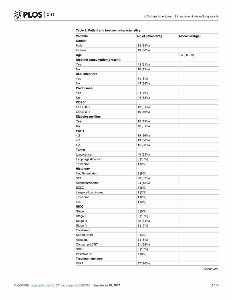
Table 1. Patient and treatment characteristics.
Variable Nr. of patients(%) Median (range)
Gender
Male 34 (64%)
Female 19 (36%)
Age 65 (30–83)
Nicotine consumption(present)
Yes 43 (81%)
No 10 (19%)
ACE-Inhibitors
Yes 8 (15%)
No 45 (85%)
Prednisone
Yes 9 (17%)
No 44 (83%)
COPD*
GOLD 0–2 43 (81%)
GOLD 3–4 10 (19%)
Diabetes mellitus
Yes 10 (19%)
No 43 (81%)
FEV 1
�2 l 19 (36%)
> 2 l 19 (36%)
n.a. 15 (28%)
Tumor
Lung cancer 44 (83%)
Esophageal cancer 8 (15%)
Thymoma 1 (2%)
Histology
Undifferentiated 3 (6%)
SCC 20 (37%)
Adenocarcinoma 24 (45%)
SCLC 3 (6%)
Large cell carcinoma 1 (2%)
Thymoma 1 (2%)
n.a. 1 (2%)
UICC
Stage I 5 (9%)
Stage II 8 (15%)
Stage III 32 (61%)
Stage IV 8 (15%)
Treatment
Neoadjuvant 2 (4%)
Adjuvant 8 (15%)
Concurrent CRT 31 (58%)
SBRT 8 (15%)
Palliative RT 4 (8%)
Treatment delivery
IMRT 37 (70%)
(Continued )
CC chemokine ligand 18 in radiation induced lung toxicity
PLOS ONE | https://doi.org/10.1371/journal.pone.0185350 September 28, 2017 5 / 14

the first (3 months from treatment) and second follow-up (6 months from treatment) the
mean CCL18 levels were 93±57 ng/ml and 104±49 ng/ml, respectively. The average slope over
the first three time points was -0.348 ng/ml/day for all patients. We could not observe an asso-
ciation between the absolute concentration and subsequent development of RILT, at any time
Table 1. (Continued)
Variable Nr. of patients(%) Median (range)
3D 16 (30%)
V20 15 (0.45–36) %
PTV Volume 368 (13–1288) ml
Physical Dose 53 (30–76) Gy
EQD210 to PTV 58.4 (34–76) Gy
Abbreviations CRT = chemoradiotherapy, RT = radiotherapy, SCC = squamous cell carcinoma,
SCLC = small cell carcinoma, IMRT: intensity modulated radiation therapy, V20 = % volume of the lung
receiving more than 20 Gy, PTV = planning target volume, EQD2 = equivalent dose in 2 Gy fraction,
COPD = chronic obstructive pulmonary disease, ACE- inhibitors = angiotensin-converting-enzyme
inhibitors, FEV 1 = Forced expiratory volume in 1 second, n.a. = not available.
*COPD was dichotomized as not significant (COPD GOLD 0–2) and significant (COPD GOLD 3–4).
https://doi.org/10.1371/journal.pone.0185350.t001
Table 2. Univariate analysis of factors prognostic for radiation pneumonitis Grade� 1.
UVA
OR (95% CI) P value
Age 0.917 (0.861–0.978) 0.008
FEV 1� 2l 1.905 (0.521–6.962) 0.330
Nicotine 1.680 (0.382–7.395) 0.493
COPD GOLD 0–2 1.680 (0.382–7.395) 0.484
Diabetes mellitus 0.316 (0.060–1.665) 0.144
Prednisone 3.870 (0.846–17.673) 0.081
ACE-Inhibitors 0.456 (0.083–2.512) 0.367
PTV Volume 1.003 (1.001–1.005) 0.010
V20 1.132(1.041–1.231) 0.004
V20>20% 4.050(1.118–14.674) 0.033
IMRT 0.542 (0.165–1.780) 0.313
Dose 1.033 (0.984–1.085) 0.179
BED 1.006 (0.960–1.053) 0.811
EQD2 1.006 (0.953–1.063) 0.824
Adjuvant treatment 1.647 (0.363–7.465) 0.518
Histology 1.034 (0,490–1,896) 0.919
T1 0.175 (0.016–1.881) 0.150
T2 0.437 (0.061–1.881) 0.413
T3 0.436 (0.100–1.916) 0.273
T4 0.729 (0.153–3.474) 0.692
Tumor progression* 0.731 (0.232–2.306) 0.593
Abbreviations: OR = odds ratio, CI: confidence interval, UVA: univariate analysis.
COPD was dichotomized as not significant (COPD GOLD 0–2) and significant (COPD GOLD 3–4).
*Within 6 months after treatment completion.
https://doi.org/10.1371/journal.pone.0185350.t002
CC chemokine ligand 18 in radiation induced lung toxicity
PLOS ONE | https://doi.org/10.1371/journal.pone.0185350 September 28, 2017 6 / 14

Fig 1. Individual trajectories of CCL18, local recurrence and distant metastases in patients without RILT.
https://doi.org/10.1371/journal.pone.0185350.g001
CC chemokine ligand 18 in radiation induced lung toxicity
PLOS ONE | https://doi.org/10.1371/journal.pone.0185350 September 28, 2017 7 / 14

point (Table 3A). The mean CCL18 levels at three and six months were in the RILT-group
94±62 ng/ml and 104±61 ng/ml and in the non-RILD-group 93±54 ng/ml and 103±39 ng/ml.
Of the three patients who developed a grade 2 pneumonitis, one patient developed a RILT 3
months after treatment at a serum concentration of 54 ng/ml and two developed clinical signs
Fig 2. Individual trajectories of CCL18, local recurrence and distant metastases in patients with RILT.
https://doi.org/10.1371/journal.pone.0185350.g002
CC chemokine ligand 18 in radiation induced lung toxicity
PLOS ONE | https://doi.org/10.1371/journal.pone.0185350 September 28, 2017 8 / 14

of pneumonitis at the second follow-up and had a serum concentration of 138ng/ml and
134 ng/ml, respectively. All patients were treated adjuvantly for pN2 NSCLC (SCC = 2,
adenocarcinoma = 1).
Changes during and after treatment. Furthermore we correlated the changes of CCL18
from baseline with subsequent development of RILT. No associations could be observed
(Table 3B). When classifying the individual courses, we found at the 2nd follow up 23 patients
with initially rather stable CCL18 concentrations remained stable, whereas 18 decreased and
13 increased more than 20% from baseline. We couldn´t observe any associations between the
different CCL18 concentrations and a higher risk to develop RILT (Table 3C).Also when clas-
sifying the course over the first 3 time points, no differences could be observed (Table 3C).
The slope did not correlate. The effect of did not change when adjusting for tumors that did
not progress within the first 6 months (n = 39) in order to exclude any possible interference of
CCL18 production due tumor progression.
When considering Cox proportional hazard models for the time until RILT, we could not
find any significant differences.
Table 3. Univariate analysis of CCL 18—Related variables predictive of radiation pneumonitis
Grade� 1.
Nr. UVA
OR (95% CI) Adj P value*
A. Absolute serum concentrations of CCL18
CCL18 1st time point* 1.002 (0.896–1.120) 1.0
CCL18 2nd time point 1.010 (0.938–1.086) 1.0
CCL18 3nd time point 1.043 (0.953–1.142) 1.0
CCL18 4nd time point 0.975 (0.883–1.077) 1.0
CCL18 5nd time point 1.022 (0.937–1.115) 1.0
B.dCCL18 compared to baseline
CCL18 2nd vs 1st 1.007 (0.927–1.093) 1.0
CCL18 3nd vs 1st 0.968 (0.847–1.107) 1.0
CCL18 4rd vs 1st 1.010 (0.891–1.145) 1.0
CCL18 5th vs 1st 0.948 (0.803–1.120) 1.0
C. Classification and summary measures of individual courses
Over 2 time points
20% decrease 18 0.308 (0.072–1.315)
20% increase 13 0.198 (0.049–0.801)
Stable (Reference) 22 0.52
Over 3 time point
20% decrease 18 0.286 (0.059–1.395)
20% increase 13 0.444 (0.087–2.276)
Stable then 20% decrease 10 2.333 (0.400–13.609)
Stable (Reference) 12 0.79
Slope 0.975 (0.797–1.080) 1.0
Abbreviations: OR = odds ratio for a 10ng/ml change of the CCL18 concentration, CI: confidence interval, *
= baseline, SD = Standard deviation, Nr: Number of patients, UVA: univariate analysis.
*Bonferroni-adjusted p-value. The p-values were adjusted by multiplication with 12 (reflecting that we
assessed significance for 12 different, potential associations).
https://doi.org/10.1371/journal.pone.0185350.t003
CC chemokine ligand 18 in radiation induced lung toxicity
PLOS ONE | https://doi.org/10.1371/journal.pone.0185350 September 28, 2017 9 / 14

CCL18 as a marker of development of RILT
Nine patients developed signs of RILT between treatment completion and first follow up and
12 patients between first and second follow up. Patients with RILT at first follow up had a
mean CCL18 concentration of 94 ±62 ng/ml whereas patients without RILT had a mean
CCL18 concentration of 93 ±54 ng/ml. These results did not vary significantly in the second
follow up (104 ±61 ng/ml vs 103 ±39 ng/ml for patients with and without RILT respectively).
Post development of RILT, CCL18 values were on average increased by 6.8ng/ml compared to
pre development of RILT. This difference was not statistically significant.
Prognostic value of CCL18 for treatment response
At the second follow-up six patients had died, all due to progression, two of whom in the first
3 months (OS at 6 months 89%) and fourteen patient developed a local progression, two of
whom during the first follow-up (LC at 6 months 74%). Neither the absolute CCL18 concen-
trations nor the changes from baseline (dCCL18) correlated with the OS and LC at any specific
time point (S2 Table).
Discussion
In patients with lung fibrosis, increases in pulmonary levers of CCL 18 occur in association
with fibrosis [10]. It has been reported that in the course of idiopathic pulmonary fibrosis, alve-
olar macrophages shift from their pro-inflammatory, classical activation (M1 type) to alterna-
tive activation, the M2 phenotype that promotes collagen production, scar formation and
angiogenesis. [7] This M2 state is induced and maintained by products of activated fibroblasts
[11]. CCL18 is abundantly produced by alternatively activated macrophages in patients with
idiopathic pulmonary fibrosis and its concentrations reflect the fibrotic lung activity in
patients with idiopathic interstitial pneumonias, systemic sclerosis and idiopathic pulmonary
fibrosis [10, 12]. That led us to the hypothesis that serum concentrations of CCL18 or their
fluctuations during therapy could be also a potential marker for predicting RILT, as radiation
pneumonitis and the ensuing pulmonary fibrosis is associated with a fibrotic remodeling of
lung tissue. [5, 25]
To our knowledge this is the first study that prospectively investigates the role of the CC
chemokine ligand 18 as a biomarker for radiation pneumonitis. We did not find any correla-
tion between increasing CCL18 concentrations and risk of RILT as initially hypothesized.
Alternatively activated macrophages are found in fibrotic diseases [13, 26] as well as in
tumors as so called tumor-associated macrophages [27]. Hence, in fibrotic diseases [6] as well
as in various cancers [22, 23, 28] CCL18 levels are increased. As CCL18 serum levels correlate
with tumor size [22, 29] we expect that CCL18 levels decrease after response to radiotherapy.
Increase of CCL18 released by alternatively activated macrophages in a pro-fibrotic environ-
ment might therefore mask this decrease, resulting to “false” stable concentrations due to two
different pathomechanisms (CCL18 decrease due to tumor regression and increase due to
RILT). Some researches for potential biomarkers for RILT suggested that the cytokines tested
could correlate more with tumor activity than with the incidence of lung injury. [30, 31].
Indeed it is known that a number of cytokines that are produced in the tumor microenviron-
ment have an important role in cancer pathogenesis.[15] Cytokines that are released in
response to infection, inflammation and immunity can function to inhibit tumor development
and progression [14] [32]. Alternatively, cancer cells can respond to host-derived cytokines
that promote growth, attenuate apoptosis and facilitate invasion and metastasis.[14]. In our
study we could not observe any effect of the different CCL18 concentrations also after
CC chemokine ligand 18 in radiation induced lung toxicity
PLOS ONE | https://doi.org/10.1371/journal.pone.0185350 September 28, 2017 10 / 14

adjusting for progression within the first 6 months after treatment completion, suggesting that
these results did not interfere with the tumor activity.
Furthermore, we could not demonstrate that elevated or rising CCL18 serum concentra-
tions correspond to tumor activity in terms of progression or worse survival, although the
aim of this study was prognostic role of CCL18 concerning RILT. However, these effects
might be masked by the heterogeneity in respect to tumor entities of our study cohort. In
previous studies it could be demonstrated that CCL18 enhances invasiveness [28] and malig-
nancy [18] and promotes angiogenesis [17] in breast cancer tissue. CCL18 is also overex-
pressed in gastric cancer cells and contributed to cell invasion and expression [19] while
Leung et al. [23] identified that high CCL18 expression levels were associated with prolonged
OS and PFS in gastric cancer patients. It has also been reported that it is a favorable prognos-
tic biomarker in patients with colorectal cancer [21] and significantly elevated in ascetic fluid
from patients with ovarian carcinoma [33]. Plones et al [22] reported that serum levels are
increased in patients with NSCLC and that its concentrations correlate with survival time in
adenocarcinomas.
A number of limitations applying to our study need to be considered, including the small
sample size tested, as only 53 patients were evaluable for the analysis, as well as the very low
incidence of clinically significant RILT (Grade ≧2) which led us to use Grade� 1 as main
outcome, providing only 21 events. We were also confronted with the challenge of the rela-
tion between CCL18 values and their course over time and the occurrence of RILT. This led
us to check several different aspects of the course for a significant association with the devel-
opment of RILT, inducing a risk of false significant results. Furthermore, our study was
limited by the inhomogeneity of histologies and treatment regimens as it was primarily
designed to identify the role of CCL18 as biomarker for predicting RILT. The heterogeneity
of our study group also limits conclusions on the prognostic value of CCL18 for treatment
response as only for adenocarcinoma a correlation between CCL18 and OS could be demon-
strated [22].
Several cytokines have been proposed as potential biomarkers in the past with conflicting
results but none of them is being used in the clinical praxis. To that concern, further progress
toward an improved understanding of the mechanisms of RILT and the regulation of CCL18
is important for the identification of appropriate targets for prediction and treatment interven-
tion in the clinical practice.
Conclusion
In this prospective study the dosimetric parameters remain the most potent predictors regard-
less their limitations. We could not demonstrate an association of different CCL18 concentra-
tions with the development of RILT. Further studies are needed to improve the understanding
of the mechanisms of RILT and complex regulation of CCL18 in pulmonary inflammation
and fibrosis.
Supporting information
S1 Table. CCL18 concentrations (ng/ml) at every time point.
(DOCX)
S2 Table. Univariate analysis of CCL 18—Related variables regarding overall survival and
local control.
(DOCX)
CC chemokine ligand 18 in radiation induced lung toxicity
PLOS ONE | https://doi.org/10.1371/journal.pone.0185350 September 28, 2017 11 / 14

Author Contributions
Conceptualization: Eleni Gkika, Werner Vach, Sonja Adebahr, Anton Brenner, Antje Prasse,
Joachim Muller-Quernheim, Anca-Ligia Grosu, Gernot Zissel, Ursula Nestle.
Data curation: Eleni Gkika, Werner Vach, Sonja Adebahr, Tanja Schimeck-Jasch, Anton
Brenner, Joachim Muller-Quernheim, Gernot Zissel, Ursula Nestle.
Formal analysis: Eleni Gkika, Werner Vach, Anton Brenner, Gernot Zissel, Ursula Nestle.
Investigation: Eleni Gkika, Sonja Adebahr, Tanja Schimeck-Jasch, Anton Brenner, Joachim
Muller-Quernheim, Gernot Zissel, Ursula Nestle.
Methodology: Eleni Gkika, Werner Vach, Sonja Adebahr, Tanja Schimeck-Jasch, Anton Bren-
ner, Klaus Kaier, Antje Prasse, Joachim Muller-Quernheim, Anca-Ligia Grosu, Gernot Zis-
sel, Ursula Nestle.
Project administration: Sonja Adebahr, Joachim Muller-Quernheim, Anca-Ligia Grosu, Ger-
not Zissel, Ursula Nestle.
Resources: Joachim Muller-Quernheim, Anca-Ligia Grosu, Gernot Zissel, Ursula Nestle.
Software: Eleni Gkika, Werner Vach.
Supervision: Eleni Gkika, Werner Vach, Sonja Adebahr, Tanja Schimeck-Jasch, Thomas Bap-
tist Brunner, Antje Prasse, Joachim Muller-Quernheim, Anca-Ligia Grosu, Gernot Zissel,
Ursula Nestle.
Validation: Eleni Gkika, Werner Vach, Anton Brenner, Gernot Zissel, Ursula Nestle.
Visualization: Eleni Gkika, Werner Vach, Sonja Adebahr, Gernot Zissel, Ursula Nestle.
Writing – original draft: Eleni Gkika, Werner Vach, Sonja Adebahr, Tanja Schimeck-Jasch,
Anton Brenner, Thomas Baptist Brunner, Klaus Kaier, Antje Prasse, Joachim Muller-Quer-
nheim, Anca-Ligia Grosu, Gernot Zissel, Ursula Nestle.
Writing – review & editing: Eleni Gkika, Werner Vach, Sonja Adebahr, Tanja Schimeck-
Jasch, Thomas Baptist Brunner, Klaus Kaier, Joachim Muller-Quernheim, Anca-Ligia
Grosu, Gernot Zissel, Ursula Nestle.
References1. Marks LB, Bentzen SM, Deasy JO, Kong FM, Bradley JD, Vogelius IS, et al. Radiation dose-volume
effects in the lung. International journal of radiation oncology, biology, physics. 2010; 76(3 Suppl):S70–
6. https://doi.org/10.1016/j.ijrobp.2009.06.091 PMID: 20171521.
2. Khalil AA, Hoffmann L, Moeller DS, Farr KP, Knap MM. New dose constraint reduces radiation-induced
fatal pneumonitis in locally advanced non-small cell lung cancer patients treated with intensity-modu-
lated radiotherapy. Acta oncologica. 2015; 54(9):1343–9. https://doi.org/10.3109/0284186X.2015.
1061216 PMID: 26198657.
3. Palma DA, Senan S, Tsujino K, Barriger RB, Rengan R, Moreno M, et al. Predicting radiation pneumoni-
tis after chemoradiation therapy for lung cancer: an international individual patient data meta-analysis.
International journal of radiation oncology, biology, physics. 2013; 85(2):444–50. https://doi.org/10.
1016/j.ijrobp.2012.04.043 PMID: 22682812.
4. Fleckenstein K, Gauter-Fleckenstein B, Jackson IL, Rabbani Z, Anscher M, Vujaskovic Z. Using biologi-
cal markers to predict risk of radiation injury. Seminars in radiation oncology. 2007; 17(2):89–98. https://
doi.org/10.1016/j.semradonc.2006.11.004 PMID: 17395039.
5. Kong FM TH R, Eisbruch A, Lawrence TS. Non-small cell lung cancer therapy-related pulmonary toxic-
ity: an update on radiation pneumonitis and fibrosi. Semin Oncol. 2005; 32(2 Suppl 3):S42–54. https://
doi.org/10.1053/j.seminoncol.2005.03.009 PMID: 16015535
6. Prasse A, Probst C, Bargagli E, Zissel G, Toews GB, Flaherty KR, et al. Serum CC-chemokine ligand
18 concentration predicts outcome in idiopathic pulmonary fibrosis. American journal of respiratory and
CC chemokine ligand 18 in radiation induced lung toxicity
PLOS ONE | https://doi.org/10.1371/journal.pone.0185350 September 28, 2017 12 / 14

critical care medicine. 2009; 179(8):717–23. https://doi.org/10.1164/rccm.200808-1201OC PMID:
19179488.
7. Wynn TA. Fibrotic disease and the T(H)1/T(H)2 paradigm. Nature reviews Immunology. 2004; 4
(8):583–94. https://doi.org/10.1038/nri1412 PMID: 15286725.
8. Hieshima K, Imai T, Baba M, Shoudai K, Ishizuka K, Nakagawa T, et al. A novel human CC chemokine
PARC that is most homologous to macrophage-inflammatory protein-1 alpha/LD78 alpha and chemo-
tactic for T lymphocytes, but not for monocytes. The Journal of Immunology. 1997; 159(3):1140–9.
PMID: 9233607
9. Lota HK, Renzoni EA. Circulating biomarkers of interstitial lung disease in systemic sclerosis. Interna-
tional journal of rheumatology. 2012; 2012:121439. https://doi.org/10.1155/2012/121439 PMID:
22988462.
10. Prasse A, Pechkovsky DV, Toews GB, Schafer M, Eggeling S, Ludwig C, et al. CCL18 as an indicator
of pulmonary fibrotic activity in idiopathic interstitial pneumonias and systemic sclerosis. Arthritis and
rheumatism. 2007; 56(5):1685–93. https://doi.org/10.1002/art.22559 PMID: 17469163.
11. Prasse A, Muller-Quernheim J. Non-invasive biomarkers in pulmonary fibrosis. Respirology. 2009; 14
(6):788–95. https://doi.org/10.1111/j.1440-1843.2009.01600.x PMID: 19703061.
12. Schupp JC B H, Jager B, Cillis G, Zissel G, Muller-Quernheim J, Prasse A. Macrophage activation in
acute exacerbation of idiopathic pulmonary fibrosis. PLoS One. 2015; 10(1):e0116775. https://doi.org/
10.1371/journal.pone.0116775 PMID: 25590613
13. Prasse A, Pechkovsky DV, Toews GB, Jungraithmayr W, Kollert F, Goldmann T, et al. A vicious circle
of alveolar macrophages and fibroblasts perpetuates pulmonary fibrosis via CCL18. American journal
of respiratory and critical care medicine. 2006; 173(7):781–92. https://doi.org/10.1164/rccm.200509-
1518OC PMID: 16415274.
14. Dranoff G. Cytokines in cancer pathogenesis and cancer therapy. Nature reviews Cancer. 2004; 4
(1):11–22. https://doi.org/10.1038/nrc1252 PMID: 14708024.
15. Balkwill F. Cancer and the chemokine network. Nature reviews Cancer. 2004; 4(7):540–50. https://doi.
org/10.1038/nrc1388 PMID: 15229479.
16. Schutyser E, Richmond A, Van Damme J. Involvement of CC chemokine ligand 18 (CCL18) in normal
and pathological processes. Journal of leukocyte biology. 2005; 78(1):14–26. https://doi.org/10.1189/
jlb.1204712 PMID: 15784687.
17. Lin L C Y, Yao YD, Chen JQ, Chen JN, Huang SY, Zeng YJ, Yao HR, Zeng SH, Fu YS, Song EW.
CCL18 from tumor-associated macrophages promotes angiogenesis in breast cancer. Oncotarget.
2015; [Epub ahead of print].
18. Gao J, Li ZH, Tang W, Wu QN, Liu GH, Zheng WB. Chemokine C-C motif ligand 18 expression corre-
lates with tumor malignancy in breast cancer. Pathologie-biologie. 2015; 63(4–5):199–203. https://doi.
org/10.1016/j.patbio.2015.07.001 PMID: 26294068.
19. Hou X, Zhang Y, Qiao H. CCL18 promotes the invasion and migration of gastric cancer cells via ERK1/
2/NF-kappaB signaling pathway. Tumour biology: the journal of the International Society for Oncodeve-
lopmental Biology and Medicine. 2015. https://doi.org/10.1007/s13277-015-3825-0 PMID: 26242263.
20. Ploenes T S B, Krohn A, Burger M, Passlick B, Muller-Quernheim J, Zissel G. CC-chemokine ligand 18
induces epithelial to mesenchymal transition in lung cancer A549 cells and elevates the invasive poten-
tial. PLoS One. 2013; 8(1):e53068. https://doi.org/10.1371/journal.pone.0053068 PMID: 23349697
21. Yuan R, Chen Y, He X, Wu X, Ke J, Zou Y, et al. CCL18 as an independent favorable prognostic bio-
marker in patients with colorectal cancer. The Journal of surgical research. 2013; 183(1):163–9. https://
doi.org/10.1016/j.jss.2013.01.017 PMID: 23433718.
22. Plones T K A, Burger M, Veelken H, Passlick B, Muller-Quernheim J, Zissel G. Serum level of CC-che-
mokine ligand 18 is increased in patients with non-small-cell lung cancer and correlates with survival
time in adenocarcinomas. PLoS One. 2012; 7(7):e41746. https://doi.org/10.1371/journal.pone.0041746
PMID: 22848587
23. Leung SY, Yuen ST, Chu K-M, Mathy JA, Li R, Chan ASY, et al. Expression profiling identifies chemo-
kine (C-C motif) ligand 18 as an independent prognostic indicator in gastric cancer. Gastroenterology.
2004; 127(2):457–69. https://doi.org/10.1053/j.gastro.2004.05.031 PMID: 15300578
24. Eisenhauer EA, Therasse P, Bogaerts J, Schwartz LH, Sargent D, Ford R, et al. New response evalua-
tion criteria in solid tumours: revised RECIST guideline (version 1.1). European journal of cancer. 2009;
45(2):228–47. https://doi.org/10.1016/j.ejca.2008.10.026 PMID: 19097774.
25. Mehta V. Radiation pneumonitis and pulmonary fibrosis in non-small-cell lung cancer: pulmonary func-
tion, prediction, and prevention. International journal of radiation oncology, biology, physics. 2005; 63
(1):5–24. https://doi.org/10.1016/j.ijrobp.2005.03.047 PMID: 15963660.
CC chemokine ligand 18 in radiation induced lung toxicity
PLOS ONE | https://doi.org/10.1371/journal.pone.0185350 September 28, 2017 13 / 14

26. Pechkovsky DV, Prasse A, Kollert F, Engel KM, Dentler J, Luttmann W, et al. Alternatively activated
alveolar macrophages in pulmonary fibrosis-mediator production and intracellular signal transduction.
Clin Immunol. 2010; 137(1):89–101. https://doi.org/10.1016/j.clim.2010.06.017 PMID: 20674506.
27. Mantovani A, Allavena P, Sica A. Tumour-associated macrophages as a prototypic type II polarised
phagocyte population: role in tumour progression. European journal of cancer. 2004; 40(11):1660–7.
https://doi.org/10.1016/j.ejca.2004.03.016 PMID: 15251154.
28. Chen J, Yao Y, Gong C, Yu F, Su S, Chen J, et al. CCL18 from tumor-associated macrophages pro-
motes breast cancer metastasis via PITPNM3. Cancer cell. 2011; 19(4):541–55. https://doi.org/10.
1016/j.ccr.2011.02.006 PMID: 21481794.
29. Schmid S, Le UT, Haager B, Mayer O, Dietrich I, Elze M, et al. Local Concentrations of CC-Chemokine-
Ligand 18 Correlate with Tumor Size in Non-small Cell Lung Cancer and Are Elevated in Lymph Node-
positive Disease. Anticancer Res. 2016; 36(9):4667–71. https://doi.org/10.21873/anticanres.11018
PMID: 27630310.
30. Rube CE, Palm J, Erren M, Fleckenstein J, Konig J, Remberger K, et al. Cytokine plasma levels: reliable
predictors for radiation pneumonitis? PloS one. 2008; 3(8):e2898. https://doi.org/10.1371/journal.pone.
0002898 PMID: 18682839.
31. Crohns M, Saarelainen S, Laine S, Poussa T, Alho H, Kellokumpu-Lehtinen P. Cytokines in bronchoal-
veolar lavage fluid and serum of lung cancer patients during radiotherapy—Association of interleukin-8
and VEGF with survival. Cytokine. 2010; 50(1):30–6. https://doi.org/10.1016/j.cyto.2009.11.017 PMID:
20044268.
32. Mantovani A, Allavena P, Sica A, Balkwill F. Cancer-related inflammation. Nature. 2008; 454
(7203):436–44. https://doi.org/10.1038/nature07205 PMID: 18650914.
33. Schutyser E, Struyf S, Proost P, Opdenakker G, Laureys G, Verhasselt B, et al. Identification of biologi-
cally active chemokine isoforms from ascitic fluid and elevated levels of CCL18/pulmonary and activa-
tion-regulated chemokine in ovarian carcinoma. The Journal of biological chemistry. 2002; 277
(27):24584–93. https://doi.org/10.1074/jbc.M112275200 PMID: 11978786.
CC chemokine ligand 18 in radiation induced lung toxicity
PLOS ONE | https://doi.org/10.1371/journal.pone.0185350 September 28, 2017 14 / 14


![STARS Journal 08 2011 [Ulrich Berding, Antje Havemann und Juliane Pegels]](https://img.pdfslide.us/doc/110x75/577d24451a28ab4e1e9c0937/stars-journal-08-2011-ulrich-berding-antje-havemann-und-juliane-pegels.jpg)