Embed Size (px)
Citation preview
Is It Necessary to Follow Patients after Resection ofa Benign Pancreatic Intraductal Papillary MucinousNeoplasm?
Jin He, MD, PhD, John L Cameron, MD, FACS, Nita Ahuja, MD, FACS, Martin A Makary, MD, FACS,Kenzo Hirose, MD, FACS, Michael A Choti, MD, FACS, Richard D Schulick, MD, FACS,Ralph H Hruban, MD, Timothy M Pawlik, MD, PhD, FACS, Christopher L Wolfgang, MD, PhD, FACS
BACKGROUND: Little is known about the risk of subsequently developing a new or progressive intraductal papil-lary mucinous neoplasm (IPMN) after partial pancreatic resection of a noninvasive IPMN.
STUDY DESIGN: One hundred thirty patients with more than 1 year of follow-up after resection were includedin this analysis.
RESULTS: At a median follow-up of 38 months, 22 (17%) developed imaging evidence of a new orprogressive IPMN. Eleven (8%) underwent completion resection. Three of the 11 patients hadinvasive adenocarcinoma. Two other patients developed metastatic pancreatic adenocarcinomaand did not undergo resection. All 5 patients (4%) with cancer had negative margins at initialoperation. Sixteen of 100 patients (16%) with negative margins for IPMN at the initialoperation developed a new IPMN vs 6 of 30 patients (20%) with margins positive for IPMN(p¼ ns). Five of 22 patients (23%) with a new IPMN had a family history of pancreatic cancer,while 8 of 108 patients (7%) without a new IPMN had a family history (p < 0.05).Overall, the chances of developing a new IPMN at 1, 5, and 10 years after the initial surgerywere 4%, 25%, and 62%, respectively, and of requiring surgery were 1.6%, 14%, and 18%,respectively. The estimated chances of developing invasive pancreatic cancer were 0%, 7%,and 38% at 1, 5, and 10 years, respectively.
CONCLUSIONS: Patients who have undergone resection for noninvasive IPMN require indefinite close surveil-lance because of the risks of developing a new IPMN, of requiring surgery, and of developingcancer. A family history of pancreatic cancer, but not margin status or degree of dysplasia, isassociated with a risk of development of a new or progressive IPMN. (J Am Coll Surg 2013;-:1e9. � 2013 by the American College of Surgeons)
Intraductal papillary mucinous neoplasms (IPMN) of thepancreas are cystic precursor lesions to invasive adenocar-cinoma (ductal adenocarcinoma). There is a stronginterest in the study of IPMNs because they representan opportunity for early detection and cancer preventionin the subset of patients with this specific type of pancre-atic neoplasm. These lesions were first described byOhhashi and colleagues in 1982, and criteria for their
Disclosure Information: Nothing to disclose.
Presented at the Southern Surgical Association 124th Annual Meeting,Palm Beach, FL, December 2012.
Received December 13, 2012; Accepted December 13, 2012.From the Department of Surgery (He, Cameron, Ahuja, Makary, Hirose,Choti, Schulick, Pawlik, Wolfgang), and the Department of Pathologyand the Sol Goldman Pancreatic Cancer Research Center (Hruban), JohnsHopkins Medical Institutions, Baltimore, MD.Correspondence address: Christopher L Wolfgang, MD, PhD, FACS,Department of Surgery, Johns Hopkins Hospital, 600 N Wolfe St, Blalock685, Baltimore, MD 21287. email: [email protected]
1ª 2013 by the American College of Surgeons
Published by Elsevier Inc.
diagnosis were established by the World Health Organi-zation (WHO) in 1996.1 Over the past decade therehave been major advances in our understanding of thebiology and natural history of IPMN as a result of exten-sive research effort in this field.2-5
Current evidence suggests that IPMNs progress to inva-sive carcinoma through a series of morphologic and geneticchanges similar to their microscopic counterpart calledpancreatic intraepithelial neoplasms (PanINs). At the lightmicroscopic level, IPMNs are associated with a spectrumof dysplastic changes of the epithelium that range fromlow- to high-grade dysplasia. High-grade dysplasia, alsoknown as carcinoma in situ, is similar in appearance toinvasive carcinoma but does not breach the basementmembrane. The entire spectrum of dysplastic changes,including carcinoma in situ, is considered to be noninva-sive, and long-term survival after resection of these lesionsis excellent in relation to invasive ductal adenocarcinoma.6
ISSN 1072-7515/12/$36.00
http://dx.doi.org/10.1016/j.jamcollsurg.2012.12.026
2 He et al Pancreatic Intraductal Papillary Mucinous Neoplasm J Am Coll Surg
An interesting feature of IPMNs is their propensitytoward multifocality. The rate of synchronous IPMNlesions has been reported in some studies to be as highas 83%.7 In addition to the finding of synchronous disease,several studies report a significant risk for developingmetachronous lesions over time.8 The association ofIPMN with nonpancreatic primary cancers such as colo-rectal cancer has also been documented.9 Taken together,these studies suggest a “field-defect,” which places theentire pancreas at risk for neoplasia. It would follow thatpatients undergoing partial pancreatic resection for nonin-vasive IPMN are at risk for developing subsequent diseasewithin their pancreas. Indeed, previous work has demon-strated that this is the case in up to 8% of patients.10-13
Because most of these studies include both malignantand benign IPMN, or are based on relatively few patients,numerous questions still remain about recurrence afterpartial pancreatectomy of a noninvasive IPMN.The goal of this study was to evaluate the risk of devel-
oping a new or progressive IPMN and invasive pancreaticcancer after resection of a noninvasive IPMN. BecauseIPMNs are known precursors to invasive cancer and aremultifocal in nature, we hypothesized that patients under-going partial resection of a noninvasive IPMN are at riskof developing subsequent clinically significant IPMNdisease. We specifically sought to quantify the risk ofdeveloping clinically significant disease and to identifyfactors associated with progression.
METHODS
Patient characteristics
A retrospective review of a prospectively collected pancre-atic resection database was performed. We identified 260patients who underwent a partial pancreatic resection fora noninvasive IPMN at the Johns Hopkins Hospitalbetween January 1995 and October 2010. Patients whohad a total pancreatectomy or a pancreatectomy forIPMN with an associated invasive carcinoma wereexcluded from this study. We further limited the studyto patients with at least 1 year of follow-up and with atleast 1 follow-up imaging study available for review.Based on this selection process, a cohort consisting of130 patients was identified and analyzed for this project.Clinicopathologic data such as patient age, sex, lesion
size on the pathologic specimen, lesion location, pathologictype, margin status, and overall survival were collected.The association between various clinicopathologic param-eters and clinical outcome was assessed. Positive familyhistory was defined as pancreatic cancer diagnosis ina first-degree family member (parent, sibling, or child) ora second-degree relative (an aunt, uncle, niece, or nephew).
Description of follow-up
Various diagnostic modalities including CT, magneticresonance cholangiopancreatography (MRCP), or endo-scopic ultrasound (EUS) were used as follow-up imagingstudies. Follow-up was performed with imaging every6 months for 2 years and annually thereafter. Data werecollected on date of last follow-up, imaging result, andsurvival.New IPMNwas defined as new radiographic evidence of
disease in the pancreatic remnant after partial pancreatec-tomy. Progression was defined as growth or developmentof a solid component in a nonresected synchronousIPMN. A clinically significant lesion was defined basedon the International Consensus Guidelines (Sendaicriteria): main duct dilation believed to not be associatedwith the operation, symptoms related to the new lesion,new lesion size >30 mm, or solid component in the newlesion, growth of >5 mm in cross section in a 6-monthperiod, or evidence of pancreatic malignancy. By defini-tion, for the purposes of this study, a clinically significantlesion warranted a completion pancreatectomy.
Pathologic examination
All pathologic specimens were reviewed by pathologists atour institution. Tumor size was referred to as themaximal size measured on the final pathologic specimen.Neoplasms were classified into 3 subtypes based on theprincipal site of tumor involvement: the main ducttype, in which the lesion was located in the dilatedmain pancreatic duct with or without involvement ofdilated branch ducts; the branch duct type, in whichthe branch ducts were dilated without involvement ofthe main pancreatic duct; and the mixed type. Noninva-sive IPMNs were classified according to standard nomen-clature as IPMN with low-grade dysplasia, IPMN withintermediate-grade dysplasia, and IPMN with high-grade dysplasia.14,15 The new classification correlateswith previous terminology in which IPMN with low-grade dysplasia, intermediate-grade dysplasia, and high-grade dysplasia was called “adenoma,” “borderline,” or“carcinoma in situ” (CIS), respectively.16
An intraoperative frozen-section examination of thepancreatic transection margin was performed, and thepancreatectomy was extended based on the presence ofintermediate- or high-grade dysplasia. In our analysis,results were based on the final margin, not the intraoper-ative frozen section.
Statistical analysis
Statistical Package for the Social Sciences software wasused to analyze data. Survival curves and recurrence curvewere estimated using the Kaplan-Meier method and
Vol. -, No. -, - 2013 He et al Pancreatic Intraductal Papillary Mucinous Neoplasm 3
compared using the Breslow test. Continuous variableswere expressed as median � standard deviation andwere compared using the Mann-Whitney test. Categor-ical variables were compared using a chi-squared test (orFisher’s exact test). Overall survival was computed fromthe time of operative resection to the date of lastfollow-up. The presence of a statistically significant differ-ence was denoted by p < 0.05.
RESULTS
Patient characteristics, surgical treatment, andpathologic results
We identified a total of 130 patients who underwentresection of a noninvasive IPMN with curative intentbetween 1995 and 2010 and had at least 1 year offollow-up. The characteristics of this cohort are summa-rized in Table 1. This study included 64 men (49%)and 66 women (51%), with a mean age of 67.5 years(range 37 to 90 years). Ninety-one (70%) of 130 patientshad their initially diagnosed disease localized within inthe head of the pancreas and underwent pancreaticoduo-denectomy. In 39 (30%) patients, their initial IPMN waslocated in the body or tail of the pancreas and they under-went distal pancreatectomy with splenectomy. Nopatients underwent an enucleation or spleen-preservingdistal pancreatectomy. There were no 30-day operativeor in-hospital deaths.
Table 1. Demographics and Clinical Characteristics of130 Patients with Noninvasive Intraductal PapillaryMucinous Neoplasm
Variable Data
Median tumor size, cm (range) 2 (0e8)
Mean age, y (range) 67.5 (37e90)
Sex, male, n (%) 64 (49)
Location in pancreas, n (%)
Body or tail 39 (30)
Head 91 (70)
Duct type, n (%)
Main duct 23 (18)
Branch duct 79 (61)
Mixed 28 (22)
Pathologic grades, n (%)
IPMN with low-grade dysplasia 23 (18)
IPMN with intermediate grade dysplasia 57 (44)
IPMN with high-grade dysplasia 43 (33)
Noninvasive IPMN 7 (5)
Resection margin status, n (%)
All negative 104 (80)
Atypia/dysplasia at surgical margin 26 (20)
IPMN, intraductal papillary mucinous neoplasm.
Pathologic features of the resected IPMN are listed inTable 1. The median tumor size of the entire cohort was2.0 cm (range 0 to 8 cm). Twenty-three patients hadan IPMN (18%) classified as a main duct type and79 (61%) had a branch duct type IPMN. The remain-ing 28 patients had an IPMN that was classified asmixed duct type. The median tumor sizes of each groupwere: 2.0 cm for the main duct group, 2.0 cm for thebranch duct group, and 2.4 cm for the mixed ducttype. The distribution of histologic grade amongthe cohort was: low-grade dysplasia in 23 (18%),intermediate-grade dysplasia in 57 (44%), and high-grade dysplasia in 43 (33%). The tumor size was notstatistically different among the various categories ofdysplasia (p ¼ 0.4). The distribution of dysplasia indifferent types of IPMN is summarized in Table 2.No significant difference in distribution of dysplasiawas identified among different types of IPMN. Consis-tent with the noninvasive nature of IPMN withhigh-grade dysplasia, no patients had lymph nodemetastases.The pancreatic resection margins were negative for
IPMN of any grade in 104 patients (80%). Atypia orlow-grade dysplasia was present in the resection marginof 26 (20%) patients.
Fate of remnant pancreas
The median follow-up for the entire cohort was 38months (range 12 to 207 months). Of the 130 patients,22 (17%) developed suspected evidence of a newIPMN in the remnant pancreas based on postoperativesurveillance imaging. The median time to developmentof a new IPMN was 46 months (range 13 to 134 months)after the initial resection. Among these 22 patients, 13developed a lesion that met the Sendai criteria17 or hadevidence of widespread pancreatic cancer. The new
Table 2. Distribution of Dysplasia among Different Typesof Intraductal Papillary Mucinous Neoplasm
Pathologic grade
Mainduct,n (%)
Branchduct,n (%)
Mixedtype,n (%)
Total,n
IPMN with low-gradedysplasia 3 (13) 18 (23) 2 (7) 23
IPMN withintermediate-gradedysplasia 10 (43) 38 (48) 9 (32) 57
IPMN with high-gradedysplasia 9 (39) 18 (23) 16 (57) 43
Noninvasive IPMN 1 (4) 5 (6) 1 (4) 7
Total 23 79 28 130
IPMN, intraductal papillary mucinous neoplasm.
4 He et al Pancreatic Intraductal Papillary Mucinous Neoplasm J Am Coll Surg
lesions in the remaining 9 patients failed to meet thecriteria for resection and were stable on follow-up.Of the other 108 patients who had no evidence of
a new IPMN or progression of an IPMN after initialresection, 15 patients (12%) had at least 1 synchronouscystic lesion that was identified before operation butnot resected because it did not meet the criteria for resec-tion and was considered not clinically significant.
Completion pancreatectomy and outcomes
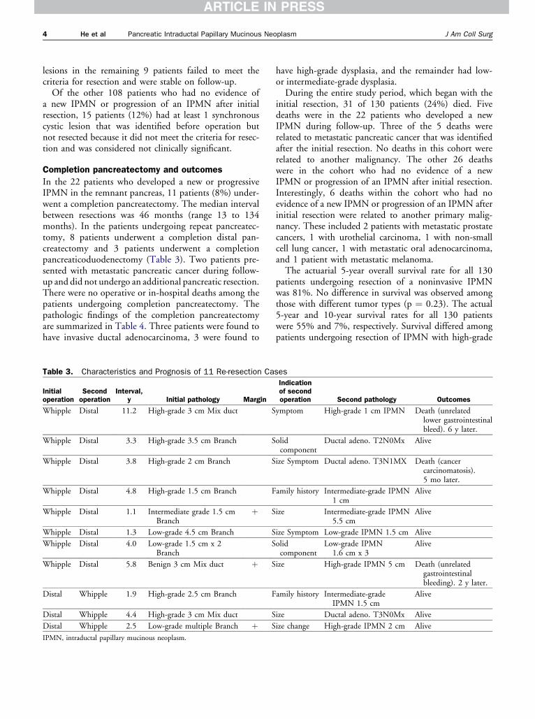
In the 22 patients who developed a new or progressiveIPMN in the remnant pancreas, 11 patients (8%) under-went a completion pancreatectomy. The median intervalbetween resections was 46 months (range 13 to 134months). In the patients undergoing repeat pancreatec-tomy, 8 patients underwent a completion distal pan-createctomy and 3 patients underwent a completionpancreaticoduodenectomy (Table 3). Two patients pre-sented with metastatic pancreatic cancer during follow-up and did not undergo an additional pancreatic resection.There were no operative or in-hospital deaths among thepatients undergoing completion pancreatectomy. Thepathologic findings of the completion pancreatectomyare summarized in Table 4. Three patients were found tohave invasive ductal adenocarcinoma, 3 were found to
Table 3. Characteristics and Prognosis of 11 Re-resection Ca
Initialoperation
Secondoperation
Interval,y Initial pathology Margin
Whipple Distal 11.2 High-grade 3 cm Mix duct S
Whipple Distal 3.3 High-grade 3.5 cm Branch S
Whipple Distal 3.8 High-grade 2 cm Branch S
Whipple Distal 4.8 High-grade 1.5 cm Branch F
Whipple Distal 1.1 Intermediate grade 1.5 cmBranch
þ S
Whipple Distal 1.3 Low-grade 4.5 cm Branch S
Whipple Distal 4.0 Low-grade 1.5 cm x 2Branch
S
Whipple Distal 5.8 Benign 3 cm Mix duct þ S
Distal Whipple 1.9 High-grade 2.5 cm Branch F
Distal Whipple 4.4 High-grade 3 cm Mix duct S
Distal Whipple 2.5 Low-grade multiple Branch þ S
IPMN, intraductal papillary mucinous neoplasm.
have high-grade dysplasia, and the remainder had low-or intermediate-grade dysplasia.During the entire study period, which began with the
initial resection, 31 of 130 patients (24%) died. Fivedeaths were in the 22 patients who developed a newIPMN during follow-up. Three of the 5 deaths wererelated to metastatic pancreatic cancer that was identifiedafter the initial resection. No deaths in this cohort wererelated to another malignancy. The other 26 deathswere in the cohort who had no evidence of a newIPMN or progression of an IPMN after initial resection.Interestingly, 6 deaths within the cohort who had noevidence of a new IPMN or progression of an IPMN afterinitial resection were related to another primary malig-nancy. These included 2 patients with metastatic prostatecancers, 1 with urothelial carcinoma, 1 with non-smallcell lung cancer, 1 with metastatic oral adenocarcinoma,and 1 patient with metastatic melanoma.The actuarial 5-year overall survival rate for all 130
patients undergoing resection of a noninvasive IPMNwas 81%. No difference in survival was observed amongthose with different tumor types (p ¼ 0.23). The actual5-year and 10-year survival rates for all 130 patientswere 55% and 7%, respectively. Survival differed amongpatients undergoing resection of IPMN with high-grade
ses
Indicationof secondoperation Second pathology Outcomes
ymptom High-grade 1 cm IPMN Death (unrelatedlower gastrointestinalbleed). 6 y later.
olidcomponent
Ductal adeno. T2N0Mx Alive
ize Symptom Ductal adeno. T3N1MX Death (cancercarcinomatosis).5 mo later.
amily history Intermediate-grade IPMN1 cm
Alive
ize Intermediate-grade IPMN5.5 cm
Alive
ize Symptom Low-grade IPMN 1.5 cm Alive
olidcomponent
Low-grade IPMN1.6 cm x 3
Alive
ize High-grade IPMN 5 cm Death (unrelatedgastrointestinalbleeding). 2 y later.
amily history Intermediate-gradeIPMN 1.5 cm
Alive
ize Ductal adeno. T3N0Mx Alive
ize change High-grade IPMN 2 cm Alive
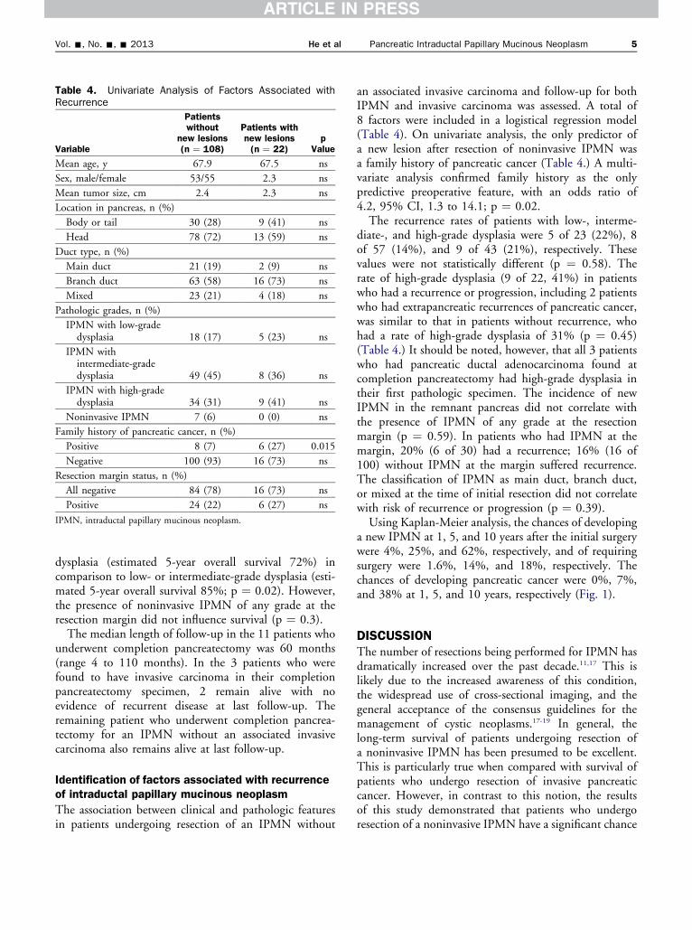
Table 4. Univariate Analysis of Factors Associated withRecurrence
Variable
Patientswithout
new lesions(n ¼ 108)
Patients withnew lesions(n ¼ 22)
pValue
Mean age, y 67.9 67.5 ns
Sex, male/female 53/55 2.3 ns
Mean tumor size, cm 2.4 2.3 ns
Location in pancreas, n (%)
Body or tail 30 (28) 9 (41) ns
Head 78 (72) 13 (59) ns
Duct type, n (%)
Main duct 21 (19) 2 (9) ns
Branch duct 63 (58) 16 (73) ns
Mixed 23 (21) 4 (18) ns
Pathologic grades, n (%)
IPMN with low-gradedysplasia 18 (17) 5 (23) ns
IPMN withintermediate-gradedysplasia 49 (45) 8 (36) ns
IPMN with high-gradedysplasia 34 (31) 9 (41) ns
Noninvasive IPMN 7 (6) 0 (0) ns
Family history of pancreatic cancer, n (%)
Positive 8 (7) 6 (27) 0.015
Negative 100 (93) 16 (73) ns
Resection margin status, n (%)
All negative 84 (78) 16 (73) ns
Positive 24 (22) 6 (27) ns
IPMN, intraductal papillary mucinous neoplasm.
Vol. -, No. -, - 2013 He et al Pancreatic Intraductal Papillary Mucinous Neoplasm 5
dysplasia (estimated 5-year overall survival 72%) incomparison to low- or intermediate-grade dysplasia (esti-mated 5-year overall survival 85%; p ¼ 0.02). However,the presence of noninvasive IPMN of any grade at theresection margin did not influence survival (p ¼ 0.3).The median length of follow-up in the 11 patients who
underwent completion pancreatectomy was 60 months(range 4 to 110 months). In the 3 patients who werefound to have invasive carcinoma in their completionpancreatectomy specimen, 2 remain alive with noevidence of recurrent disease at last follow-up. Theremaining patient who underwent completion pancrea-tectomy for an IPMN without an associated invasivecarcinoma also remains alive at last follow-up.
Identification of factors associated with recurrenceof intraductal papillary mucinous neoplasm
The association between clinical and pathologic featuresin patients undergoing resection of an IPMN without
an associated invasive carcinoma and follow-up for bothIPMN and invasive carcinoma was assessed. A total of8 factors were included in a logistical regression model(Table 4). On univariate analysis, the only predictor ofa new lesion after resection of noninvasive IPMN wasa family history of pancreatic cancer (Table 4.) A multi-variate analysis confirmed family history as the onlypredictive preoperative feature, with an odds ratio of4.2, 95% CI, 1.3 to 14.1; p ¼ 0.02.The recurrence rates of patients with low-, interme-
diate-, and high-grade dysplasia were 5 of 23 (22%), 8of 57 (14%), and 9 of 43 (21%), respectively. Thesevalues were not statistically different (p ¼ 0.58). Therate of high-grade dysplasia (9 of 22, 41%) in patientswho had a recurrence or progression, including 2 patientswho had extrapancreatic recurrences of pancreatic cancer,was similar to that in patients without recurrence, whohad a rate of high-grade dysplasia of 31% (p ¼ 0.45)(Table 4.) It should be noted, however, that all 3 patientswho had pancreatic ductal adenocarcinoma found atcompletion pancreatectomy had high-grade dysplasia intheir first pathologic specimen. The incidence of newIPMN in the remnant pancreas did not correlate withthe presence of IPMN of any grade at the resectionmargin (p ¼ 0.59). In patients who had IPMN at themargin, 20% (6 of 30) had a recurrence; 16% (16 of100) without IPMN at the margin suffered recurrence.The classification of IPMN as main duct, branch duct,or mixed at the time of initial resection did not correlatewith risk of recurrence or progression (p ¼ 0.39).Using Kaplan-Meier analysis, the chances of developing
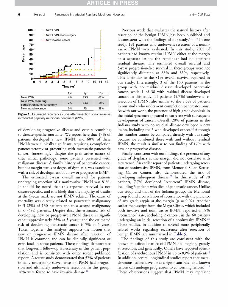
a new IPMN at 1, 5, and 10 years after the initial surgerywere 4%, 25%, and 62%, respectively, and of requiringsurgery were 1.6%, 14%, and 18%, respectively. Thechances of developing pancreatic cancer were 0%, 7%,and 38% at 1, 5, and 10 years, respectively (Fig. 1).
DISCUSSIONThe number of resections being performed for IPMN hasdramatically increased over the past decade.11,17 This islikely due to the increased awareness of this condition,the widespread use of cross-sectional imaging, and thegeneral acceptance of the consensus guidelines for themanagement of cystic neoplasms.17-19 In general, thelong-term survival of patients undergoing resection ofa noninvasive IPMN has been presumed to be excellent.This is particularly true when compared with survival ofpatients who undergo resection of invasive pancreaticcancer. However, in contrast to this notion, the resultsof this study demonstrated that patients who undergoresection of a noninvasive IPMN have a significant chance
0 1 2 3 4 5 6 7 8 9 10 11 12
0
10
20
30
40
50
60
70
80
90
100 New IPMN
New IPMN needs surgery
New invasive cancer
Time (yr)
Pe
rc
en
ta
ge
Figure 1. Estimated recurrence curve after resection of noninvasiveintraductal papillary mucinous neoplasm (IPMN).
6 He et al Pancreatic Intraductal Papillary Mucinous Neoplasm J Am Coll Surg
of developing progressive disease and even succumbingto disease-specific mortality. We report here that 17% ofpatients developed a new IPMN, and 60% of theseIPMNs were clinically significant, requiring a completionpancreatectomy or presenting with metastatic pancreaticcancer. Interestingly, despite the preinvasive nature oftheir initial pathology, some patients presented withmalignant disease. A family history of pancreatic cancer,but not margin status or degree of dysplasia, was associatedwith a risk of development of a new or progressive IPMN.The estimated 5-year overall survival for patients
undergoing resection of a noninvasive IPMN was 81%.It should be noted that this reported survival is notdisease-specific, and it is likely that the majority of deathsat the 5-year mark are not IPMN related. The cause ofmortality was directly related to pancreatic malignancyin 3 (2%) of 130 patients and to a second malignancyin 6 (4%) patients. Despite this, the estimated risk ofdeveloping new or progressive IPMN disease is signifi-cantdapproximately 25% at 5 yearsdand the estimatedrisk of developing pancreatic cancer is 7% at 5 years.Taken together, this analysis supports the notion thatnew or progressive IPMN disease after resection ofIPMN is common and can be clinically significant oreven fatal in some patients. These findings demonstratethat long-term follow-up is necessary in this patient pop-ulation and is consistent with other recent publishedreports. A recent study demonstrated that 57% of patientsinitially undergoing surveillance of IPMN had progres-sion and ultimately underwent resection. In this group,18% were found to have invasive disease.20
Previous work that evaluates the natural history afterresection of the benign IPMN has been published andis consistent with the findings of our study.12,21,22 In onestudy, 191 patients who underwent resection of a nonin-vasive IPMN were evaluated. In this study, 20% ofpatients had known residual IPMN either at the marginor a separate lesion; the remainder had no apparentresidual disease. The estimated overall survival and5-year progression-free survival in these groups were notsignificantly different, at 88% and 83%, respectively.This is similar to the 81% overall survival reported inour study. Interestingly, 3 of the 153 patients in thegroup with no residual disease developed pancreaticcancer, while 1 of 38 with residual disease developedcancer. In this study, 11 patients (5.7%) underwent re-resection of IPMN, also similar to the 8.5% of patientsin our study who underwent completion pancreatectomy.As with our work, the presence of high-grade dysplasia inthe initial specimen appeared to correlate with subsequentdevelopment of cancer. Overall, 20% of patients in theIndiana study with no residual disease developed a newlesion, including the 3 who developed cancer.12 Althoughthis number cannot be compared directly with our studybecause we combined those with and without residualIPMN, the result is similar to our finding of 17% withnew or progressive disease.Finally, consistent with our findings, the presence of any
grade of dysplasia at the margin did not correlate withrecurrence. An earlier report of patients undergoing resec-tion of noninvasive IPMN, from Memorial Sloan Ketter-ing Cancer Center, also demonstrated the risk ofdeveloping subsequent disease.22 In this study of 78patients, 7.7% developed “recurrent” IPMN disease,including 3 patients who died of pancreatic cancer. Unlikeour study and that of the Indiana group, the Memorialgroup found a correlation of recurrence with the presenceof any grade atypia at the margin (p ¼ 0.02). Anotherearlier manuscript from the Mayo Clinic, which includedboth invasive and noninvasive IPMN, reported an 8%“recurrence” rate, including 2 cancers, in the 60 patientsundergoing an initial resection of a noninvasive IPMN.21
These studies, in addition to several more peripherallyrelated works regarding recurrence after resection ofbenign IPMN, are summarized in Table 5.The findings of this study are consistent with the
known multifocal nature of IPMN on imaging, grosslyat resection, and genetically. Others have reported identi-fication of synchronous IPMN in up to 83% of patients.8
In addition, several longitudinal studies report that meta-chronous lesions develop at a significant rate, and knownlesions can undergo progression to concerning lesions.11,23
These observations suggest that IPMN may represent
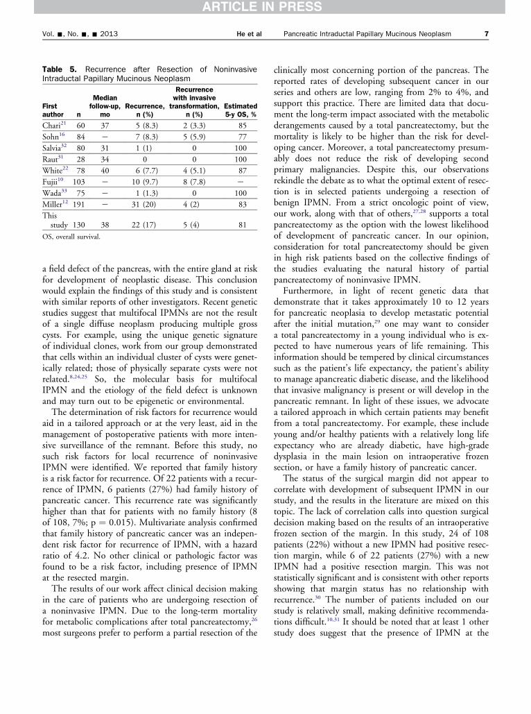
Table 5. Recurrence after Resection of NoninvasiveIntraductal Papillary Mucinous Neoplasm
Firstauthor n
Medianfollow-up,
moRecurrence,
n (%)
Recurrencewith invasive
transformation,n (%)
Estimated5-y OS, %
Chari21 60 37 5 (8.3) 2 (3.3) 85
Sohn16 84 e 7 (8.3) 5 (5.9) 77
Salvia32 80 31 1 (1) 0 100
Raut31 28 34 0 0 100
White22 78 40 6 (7.7) 4 (5.1) 87
Fujii10 103 e 10 (9.7) 8 (7.8) e
Wada33 75 e 1 (1.3) 0 100
Miller12 191 e 31 (20) 4 (2) 83
Thisstudy 130 38 22 (17) 5 (4) 81
OS, overall survival.
Vol. -, No. -, - 2013 He et al Pancreatic Intraductal Papillary Mucinous Neoplasm 7
a field defect of the pancreas, with the entire gland at riskfor development of neoplastic disease. This conclusionwould explain the findings of this study and is consistentwith similar reports of other investigators. Recent geneticstudies suggest that multifocal IPMNs are not the resultof a single diffuse neoplasm producing multiple grosscysts. For example, using the unique genetic signatureof individual clones, work from our group demonstratedthat cells within an individual cluster of cysts were genet-ically related; those of physically separate cysts were notrelated.8,24,25 So, the molecular basis for multifocalIPMN and the etiology of the field defect is unknownand may turn out to be epigenetic or environmental.The determination of risk factors for recurrence would
aid in a tailored approach or at the very least, aid in themanagement of postoperative patients with more inten-sive surveillance of the remnant. Before this study, nosuch risk factors for local recurrence of noninvasiveIPMN were identified. We reported that family historyis a risk factor for recurrence. Of 22 patients with a recur-rence of IPMN, 6 patients (27%) had family history ofpancreatic cancer. This recurrence rate was significantlyhigher than that for patients with no family history (8of 108, 7%; p ¼ 0.015). Multivariate analysis confirmedthat family history of pancreatic cancer was an indepen-dent risk factor for recurrence of IPMN, with a hazardratio of 4.2. No other clinical or pathologic factor wasfound to be a risk factor, including presence of IPMNat the resected margin.The results of our work affect clinical decision making
in the care of patients who are undergoing resection ofa noninvasive IPMN. Due to the long-term mortalityfor metabolic complications after total pancreatectomy,26
most surgeons prefer to perform a partial resection of the
clinically most concerning portion of the pancreas. Thereported rates of developing subsequent cancer in ourseries and others are low, ranging from 2% to 4%, andsupport this practice. There are limited data that docu-ment the long-term impact associated with the metabolicderangements caused by a total pancreatectomy, but themortality is likely to be higher than the risk for devel-oping cancer. Moreover, a total pancreatectomy presum-ably does not reduce the risk of developing secondprimary malignancies. Despite this, our observationsrekindle the debate as to what the optimal extent of resec-tion is in selected patients undergoing a resection ofbenign IPMN. From a strict oncologic point of view,our work, along with that of others,27,28 supports a totalpancreatectomy as the option with the lowest likelihoodof development of pancreatic cancer. In our opinion,consideration for total pancreatectomy should be givenin high risk patients based on the collective findings ofthe studies evaluating the natural history of partialpancreatectomy of noninvasive IPMN.Furthermore, in light of recent genetic data that
demonstrate that it takes approximately 10 to 12 yearsfor pancreatic neoplasia to develop metastatic potentialafter the initial mutation,29 one may want to considera total pancreatectomy in a young individual who is ex-pected to have numerous years of life remaining. Thisinformation should be tempered by clinical circumstancessuch as the patient’s life expectancy, the patient’s abilityto manage apancreatic diabetic disease, and the likelihoodthat invasive malignancy is present or will develop in thepancreatic remnant. In light of these issues, we advocatea tailored approach in which certain patients may benefitfrom a total pancreatectomy. For example, these includeyoung and/or healthy patients with a relatively long lifeexpectancy who are already diabetic, have high-gradedysplasia in the main lesion on intraoperative frozensection, or have a family history of pancreatic cancer.The status of the surgical margin did not appear to
correlate with development of subsequent IPMN in ourstudy, and the results in the literature are mixed on thistopic. The lack of correlation calls into question surgicaldecision making based on the results of an intraoperativefrozen section of the margin. In this study, 24 of 108patients (22%) without a new IPMN had positive resec-tion margin, while 6 of 22 patients (27%) with a newIPMN had a positive resection margin. This was notstatistically significant and is consistent with other reportsshowing that margin status has no relationship withrecurrence.30 The number of patients included on ourstudy is relatively small, making definitive recommenda-tions difficult.10,31 It should be noted that at least 1 otherstudy does suggest that the presence of IPMN at the
8 He et al Pancreatic Intraductal Papillary Mucinous Neoplasm J Am Coll Surg
margin may correlate with development of new orprogressive IPMN disease. Therefore, despite the lack ofcorrelation in our series, we believe that the intraoperativefrozen section is informative and in some cases, may alteroperative decision making. One may want to morestrongly consider a total pancreatectomy in a youngpatient with high-grade dysplasia at the margin vs thesame patient with low-grade dysplasia at the margin.This is based on the premise that recurrences are due tomultifocal disease, with a synchronous IPMN presentwithin the pancreatic remnant or the development ofa second metachronous IPMN, rather than progressionof margin-positive disease. In this regard, the margin isused as a marker of residual disease throughout theremnant. The International Consensus Guidelines forManagement of Intraductal Papillary Mucinous Neo-plasms and Mucinous Cystic Neoplasms of the Pancreas17
are reasonable to guide surgical decisions based on thisargument. These guidelines state that benign adenomas(low-grade dysplasia) at the resected margin do notwarrant further resection because they are at minimalrisk of progression to cancer. However, patients with anIPMN with an associated invasive carcinoma or anIPMN with high-grade dysplasia would benefit from totalpancreatectomy.The relatively high rate of developing a new and
progressive IPMN and the potential to develop invasivecancer reported in this study demonstrate the need forcontinued surveillance in patients with resected noninva-sive IPMN. Currently, there are no clear guidelinesregarding the frequency, duration, or methods of postop-erative surveillance in these patients. Based on our study,it seems prudent to manage these patients in a mannersimilar to that in patients not undergoing resection ofIPMN. One should be cautioned, however, that themere fact that patients were previously selected for resec-tion suggests that they have a more aggressive naturalhistory than patients with IPMN that never met criteriafor surgical resection. We recommend cross-sectionalimaging (CT or MR) every 6 to 12 months for the first5 years, and annually thereafter. Endoscopic ultrasoundcan be used in selected cases. Meanwhile, becauseIPMN patients have been shown to have increased riskof developing extrapancreatic malignancies, general rec-ommended cancer screening guidelines should bestrongly encouraged.6
This study had several limitations. Because determina-tion of the development of a new IPMN is made onradiographic imaging studies, it is likely that we underes-timated the true extent of disease and recurrence. Forexample, it is possible that some patients progress to inva-sive carcinoma in the absence of a detectable cyst, as
evidenced by our patients who presented with an invasivecarcinoma. In this regard, patients with resected IPMNmight harbor synchronous invasive carcinoma, but weare unable to identify their lesion as clinically significantbased on imaging. In addition, 11 recurrence patientsdiagnosed by CT did not undergo resection, and we donot have pathologic confirmation that their clinicalprogression correlated with histologic progression. More-over, this was a retrospective study, and given that ourhospital is a tertiary referral center, there is likely selectionbias and heterogeneity in management. Finally, the clin-ically significant recurrence does not allow strict determi-nation of risk factors for recurrence.
CONCLUSIONSIn conclusion, we have demonstrated that patients under-going resection of a noninvasive IPMN have a high risk ofdeveloping a new IPMN, additional malignancies, and areat risk for disease-specific mortality. It should be notedthat their long-term survival is clearly favorable incomparison to pancreatic ductal adenocarcinoma. Thehigh rate of recurrence and progression calls for closesurveillance in the postoperative period and supportsconsideration of a total pancreatectomy in some patients.Although margin status can be helpful in the surgical deci-sion making, it did not correlate with outcomes in ourstudy. A family history of pancreatic cancer was the onlyrisk factor for development of a new IPMN in our study.
Author Contributions
Study conception and design: He, Cameron, WolfgangAcquisition of data: He, Cameron, Ahuja, Makary,Hirose, Choti, Schulick, Hruban, Pawlik, Wolfgang
Analysis and interpretation of data: He, Cameron,Wolfgang
Drafting of manuscript: He, Cameron, WolfgangCritical revision: Cameron, Ahuja, Makary, Hirose,Choti, Schulick, Hruban, Pawlik, Wolfgang
Acknowledgment: The authors would like to thankDr. Donghang Huang for his help in collecting data fromtheir database.
REFERENCES
1. Tollefson MK, Libsch KD, Sarr MG, et al. Intraductal papil-lary mucinous neoplasm: did it exist prior to 1980? Pancreas2003;26:e55e58.
2. Fernandez-del Castillo C, Adsay NV. Intraductal papillarymucinous neoplasms of the pancreas. Gastroenterology 2010;139:708e713.e1e2.
Vol. -, No. -, - 2013 He et al Pancreatic Intraductal Papillary Mucinous Neoplasm 9
3. Allen PJ. The management of intraductal papillary mucinousneoplasms of the pancreas. Surg Oncol Clin North Am2010;19:297e310.
4. Gourgiotis S, Ridolfini MP, Germanos S. Intraductal papillarymucinous neoplasms of the pancreas. Eur J Surg Oncol 2007;33:678e684.
5. Reid-Lombardo KM, St Sauver J, Li Z, et al. Incidence, prev-alence, and management of intraductal papillary mucinousneoplasm in Olmsted County, Minnesota, 1984-2005: a popu-lation study. Pancreas 2008;37:139e144.
6. Poultsides GA, Reddy S, Cameron JL, et al. Histopathologicbasis for the favorable survival after resection of intraductalpapillary mucinous neoplasm-associated invasive adenocarci-noma of the pancreas. Ann Surg 2010;251:470e476.
7. Bendix Holme J, Jacobsen NO, Rokkjaer M, Kruse A. Totalpancreatectomy in six patients with intraductal papillarymucinous tumour of the pancreas: the treatment of choice.HPB (Oxford) 2001;3:257e262.
8. Matthaei H, Norris AL, Tsiatis AC, et al. Clinicopathologicalcharacteristics and molecular analyses of multifocal intraductalpapillary mucinous neoplasms of the pancreas. Ann Surg 2012;255:326e333.
9. Kawakubo K, Tada M, Isayama H, et al. Incidence of extrap-ancreatic malignancies in patients with intraductal papillarymucinous neoplasms of the pancreas. Gut 2011;60:1249e1253.
10. Fujii T, Kato K, Kodera Y, et al. Prognostic impact of pancre-atic margin status in the intraductal papillary mucinousneoplasms of the pancreas. Surgery 2010;148:285e290.
11. Moriya T, Traverso W. Fate of the pancreatic remnant afterresection for an intraductal papillary mucinous neoplasm:a longitudinal level II cohort study. Arch Surg 2012;147:528e534.
12. Miller JR, Meyer JE, Waters JA, et al. Outcome of the pancre-atic remnant following segmental pancreatectomy for non-invasive intraductal papillary mucinous neoplasm. HPB(Oxford) 2011;13:759e766.
13. Cauley CE, Waters JA, Dumas RP, et al. Outcomes of primarysurveillance for intraductal papillary mucinous neoplasm.J Gastrointest Surg 2012;16:258e267; discussion 266.
14. Hruban RH, Pitman MB, Klimstra DS. Tumors of thepancreas. Washington, DC: American Registry of Pathologyand Armed Forces Institute of Pathology; 2007.
15. Hruban RH, Takaori K, Klimstra DS, et al. An illustratedconsensus on the classification of pancreatic intraepithelialneoplasia and intraductal papillary mucinous neoplasms. AmJ Surg Pathol 2004;28:977e987.
16. Sohn TA, Yeo CJ, Cameron JL, et al. Intraductal papillarymucinous neoplasms of the pancreas: an updated experience.Ann Surg 2004;239:788e797; discussion 797e799.
17. Tanaka M, Fernandez-del Castillo C, Adsay V, et al. Interna-tional consensus guidelines 2012 for the management ofIPMN and MCN of the pancreas. Pancreatology 2012;12:183e197.
18. Tanaka M. Controversies in the management of pancreaticIPMN. Nat Rev Gastroenterol Hepatol 2011;8:56e60.
19. Tanaka M, Chari S, Adsay V, et al. International consensusguidelines for management of intraductal papillary mucinousneoplasms and mucinous cystic neoplasms of the pancreas.Pancreatology 2006;6:17e32.
20. Lafemina J, Katabi N, Klimstra D, et al. Malignant progres-sion in IPMN: a cohort analysis of patients initially selectedfor resection or observation. Ann Surg Oncol 2012 Oct 31[Epub ahead of print].
21. Chari ST, Yadav D, Smyrk TC, et al. Study of recurrence aftersurgical resection of intraductal papillary mucinous neoplasmof the pancreas. Gastroenterology 2002;123:1500e1507.
22. White R, D’Angelica M, Katabi N, et al. Fate of the remnantpancreas after resection of noninvasive intraductal papillarymucinous neoplasm. J Am Coll Surg 2007;204:987e993;discussion 993e995.
23. Fritz S, Klauss M, Bergmann F, et al. Small (Sendai negative)branch-duct IPMNs: not harmless. Ann Surg 2012;256:313e320.
24. Dal Molin M, Hong SM, Hebbar S, et al. Loss of expression ofthe SWI/SNF chromatin remodeling subunit BRG1/SMARCA4 is frequently observed in intraductal papillarymucinous neoplasms of the pancreas. Hum Pathol 2012;43:585e591.
25. Wu J, Matthaei H, Maitra A, et al. Recurrent GNAS muta-tions define an unexpected pathway for pancreatic cyst devel-opment. Sci Transl Med 2011;3:92ra66.
26. Billings BJ, Christein JD, Harmsen WS, et al. Quality-of-lifeafter total pancreatectomy: is it really that bad on long-termfollow-up? J Gastrointest Surg 2005;9:1059e1066; discussion1066e1067.
27. Jamil LH, Chindris AM, Gill KR, et al. Glycemic control aftertotal pancreatectomy for intraductal papillary mucinousneoplasm: an exploratory study. HPB Surg 2012;2012:381328.
28. Fujino Y, Matsumoto I, Ajiki T, Kuroda Y. Clinical reap-praisal of total pancreatectomy for pancreatic disease. Hepato-gastroenterology 2009;56:1525e1528.
29. Yachida S, Jones S, Bozic I, et al. Distant metastasis occurs lateduring the genetic evolution of pancreatic cancer. Nature2010;467:1114e1117.
30. Park J, Lee KT, Jang TH, et al. Risk factors associated with thepostoperative recurrence of intraductal papillary mucinousneoplasms of the pancreas. Pancreas 2011;40:46e51.
31. Raut CP, Cleary KR, Staerkel GA, et al. Intraductal papillarymucinous neoplasms of the pancreas: effect of invasion andpancreatic margin status on recurrence and survival. AnnSurg Oncol 2006;13:582e594.
32. Salvia R, Fernandez-del Castillo C, Bassi C, et al. Main-ductintraductal papillary mucinous neoplasms of the pancreas:clinical predictors of malignancy and long-term survivalfollowing resection. Ann Surg 2004;239:678e685; discussion685e687.
33. Wada K, Kozarek RA, Traverso LW. Outcomes followingresection of invasive and noninvasive intraductal papillarymucinous neoplasms of the pancreas. Am J Surg 2005;189:632e636; discussion 637.


![Mucinous Neoplasm: A Case Report A Rare Case of Low-grade ... · cell adenocarcinoma, or neuroendocrine carcinoma [3]. Mucinous adenocarcinoma accounts for Mucinous adenocarcinoma](https://img.pdfslide.us/doc/110x75/5d66f73588c993283a8b59a1/mucinous-neoplasm-a-case-report-a-rare-case-of-low-grade-cell-adenocarcinoma.jpg)