Embed Size (px)
Citation preview

0013-7227/93/1324-1421$03.c@/O Endocrinology Copyright Q 1993 by The Endocrine Society
Vol. 132, No. 4 Printed in U.S.A.
IRS-l Is a Common Element in Insulin and Insulin-Like Growth Factor-I Signaling to the Phosphatidylinositol 3’-Kinase*
MARTIN G. MYERS, JR.t, XIAO JIAN SUN, BENTLEY CHEATHAM, BOZENA R. JACHNA, ERIN M. GLASHEEN, JONATHAN M. BACKER, AND MORRIS F. WHITE
Research Division, Joslin Diabetes Center and Program in Cell and Developmental Biology, Harvard Medical School, Boston, Massachusetts 02215
ABSTRACT IRS-l is a unique cytosolic protein that becomes tyrosine phosphor-
ylated during insulin stimulation of intact cells and immediately asso- ciates with the phosphatidylinositol 3’-kinase (PtdIns 3’-kinase). The insulin-like growth factor-I (IGF-I) receptor also mediated the tyrosine phosphorylation of IRS-l and increased the amount of PtdIns 3’- kinase activity bound to IRS-1 in Chinese hamster ovary cells. Purified insulin receptor and IGF-I receptor phosphorylated recombinant bac- ulovirus-produced IRS-1 on similar sites in uitro, and phosphorylated
baculovirus-produced IRS-l bound PtdIns 3’-kinase activity from ly- sates of quiescent cells. Treatment of cells with IGF-I activated the PtdIns 3’-kinase, suggesting that IGF-I activates the PtdIns 3’-kinase through IRS-1 binding to p85 in a manner similar to insulin. Chinese hamster ovary cells overexpressing IRS-1 demonstrated increased ty- rosine phosphorylation of IRS-l, and more PtdIns 3’-kinase activity associated with IRS-1 in these cells. These data demonstrate that IRS- 1 is a common element for signal transmission by the IGF-I and insulin receptors. (Endocrinology 132: 1421-1430,1993)
I NSULIN and Insulin-like growth factor-I (IGF-I) are ho- mologous growth factors which control cellular growth
and metabolism. The cell surface receptors for insulin (IR) and IGF-I (IGF-Ir) are highly homologous transmembrane glycoproteins which are composed of two LY- and two /3- subunits (1, 2). Whereas the a-subunits contain distinct li- gand-binding domains, the cytoplasmic domain of the ,& subunits contains homologous tyrosine kinases (1, 3, 4). Binding of insulin and IGF-I to their cognate receptors acti- vates the tyrosine kinases, causing a cascade of tyrosine autophosphorylation in the P-subunit and further stimula- tion of the receptor kinase activity (5). Both IR and IGF-lr activation result in the tyrosine phosphoryation of ~~185, thought to be the major receptor kinase substrate (6-8).
Recently, we purified pp185 from insulin-stimulated rat liver by affinity chromatography on immobilized antiphos- photyrosine antibody (9) and isolated a complementary DNA (cDNA) molecule encoding this protein, which we call IRS- 1 (10, 11). IRS-l is a unique hydrophilic phosphoprotein with a calculated molecular mass of 131 kilodaltons (kDa); however, IRS-l migrates between 170-185 kDa when ex- pressed in Chinese hamster ovary (CHO) cells (10, 12). IRS- 1 contains more than 30 potential serine and threonine phosphorylation sites and 15 potential tyrosine phosphoryl- ation sites (10, 13). Six of the tyrosine residues are found in
Received November 13, 1992. Address all correspondence and requests for reprints to: Dr. Morris
F. White, Research Division, Joslin Diabetes Center, 1 Joslin Place, Boston, Massachusetts 02215.
* The work was suuuorted bv NIH Grants DK-38712 and DK-43808 (to M.F.W.), postdoct&al fello&hips from the Juvenile Diabetes Foun- dation (to J.M.B. and X.J.S.), and Diabetes and Endocrinology Research Grant DK-36836.
t Supported in part by the Medical Scientist Training Program at Harvard Medical School.
YMXM (TyrMetXaaMet) motifs, three in YXXM motifs, and several in other related motifs (e.g. YVNI) which are thought to associate with proteins containing certain isoforms of the src homology-2 (SH2) domain (10, 14). There is a 90.5% amino acid sequence identity between rat and human IRS-I, and all of the potential tyrosine phosphorylation sites are conserved, suggesting that they may be important in signal- ing (10, 15).
IRS-1 undergoes tyrosine phosphorylation immediately after insulin stimulation and associates with the phosphati- dylinositol 3’-kinase (PtdIns 3’-kinase) (10). The PtdIns 3’- kinase phosphorylates the D-3 position of phosphatidylino- sitol, forming PtdIns(3)P, Ptdlns(3,4)P,, and PtdIns(3,4,5)P, in the intact cell, which may control certain aspects of cellular growth and metabolism (14, 16-18). The PtdIns 3’-kinase is activated when the SH2 domains in its 85-kDa subunit (~85) bind to phosphotyrosine residues in IRS-l (19-23). Together these results suggest that IRS-l is a regulatory docking pro- tein that is activated by tyrosine phosphorylation. The asso- ciation of IRS-l with cellular enzymes or adapter molecules that contain the correct SH2 domain isoform may play an important role in the molecular link between receptor tyro- sine kinases and enzymes controlling cellular growth and metabolism.
As the receptors for insulin and IGF-I are highly homolo- gous, we examined whether IRS-l was involved in IGF-1 signaling. In this report we show that IRS-l is a substrate for the IGF-Ir and that IRS-1 is a common element in the IGF-1 and insulin signaling pathways involving the PtdIns 3’- kinase.
Materials and Methods
Cell lines and growth factors
Untransfected CHO cells expressing approximately 30,000 endoge- nous IRS and 200,000 endogenous IGF-Irs or CHO cells expressing lo6
1421 on May 4, 2005 endo.endojournals.orgDownloaded from

1422 IRS-l MEDIATES IGF-I SIGNALING Endo - 1993 Vol132 * No 4
copies of the human IR (CHO/IR) were previously described (24). CHO cells overexpressing rat IRS-l (rIRS-1) (CHO/IRS-1) and control cells transfected with the histidinol resistance plasmid pCMVHis (CHO/HIS) have also been described (10, 12). Cells were grown in Ham’s F-I 2 media suuulemented with 10% fetal bovine serum (Sigma, St. Louis, MO) (24): For insulin stimulation, recombinant‘ hvuman insulin (ELANCO, Indianapolis, IN) was added to a final concentration of 100 nM, and recombinant human IGF-I (Calbiochem, La Jolla, CA) was used at 130 nM, final concentration, unless otherwise noted.
Antibodies
Anti-IRS-l antibodies were protein A-purified polyclonal antibodies from rabbits immunized with recombinant baculovirus-produced IRS-l protein (aIRS-lb”) or affinity-purified antipeptide antidodies (apep80) (101. Monoclonal anti-IRS-l antibodies (lM92-7) were used for immu- nodlotting; these are mouse monocloAa1 imm&oglobulin Gs raised against recombinant IRS-I protein (22). Supernatants from cultured lM92-7 hybridoma cells were used for immunoblotting. Affinity-puri- fied polyclonal antiphosphotyrosine (olPY) antibodies were described previously (25). Immunoprecipitating antibodies specific for the p85 subunit of the PtdIns 3’-kinase were protein A-purified polyclonal antibodies against the C-terminal two-thirds of p85 expressed as a GST fusion protein (26) (Backer J. M., M. F. White, unpublished data).
Immunoprecipitation
Cells were grown to 80% confluence on 15.cm dishes (Costar, Cam- bridge, MA) and made quiescent overnight in serum-free media contain- ing 0.5% BSA (Fluka, Ronkonkoma, NY). Cells were stimulated with growth factor for 1 min, frozen in liquid nitrogen, and extracted in 100 mM Tris, pH 8.0, containing 100 mM NaF, 1% Triton X-100, 1 mM Na,VO,, 1 rnM phenylmethylsulfonylfluoride, 10 pg/ml Aprotinin, and 10 loll NaP204. Insoluble material was pelleted at 100,000 X g for 1 h, and supematants were incubated with aPY (3 pg/ml) or otIRS-lb”’ (10 pg/ml) antibodies. Immunecomplexes were collected with Pansorbin cells (Calbiochem), and complexes were washed three times in 50 mM HEPES, pH 7.4, containing 1% Triton X-100, 0.1% sodium dodecvl sulfate (S’DS), 150 mM NaCI] and 2 mM Na3V04 before being denatured in Laemmli Sample buffer containing 10 rnM dithiothreitol (DTT) and resolved by SDS-polyacrylamide gel electrophoresis (PAGE).
Immunoblotting
Proteins were resolved by 7.5% SDS-PAGE on a generic apparatus at 5 mA or in Bio-Rad (Richmond, CA) miniprotean apparatuses at 100 V. Gels were transferred to nitrocellulose membranes (Schleicher & Schuell, Keene, NH) for aPY and cup85 immunoblotting and to polyvi- nvldifluoridene membranes (Millioore. Milford. MA) for LuIRS-1 (lM92- 7j immunoblotting. Proteins we;e transferred for’l.5 h at IdO V in Towbin buffer containing 0.02% SDS and 20% methanol (27). Mem- branes were blocked overnight at 4 C in wash buffer (25 mM Tris-HCl, pH 7.4, 150 mM NaCl, and 0.01% Tween-20) supplemented with 3% BSA (Fluka). Membranes were then incubated for 2 h at room temper- ature in wash buffer containing 3% BSA and either &Y (3 rg/ml) or &RS-1 (lM92-7,1:500 dilution of tissue culture supernatant) antibodies. The membranes were subsequently washed three times in wash buffer, and reblocked for 1 h at room temperature in wash buffer containing 3% BSA. For blots with lM92-7, membranes were subsequently incu- bated for 1 h with wash buffer containing 3% BSA and 2.5 pg/ml rabbit antimouse immunoglobulin G (H + L; Pierce, Rockford, IL) and washed twice in wash buffer with 3% BSA. Blots were then incubated with [?I protein A (ICN, Costa Mesa, CA; 0.2 pCi/ml) for 1 h in wash buffer containing 3% BSA. Blots were washed four or five times in wash buffer, dried, and exposed to autoradiography with Kodak X-AR film (Eastman Kodak Co., Rochester, NY) or phosphorimager screens (Molecular Dy- namics, Sunnyvale, CA).
Production of recombinant IRS-l protein
The rIRS-I cDNA, with the 5’. and 3’-untranslated regions removed, was subcloned into the pBlueBac transfer vector (Invitrogen, San Diego,
CA) using standard techniques. pBlueBac containing IRS-l cDNA was then recombined with the wild type AcNPV baculovirus genome in Sf9 insect cells and recombinant baculoviruses were selected (23, 28, 29). Sf9 cells were infected for 54-56 h at high multiplicity of infection of recombinant virus as described (28, 29). Cells were collected bv centrif- ugation and lysed by douncing in B biffer (50 mM Tris-HCI, GH 7.8, 1 M NaCI) supplemented with 10 pg/ml Aprotinin, 10 mM benzamidine, 10 wg/ml leupeptin, 350 fig/ml phenylmethylsulfonylfluoride, and 10 rnM DTT. Insoluble material was removed by sedimentation at 100,000 x g for 1 h. Recombinant IRS-1 protein (IRS-lb”‘) accounted for approx- imately 15% of total protein in clarified lysates and was subsequently purified to approximately 90% homogeneity by gel filtration chromatog- raphy on SK 300 HR media (Pharmacia, Piscataway, NJ) in B buffer (Myers, Jr., M. G., M. F. White, unpublished observations).
In vitro tyrosine phosphorylation of IRS-lb”’
Wheat germ agglutinin (WGA)-purified IR (WGA-IR) was prepared from CHO/IR cells (24, 30), and WGA-purified IGF-Ir (WGA-IGF-Ir) was prepared from CHO cells overexpressing the human IGF-ir (gen- erously provided by Drs R. J. Smith and G. Condorelli, Joslin Diabetes Cente;) &as descrided (31). IRS-lb”’ (-1 FM) was incubated for the indicated time in the presence of 4 ILE WGA-IR or WGA-IGF-Ir in 60 ul B Buffer containing S’O mM ATP, 5 LM MnC12, and 100 nM insulin or IGF-I. For some experiments, 60 &i [y-32P]ATP (Du Pont-New England Nuclear, Boston, MA) was added as tracer.
Trypsin digestion and HPLC analysis of IRS-l phosphopeptides
IRS-lba’ from 3-h in vitro phosphorylation reactions was boiled in Laemmli sample buffer containing 10 II~M DTT and separated by SDS- PAGE. Gels were exposed to autoradiography, and bands corresponding to IRS-l were excised and washed in 20% (vol/vol) methanol overnight at 37 C. Gel slices were dried and incubated in 50 mM NHIHC03 containing 10 mM DTT and 6 M guanidine-HC1 for 5 h at 55 C. Iodoacetamide (25 mg; Pierce) was added to slices, and incubation was continued in the dark for 30 min at room temperature before neutralizing the iodoacetamide with 10 ~1 2-mercaptoethanol for 1 h at room tem- perature. Slices were then washed five times in 50 rnM NHIHCO1, dried, and incubated overnight at 37 C in 1 ml 50 mM NHIHCO, containing 0.1 mg/ml tosylphenylalanine chloromethylketone trypsin (Worthington Enzymes, Freehold, NJ). An additional 100 ~1 50 rnM NHIHCO, con- taining 1 .O mg/ml tosylphenylalanine chloromethylketone trypsin were added, and the incubation was continued overnight. Supernatants were dried, resuspended in 0.055% trifluoroacetic acid (TFA) and analyzed by reversed-phase HF’LC on a Beckman System Gold (Beckman Instru- ments, Palo Alto, CA) equipped with a Hi-Pore reversed phase RP-318 column (Beckman) eluted at a flow rate of 1 ml/min with 0.055% TFA modified with 75% acetonitrile-0.05% TFA solution as described (12). Eluted radioactivity was collected in 0.5-ml fractions and quantitated (Cerenkov counting) in a Beckman LS 1801 scintillation counter.
Ptdlns 3’-kinase activity
In vitro phosphorylation of PtdIns was carried out in the immune complexes as described previously (32). Subconfluent CHO cells grown in IOO-mm dishes were made quiescent by an overnight incubation in F-12 medium containing 0.5% BSA. The quiescent cells were incubated with insulin or IGF-I for 10 min and washed once with ice-cold PBS and twice with 20 mM Tris-HCl, pH 7.5, containing 137 mM NaCI, 1 rnM MgCl,, 1 mM CaCl,, and 100 ;M Na3V0, (buff& A). The cells were solubilized in 1 ml buffer A containing 1% NP-40 (Siema) and 10% glycerol, and insoluble material was rem&ed by centri&$io~ at 13,000 X g for 10 min. Supematant was incubated with antibody overnight at 4 C, and immune complexes were precipitated from the supernatant with Protein A-Sepharose (Pharmacia) and washed successively in PBS containing 1% NP-40 and 100 PM Na3V0, (three times), 100 mM Tris- HCl, pH 7.5, containing 500 mM LiCl and 100 WM Na,VO, (three times), and 10 mM Tris-HCI, pH 7.5, containing 100 mM NaCl, 1 rnM EDTA, and 100 PM Na3V04 (two times). The pellets were resuspended in 50 ~1
on May 4, 2005 endo.endojournals.orgDownloaded from
IRS-l MEDIATES IGF-I SIGNALING 1423
10 mu Tris-HCl, pH 7.5, containing 100 rnM NaCl and 1 mM EDTA, and combined with 10 vi 100 rnM MnCl, and 10 III 2 uelul PtdIns (Avanti, Pelham, AL) somcated in 10 mM Tris-HCl (pH 7.$“cbntaining 1 mr.t EGTA. The phosphorylation reaction was started by adding 10 ~1 440 NM ATP containing 30 pCi [@‘P]ATP. After 10 min at 22 C, the reaction was stopped with 20 ~1 8 N HCl and 160 ~1 CHCla:methanol (1:l). The samples were centrifuged, and the lower organic phase was removed and applied to a silica gel TLC plate (Merck, Rahway, NJ) which had been-coated with 1% potassium oxalate. TLC plates were develoued in CHCl,:CH~OH:H,O:NH,OH f60:47:11.3:2). dried, and ~~-- - ,. visual&d by autoradiography. The radioactivity in spots which comi- grated with PtdIns-4P standard (Sigma) was measured by Cerenkov counting as previously described (32).
In vitro association of PtdIns 3’-kinase with IRS-lbaC
IR’SI-~~‘~ incubated overnight with activated WGA-IR, WGA-IGF-Ir, or buffer alone was immunoprecipitated for 2 h at 4 C with cupep80 immobilized on Protein A-Sepharose (Pharmacia). Immunoprecipitates were washed twice with PtdIns 3’kinase cell lysis buffer and incubated with lysates from unstimulated CHO cells for 30 min. Immunoprecipi- tates were then washed and assayed for associated PtdIns 3’kinase activity as described above.
Results IR/IGF-lr Jkta--:
IRS-1 is tyrosine phosphoryluted after stimulation of CHO celb with IGF-I or insulin
We assessed the tyrosine phosphorylation state of cellular proteins in CHO and CHO/IR cells which had been incu- bated in the absence or presence of insulin, IGF-I, or both. Cell extracts were incubated with antiphosphotyrosine anti- body ((YPY), and immunoprecipitated phosphotyrosine-con- taining proteins were separated by SDS-PAGE and detected by immunoblotting with CVPY antibodies (Fig. 1, A, lanes a- d and B, lanes a-d). The P-subunits of the insulin and IGF-I receptors were tyrosine phosphorylated immediately after insulin or IGF-I stimulation, respectively (Fig. 1, A and B, lanes a-d). Moreover, ppl85 was detected with CVPY in CHO and CHO/IR cells only after stimulation with insulin, IGF-I, or both. There are significantly more receptors for IGF-I than insulin in CHO cells, and there was correspondingly greater stimulation of pp185 phosphorylation by IGF-I than insulin (Fig. lA, compare lanes b and c). In contrast, the insulin stimulation of pp185 phosphorylation was greater in CHO/ IR cells due to the overexpression of insulin receptors (Fig. 1, A and 8, lanes a and b).
FIG. 1. Tyrosine-phosphorylated proteins in olPY and (uIRS-1 immu- noprecipitates of cells stimulated with insulin and/or IGF-I. CHO (A) and CHO/IR (B) cells were incubated in the absence of insulin and IGF-I (lanes a and e) or with insulin (lanes b and f), IGF-I (lanes c and g), or both insulin and IGF-I (lanes d and h). Cell lysates were immunoprecipitated with crPY (lanes a-d) or (YIRS-1 (lanes e-h) anti- bodies. Immunoprecipitates were resolved by SDS-PAGE, transferred to nitrocellulose, and immunoblotted with (YPY. Migration of IRS-l/ ~~185, the &subunits of IR and IGF-Ir, and molecular mass standards are indicated.
cells and that this effect is predominantly mediated via IGF- I receptors.
Recombinant IRS-l protein is phosphorylated by the IR and IGF-Ir in vitro
In order to directly assess whether IRS-l is phosphorylated by the IGF-Ir, we used anti-IRS-l (nIRS-lb”‘) antibodies to immunoprecipitate the endogenous hamster IRS-l from CHO or CHO/IR cells incubated in the absence or presence of insulin, IGF-I, or both. Immunoblots with (uPY indicated that IRS-l was tyrosine phosphorylated during stimulation with insulin and IGF-I, alone or in combination (Fig. 1, A and B, lanes e-h). Tyrosine phosphorylation of IRS-l in CHO cells was greater with IGF-I than insulin, consistent with the greater number of IGF-I receptors than insulin receptors in CHO cells (Fig. lA, lanes e-h). In contrast, insulin stimulated more phosphorylation of IRS-l in CHO/ IR cells (Fig. lB, lanes e-h). These data suggest that IGF-I stimulates the tyrosine phosphorylation of IRS-l in CHO
Recombinant IRS-l protein (IRS-lb”‘) produced in a bac- ulovirus expression system (23) was incubated in vitro with WGA-purified insulin or IGF-I receptors. Phosphorylation of the WGA-purified receptors and IRS-1 was analyzed by SDS- PAGE and autoradiography. Insulin and IGF-I stimulated in vitro phosphorylation of the P-subunit of the IR and IGF-Ir, respectively (Fig. 2, lanes a and b). Reduction and seperation of the receptors by SDS-PAGE confirmed that the P-subunit of the IGF-Ir migrated more slowly than the IR (31). Incu- bation of IRS-lbaC alone resulted in little phosphate incorpo- ration into IRS-l (Fig. 2, lane e). However, after incubation of IRS-lbac with the IR (lane c) or the IGF-Ir (lane d), both IRS-lbac and the receptor P-subunit were phosphorylated (Fig. 2, lanes c and d). Phosphoamino acid analysis demon- strated that IRS-l was phosphorylated on tyrosine residues (data not shown). The tyrosine phosphorylation sites of IRS- 1 were identical during phosphorylation by insulin and IGF- I receptors, as a similar pattern of tryptic fragments was
A IP: aPY a IRS-1 I I
INSULIN: - + - + - + - + M r IGF-I: - - -I- + - - + + (kDa)
pplS!VIRS-1 ---)I
IR/IGF-lr Beta+ -116
- 80
abcdefgh
B IP: CXPY a IRS-1 I
INSULIN: - + - c - -I- - + M ,. IGF-1: - - + + - - + + (kDi) . .
-200 pplWIRS-1 -
abcdefgh
-116 - 80
on May 4, 2005 endo.endojournals.orgDownloaded from
1424 IRS-l MEDIATES IGF-I SIGNALING Endo * 1993 Vol 132 *No 4
IRS-l -
IGFr Beta
IR Beta
- 116
- 50
a b c d e FIG. 2. In vitro tyrosine phosphorylation of IRS-lb” by the IR and IGF-Ir. WGA-purified proteins containing ligand-activated IR (lanes a and c) or IGF-Ir (lanes b and d) were incubated with [r3*P]ATP in the absence (lanes a and b) or presence of IRS-lb”” (lanes c and d), or IRS- lb”’ was incubated alone (lane e) in the presence of [r3*P]ATP. Proteins were resolved by SDS-PAGE and detected by autoradiography. Migra- tion of IRS-lb”‘, the p-subunits of IR and IGF-Ir, and molecular mass standards are indicated.
FIG. 3. Reversed-phase HPLC analysis of IRS-lbac phosphopeptides after incubation alone (IRS) or with the IR (IR/IRS) or IGF-Ir (IGFr/ IRS) in uitro. Bands containing labeled IRS-l”“’ were excised from gels such as that in Fig. 2 and subjected to reduction carboxymethylation and tryptic digestion. The resulting phosphopeptides were analyzed by reversed-phase HPLC, and radiation in each 0.5-min fraction was determined by Cerenkov counting.
obtained in each case when analyzed by reversed-phase HPLC (Fig. 3); the minor differences in the phosphopeptide maps shown are not reproducible and are primarily due to the splitting of peaks into two fractions during the collection process. We conclude that IRS-l is phosphorylated on iden- tical sites by each receptor. No significant peaks were seen in the absence of WGA receptors.
Insulin and IGF-I stimulate the association of the PtdIns 3- kinase with IRS-l
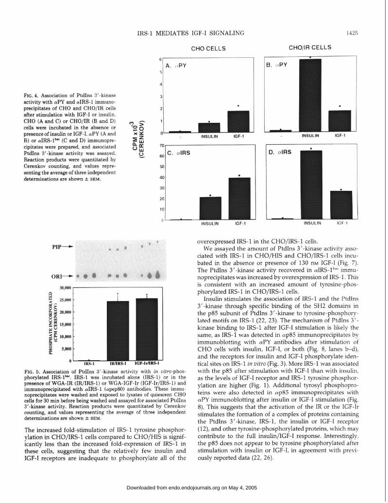
We measured the amount of PtdIns 3’-kinase activity detectable in rvPY or (uIRS-lb”’ immunoprecipitates from CHO and CHO/IR cells after stimulation with insulin or IGF- I (Fig. 4). Both insulin and IGF-I increased the amount of PtdIns 3’-kinase activity in (uPY and cuIRS-lb”’ immunopre- cipitates. The amount of PtdIns 3’-kinase in aIRS-1 immu- noprecipitates was lo-fold greater than the activity found in &Y immunoprecipitates, reflecting the greater ability of aIRS-lb”’ to immunoprecipitate IRS-l, as seen by immuno- blotting (Fig. 1). The ratios of cuPY/aIRS-lb”’ associated PtdIns 3’-kinase activity were nearly identical after stimula- tion by insulin or IGF-I, suggesting that the PtdIns 3’-kinase binds IRS-l similarly after stimulation with IGF-I or insulin. In CHO cells, more PtdIns 3’-kinase activity associated with IRS-l after IGF-I stimulation than with insulin stimulation (Fig. 4C), which reflects the lesser number of IRS and the lower incorporation of phosphate into IRS-l in CHO cells after insulin treatment (Fig. 1). The amount of PtdIns 3’- kinase activity associated with IRS-l from CHO/IR cells after insulin stimulation was greater than that after IGF-I stimu- lation, which reflected the higher phosphorylation of IRS-l in cells overexpressing the IR (Fig. 4D). These data suggest that the stimulation of IRS-l association with the PtdIns 3’- kinase after IGF-I or insulin stimulation is predominantly mediated by the IGF-Ir and IR, respectively.
The association of the PtdIns 3’-kinase with IRS-1 was reconstituted in vitro using IRS-lbac that was phosphorylated by WGA-IR or WGA-IGF-Ir (Fig. 5). Phosphorylated IRS- 1 bar was immobilized on cuIRS-1 (apep80) immunoprecipi- tates and incubated with lysates of quiescent CHO cells. No PtdIns 3’-kinase activity associated with the unphosphory- lated IRS- 1 bat; however, large amounts of activity associated with IRS-lbac which had been phosphorylated by the IR or IGF-Ir (Fig. 5).
Overexpression of IRS-l in CHO cells enhances tyrosine phosphorylation of IRS-l and association of the Ptdlns 3’- kinase with IRS-l after stimulation with IGF-I
CHO cells overexpressing rIRS-1 (CHO/IRS-1) and control CHO cells (CHO/HIS) (12) were stimulated with IGF-I or insulin, and lysates were analyzed by immunoblotting with aIRS-1 (lM92-7) (Fig. 6A) or aPY antibodies (Fig. 6B). This analysis demonstrated that CHO/IRS-1 cells express at least 20-fold more IRS-l than control CHO/HIS cells (Fig. 6A). The amount of tyrosine-phosphorylated IRS-l was increased approximately 2-fold in CHO/IRS-1 cells (Fig. 68, lanes d- f) compared to control CHO/HIS cells (Fig. 6B, lanes a-c) after growth factor stimulation. Thus, this overexpressed IRS- 1 acts as a substrate for the IGF-Ir and the IR after stimulation of cells with growth factor. The amount of tyrosine-phos- phorylated IRS-l was greater after IGF-I stimulation than insulin stimulation (Fig. 68, lanes e and f), reflecting the greater amount of IGF-Ir than IR in these cells. Thus, both the amount of IRS-l and amount of receptor expressed are important determinants of IRS-1 tyrosine phosphorylation.
on May 4, 2005 endo.endojournals.orgDownloaded from
CHO CELLS CHO/IR CELLS
FIG. 4. Association of PtdIns 3’-kinase activity with (YPY and oIRS-1 immuno- precipitates of CHO and CHO/IR cells after stimulation with IGF-I or insulin. CHO (A and C) or CHO/IR (B and D) cells were incubated in the absence or presence of insulin or IGF-I. (uPY (A and B) or cuIRS-lb” (C and D) immunopre- cipitates were prepared, and associated PtdIns 3’-kinase activity was assayed. Reaction products were quantitated by Cerenkov counting, and values repre- senting the average of three independent determinations are shown + SEM.
PIP-
61 I
IRS-l MEDIATES IGF-I SIGNALING 1425
INSULIN IGF-1 INSULIN IGF~l
ORI- @ i
0 I-- IRS-1
O- IRS-1 IRIIRS-1 !/IRS.
FIG. 5. Association of PtdIns 3’-kinase activity with in uitro-phos- nhorvlated IRS-lb”‘. IRS-1 was incubated alone (IRS-l) or in the pre&nce of WGA-IR (IR/IRS-1) or WGA-IGF-Ir (IGF-Ir/IRS-1) and immunoprecipitated with (YIRS-1 (cupep80) antibodies. These immu- noprecipitates were washed and exposed to lysates of quiescent CHO cells for 30 min before being washed and assayed for associated PtdIns 3’-kinase activity. Reaction products were quantitated by Cerenkov counting, and values representing the average of three independent determinations are shown + SEM.
The increased fold-stimulation of IRS-l tyrosine phosphor- ylation in CHO/IRS-1 cells compared to CHO/HIS is signif- icantly less than the increased fold-expression of IRS-l in these cells, suggesting that the relatively few insulin and IGF-I receptors are inadequate to phosphorylate all of the
INSULIN IGF-1
overexpressed IRS-l in the CHO/IRS-1 cells. We assayed the amount of PtdIns 3’kinase activity asso-
ciated with IRS-l in CHO/HIS and CHO/IRS-1 cells incu- bated in the absence or presence of 130 nr.4 IGF-1 (Fig. 7). The PtdIns 3’-kinase activity recovered in aIRS-lb”’ immu- noprecipitates was increased by overexpression of IRS-l. This is consistent with an increased amount of tyrosine-phos- phorylated IRS-l in CHO/IRS-1 cells.
Insulin stimulates the association of IRS-l and the PtdIns 3’-kinase through specific binding of the SH2 domains in the ~85 subunit of PtdIns 3’kinase to tyrosine-phosphory- lated motifs on IRS-l (22, 23). The mechanism of PtdIns 3’- kinase binding to IRS-l after IGF-I stimulation is likely the same, as IRS-l was detected in (~~85 immunoprecipitates by immunoblotting with c&‘Y antibodies after stimulation of CHO cells with insulin, IGF-I, or both (Fig. 8, lanes b-d), and the receptors for insulin and IGF-I phosphorylate iden- tical sites on IRS-l in vitro (Fig. 3). More IRS-l was associated with the ~85 after stimulation with IGF-I than with insulin, as the levels of IGF-I receptor and IRS-l tyrosine phosphor- ylation are higher (Fig. 1). Additional tyrosyl phosphopro- teins were also detected in (up85 immunoprecipitates with (YPY immunoblotting after insulin or IGF-I stimulation (Fig. 8). This suggests that the activation of the IR or the IGF-Ir stimulates the formation of a complex of proteins containing the PtdIns 3’-kinase, IRS-l, the insulin or IGF-I receptor (12), and other tyrosine-phosphorylated proteins, which may contribute to the full insulin/IGF-I response. Interestingly, the p85 does not appear to be tyrosine phosphorylated after stimulation with insulin or IGF-I, in agreement with previ- ously reported data (22, 26).
on May 4, 2005 endo.endojournals.orgDownloaded from

1426 IRS-l MEDIATES IGF-I SIGNALING Endo. 1993
A
INSULIN (100nM): IGF-1 (130nM):
IRS-1 -
CHO/HlS CHOjlRS -r-----l
- +--+- m -+m-+ Mr
\ WW
-200
-116
- 80
: \ - 49 ,. ‘_ _ \ ‘,,
a bcdef
B CHO/HiS CHO/IRS II
INSULIN(lOOnM): - + - - + - Mr IGF-1 (130nM): - - + - - +
&Da)
IRS-1 -
P-SUBUNIT -
-200
-116
- 80
- 49
a bcdef FIG. 6. Tyrosine phosphorylation of proteins in CHO/HIS and CHO/ IRS-l cells after stimulation with IGF-I or insulin. CHO/HIS (lanes a-c) or CHO/IRS-1 (lanes d-f) cells which had been incubated in the absence (lanes a and d) or presence of insulin (lanes b and e) or IGF-I (lanes c and f) for 1 min were lysed in Laemmli sample buffer. Lysates were resolved by SDS-PAGE, transfered to nitrocellulose membranes, and analyzed by immunoblotting with aIRS-1 (lM92-7) (A) or (uPY antibodies and [iz51]protein A (B). Membranes were dried and exposed to autoradiography or phosphorimager screens. Positions of IRS-l, receptor P-subunits, and molecular mass standards are shown.
160.000 -
140.000
120.000
F 100,000 $5 % 80,000
E 60,000
40,000
20,000
IGF-1 (130 nM;
i
CHOlHlS CHOWS
FIG. 7. Association of PtdIns 3’-kinase activity with IRS-1 in CHO/ HIS and CHO/IRS-1 cells. CHO/HIS and CHO/IRS-1 cells were incubated in the absence (-) or presence (+) of 130 nM IGF-I. PtdIns 3’-kinase activity in (YIRS-~~~’ immunoprecipitates was assayed and quantitated (Cerenkov counting).
INSULIN (IOOnM): - + - + Mr IGF-1 (13OnMj: - - + + WW
IRS-1 * -200
-116 \ :
- 80
a b c d
WC. 8. IRS-1 is found in cup85 immunoprecipitates from CHO cells stimulated with IGF-I or insulin. Quiescent CHO cells (lane al or CHO cells stimulated with insulin (lane b), IGF-I (lane c), or both (lane d) were lysed, and p85 was immunoprecipitated as for a PtdIns 3’-kinase assay. Immunoprecipitated proteins were resolved by SDS-PAGE, transferred to nitrocellulose, and analyzed by immunoblotting with CVPY antibodies and [iz51]protein A. Membranes were dried and exposed to phosphorimager screens. Positions of IRS-1 and molecular mass standards are shown.
IGF-I activates the Ptdlns 3’-kinase The PtdIns 3’-kinase is activated by interacting with IRS-
1 after insulin stimulation (22, 23). Stimulation of cells with growth factors was initially thought to cause the tyrosine phosphorylation and subsequent activation of the PtdIns 3’- kinase (16). However, we and others have been unable to detect tyrosine phosphorylation of ~85 after stimulation with a variety of growth factors (Fig. 8) (12, 26). Both IGF-I and insulin stimulate the PtdIns 3’-kinase in ~yp85 immunopre- cipitates (Fig. 9). The activity of the PtdIns 3’-kinase was stimulated approximately 2.5-fold in CHO cells after stimu- lation with insulin and 3.5-fold after stimulation with IGF-I alone or with both insulin and IGF-I. Thus, activation of the PtdIns 3’-kinase by insulin and IGF-I was not additive, suggesting that a similar mechanism was involved. Activa- tion of the PtdIns 3’-kinase correlated with increased amounts of tyrosine-phosphorylated IRS-l in CHO cells.
Discussion
The receptors for IGF-I and insulin possess a homologous tyrosine kinase that is required for cellular signaling (1, 3, 4). However, the common and distinct signaling pathways used by the insulin and IGF-I receptors have remained elusive. The discovery that pp185 is a substrate for the insulin and IGF-I receptors provided the first evidence for the existence of a common cellular substrate for these receptors (6-8). Recently, we cloned IRS-l from the partial amino acid se- quence of pp185 (9-11). IRS-l is a substrate for the insulin receptor tyrosine kinase and acts as a docking protein which binds and regulates the activity of cellular enzymes such as the PtdIns 3’-kinase (10, 12, 22, 23). Our current study suggests that IRS-l is a common element in signal transmis- sion by both the insulin and IGF-I receptors.
IRS-1 is tyrosine phosphorylated in CHO cells after IGF-I
on May 4, 2005 endo.endojournals.orgDownloaded from

IRS-1 MEDIATES IGF-I SIGNALING 1427
NONE INSUI,IN IGF- I
FIG. 9. The PtdIns 3’.kinase is activated in CHO cells after stimula- tion with insulin or IGF-I. aP85 immunoureciuitates were ureoared from unstimulated CHO cells or CHO celis stimulated with insulin, IGF-I, or both, and assayed for associated PtdIns 3’-kinase activity. Reaction products were quantitated by Cerenkov counting, and values representing the average of three independent determinations are shown _’ SEM.
and insulin stimulation. However, the phosphorylation is significantly stronger during IGF-I stimulation, reflecting the relatively greater number of IGF-I receptors in these cells; this pattern is reversed in CHO/IR cells which overexpress the human insulin receptor. Similarly, the activated IGF-I receptor tyrosine kinase phosphorylates recombinant IRS- lhnc protein in vitro on sites identical to those phosphorylated by the IR. As tyrosine-phosphorylated sites on IRS-l create binding sites for certain cellular proteins containing SH2 domains (23), these results suggest that IRS-l interacts with similar downstream elements after IGF-I or insulin stimula- tion Indeed, insulin or IGF-I treatment of CHO cells recruit similar proteins into a complex or complexes including IRS- 1, ~85, and other unidentified phosphotyrosine-containing proteins, probably including the insulin or IGF-I receptor (12). A molecular complex containing several distinct com- ponents, including tyrosyl-phosphorylated IRS-l at the core, may provide a molecular explanation for the pleotropic ef- fects of insulin and IGF-I.
PtdIns 3’-kinase is a heterodimeric enzyme consisting of a 1 IO-kDa catalytic subunit which is regulated by an 85-kDa subunit containing two SH2 domains and an SH3 domain (19-23, 33). IGF-I stimulates the association of the PtdIns 3’-kinase with IRS-1 in CHO cells. This association is greater in IGF-I-treated CHO cells than in insulin-treated CHO cells, which reflects the stronger phosphorylation of IRS-l due to the higher level of IGF-I receptors than insulin receptors in these cells. Overexpression of IRS-l in CHO cells also in- creases the amount of tyrosine-phosphorylated IRS-l during IGF-I stimulation, suggesting that there is a direct kinase- substrate relationship between the IGF-Ir and IRS-l. This
increase in tyrosine-phosphorylated IRS-1 correlates with the increase in PtdIns 3-kinase activity associated with IRS- 1 after stimulation with IGF-I. Also, tyrosyl-phosphorylated IRS-lb”’ binds to PtdIns 3’-kinase activity from extracts of quiescent CHO cells. Both insulin and IGF-I receptor-cata- lyzed phosphorylation of IRS-l produce an identical result, suggesting that the mechanism of association between IRS- 1 and the PtdIns 3’-kinase is similar after insulin or IGF-I stimulation. As PtdIns 3’-kinase binds to tyrosyl-phosphor- ylated sequences (YMXM motifs) in IRS-1 through the SH2 domains of the ~85 subunit during insulin stimulation (22, 23), it is likely that this mechanism accounts for the associ- ation during IGF-I stimulation as well.
It has been reported that the PtdIns 3-kinase associates with the receptors for insulin and IGF-I after ligand stimu- lation (32, 34, 35). The association between IGF-Ir and the Ptdlns 3’-kinase has been studied in vitro using tyrosine- phosphorylated, immunoprecipitated IGF-I receptors and fu- sion proteins containing the SH2 domains of ~85 (35). Im- mobilized IGF-Ir binds PtdIns 3’-kinase activity from cell lysates. Furthermore, this in vitro association is blocked by incubation with SH2 domain fusion proteins from the PtdIns 3-kinase p85 subunit (35). We have observed similar results with the insulin receptor; however, the amount of PtdIns 3’- kinase associated with IRS-l is much greater than with the insulin receptor in vitro as well as in vim (10, 49). Further- more, the receptors for insulin and IGF-I stably associate with IRS-l after ligand binding and receptor activation in intact cells, suggesting that the PtdIns 3’-kinase associated with the IR and IGF-Ir in viva may actually represent PtdIns 3’-kinase bound to receptor-associated IRS-1 (12). We have also observed the association of IRS-l from cell lysates with the activated insulin receptor in vitro (49), suggesting that IRS-l can account for receptor-associated PtdIns 3’-kinase activity in vitro as well as in uivo.
Treatment of cells with growth factors such as insulin and IGF-I increases the amount of PtdIns 3-phosphate
[PtdIns(3)P], PtdIns(3,4)P2, and PtdIns(3,4,5)P, in cells, sug- gesting that the PtdIns 3’-kinase is activated during activa- tion of receptor tyrosine kinases (14, 16-18). This activation was originally thought to be secondary to tyrosine phos- phorylation of the ~85 subunit of the Ptdlns 3’-kinase (16, 36-38), but tyrosine phosphorylation of the ~85 is not de- tected after stimulation by a variety of growth factors (22, 26). We have observed activation of the PtdIns 3’-kinase in ap85 immunoprecipitates from insulin-stimulated cells (22); in these immunoprecipitates, p85 is not tyrosine phosphor- ylated but is associated with tyrosine-phosphorylated IRS- 1, Furthermore, tyrosine-phosphorylated IRS-l activates the PtdIns 3’-kinase in immunoprecipitates from unstimulated cells (22), an effect which appears to be mediated by the binding of tyrosine-phosphorylated IRS-l to the SH2 do- mains of the PtdIns 3’-kinase (22, 23). Similarly, PtdIns 3’- kinase is activated in immunoprecipitates from IGF-I-treated CHO cells, even though p85 is not tyrosine phosphorylated. The presence of tyrosine-phosphorylated IRS-l in ~up85 im- munoprecipitates suggests that the PtdIns 3’-kinase is simi- larly regulated after stimulation with insulin or IGF-I.
on May 4, 2005 endo.endojournals.orgDownloaded from

1428 IRS-l MEDIATES IGF-I SIGNALING Endo. 1993
IRS-l may bind several distinct SH2 domain-containing proteins in addition to ~85, contributing to the transmission of the full insulin signal. SH2 domains are thought to mediate protein-protein interactions by binding to tyrosine-phos- phorylated motifs; different isoforms of the SH2 domain found in different proteins are thought to mediate binding to distinct tyrosine-phosphorylated motifs. The platelet-de- rived growth factor (PDGF) receptor and similar receptors, such as the fibroblast growth factor receptor, bind to phos- pholipase C, and ras-GTPase activating protein (as well as the PtdIns 3’-kinase) after ligand-stimulated tyrosine phos- phorylation (39-42). We have been unable to detect an association between either of these enzymes and IRS-l (Myers, Jr., M. G., 8. C. Cheatham, and M. F. White, unpub- lished observations). The tyrosine-phosphorylated motifs YLDL (in the fibroblast growth factor receptor) and YMAP (in the PDGF receptor) are thought to be the preferred binding sites for the SH2 domains of phospholipase C, and ras-GTPase activating protein, respectively (43, 44). Phos- phorylation sites in these motifs are not found in IRS-l (10); thus, it is not surprising that these molecules do not associate with IRS-l. In contrast, IRS-1 contains 15 potential sites of tyrosine phosphorylation. In addition to the consensus PtdIns 3’-kinase binding sites in YMXM and YXXM motifs, IRS-1 also contains consensus tyrosine phosphorylation sites in the motifs YQAL, YDTG, YVNI, and YPEE (10). Phos- phorylation of these or other sites could create binding sites on IRS-l for proteins containing distinct isoforms of the SH2 domain, contributing to the full insulin/IGF-I signal.
Insulin and IGF-I perform different functions in the intact organism, and studies with chimeric receptors have shown that the IR and IGF-Ir P-subunits have different signaling functions in cultured cells (45). We have not observed dif- ferences in signaling by the IGF-Ir us. the insulin receptor at the level of IRS-l. It is therefore likely that differences in signaling by the IR and the IGF-Ir are not mediated by IRS- 1 but are mediated by other cellular proteins. As IRS-l appears to be expressed in most tissues (Araki, E., X. J. Sun, M. F. White, submitted), the divergence between insulin and IGF-I actions may arise through differential expression of the receptors for insulin and IGF-I and through unique down- stream elements which are selectively expressed in various tissues. For instance, although the PtdIns 3’-kinase appears to be ubiquitously expressed, other SH2 domain-containing signaling enzymes which associate with IRS-l could be ex- pressed in concert with either the insulin or IGF-I receptor. Also, the possibility that IRS-l is differentially phosphory- lated by the insulin and IGF-I receptors in vim is not entirely ruled out by our in vitro observations. Thus, differential tyrosine phosphorylation of IRS-l by these receptors could also account for the differences in signal transmission by these receptors observed in intact cells.
IRS-l may not be the sole substrate and mediator of IGF- I/insulin receptor signaling; careful analysis of pp185 has shown that this band of protein likely represents several proteins of similar molecular weights (11). The lower portion of this band is IRS-l, whereas the upper portion is a distinct substrate for the insulin (and possibly IGF-I) receptors, which
Vol 132. No 4
does not associate with the PtdIns 3’-kinase (11). Thus, tyrosine kinase substrate signaling pathways divergent from IRS-l may exist downstream of the insulin and IGF-I recep- tors and could account for the differences in insulin and IGF- I signaling.
Our data show that IRS-l is a substrate and mediator for both the IGF-I and insulin receptor systems, which are highly homologous. Similarly, IRS-1 may be involved in signaling by other systems. Likely tyrosine kinases include the Insulin receptor-related receptor, ros, and trk, all of which possess degrees of homology to the receptors for insulin and IGF-I (46-48). IRS-1 may also mediate signaling by some less- related systems, although it does not function downstream of the PDGFr (Myers, Jr., M. G., B. Cheatham, and M. F. White, in preparation). Thus, IRS-l may act to distinguish between the signals generated by different classes of tyrosine kinases. For the insulin and IGF-I receptors, IRS-l acts sim- ilarly as a docking protein which binds and regulates the PtdIns 3’-kinase and potentially other SH2 domain-contain- ing molecules. IRS-l also appears to be a common element controlling cell proliferation in the IR and IGF-Ir signaling cascades.
Acknowledgments
We thank Dr. Ellis Reinherz and the members of his laboratory, especially Rebecca Hussey, for invaluable assistance in the production of monoclonal antibodies, and Monika Kellerer for help in characterizing lM92-7. We gratefully acknowledge Drs. R. J. Smith and G. Condorelli for the generous gift of WGA-pu&ed IGF-I receptors, and we thank Dr. M. Miralpeix for the preparation of cypep80 antibodies,
1.
2.
3.
4.
5.
6.
7.
8.
9.
References
Yarden Y, Ullrich A 1988 Growth factor receptor tyrosine kinases. Annu Rev Biochem 57:443-478 Abbott AM, Bueno R, Pedrin MT, Murray JM, Smith RJ 1992 Insulin-like growth factor I receptor gene structure. J Biol Chem 267:10759-10763 Rubin J, Shia MA, Pilch P 1983 Stimulation of tyrosine-specific phosphorylation in vitro by insulin-like growth factor I. Nature 305:338-340 Ullrich A, Bell JR, Chen EY, Herrera R, Petruzzelli LM, Dull TJ, Gray A, Coussens L, Liao Y-C, Tsubokawa M, Mason A, Seeburg PH, Grunfeld C, Rosen OM, Ramachandran J 1985 Human insulin receptor and its relationship to the tyrosine kinase family of onco- genes. Nature 313:756-761 White MF, Shoelson SE, Keutmann H, Kahn CR 1988 A cascade of tyrosine autophosphorylation in the b-subunit activates the in- sulin receptor. J Biol Chem 263:2969-2980 White MF, Maron R, Kahn CR 1985 Insulin rapidly stimulates tyrosine phosphorylation of a Mr 185,000 protein in intact cells. Nature 318:183-186 White MF, Stegmann EW, Dull TJ, Ullrich A, Kahn CR 1987 Characterization of an endogenous substrate of the insulin receptor in cultured cells. J Biol Chem 262:9769-9777 Izumi T, White MF, Kadowaki T, Takaku F, Akanuma Y, Kasuga M 1987 Insulin-like growth factor I rapidly stimulates tyrosine phosphorylation of a Mr 185,000 protein in intact cells. J Biol Chem 262:1282-1287 Rothenberg PL, Lane WS, Backer JM, White MF, Kahn CR 1991 Purification and partial sequence analysis of ~~185, the major cellular substrate of the insulin receptor tyrosine kinase. J Biol Chem 266:8302-8311
on May 4, 2005 endo.endojournals.orgDownloaded from

IRS-l MEDIATES IGF-I SIGNALING 1429
10. Sun XJ, Rothenberg P, Kahn CR, Backer JM, Araki E, Wilden PA, Cahill DA, Goldstein BJ, White MF 1991 The structure of the insulin receptor substrate IRS-l defines a unique signal transduction protein. Nature 352:73-77
11. Miralpeix M, Sun XJ, Backer JM, Myers Jr MG, Araki E, White MF 1992 Insulin stimulates tyrosine phosphorylation of multiple high molecular weight substrates in FAO hepatoma cells. Biochem- istry 31:9031-9039
12. Sun XJ, Miralpeix M, Myers Jr MG, Glasheen EM, Backer JM, Kahn CR, White MF 1992 The expression and function of IRS-1 in insulin signal transmission. J Biol Chem 267:22662-22672
13. Shoelson SE, Chatterjee S, Chaudhuri M, White MF 1992 YMXM motifs of IRS-l define the substrate specificity of the insulin receptor kinase. Proc Nat1 Acad Sci USA 89:2027-2031
14. Auger KR, Carpenter CL, Shoelson SE, Piwnica-Worms H, Can- tley LC 1992 Polyoma virus middle T antigen-pp60c-src complex associates with purified phosphatidylinositol 3-kinase iri vitro. J Biol Chem 267:5408-5415
15. Nishiyama M, Wands JR 1992 Cloning and increased expression of an insulin receptor substrate-l -like gene in human hepatocellular carcinoma. Biochem Biophys Res Commun 183:280-285
16. Cantley LC, Auger KR, Carpenter C, Duckworth B, Kapeller R, Soltoff S 1991 Oncogenes and signal transduction. Cell 64:281-302
17. Kapeller R, Chem KS, Yoakim M, Schaffhausen BS, Backer JM, White MF, Cantley LC, Ruderman NB 1991 Mutations in the juxtamembrane region of the insulin receptor impair activation of phosphatidylinositol3-kinase by insulin, Mol Endocrinol5:769-777
18. Hawkins PT, Jackson TR, Stephens LR 1992 Platlet-derived growth factor stimulates synthesis of Ptdlns(3,4,5)P3 by activating a PtdIns(4,5)P2 kinase. Nature 358:157-159
19. Escobedo JA, Navankasattusas S, Kavanaugh WM, Milfay D, Fried VA, Williams LT 1991 cDNA cloning of a novel 85 kD protein that has SH2 domains and regulates binding of PI3-kinase to the PDGF p-receptor. Cell 65:75-82
20. Skolnik EY, Margolis B, Mohammadi M, Lowenstein E, Fischer R, Drepps A, Ullrich A, Schlessinger J 1991 Cloning of PI3 kinase- associated ~85 utilizing a novel method for expression/cloning of target proteins for receptor tyrosine kinases. Cell 65:83-90
21. Otsu M, Hiles I, Gout I, Fry MJ, Ruis-Larrea F, Panayotou G, Thompson A, Dhand R, Hsuan J, Totty N, Smith AD, Morgan SJ, Courtneidge SA, Parker PJ, Waterfield MD 1991 Characterization of two 85 kD proteins that associate with receptor tyrosine kinases, middle-T/pp60c-src complexes and PI3-kinase. Cell 65:91-104
22. Backer JM, Myers Jr MG, Shoelson SE, Chin DJ, Sun XJ, Miral- peix M, Hu P, Margolis B, Skolnik EY, Schlessinger J, White MF 1992 The phosphatidylinositol 3’.kinase is activated by association with IRS-1 during insulin stimulation. EMBO J 11:3469-3479
23. Myers Jr MG, Backer JM, Sun XJ, Shoelson SE, Hu P, Schlessinger J, Yoakim M, Schaffhausen B, White MF 1992 IRS-1 activates the phosphatidylinositol3’-kinase by associating with the src homology 2 domains of ~85. Proc Nat1 Acad Sci USA 89:10350-10354
24. White MF, Livingston JN, Backer JM, Lauris V, Dull TJ, Ullrich A, Kahn CR 1988 Mutation of the insulin receptor at tyrosine 960 inhibits signal transmission but does not affect its tyrosine kinase activity. Cell 54:641-649
25. White MF, Backer JM 1991 Preparation and use of antiphospho- tyrosine antibodies to study structure and function of insulin recep- tors. In: Hunter T, Sefton B (eds) Methods of Enzymology. Academic Press, New York, pp 65-79
26. Hu P, Margolis B, Skolnik EY, Lammers R, Ullrich A, Schlessin- ger J 1992 Interactions of PI 3-kinase-associated p85 with EGF and PDGF receptors. Mol Cell Biol 12:981-990
27. Towbin H, Staehelin T, Gordon G 1979 Electrophoretic transfer of proteins from polyacrylamide gels to nitrocellulose sheets: procedure and some applications. Proc Nat1 Acad Sci USA 76:4350-4354
28. Webb NR, Summers MD 1990 Expression of proteins using recom- binant baculovirus. Technique 2:173-l 88
29. Summers MD, Smith GE 1988 A Manual of Methods for Baculo-
virus Vectors and Insect Cell Culture Procedures. Texas Agricultural Experiment Station, Bulletin 1555:1-57
30. White MF 1990 The insulin receptor tyrosine kinase. In: Siddle K, Hutton JC (eds) Peptide Hormone Action, a Practical Approach. IRL Press, Oxford, pp 223-250
3 1. Condorelli G, Bueno R, Smith RJ, Two alternatively spliced forms of the IGF-1 receptor have distinct biological activities in transfected CHO cells. Program of the 74th Annual Meeting of The Endocrine Society, San Antonio, TX, 1992, p 122 (Abstract)
32. Backer JM, Schroeder G, Kahn CR, Myers Jr MG, Wilden PA, Cahill DA, White MF 1992 Insulin stimulation of phosphatidyli- nositol 3-kinase activity maps to insulin receptor regions required for endogenous substrate phosphorylation. J Biol Chem 267:1367- 1374
33. Hiles ID, Otsu M, Volinna S, Fry MJ, Gout I, Dhand R, Panayotou G, Ruiz-Larrea F, Thompson A, Totty NF, Hsuan JJ, Courtneidge SA, Parker PJ, Waterfield MD 1992 Phosphatidylinositol3-kinase: structure and expression of the 110 kd catalytic subunit. Cell 70:419- 429
34. Ruderman N, Kapeller R, White MF, Cantley LC 1990 Activation of phosphatidylinositol-3-kinase by insulin. Proc Nat1 Acad Sci USA 87:1411-1415
35. Yamamoto K, Altschuler D, Wood E, Horlick K, Jacobs S, Lapetina EG 1992 Association of phosphorylated insulin-like growth factor- 1 receptor with the SH2 domains of the phosphatidylinositol 3- kinase ~85. J Biol Chem 267:11337-l 1343
36. Cohen B, Yoakim M, Piwnica-Worms H, Roberts TM, Schaffhau- sen BS 1990 Tyrosine phosphorylation is a signal for the trafficking of pp85, an 85.kDa phosorylated polypeptide associated with phos- phatidylinositol kinase activity. Proc Nat1 Acad Sci USA 87:4458- 4462
37. Cohen B, Liu Y, Drucker B, Roberts TM, Schaffhausen BS 1990 Characterization of pp85, a target of oncogenes and growth factor receptors, Mol Cell Biol 10:2909-2915
38. Kaplan DR, Whitman M, Schaffhausen 8, Pallas DC, White MF, Cantley L, Roberts TM 1987 Common elements in growth factor stimulation and oncogenic transformation: 85 kDa phosphoprotein and phosphatidylinositol kinase activity. Cell 50:1021-1029
39. Morrison DK, Kaplan DR, Rhee SG, Williams LT 1990 Platelet- derived growth factor (PDGF)-receptor-dependent association of phospholipase C-gamma with the PDGF receptor signaling com- plex. Mol Cell Biol 10:2359-2366
40. Kazlauskas A, Ellis C, Pawson T, Cooper JA 1990 Binding of GAP to activated PDGF receptors. Science 247:1578-1581
41. Kazlauskas A, Cooper JA 1989 Autophosphorylation of the PDGF receptor in the kinase insert region regulates interactions with cell proteins. Cell 58:1121-1133
42. Kazlauskas A, Cooper JA 1990 Phosphorylation of the PDGF receptor &subunit creates a tight binding site for phosphatidylino- sitol 3 kinase. EMBO J 9:3279-3286
43. Mohammadi M, Honegger AM, Rotin D, Fischer R, Bellot F, Li W, Dionne CA, Jaye M, Rubinst&in M, Schlessinger J 1992 A tyrosine-phosphorylated carboxy-terminal peptide of the fibroblast growth factor receptor (Flg) is a binding site for the SH2 domain of phospholipase C-gammal. Mol Cell Biol 11:5068-5078
44. Fantl WJ, Escobedo JA, Martin GA, Turck CW, de1 Rosario M, McCormick F, Williams LT 1992 Distinct phosphotyrosines on a growth factor receptor bind to specific molecules that mediate different signalling pathways. Cell 69:413-423
45. Lammers R, Gray A, Schlessinger J, Ullrich A 1989 Differential signalling potential of insulin- and IGF-l-receptor cytoplasmic do- mains, EMBO J 8:1369-1375
46. Ullrich A, Bell JR, Chen EY, Herrera R, Petruzzelli LM, Dull TJ, Gray A, Coussens L, Liao Y-C, Tsubokawa M, Mason A, Seeburg PH, Grunfeld C, Rosen OM, Ramachandran J 1985 Human insulin receptor, its relationship to the tyrosine kinase family of oncogenes. Nature 313:756-761
47. Zhang 8, Roth RA 1992 The insulin receptor-related receptor: tissue
on May 4, 2005 endo.endojournals.orgDownloaded from

1430 IRS-l MEDIATES IGF-I SIGNALING Endo. 1993 Voll32. No 4
expression, ligand binding specificity, signaling capabilities. J Biol Chem 267:18320-18328
48. Hempstead BL, Martin-Zanca D, Kaplan DR, Parada LF, Chao MV 1991 High-affinity NGF binding requires coexpression of the trk proto-oncogene, the low-affinity NGF receptor. Nature 350:678-
683 49. Backer JM, Myers Jr MG, Sun X-J, Chin DJ, Shoelson SE, Miral-
peix M, White MF, Association of IRS-l with the insulin receptor and the PtdIns 3’-kinase: formation of binary and tertiary signaling complexes in vim and in vitro. J Biol Chem, in press.
on May 4, 2005 endo.endojournals.orgDownloaded from


![Insulin Receptor Substrate (IRS)-2 phosphorylation is ... · Insulin promotes the dephosphorylation of glycogen synthase (GS) and consequent stimulation of glycogen synthesis [10-12]](https://img.pdfslide.us/doc/110x75/5f0a00567e708231d4298871/insulin-receptor-substrate-irs-2-phosphorylation-is-insulin-promotes-the-dephosphorylation.jpg)