Embed Size (px)
Citation preview
Urolithiasis
Sarel Van Amstel, Karen Tobias, David Anderson, Ricardo Videla,
Ellie Cypher
Large Animal Clinical Sciences
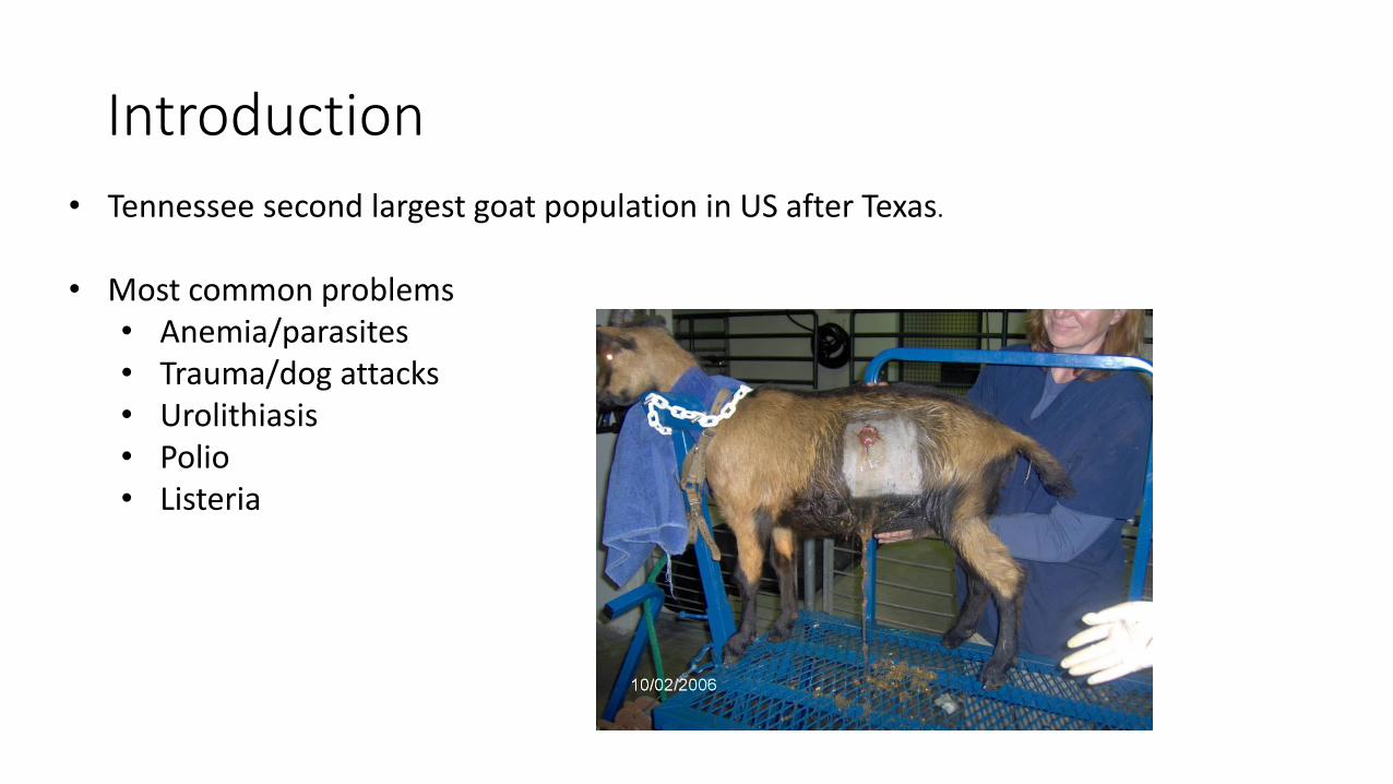
Introduction
• Tennessee second largest goat population in US after Texas.
• Most common problems• Anemia/parasites• Trauma/dog attacks• Urolithiasis• Polio• Listeria
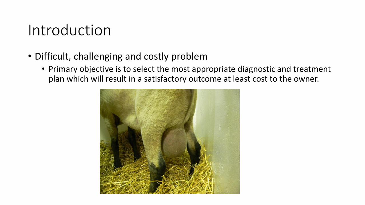
Introduction
• Difficult, challenging and costly problem• Primary objective is to select the most appropriate diagnostic and treatment
plan which will result in a satisfactory outcome at least cost to the owner.

Identifying the Case
• Male, castrate?• Urethral diameter
• Increase with age
• Calcium stones often very large anyway
• Late castration• Unwanted pregnancies
• Welfare – long recovery time

Identifying the Case
• Breed• Typical profile
• Neutered goat African descent > I year old
• African origin breeds: Pygmy; Nigerian dwarf;
Boer 146/275 (53%). Higher risk of calcium stones
• Odds lowest for Anglo Nubian and Toggenburg.
• Diet• Very important indicator of type of stone
• Struvite
• Calcium

Identifying the Case
• Off-feed, Depressed• Directly related to systemic/metabolic problems
• Hyperkalemia/hyponatremia/dehydration/azotemia
• Straining/ Posturing/Vocalization• Dribbling
• Look for stones/gravel sticking to hair• Measure solubility in acid
• Tail switching• Swelling. Perineal area/”water belly”
• Urethral rupture• Necrotizing inflammation• Abscess formation


Factors influencing outcome
•How long has it been going on for• Likelihood of complications
• Hydronephrosis
• Ruptured bladder; azotemia; electrolyte changes
• Is the animal used for breeding• Look for signs of urethral rupture
• Perineal/ventral swelling

Diagnostic approach
• Confirm diagnosis Palpation or Ultrasound• Check kidneys – hydronephrosis. US & BUN/Creatinine• Check for bladder rupture. Can cause hyperkalemia
/hyponatremia• Check K If > 6 give 50% dextrose (0.5ml/kg) and insulin (0.1 – 0.2 units/kg)• During surgery: If hypotensive, tend towards arrhythmia
can give dobutamine (0.5 - 5µg/kg/min) and calcium (23% at 0.5ml – 1ml/kg over 1 hour or 1ml/10kg 10% CaCl during anesthesia
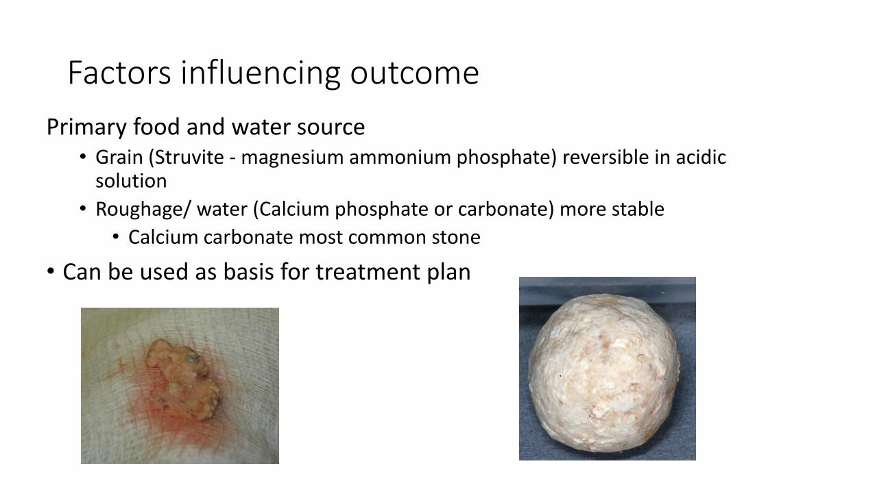
Factors influencing outcome
Primary food and water source• Grain (Struvite - magnesium ammonium phosphate) reversible in acidic
solution
• Roughage/ water (Calcium phosphate or carbonate) more stable
• Calcium carbonate most common stone
• Can be used as basis for treatment plan

Urethral process
• Remove. • Rarely successful as only treatment with or without ammonium chloride• Place animal in sitting position• Instill 3-5 ml lidocaine in prepuce• Gentle traction with sponge forceps whilepulling prepuce down (unrolling a sock)
• Persistent vermiform process• Gently peel off attachment
• Necrosis of the tip of the penis in
some cases
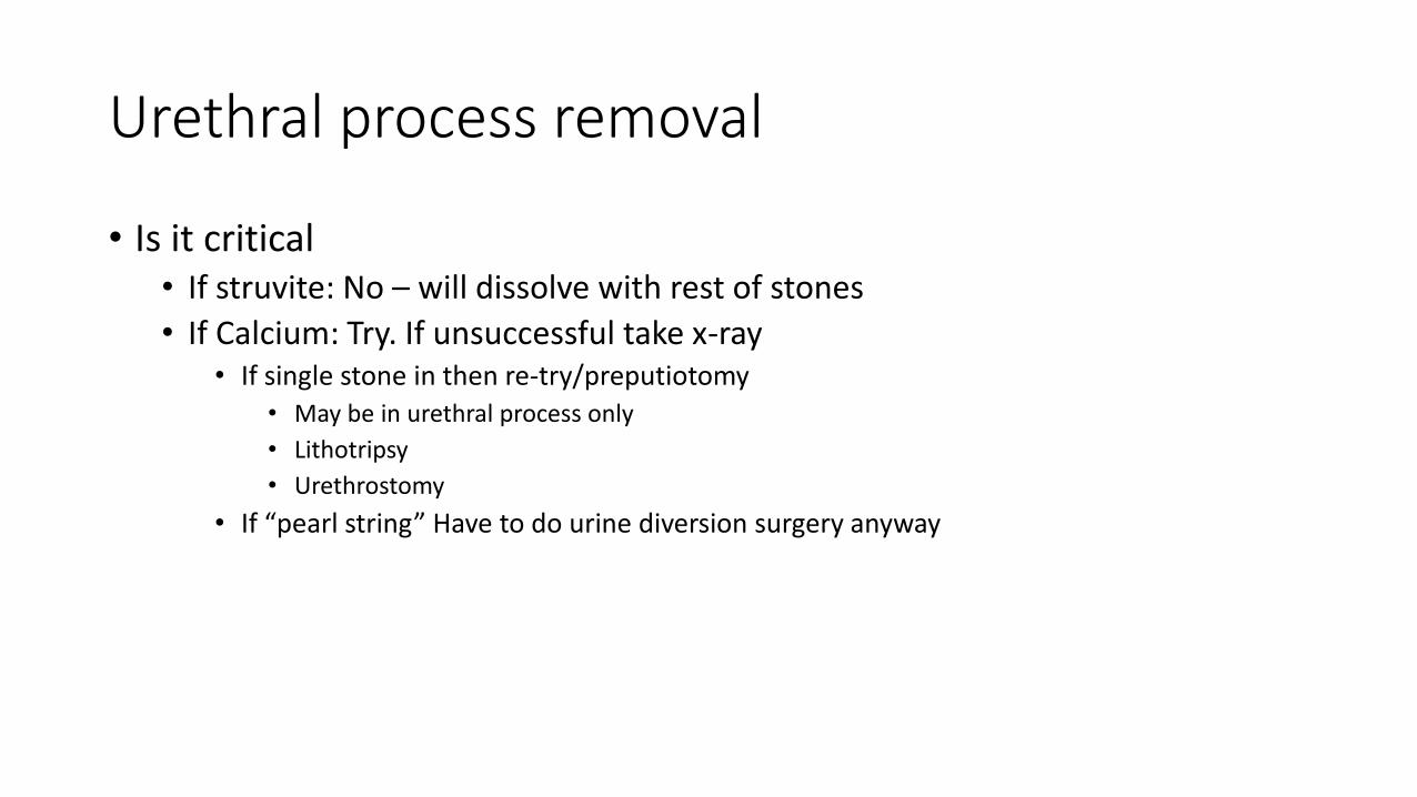
Urethral process removal
• Is it critical• If struvite: No – will dissolve with rest of stones
• If Calcium: Try. If unsuccessful take x-ray• If single stone in then re-try/preputiotomy
• May be in urethral process only
• Lithotripsy
• Urethrostomy
• If “pearl string” Have to do urine diversion surgery anyway
Medical Treatment
• Ammonium Chloride • 0.5%-1% of the ration
• 200- 300 mg/kg/day PO SID
• May take up to 5 days before effective decrease in urine pH
• Toxicity. Can cause a hyperchloremic metabolic acidosis
• Acidic urine pH not maintained - renal adaptation through bicarb
• Can cause severe chemical pharyngitis
• Owner compliance

Important issues
• How long has it been going on for?
• Is animal used for breeding?
• Is the urethra ruptured?
• Urethral process amputation/ammonium chloride/retrograde catheterization rarely successful.
• What is the diet? Struvite vs Calcium stones?
• Prognosis and cost

Medical Treatment: Bladder lavage with acidic solution (Walpole’s; Renacidin)
• Obtain stone if possible to see if dissolves
• Ultrasound bladder
• Cystocentesis after sterile prep
• Lavage bladder with 50 ml multiple times
• Monitor urine pH- stop lavage when pH< 4-5
• Leave 50 ml in bladder
“Use of Walpole’s solution for treatment of goats with urolithiasis: 25 cases (2001-2006)
Janke J., Osterstock J., Washburn K, et alJAVMA, Vol 234, (2) Jan. 15, 2009, 249-252.
Grain diet. Struvite suspected.Financial constraints
Medical Treatment
•Results of Walpole’s solution study• 20/25 (80%) goats urethral obstruction resolved and were
discharged• 5/25 (20%) euthanized due to failure to relieve
obstruction• 6/20 (30%) that were discharged were reexamined due to
recurrence of urethral obstruction
“Use of Walpole’s solution for treatment of goats with urolithiasis: 25 cases (2001-2006)
Janke J., Osterstock J., Washburn K, et alJAVMA, Vol 234, (2) Jan. 15, 2009, 249-252.

Percutaneous tube cystostomy

Grain dietStruvite suspected
Tube cystostomyvia laparotomy
Normograde catheterization and flushuroliths
Unblock50/63 (76%) goats: Urination 11daysHospital 14 days< 20% re-obstruction
Failure to unblock
Continue flushing
Long term FoleyPUVesico-preputial anastomosis
Unblock
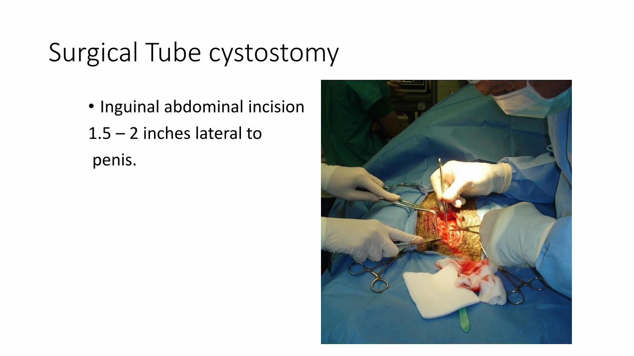
Surgical Tube cystostomy
• Inguinal abdominal incision
1.5 – 2 inches lateral to
penis.

Exteriorize bladder
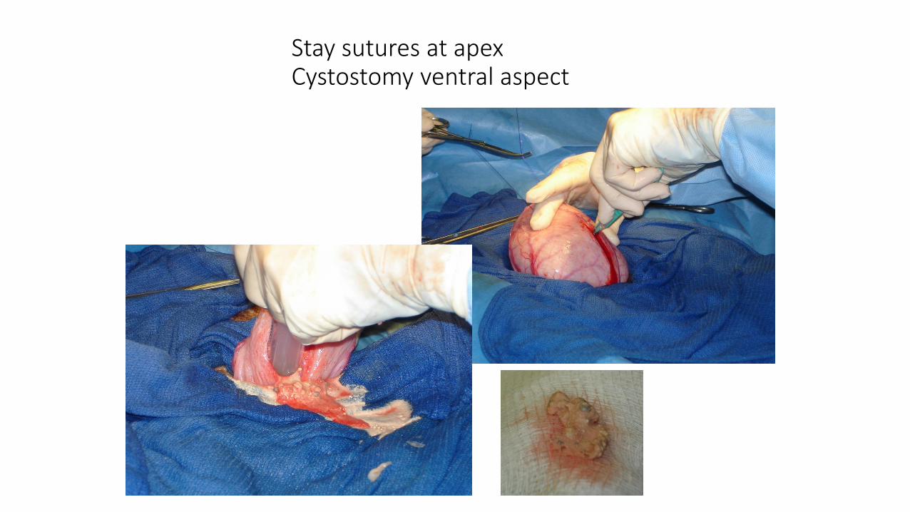
Stay sutures at apexCystostomy ventral aspect

Flush bladder and urethra

Foley catheter placement

Foley catheter placement

Foley catheter placement


Continue flushing daily with Walpole solution

Renacidin Irrigation
• Contains Citric Acid, Glucono-delta-lactone and Magnesium Carbonate
• Targets apatite and struvite:
• Apatite calculi are mainly calcium: action results from an exchange of Magnesium from the solution for calcium in the stone; the magnesium salts are soluble in gluconocitrate solution = dissolution of the calculi
• Struvite calculi are composed of mainly magnesium ammonium phosphates, which are solubilized by the solution due to the acidic pH

Long term tube cystostomy
• Success rate, 84% to 86%.
• Problems:• Client compliance
• Tube becomes dislodged• Has to be replaced quickly
• Use short tube 8.5inch/22 Fr
• Urine scald – usually not serious
• Persistent low grade cystitis
• Tube becomes obstructed with cellular and mucoid debri


calcium carbonate based on water and food supply
Large bladder on palpation or US
Survey radiographs29 cases 85% positive
Few uroliths
PU/ Vesico-preputial anastomosis/Marsupialization
LithotripsyEndoscopic removal
Cystocentesis
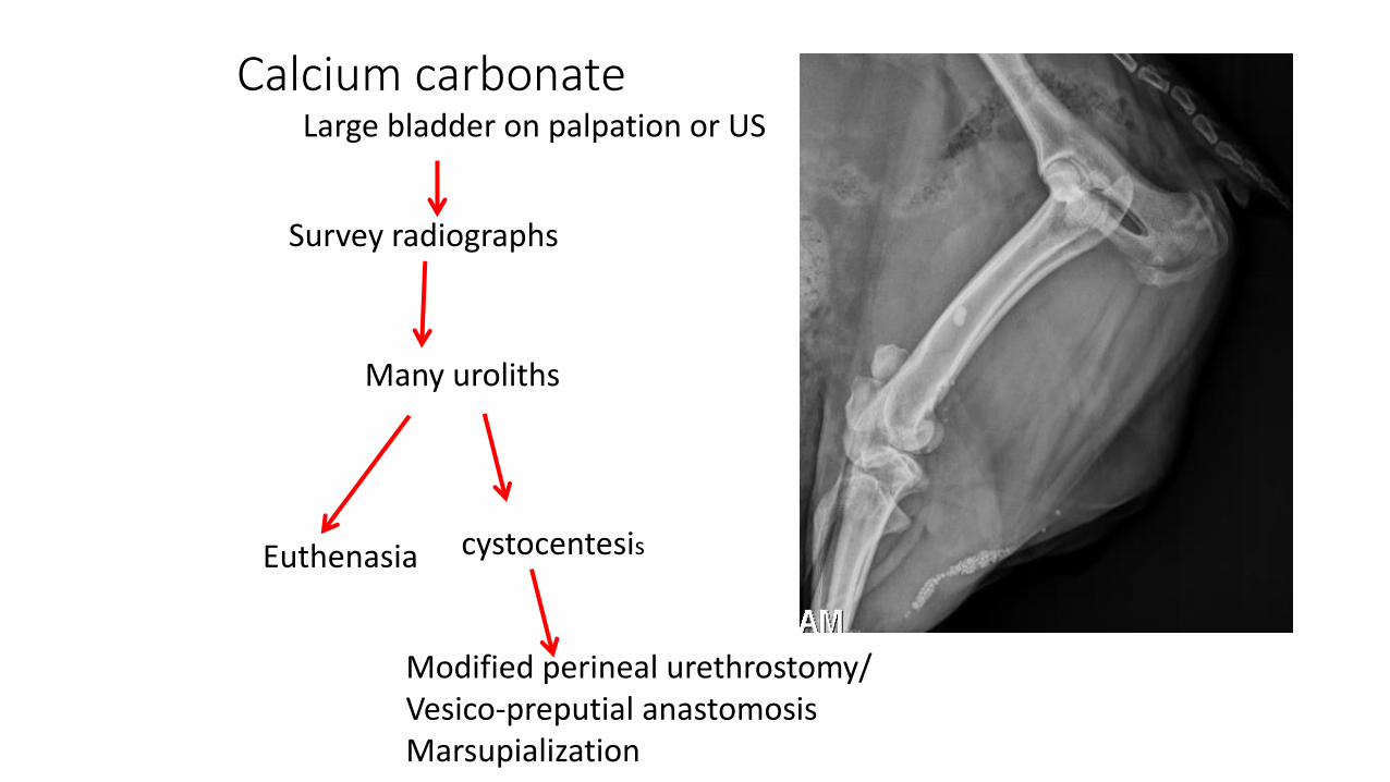
Calcium carbonateLarge bladder on palpation or US
Survey radiographs
Many uroliths
Modified perineal urethrostomy/Vesico-preputial anastomosisMarsupialization
Euthenasia cystocentesis

•Advantages• Reduce/eliminate urine scald
•Disadvantages• Stricture at anastomosis site
•4 Cases completed• 3 cases OK.• 1 case died of azotemia after stricture
Vesico-preputial anastomosis

• Determine margins of abdominal incision
• Center should align with end of preputial cavity

• Place stay sutures in apex of bladder
• Pull apex through the abdominal incision 1.5 – 2 cm
• Anchor the bladder wall to the abdominal wall using simple interrupted; horizontal mattress or cruciate sutures

• Bladder anchored to abdominal wall. • Should be leakage proof

• Isolate penis and amputate• Undermine and dissect the prepuce away from
the skin and abdominal wall. • Make a circular incision around thepenis at the point of entry into the preputial
cavity. Remove penile stump


• Cut an appropriate size (slightly larger than 22 Fr Foley)circular opening into the bladder
• Enlarge the opening in the prepuce to match that
in the bladder• Use simple interrupted sutures
to facilitate mucosa to mucosa apposition

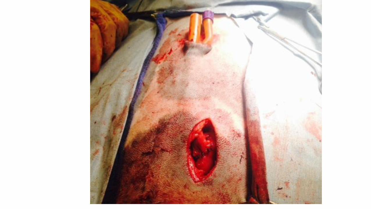


Perineal urethrostomy
• Traditional perineal urethrostomy• Salvage: Stricture in 45% to 78% within 8 months
• Modified perineal urethrostomy
• No long term strictures or recurrences in 3 goats (14, 15, and 26
months).• 1 goat urinated to the left.• Severe hemorrhage in one goat.
• Additional findings:• Easy catheterization in all goats.

Modified proximal perineal urothrostomy
•Problems• Immediately post op
• Hemorrhage. May be severe/intermittent
• Diaper type bandage sometimes necessary
• 2-3 weeks• Wound dehiscence/ tissue necrosis
• Long term• Stricture
• Re-obstruction due to more stone formation

Surgical positioning
• Raise table as high as practically possible. Lower head.
• Let legs hang over end of table
• Prop hind quarters up with towels
• Make 4cm incision from 1cm below anus to halfway down theperineum
• Isolate penis with blunt dissection
and free from surrounding tissues
to pelvic attachments

Transect ischiocavernosus muscles taking care not to cut urethra
Penile body
Dr Karen TobiasUT

Dorsal penile vessels Penile stump. Leave 1-1.5 inches
Dr Karen TobiasUT

Elevate any remaining ischial attachments.
Left Ischial tuberosity
Right Ischial tuberosity
Ischiocavernosus muscles
Ischiocavernosusattachment
Dorsal penile vessels
Penile body
Dr Karen TobiasUT

Cutting urethra open (side view)
Dr Karen TobiasUT.

Incise urethra, and suture mucosa to skin.
3-0 or 4-0 absorbable monofilament
Note mucosal edge
Dr Karen TobiasUT

Add sutures as needed to appose mucosa to skin, and close any remaining skin wounds.
Anus
Simple interrupted or continuous pattern
Dr Karen TobiasUT

Dr Karen TobiasUT

Conclusions
• Risk of postoperative stricture in nonbreeding goats with recurrent urethral obstruction may be reduced with modified proximal perineal urethrostomy• Transection of ischiocavernosus muscles.
• Careful, accurate apposition of mucosa to skin.
• Long term study on a larger number of goats is needed to verify these findings.

•Bladder marsupialization• Success rate, 66% to 94%.• Incontinence, urine scald, infection, mucosal
prolapse.