Embed Size (px)
Citation preview
40 American Society of Hematology
Iron Deficiency and Overload
Ernest Beutler, A. Victor Hoffbrand, and James D. Cook
In the past seven years numerous genes thatinfluence iron homeostasis have been discovered.Dr. Beutler provides a brief overview of thesegenes, genes that encode HFE, DMT-1, ferroportin,transferrin receptor 2, hephaestin, and hepcidin tolay the groundwork for a discussion of the variousclinical forms of iron storage disease and howthey differ from one another.
In Section I, Dr. Beutler also discusses thetypes of hemochromatosis that exist as acquiredand as hereditary forms. Acquired hemochromato-sis occurs in patients with marrow failure, particu-larly when there is active ineffective erythropoie-sis. Hereditary hemochromatosis is most com-monly due to mutations in the HLA-linked HFEgene, and hemochromatosis clinically indistin-guishable from HFE hemochromatosis is theconsequence of mutations in three transferrinreceptor-2 gene. A more severe, juvenile form ofiron storage disease results from mutations of thegene encoding hepcidin or of a not-yet-identifiedgene on chromosome 1q. Autosomal dominantiron storage disease is a consequence offerroportin mutations, and a polymorphism in theferroportin gene appears to be involved in theAfrican iron overload syndrome.
Evidence regarding the biochemical andclinical penetrance of hemochromatosis due tomutations of the HFE gene is rapidly accumulat-ing. These studies, emanating from several centersin Europe and the United States, all agree that thepenetrance of hemochromatosis is much lowerthan had previously been thought. Probably only1% of homozygotes develop clinical findings. Theimplications of these new findings for the manage-ment of hemochromatosis will be discussed.
In Section II, Dr. Victor Hoffbrand discussesthe management of iron storage disease bychelation therapy, treatment that is usually re-served for patients with secondary hemochroma-tosis such as occurs in the thalassemias and in
patients with transfusion requirements due tomyelodysplasia and other marrow failure states.Tissue iron can be estimated by determiningserum ferritin levels, measuring liver iron, and bymeasuring cardiac iron using the MRI-T2* tech-nique. The standard form of chelation therapy isthe slow intravenous or subcutaneous infusion ofdesferoxamine. An orally active bidentate ironchelator, deferiprone, is now licensed in 25 coun-tries for treatment of patients with thalassemiamajor. Possibly because of the ability of thiscompound to cross membranes, it appears tohave superior cardioprotective properties. Agranu-locytosis is the most serious complication ofdeferiprone therapy and occurs in about 1% oftreated patients. Deferiprone and desferoxaminecan be given together or on alternating schedules.A new orally active chelating agent ICL 670 seemspromising in early clinical studies.
In Section III, Dr. James Cook discusses themost common disorder of iron homeostasis, irondeficiency. He will compare some of the standardmethods for identifying iron deficiency, the hemo-globin level, transferrin saturation, and meancorpuscular hemoglobin and compare these withsome of the newer methods that have beenintroduced, specifically the percentage of hypo-chromic erythrocytes and reticulocyte hemoglobincontent. The measurement of storage iron isachieved by measuring serum ferritin levels. Thesoluble transferrin receptor is a truncated form ofthe cellular transferrin receptor and the possiblevalue of this measurement in the diagnosis of irondeficiency will be discussed. Until recently irondextran was the only parental iron preparationavailable in the US. Sodium ferric gluconate, whichhas been used extensively in Europe for manyyears, is now available in the United States. Itseems to have a distinct advantage over irondextran in that anaphylactic reactions are muchless common with the latter preparation.
Hematology 2003 41
I. HEMOCHROMATOSIS
Ernest Beutler, MD*
DefinitionHemochromatosis is generally considered to be a dis-ease in which increased iron storage causes pathologicchanges. However, the definition of hemochromatosishas undergone considerable evolution since the disor-der was recognized as a distinct clinical syndrome inthe late 19th century. Until the performance of plasmairon and ferritin determinations became commonplace,the designation was reserved for patients who had, as aresult of iron deposition, frank cirrhosis of the liver andusually diabetes, bronzing of the skin, and cardiac dis-ease. The disease was sometimes called “bronzed dia-betes.” But in the 1970s the definition of the diseasegradually changed. Instead of being applied only topatients who had severe clinical manifestations of ironstorage, patients who had elevated serum transferrinsaturation and ferritin levels were also designated ashaving hemochromatosis, especially if they had the HLAtype A4 B14 that was commonly associated with the dis-ease. Even larger numbers of patients were considered tohave hemochromatosis after the HLA-linked gene thatwas associated with the disease, HFE, was discovered.
But who, then, should be considered to have hemo-chromatosis? Only those with disease? Or those whomerely have the genotype? No consensus exists con-cerning what definition is to be used.
ClassificationHemochromatosis may be divided into primary (or genetic)hemochromatosis and secondary hemochromatosis.
Primary (hereditary) hemochromatosisAs shown in Table 1, the hereditary form can be di-vided into several subgroups. A numerical classifica-tion has been proposed (http://www.ncbi.nlm.nih.gov/htbin-post/Omim/dispmim?235200), but such a systemdoes not add to our understanding, since most of theforms of hemochromatosis can simply be designatedby the mutation that is their cause, and some forms ofiron storage disease (e.g., African iron overload) arenot included.
Secondary hemochromatosisSecondary hemochromatosis can arise in many disor-ders, inborn or acquired. These disorders have in com-mon the fact that the patient is anemic. Transfusions oferythrocytes add, nearly stoichiometrically, to the bodyiron burden: each milliliter of red cells contains onemilligram of iron. When anemia is accompanied byineffective erythropoiesis, inappropriate absorption ofiron from the gastrointestinal tract seems to be acti-vated. Patients with anemias in which ineffective eryth-ropoiesis does not play a role seem much less prone tohyperabsorb iron. Thus, it is patients with ineffectiveerythropoiesis who develop the largest iron burdens.Among the hereditary forms, the most common are thethalassemias; among the acquired forms, the acquiredsideroblastic anemias predominate. Table 2 lists someof the diseases with which hemochromatosis has beenassociated.
PenetranceThe penetrance of a mutation may be defined as theextent to which a phenotypic effect is exerted in indi-viduals carrying the mutation. But which phenotypiceffect? Clearly, the penetrance is a function of whichendpoint is selected.
* The Scripps Research Institute, 10550 North Torrey PinesRoad, La Jolla CA 92037
Acknowledgments: This is manuscript number 15873-MEM.Supported by National Institutes of Health grants DK3505-06and RR00833 and the Stein Endowment Fund.
Table 1. Classification of hemochromatosis.
A. Hereditary hemochromatosis
1. Classical hemochromatosis (hereditary hemochromato-sis; HFE hemochromatosis) (Type 1)
2. Juvenile hemochromatosis (Type 2)
a. Chromosome 1q-linked
b. Abnormality of hepcidin
3. Transferrin receptor-2 deficiency (Type 3)
4. Ferroportin deficiency (includes some cases of Africaniron overload1,2) (Type 4)
5. African iron overload
B. Secondary hemochromatosis
Table 2. Some causes of secondary hemochromatosis.
A. Hereditary disorders1. Thalassemia3,4
2. Pyruvate kinase deficiency5,6
3. Dyserythropoietic anemia7-11
4. Glucose-6-phosphate dehydrogenase (G6PD) deficiency12
5. Hereditary spherocytosis13,14
6. Sideroblastic anemia (ALA-S deficiency)
B. Acquired disorders1. Sideroblastic and other dyserythropoietic anemias15
2. Any anemia, except for that due to blood loss, in whichmultiple transfusions are required.
42 American Society of Hematology
Penetrance of the homozygous HFE mutationThe C282Y mutation of the HFE gene is a very com-mon one. About 15% of the northern European popu-lation is heterozygous; accordingly, one would expectover 5 per 1000 in the population to be homozygous,and this is, indeed, the case.
Biochemical penetrance: Relatively few studieshave been conducted in which an unbiased populationwas screened for the C282Y mutation and the transfer-rin saturation and ferritin levels of the homozygoteswere determined. Deugnier et al16 screened over 9000individuals (3367 men and 6029 women) in France andfound 10 homozygous men and 44 homozygous women.Although the population was relatively young, 80% ofthe men had transferrin saturations over 55%, and 44%of the women had transferrin saturations over 50%. Inour study of patients in the health appraisal clinic ofKaiser Permanente in San Diego we found that among152 homozygotes, 75% of men and 40% of women hada transferrin saturation higher than 50%.17-19 Serumferritin levels were increased in 76% of the men and54% of the women. In another small study all 5 ho-mozygotes detected have transferrin saturations greaterthan 55%.20 Thus, there is agreement that homozygotesfor the HFE C282Y mutation usually have increasedserum transferrin saturation levels and increased serumferritin levels. Clearly, there is a subset of homozygoteswho do not show these biochemical stigmata. A few ofthese prove to be frequent blood donors, but most ofthem are not. It is simply that even on a biochemicallevel the homozygous state is not always expressed.
Clinical penetrance: Clinicians do not encountermany cases of full-blown hemochromatosis. Our at-tempts to obtain samples from patients with iron over-load who manifested diabetes, cirrhosis, cardiomyopa-thy, and darkening of the skin from our own clinic andfrom major centers in which many patients with hemo-
chromatosis are treated have met with very little suc-cess. Most of the many patients that have been diag-nosed as having hemochromatosis have been diagnosedon the basis of biochemical changes and non-specificsymptoms, such as fatigue and arthropathy. Neither com-mon clinical experience21,22 nor autopsy series21,23-25 sug-gest that hemochromatosis is a common cause of death.However, it has been a common belief that milder symp-toms are, in contrast, very common in patients homozy-gous for the C282Y mutation, and it has been suggestedthat most of the homozygous males will develop symp-toms by the time they are 40 years of age.26 This im-pression that a mild phenotype exists (and the accom-panying assumption that this leads to the more severephenotype if not treated) has been based largely on un-controlled observations in which the patients being as-sessed and the physician performing the assessment knewthe diagnosis and could well have been influenced by it.
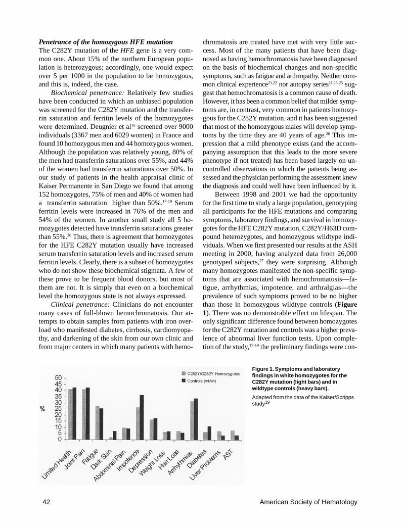
Between 1998 and 2001 we had the opportunityfor the first time to study a large population, genotypingall participants for the HFE mutations and comparingsymptoms, laboratory findings, and survival in homozy-gotes for the HFE C282Y mutation, C282Y/H63D com-pound heterozygotes, and homozygous wildtype indi-viduals. When we first presented our results at the ASHmeeting in 2000, having analyzed data from 26,000genotyped subjects,27 they were surprising. Althoughmany homozygotes manifested the non-specific symp-toms that are associated with hemochromatosis—fa-tigue, arrhythmias, impotence, and arthralgias—theprevalence of such symptoms proved to be no higherthan those in homozygous wildtype controls (Figure1). There was no demonstrable effect on lifespan. Theonly significant difference found between homozygotesfor the C282Y mutation and controls was a higher preva-lence of abnormal liver function tests. Upon comple-tion of the study,17-19 the preliminary findings were con-
Figure 1. Symptoms and laboratoryfindings in white homozygotes for theC282Y mutation (light bars) and inwildtype controls (heavy bars).
Adapted from the data of the Kaiser/Scrippsstudy19
Hematology 2003 43
firmed. Only one of 152 homozygotes had the typicalclinical syndrome of hemochromatosis and we esti-mated the clinical penetrance of the homozygous stateto be of the order of 1% (Figure 1).
No one had expected the penetrance to be so low,and predictably, the results were greeted with consid-erable skepticism. In attempting to reconcile our datawith the concept that the homozygous state had a muchhigher penetrance, it was suggested that the data were“flawed” in a number of respects. It was proposed thatwe were dealing with an unusually healthy population28
or a population with an extraordinarily healthy life style.Alternatively, it was proposed that our population wasunusually “sickly” and that the manifestations of hemo-chromatosis had been obscured by the poor health ofthe controls.29 Obviously it is impossible to reconcile thesetwo objections: the population cannot be too well and toosickly at the same time. But, in fact, neither criticism ap-plies. The most cogent objection was that our study wasbiased by selecting a healthy population. If, indeed, pa-tients with symptoms had been excluded because theydid not attend a health appraisal clinic, having died orbeing taken care of in a more intense medical setting,we might have erroneously concluded that the pen-etrance of the homozygous state is very low. But thereis a straightforward way to address this problem. Ifhomozygotes were systematically excluded then thenumber found in the population should fall short of thenumber predicted by the Hardy-Weinberg equilibriumbased on the gene frequency in the population. But infact, the number of homozygotes actually exceeds thepredicted number.17-19,30-32 Another way to examine the
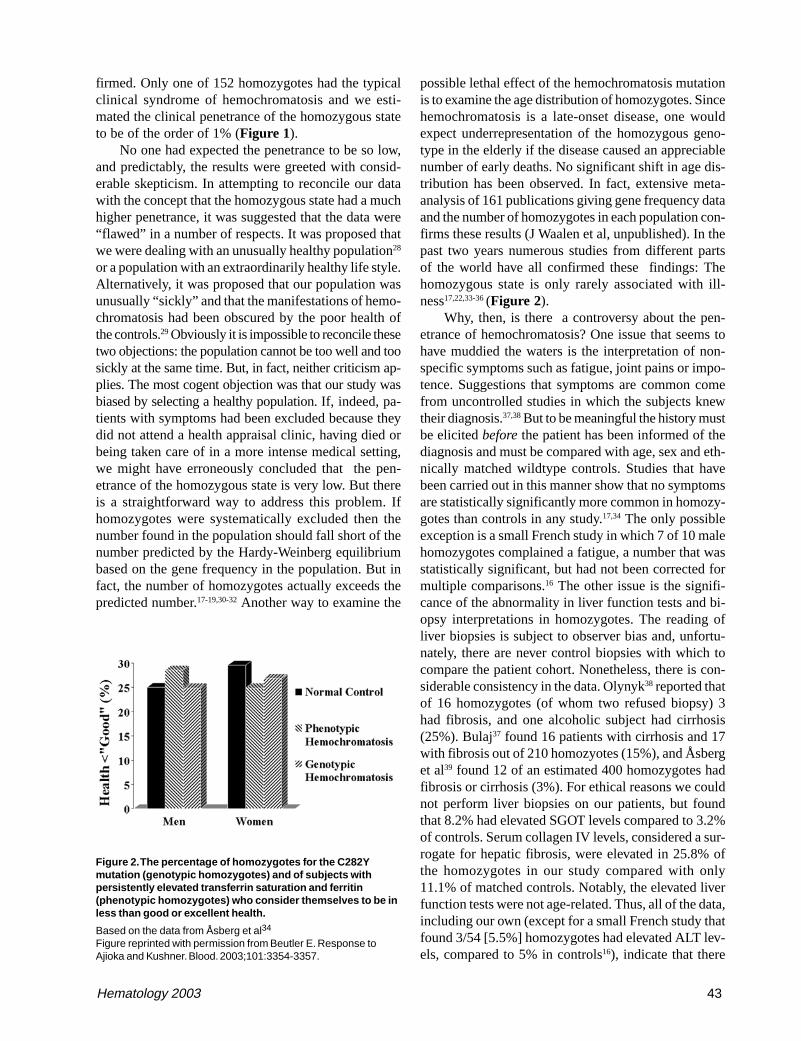
possible lethal effect of the hemochromatosis mutationis to examine the age distribution of homozygotes. Sincehemochromatosis is a late-onset disease, one wouldexpect underrepresentation of the homozygous geno-type in the elderly if the disease caused an appreciablenumber of early deaths. No significant shift in age dis-tribution has been observed. In fact, extensive meta-analysis of 161 publications giving gene frequency dataand the number of homozygotes in each population con-firms these results (J Waalen et al, unpublished). In thepast two years numerous studies from different partsof the world have all confirmed these findings: Thehomozygous state is only rarely associated with ill-ness17,22,33-36 (Figure 2).
Why, then, is there a controversy about the pen-etrance of hemochromatosis? One issue that seems tohave muddied the waters is the interpretation of non-specific symptoms such as fatigue, joint pains or impo-tence. Suggestions that symptoms are common comefrom uncontrolled studies in which the subjects knewtheir diagnosis.37,38 But to be meaningful the history mustbe elicited before the patient has been informed of thediagnosis and must be compared with age, sex and eth-nically matched wildtype controls. Studies that havebeen carried out in this manner show that no symptomsare statistically significantly more common in homozy-gotes than controls in any study.17,34 The only possibleexception is a small French study in which 7 of 10 malehomozygotes complained a fatigue, a number that wasstatistically significant, but had not been corrected formultiple comparisons.16 The other issue is the signifi-cance of the abnormality in liver function tests and bi-opsy interpretations in homozygotes. The reading ofliver biopsies is subject to observer bias and, unfortu-nately, there are never control biopsies with which tocompare the patient cohort. Nonetheless, there is con-siderable consistency in the data. Olynyk38 reported thatof 16 homozygotes (of whom two refused biopsy) 3had fibrosis, and one alcoholic subject had cirrhosis(25%). Bulaj37 found 16 patients with cirrhosis and 17with fibrosis out of 210 homozyotes (15%), and Åsberget al39 found 12 of an estimated 400 homozygotes hadfibrosis or cirrhosis (3%). For ethical reasons we couldnot perform liver biopsies on our patients, but foundthat 8.2% had elevated SGOT levels compared to 3.2%of controls. Serum collagen IV levels, considered a sur-rogate for hepatic fibrosis, were elevated in 25.8% ofthe homozygotes in our study compared with only11.1% of matched controls. Notably, the elevated liverfunction tests were not age-related. Thus, all of the data,including our own (except for a small French study thatfound 3/54 [5.5%] homozygotes had elevated ALT lev-els, compared to 5% in controls16), indicate that there
Figure 2. The percentage of homozygotes for the C282Ymutation (genotypic homozygotes) and of subjects withpersistently elevated transferrin saturation and ferritin(phenotypic homozygotes) who consider themselves to be inless than good or excellent health.
Based on the data from Åsberg et al34
Figure reprinted with permission from Beutler E. Response toAjioka and Kushner. Blood. 2003;101:3354-3357.
44 American Society of Hematology
is a subset of patients, considerably larger than 1% whohave abnormal liver function tests. Those who hold thatthe penetrance of hemochromatosis is higher than theapproximately 1% estimate can point to the presenceof hepatic fibrosis as an indication that iron overload isclinically important. We, however, take the point of viewthat since the fibrosis did not produce any clinical symp-toms in the vast majority of subjects, and that it doesnot appear progressive, it is not important for the per-son to whom it should matter the most, the patient. Thus,to some degree the disagreement about penetrancecomes down to the single issue of whether hepatic fi-brosis seen on liver biopsy by pathologists or abnormalliver function is important if it is not associated withmeasurable morbidity or mortality.
Penetrance of the compound heterozygousC282Y/H63D HFE mutationOn the average, compound heterozygotes manifest sig-nificantly higher transferrin saturations and serum fer-ritin levels than do individuals with the wildtype geno-type. Because the H63D mutation is very prevalent inthe population, this compound heterozygous genotypeis very common in the population. Among patients whohad been classified as having “hemochromatosis” onthe basis of increased biochemical parameters there isan increased number of compound heterozygotes, andit has been calculated that the biochemical penetranceof this genotype is only about 1% of that of the ho-mozygous genotype.40 Accordingly, patients with thisgenotype who develop severe cirrhosis and other clini-cal manifestations of hemochromatosis are very rare.
Penetrance of the simple heterozygous genotypeIt is clear from large studies that simple heterozygotesfor the C282Y or H63D mutations have, on the aver-age, very slightly higher transferrin saturations and fer-ritin levels than do homozygotes for the wildtype. Nu-merous claims have been made that these minor changestranslate into increased prevalence of a variety disor-ders including diabetes,41,42 heart disease,43.44 and can-cer.45,46 None of these claims has been widely substan-tiated,18,36,47,48 and it seems unlikely that the heterozy-gote for these common mutations suffers ill health be-cause of them with one notable, rather uncommon ex-ception. Carrying either the C282Y or H63D does ap-pear to be a risk factor for porphyria cutanea tarda.49,50
In general, however, it is much more likely that muta-tions that have gained a high prevalence in the genomehave a beneficial effect, i.e., that they constitute a bal-anced polymorphism. Their beneficial effect is prob-ably that of preventing iron deficiency in women.51,52
DiagnosisIt is a tragedy when a patient dies of a treatable diseasesuch as hemochromatosis without receiving the ben-efits of therapy. However, the enthusiasm for generalpopulation screening for hereditary hemochromatosishas abated with the realization that the penetrance isvery low.53 The burden for making the diagnosis ofhemochromatosis therefore falls squarely upon the phy-sician. The diagnosis of hemochromatosis should beconsidered in patients with cirrhosis of the liver, par-ticularly those with diabetes. Alcoholism is no bar toconsideration of hemochromatosis in a cirrhotic patient;indeed, a high proportion of patients with clinical hemo-chromatosis ingest excessive amounts of alcohol54,55 andcirrhosis is much more common in patients with thehemochromatosis genotype who have heavy alcoholintake.56
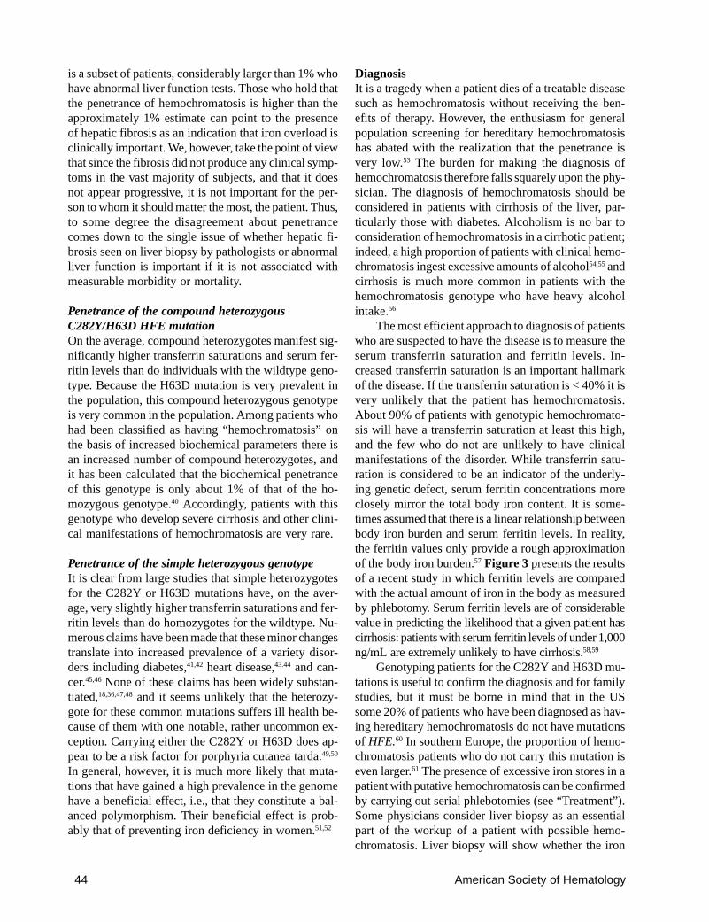
The most efficient approach to diagnosis of patientswho are suspected to have the disease is to measure theserum transferrin saturation and ferritin levels. In-creased transferrin saturation is an important hallmarkof the disease. If the transferrin saturation is < 40% it isvery unlikely that the patient has hemochromatosis.About 90% of patients with genotypic hemochromato-sis will have a transferrin saturation at least this high,and the few who do not are unlikely to have clinicalmanifestations of the disorder. While transferrin satu-ration is considered to be an indicator of the underly-ing genetic defect, serum ferritin concentrations moreclosely mirror the total body iron content. It is some-times assumed that there is a linear relationship betweenbody iron burden and serum ferritin levels. In reality,the ferritin values only provide a rough approximationof the body iron burden.57 Figure 3 presents the resultsof a recent study in which ferritin levels are comparedwith the actual amount of iron in the body as measuredby phlebotomy. Serum ferritin levels are of considerablevalue in predicting the likelihood that a given patient hascirrhosis: patients with serum ferritin levels of under 1,000ng/mL are extremely unlikely to have cirrhosis.58,59
Genotyping patients for the C282Y and H63D mu-tations is useful to confirm the diagnosis and for familystudies, but it must be borne in mind that in the USsome 20% of patients who have been diagnosed as hav-ing hereditary hemochromatosis do not have mutationsof HFE.60 In southern Europe, the proportion of hemo-chromatosis patients who do not carry this mutation iseven larger.61 The presence of excessive iron stores in apatient with putative hemochromatosis can be confirmedby carrying out serial phlebotomies (see “Treatment”).Some physicians consider liver biopsy as an essentialpart of the workup of a patient with possible hemo-chromatosis. Liver biopsy will show whether the iron
Hematology 2003 45
is deposited chiefly in liver parenchymal cells, whichis characteristic of the disease, it will allow quantitationof liver iron, and it will establish whether the patienthas cirrhosis. However, serial phlebotomy will quanti-tate the iron. Therefore, the major benefit to the patientof undergoing liver biopsy is to establish whether cir-rhosis is present. Hepatic cirrhosis affects prognosis,but not treatment, and many or most patients may notbe eager to know what their estimated lifespan will be.Cirrhosis also increases greatly the risk of hepatocellu-lar carcinoma, an important complication in patientswith hemochromatosis and cirrhosis. Patients with cir-rhosis may therefore be better candidates for periodicscreening for this disease. As treatment for hepatocel-lular carcinoma becomes more effective, establishingwhether or not cirrhosis is present may be importantfor optimal management and may therefore be a rea-son for performing a biopsy.
TreatmentThe aim of treatment of iron storage disease is to re-move from the body the excess iron that has accumu-lated. In the case of patients without primary disordersof hematopoiesis (i.e., patients with one of the formsof primary hemochromatosis), this is best achieved byphlebotomy, since regeneration of erythrocytes by themarrow utilizes iron, which is therefore withdrawn fromvarious body pools. Phlebotomy is only occasionally
feasible in patients who have augmented iron storesbecause of ineffective erythropoiesis and is generallyprecluded in those in whom the iron overload is theresult of multiple transfusions. Such patients requiretreatment with an iron chelating agent, as discussed inSection II.
One ml of erythrocytes contains 1 mg of iron.Hence, removing one unit (~450 mL) of blood with ahematocrit of 45% removes approximately 200 mg ofiron from the body. Iron absorption is increased in pa-tients with hemochromatosis who are undergoing phle-botomy, and may be more than 5 mg per day.62 But witheven this level of iron absorption, it is relatively simpleto achieve a negative iron balance by instituting a phle-botomy program. Initially, some patients feel enervatedby phlebotomies, and I find that compliance is improvedby initially phlebotomizing every two weeks. But afterthis has been done for a month or two, weekly phle-botomies are almost always well tolerated. The aim ofphlebotomy is to deplete the body iron stores, and themost easily appreciated endpoint is mild iron deficiencyanemia. One of the easiest guides to incipient iron deple-tion is a falling MCV.63 The other useful parameter isthe serum ferritin level, which should be brought tounder 10 ng per ml. Some clinicians prefer a highercutoff, but there is usually appreciable residual, diffi-cult-to-mobilize tissue iron even when the erythron isiron depleted, and the mobilization of this iron and en-hanced gastrointestinal iron absorption that is presentin iron-depleted patients with hemochromatosis willquickly correct any deficiency that may have been in-duced. Maintenance phlebotomy is initiated when theferritin level rises to 80 or 100 ng/ml, and the rate ofmaintenance phlebotomy required varies widely be-tween patients. Some physicians prescribe an iron-poordiet, but this seems to me to represent more interfer-ence with the patient’s lifestyle than is justified by theslight decrease in intervals between phlebotomies thatmay be achieved by restricting iron intake.
One of the manifestations of hereditary hemochro-matosis is increased susceptibility to infection. Deathdue to overwhelming sepsis is not uncommon in se-verely affected patients. Because of the risk of infec-tion with organisms such as Yersinia enterocolitica orVibrio vulnificus, patients are usually advised to avoidthe ingestion of raw shellfish. Because of the fulminat-ing nature of some infections in patients with hemo-chromatosis, vigorous early treatment of febrile diseasesis recommended.
The prevalence of hepatomas is increased in pa-tients with hemochromatosis, particularly those with cir-rhosis; a relative risk of 1.8 (CI 1.1–2.9) has been re-ported.64 Some cases have been documented in non-
Figure 3. The relationship between serum ferritin levels andstorage iron as determined by serial phlebotomy.
The dots represent patients homozygous for the C282Y mutation.Squares represent patients with a diagnosis of hemochromatosiswho are not homozygous for the C282Y mutation. The plus signsrepresent those whose phlebotomy program had not yet beencompleted.
Figure reprinted with permission from Beutler E, Felitti V, Ho N,Gelbart T. Relationship of body iron stores to levels of serumferritin, serum iron, unsaturated iron binding capacity andtransferrin saturation in patients with iron storage disease. ActaHaematol (Basel). 2002;107:145-149.
46 American Society of Hematology
cirrhotic patients,65,66 but these are only case reports,and it is unclear whether the relative risk is increased.It is probably prudent to monitor the liver by ultrasoundevery 6 to 12 months, although the value of this proce-dure is in some doubt since the treatment results forhepatoma are so unsatisfactory.
It is universally stated that phlebotomy improvesthe health and increases the lifespan of patients withhemochromatosis. This assumption is based upon astudy in which it was shown that phlebotomized pa-tients with “hemochromatosis” had a normal lifespan.67
We now recognize that unphlebotomized patients withhemochromatosis also have a normal lifespan when theselection criterion is based on either genotype or chemi-cal phenotype. The fact is that there are no data thatallow us to show definitively that phlebotomy improvessurvival. No such data can be obtained because of ethi-cal issues. Nonetheless, the concept that removing ironfrom a patient whose disease is due to an excess of ironwill be helpful is compelling, and therefore treatingpatients with hemochromatosis who have clinical dis-ease is mandatory. The recognition that most patientswith the genetic and biochemical stigmata of hemo-chromatosis will have a normal lifespan without inter-vention raises the question of whether all patients withhemochromatosis should be phlebotomized. Fortu-nately, the treatment is almost entirely risk free andpotentially beneficial to society. Moreover, our currentstate of knowledge is such that we cannot predict whichof the patients with hemochromatosis will be the rareones who develop serious clinical consequences. Thus,it seems prudent to phlebotomize all patients with hemo-chromatosis who have elevated body iron stores as es-timated by serum ferritin levels. It is useful in this re-gard to consider the fact that cirrhosis is very largelylimited to patients with the serum ferritin levels higherthan 1000 ng/mL.59,68 There is no harm, however, inphlebotomizing patients with less elevated ferritin lev-els. Family studies are also potentially useful, since ho-mozygous relatives of non-expressing homozygotes arepotentially more seriously affected.
The management of patients with the juvenilehemochromatosis is similar to that of patients with HFEhemochromatosis, but knowing that this is an aggres-sive form of disease, a vigorous phlebotomy programshould be initiated early.
SummaryThere are many forms of iron storage disease, primary(or hereditary) and secondary to various hematologicdisorders. The most common of the primary forms areassociated with mutations of an HLA-linked gene des-ignated HFE. Five per thousand northern Europeans
are homozygous for the common C282Y mutation ofHFE. Very few of these develop any clinical symptoms,but uncommonly cirrhosis, diabetes, arthropathies, andbronzing of the skin may result, and approximately 25%will have elevated serum collagen IV levels, a surro-gate marker for hepatic fibrosis, compared with 11%of controls. Hereditary hemochromatosis is character-ized by an increased serum transferrin saturation. It istreated by serial phlebotomy to remove the accumu-lated iron. Secondary hemochromatosis has clinicalmanifestations similar to those of primary hemochro-matosis, but by necessity treatment usually consists ofthe administration of iron chelating agents.
II. IRON CHELATION THERAPY
A. Victor Hoffbrand, DM, FRCP*
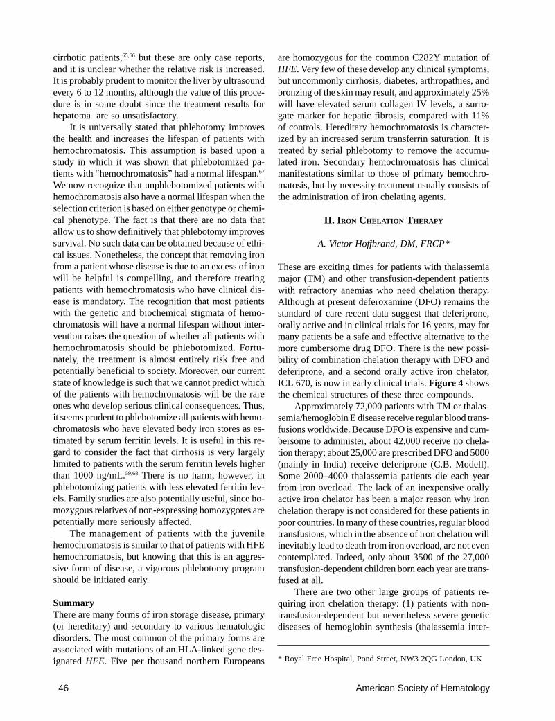
These are exciting times for patients with thalassemiamajor (TM) and other transfusion-dependent patientswith refractory anemias who need chelation therapy.Although at present deferoxamine (DFO) remains thestandard of care recent data suggest that deferiprone,orally active and in clinical trials for 16 years, may formany patients be a safe and effective alternative to themore cumbersome drug DFO. There is the new possi-bility of combination chelation therapy with DFO anddeferiprone, and a second orally active iron chelator,ICL 670, is now in early clinical trials. Figure 4 showsthe chemical structures of these three compounds.
Approximately 72,000 patients with TM or thalas-semia/hemoglobin E disease receive regular blood trans-fusions worldwide. Because DFO is expensive and cum-bersome to administer, about 42,000 receive no chela-tion therapy; about 25,000 are prescribed DFO and 5000(mainly in India) receive deferiprone (C.B. Modell).Some 2000–4000 thalassemia patients die each yearfrom iron overload. The lack of an inexpensive orallyactive iron chelator has been a major reason why ironchelation therapy is not considered for these patients inpoor countries. In many of these countries, regular bloodtransfusions, which in the absence of iron chelation willinevitably lead to death from iron overload, are not evencontemplated. Indeed, only about 3500 of the 27,000transfusion-dependent children born each year are trans-fused at all.
There are two other large groups of patients re-quiring iron chelation therapy: (1) patients with non-transfusion-dependent but nevertheless severe geneticdiseases of hemoglobin synthesis (thalassemia inter-
* Royal Free Hospital, Pond Street, NW3 2QG London, UK
Hematology 2003 47
media) who become iron overloaded because of in-creased iron absorption but are too anemic to undergophlebotomy to reduce iron overload; and (2) regularlytransfused patients with, for instance, sickle cell ane-mia, myelodysplasia, myelofibrosis, red cell aplasia,aplastic anemia, congenital dyserythropoietic anemia,and congenital sideroblastic anemia.
Requirements of Iron Chelation TherapyThe chelator must result in excretion of sufficient ironto prevent damage to the endocrine organs, liver, andmost importantly heart. In TM, about 100–200 mL ofpure red cells/kg/y are transfused, equivalent to 0.32–0.64 mg/kg/d of iron.1 In thalassemia intermedia, ironabsorption is about 5–10 times the normal amount,around 0.1 mg/kg/d. Excretion levels of these rates mustbe achieved to maintain a “safe” level of body iron.Monitoring of iron chelation therapy requires: (1) esti-mation of the iron content of different organs, and (2)assessment of the function of the heart, liver, and endo-crine glands, the organs particularly damaged by ironoverload. These aspects have recently been extensivelyreviewed and are only briefly discussed here.2,3
Estimation of Tissue Iron
Serum ferritinThis is a useful technique for assessing changes in bodyiron, although the absolute level is an imprecise mea-sure of body iron. This is partly because inflammation—for example, hepatitis C—raises the level, while vita-
min C deficiency lowers it, both frequent complicationsof TM. Most studies have found a wide range in liveriron at any given serum ferritin level. The ThalassemiaInternational Federation guidelines1 recommend main-taining serum ferritin levels around 1000 µg/L; never-theless, levels below this may in some individuals beassociated with cardiac complications. One study in TMpatients receiving DFO found that those with at leasttwo-thirds of serial serum ferritin estimations less than2500 µg/L had significantly less cardiac disease thanthose with higher levels.4 More recently, a level consis-tently below 1500 µg/L was found to be associated withfew complications in 32 patients with TM followed forapproximately 15 years.5 When effective chelationtherapy is initiated, the serum ferritin falls more rap-idly than body iron. This may happen partly because ofimprovement in liver function and partly because se-rum ferritin may reflect predominantly reticular endo-thelial iron rather than parenchymal iron in the liverand other organs.6
Liver ironLiver iron has been described as the “gold standard”for determining body iron and has been recently shownto correlate with total body iron stores.7 It can be mea-sured chemically after liver biopsy (which can be inac-curate because of fibrosis, cirrhosis, or uneven distri-bution of iron) or noninvasively by the superconduct-ing quantum interface device (SQUID) (available inonly a few centers) or by magnetic resonance imaging(MRI). Brittenham et al8 studied 59 TM patients whowere more than 7 years old. All patients who died hadliver iron concentrations > 15 mg/g dry weight, andthis level has been subsequently regarded as an indexof high risk of death from cardiac disease. More re-cently, Angelucci et al7 have shown that this level isalso associated with liver fibrosis and cirrhosis. Thelevel of 7 mg/g is the upper limit found in carriers ofgenetic hemochromatosis. For levels between 7 and 15mg/g, Angelucci et al found no evidence of liver dam-age except in patients who had hepatitis C and weremessenger RNA positive; the combination of iron over-load and hepatitis C infection is particularly damagingto the liver.
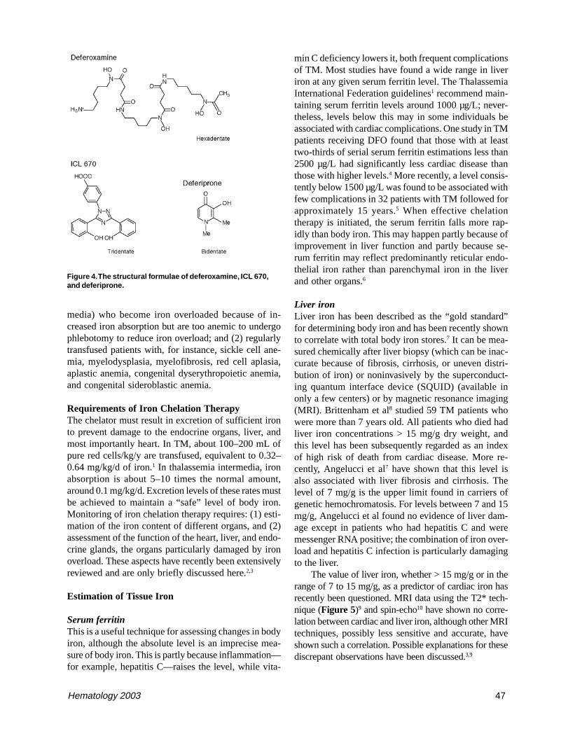
The value of liver iron, whether > 15 mg/g or in therange of 7 to 15 mg/g, as a predictor of cardiac iron hasrecently been questioned. MRI data using the T2* tech-nique (Figure 5)9 and spin-echo10 have shown no corre-lation between cardiac and liver iron, although other MRItechniques, possibly less sensitive and accurate, haveshown such a correlation. Possible explanations for thesediscrepant observations have been discussed.3,9
Figure 4. The structural formulae of deferoxamine, ICL 670,and deferiprone.
48 American Society of Hematology
Cardiac ironDirect measurement of cardiac iron by endomyocardialbiopsy of the right atrium is inappropriate since ironlocates mainly to the myocardium of the ventricles. Therecent development of a reproducible, sensitive, andaccurate indirect measure of cardiac iron using the MRIT2* technique11 has provided substantial important newdata. A T2* value less than 20 ms has been found tocorrelate with the presence of cardiac dysfunction, de-tected by echocardiography, 24-hour monitoring, or theneed for cardiac therapy. It is also valuable for moni-toring changes in cardiac iron during intensive chela-tion therapy.12
Non-transferrin-bound ironIn severely iron-loaded patients, non-transferrin-boundiron (NTBI) is present in plasma. It occurs in 80% ofpatients with TM and represents a highly toxic speciescausing tissue iron loading. NTBI is also found in pa-tients receiving chemotherapy or undergoing heart by-pass operations and those having other conditions inwhich large amounts of iron from hemoglobin break-down are released into the circulation.13 NTBI is re-moved by administration of DFO or DFP but reappearsrapidly (i.e., usually within 1 hour of discontinuing in-travenous DFO therapy) unless body iron burden is sub-stantially reduced.
Urine iron excretionIron excreted in response to a single dose of DFO ordeferiprone has been taken as an index of body iron bur-
den. It will vary, however, with the dose of chelator usedand, for DFO, whether vitamin C is also given and withthe hemoglobin level. There is also considerable day-to-day variation, even with apparently the same conditions.2
Nevertheless, in one recent study it has been found tocorrelate closely with cardiac iron measured by MRI.10
Estimation of Iron-Induced Tissue DamageIn addition to measuring iron status, it is important toassess the function of the heart, liver, and endocrineglands, the organs particularly damaged by iron over-load. Early detection of cardiac dysfunction is espe-cially important so that increased chelation therapy canbe instituted before cardiac damage is irreversible. Oncethe patient has started chelation therapy, it will also benecessary to monitor for potential side effects of theiron chelator being used (Table 3).
Chelation Therapy
DeferoxamineThe management of iron overload using subcutaneousDFO has been extensively reviewed.2 DFO is hexa-dentate, 1 molecule binding 1 atom of iron (Table 3).Standard therapy is with 40 mg/kg infused subcutane-ously over a period of 8–12 hours on 5–7 nights eachweek using a battery-operated infusion pump. Therapyis usually begun in children after 10–20 transfusionshave been given or when serum ferritin levels reach1000 µg/L. Vitamin C, 200 mg, given orally when theinfusion is started, enhances urine iron excretion. Al-
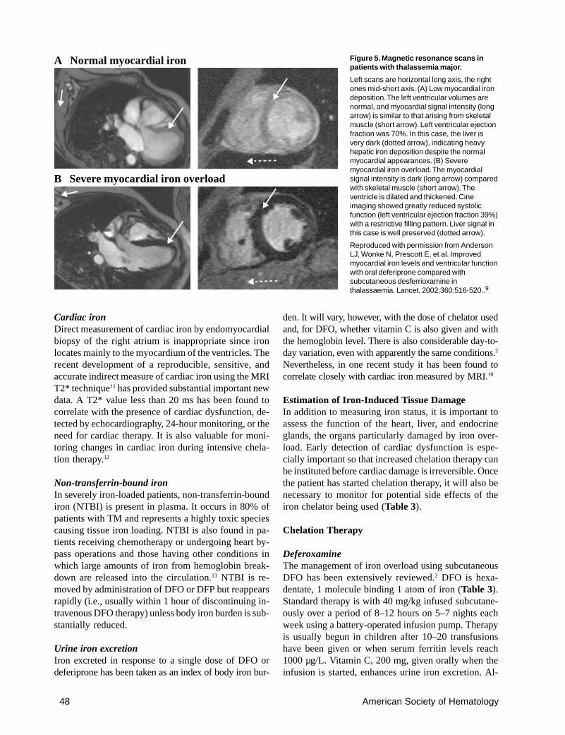
Figure 5. Magnetic resonance scans inpatients with thalassemia major.
Left scans are horizontal long axis, the rightones mid-short axis. (A) Low myocardial irondeposition. The left ventricular volumes arenormal, and myocardial signal intensity (longarrow) is similar to that arising from skeletalmuscle (short arrow). Left ventricular ejectionfraction was 70%. In this case, the liver isvery dark (dotted arrow), indicating heavyhepatic iron deposition despite the normalmyocardial appearances. (B) Severemyocardial iron overload. The myocardialsignal intensity is dark (long arrow) comparedwith skeletal muscle (short arrow). Theventricle is dilated and thickened. Cineimaging showed greatly reduced systolicfunction (left ventricular ejection fraction 39%)with a restrictive filling pattern. Liver signal inthis case is well preserved (dotted arrow).
Reproduced with permission from AndersonLJ, Wonke N, Prescott E, et al. Improvedmyocardial iron levels and ventricular functionwith oral deferiprone compared withsubcutaneous desferrioxamine inthalassaemia. Lancet. 2002;360:516-520..9
A Normal myocardial iron
B Severe myocardial iron overload
Hematology 2003 49
ternative routes of administration that have been triedinclude twice-daily bolus subcutaneous injections,14
continuous infusions over 24 or 48 hours using dispos-able prefilled balloons,15 and continuous intravenousinfusion using an indwelling central line or Portacath.16
First introduced in 1976 as subcutaneous treatmentfor TM, DFO has substantially improved the life ex-pectancy in the disease.4,8,17 Deaths continue to occurfrom cardiac failure due to iron overload, but these aremainly caused by lack of compliance.17,18 Defining com-pliance as more than 250 infusions a year, Gabutti andPiga17 found that 95% of compliant patients are alive at30 years of age, compared with only 12% of non-compliant patients. Modell et al18 reported that only 50%of TM patients in the UK reach 35 years, the poor re-sult again being attributable to cardiac failure due topoor compliance.
DFO can reverse iron-induced cardiomyopathy insome but not all patients.16,19 Continuous intravenousDFO results in comparatively rapid improvement inventricular function compared with the slow clearanceof cardiac iron, which can remain high even after 1year.12 Recent studies show that liver iron clears morerapidly and, despite severe iron overload initially, maybe normal at 6 months.12
Although cost and lack of compliance are the mainobstacles to DFO therapy, complications may also ex-clude some patients. High-frequency hearing loss, deaf-ness, and retinal damage with impaired vision (e.g.,night blindness) can occur when large doses of the drugare given to less severely iron-loaded patients, especiallychildren, in whom growth retardation and skeletal dam-age have also been reported. Generalized hypersensitiv-
ity is rare, but painful local reactions atthe injection site are common and oftenlead to lack of compliance. Infection withYersinia is increased, and on rare occasionsother infections (e.g. Klebsiella) are pre-cipitated.
DeferiproneThe orally active bidentate iron chelatordeferiprone (1,2 dimethyl-3-hydroxy-pyrid-4-one, also known as L1, CP20,Ferriprox, or Kelfer) was designed in R.C.Hider’s laboratories (Figure 4).20 Firsttested clinically in 1987, this drug is nowlicensed in 25 countries for patients withTM unable to be effectively treated withDFO. Reviews of its chemistry, pharma-cology, and clinical results have recentlybeen or are being published.20-22
PharmacokineticsDeferiprone is rapidly absorbed, appearing in plasmawithin 15 minutes of ingestion, with a peak plasma levelwithin 45–60 minutes (Table 3). It forms a 3:1chelator:iron complex that is excreted with the free drugin urine. Only 4% of a single oral dose of the drug isexcreted bound to iron, even in heavily iron-loadedpatients. Its iron chelation site is inactivated byglucuronidation, the speed of which varies from pa-tient to patient. This explains much of the individualvariation in response, the area under the curve of theconcentration of free drug in plasma being related tothe amount of iron excreted.23 Deferiprone mobilizesiron from parenchymal and reticuloendothelial poolsand from transferrin, ferritin, and hemosiderin. UnlikeDFO, it is also capable of chelating iron from intact redcells in vitro and in vivo, shown in patients with sicklecell anemia24 and thalassemia intermedia.25 The en-hanced ability of DFP to cross cell membranes mayunderlie what is emerging as its superior ability, com-pared with DFO, to protect the heart from iron and alsothe “shuttle effect” for iron when the 2 drugs are givensimultaneously (Figure 6).
Clinical studiesShort-term studies showed that iron excretion occursin the urine with negligible amounts in feces, althoughsome subsequent studies have suggested excretion upto 33%. Iron excretion increases with the dose of thedrug and the transfusional iron load of the patients.Although initial studies showed that 100 mg/kg/d wasmore effective,26,27 the dose used in most trials has been75 mg/kg/day fractionated into 3 doses. This dose was
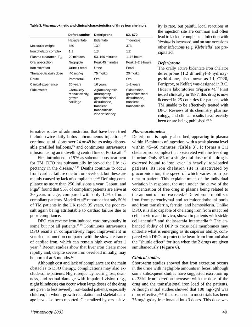
Table 3. Pharmacokinetic and clinical characteristics of three iron chelators.
Deferoxamine Deferiprone ICL 670
Hexadentate Bidentate Tridentate
Molecular weight 560 139 373
Iron:chelator complex 1:1 1:3 1:2
Plasma clearance, T½ 20 minutes 53–166 minutes 1–16 hours
Oral absorption Negligible Peak 45 minutes Peak 1–2.9 hours
Iron excretion Urine + fecal Urine Fecal
Therapeutic daily dose 40 mg/kg 75 mg/kg 20 mg/kg
Route Parenteral Oral Oral
Clinical experience 30 years 16 years 1–2 years
Side effects Ototoxicity, Agranulocytosis, Skin rashes,retinal toxicity, arthropathy, gastrointestinalgrowth, gastrointestinal disturbance,cartilage disturbance, transient
transient transaminitistransaminitis,zinc deficiency
50 American Society of Hematology
reported in early studies to be as effective as standard-dose DFO at increasing urine iron excretion.28,29 In mostpatients urine iron excretion around 0.5 mg/kg/day wasachieved with no indication of a diminishing responsewith time. There were early concerns that doses higherthan 75 mg/kg might produce more side effects, e.g.,arthropathy.27 Balance studies suggest that total iron ex-cretion with 75 mg/kg deferiprone is somewhat less thanthat with 40 mg/kg DFO given over 8 hours subcutane-ously, but only small numbers of patients have beenstudied and there is wide patient-to-patient variability.30
Final mean serum ferritin concentrations in 9 pub-lished trials in patients (mainly DFO ‘failures’ with TM)treated from 1 to 56 months with deferiprone haveranged from 1779 to 3273 µg/L.22 In 7 of the trials, therewas a significant fall in serum ferritin, and in 2 therewas no significant change. Serum ferritin levels fellmainly in the patients starting with the highest levels.In 6 studies in which serial liver iron determinationswere made, hepatic iron fell significantly in 1, rose sig-nificantly in 1, and did not change significantly in theother 4.22 When SQUID technique was used in 54thalassemia patients (aged 7–22 years), median liveriron concentration rose from 1456 mg/g liver to 2029mg at 2 years and 2449 mg at 3.2 years.6 These patientswere well chelated before starting deferiprone, but theiriron intake from transfusions rose substantially duringthe study. Recent MRI data retrospectively comparing15 patients treated long term with deferiprone with 30matched patients treated with DFO, in which liver MRIT2* was converted to estimated dry-weight liver iron,showed significantly higher liver iron: 5.1 versus 3.5mg/g in the deferiprone compared with the DFO group.9
In a short-term study, raising the dose ofdeferiprone to around 100 mg/kg led to reductionof serum ferritin levels in patients inadequatelychelated at 75 mg/kg.31 Prospective trials areneeded to assess whether doses of deferiprone(around 100 mg/kg daily) can safely be given longterm and will result in more effective iron chela-tion in patients inadequately chelated on 75 mg/kg daily. There is also a need for further (> 3 years)long-term studies to determine liver iron levels inlarge numbers of patients receiving deferiproneto determine more accurately the proportion ofpatients in whom liver iron is adequately con-trolled.
The most important aspect of iron chelationtherapy is protection of the heart. Two recent stud-ies, albeit retrospective, show significant benefitfor the patients receiving deferiprone comparedwith DFO. In a London study, 15 TM patients whohad received deferiprone 75 mg/kg/d for 3 years
showed a lower incidence of cardiac disease (assessedby echocardiography and need for cardiac drug therapy)and lower cardiac iron estimated indirectly by MRI T2*than 30 age- and sex-matched patients who had receivedDFO 40 mg/kg/d subcutaneously on 5-7 days each week(median myocardial T2* 34.0 vs 11.4 ms, P = .02).9
Excess myocardial iron (T2* < 20 ms) was significantlyless common in the deferiprone group (27%) than inthe DFO group (67%), P = .025.
In a Turin study, 54 patients who had receiveddeferiprone were compared retrospectively with 75patients who had received DFO; the drugs were givenat standard doses over an average of 6 years.32 No pa-tient in the deferiprone group compared with 3 in theDFO group died of cardiac failure. Deterioration ofpreexisting cardiac dysfunction or new cardiac diseaseoccurred in 2 (4%) of the deferiprone-treated patientscompared with 15 (20%) of the DFO group (P = .007).32
Formal prospective studies are needed to confirm theapparent greater cardioprotective effect against irontoxicity of deferiprone compared with subcutaneousDFO suggested by these two retrospective studies. In ashort-term (12 months) prospective randomized study,Maggio et al33 found no difference in any of the param-eters used to detect cardiac abnormalities between pa-tients receiving DFO and patients receiving deferiprone.
ComplicationsThe incidence of the now well-established complica-tions of deferiprone therapy—agranulocytosis, neutro-penia, arthralgia, gastrointestinal symptoms, transientchanges in liver enzymes, and zinc deficiency—hasbeen established in recent prospective trials34-36 and re-
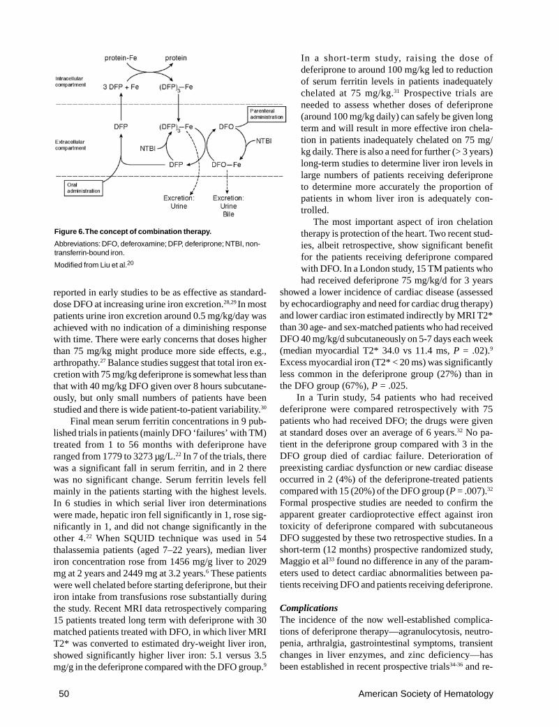
Figure 6. The concept of combination therapy.
Abbreviations: DFO, deferoxamine; DFP, deferiprone; NTBI, non-transferrin-bound iron.
Modified from Liu et al.20
Hematology 2003 51
viewed.22 Hepatic fibrosis has also been suggested inone small retrospective study to be a consequence ofdeferiprone therapy.37 However, recent evidence basedon 56 repeat biopsies in patients treated for a mean of3.1 years shows no evidence for this.38 No other studyhas reported significant increase in hepatic fibrosis as-cribed to deferiprone. Transient changes in alanineaminotransferase (ALT) levels, especially in the firstfew months of therapy and in hepatitis C antibody–posi-tive patients, have been observed. The mean ALT lev-els did not increase among 151 patients treated for 3years.36 Occasional patients have, however, been with-drawn from therapy in some trials because of concernsabout raised ALT levels.34
Agranulocytosis, the most serious complication ofdeferiprone, occurs in about 1% of patients and appearsto be idiosyncratic; it is probably more frequent in fe-males. Patients with agranulocytosis should be perma-nently withdrawn from therapy, although a proportionof patients with less severe degrees of neutropenia havesuccessfully been re-exposed to the drug. Most patientswith the other side effects can usually continue withthe drug, often after a period of withdrawal andretreatment initially at a lower dose.
Combination therapyCombination therapy with DFO and deferiprone com-menced in 1998, when it was reported that DFO anddeferiprone could be safely given simultaneously andthat the urine iron excretion achieved is at least equiva-lent to the iron excretion resulting when the 2 drugs aregiven on separate days.31 Six clinical studies of combi-nation therapy have now been reported (Table 4). Allshow decreasing serum ferritin levels and, where mea-sured, decreasing liver iron. Mourad et al,40 for instance,report that deferiprone 75 mg/kg 7 days a week andDFO 40 mg/kg subcutaneously over 8-12 hours 2 daysa week gives approximately equivalent iron chelation,based on serum ferritin levels, to 5 days a week of DFO.Compliance is likely to be improved longer term for apatient needing 2 rather than 5 days of subcutaneousinfusions.
The basis for this additive or synergistic effect isgiven by the studies of Grady et al30 and Breuer et al.45
These suggest that deferiprone enters cells and chelatesiron, which it brings into plasma. The iron is then trans-ferred to DFO for excretion in urine and feces (Figure6). If combination therapy in longer-term studies doesnot show any unexpected toxicity, it is an exciting thera-peutic advance for improving compliance and avoid-ing large, potentially toxic doses of either drug.
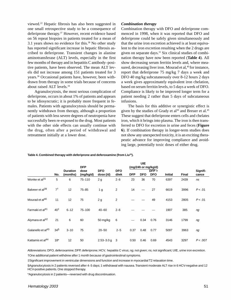
Table 4. Combined therapy with deferiprone and deferoxamine (from Liu20).
UIEDFP (mg/24h or mg/kg/d)
Duration dose DFO DFO DFP + Signifi-No. (months) (mg/kg/d) dose (/d) d/wk DFP DFO DFO Initial Final cance
Wonke et al31 5 6 75–110 2 g 2–6 23 36 70 6397 2439 ns
Balveer et al39 7† 12 75–85 1 g 2 14 — 27 6619 3996 P < .01
Mourad et al40 11 12 75 2 g 2 — — 49 4153 2805 P < .01
Farmaki et al41 40‡ 6–12 75–100 40–60 2–6 — — — 1907 385 ng
Alymara et al42 21 6 60 50 mg/kg 6 — 0.34 0.76 3146 1799 ng
Galanello et al43 34§ 3–10 75 20–50 2–5 0.37 0.48 0.77 5097 3963 ng
Kattamis et al44 18* 12 50 2.53–3.0 g 3 0.50 0.46 0.69 4543 3297 P < .007
Abbreviations: DFO, deferoxamine; DFP, deferiprone; HCV, hepatitis C virus; ng, not given; ns, not significant; UIE, urine iron excretion.
†One additional patient withdrew after 1 month because of gastrointestinal symptoms.
‡Significant improvement in ventricular dimensions and function and increase in myocardial T2 relaxation time.
§Agranulocytosis in 2 patients reversed after 4–5 days; 1 withdrawal with nausea. Transient moderate ALT rise in 6 HCV-negative and 12HCV-positive patients. One stopped therapy.
*Agranulocytosis in 2 patients—reversed with drug discontinuation.
52 American Society of Hematology
Alternating (sequential) therapy with DFO anddeferiprone has also been studied in 7 children non-compliant to DFO.46 Compliance was improved whendeferiprone was given for 4 days and then DFO for 2days each week. Over 6 months, liver iron fell signifi-cantly and there was a nonsignificant fall in mean serumferritin from 5536 to 3778 µg/L. More prolonged studiesare needed to determine the place of this approach.
Thalassemia intermediaOral iron chelation therapy is a potentially attractiveoption for patients with iron overload who are too ane-mic for phlebotomy. Olivieri et al47 first reported a pa-tient with thalassemia intermedia in whom deferipronewas effective in reducing both liver iron and serum fer-ritin to normal within 12 months of therapy. Pootrakulet al25 have recently extended these observations. Theyfound in 8 thalassemia intermedia patients in Thailand(mainly suffering from thalassemia/hemoglobin E) thatdeferiprone at the low dose of 50 mg/kg/d not only sig-nificantly reduced serum ferritin and red cell membraneliver iron over 12 months but also resulted in an in-crease in hemoglobin and serum erythropoietin levelsand improvement in weight and appetite. No side ef-fects requiring drug withdrawal were encountered.
Other transfusion-dependent anemiasSimilar results to those in TM have been obtained withdeferiprone in patients with myelodysplasia, myelofi-brosis, and other acquired marrow diseases. Althoughthere has been theoretical concern that agranulocytosismay be more frequent in these acquired bone marrowdisorders than in TM, there are no data to suggest this.
ICL 670ICL 670 (4-[3,5-bis(2-hydroxyphenyl)-1,2,4-triazol-1-y1] benzoic acid) (Figure 4) was developed by NovartisPharma AG after many hundreds of potential orallyactive iron chelators were screened. Preclinical studiesshow that it forms a 2:1 chelator:iron complex and pro-duces an increase predominantly in fecal iron excre-tion after a single oral dose, only 6% of iron excretionaccruing in the urine (Table 3). It is highly selectivefor iron, is rapidly absorbed, and circulates for severalhours. In the non-iron-loaded marmoset and rat, its maintoxic effect was on the renal tubular epithelial cells,but this effect was abrogated in iron-loaded marmosetsand substantially reduced in iron-loaded rats.48
Short-term clinical trials have recently been re-ported.49 Single daily doses of 10, 20, and 40 mg/kgbody weight were studied. Peak plasma concentrationafter a single oral dose occurred at about 2 hours, andthe drug was still detectable in plasma in almost all
patients at 24 hours; the mean elimination half-life wasbetween 11–16 hours after multiple dose administra-tion. Net iron excretion after 6 days of exposure waslinearly related to the dose of the drug. Iron excretionat 12 days was related to the area under the curve of theconcentration of free drug in plasma. Five of 6 patientsreceiving 20 mg/kg were calculated to excrete ironequivalent to the amount received in blood transfusions.The main side effect was skin rashes that required with-drawal of 4 patients given the highest dose of 40 mg/kgover 8–10 days. Sporadic transaminase rises occurredin 1 of these patients and in 4 other patients. Mild nau-sea, diarrhea, and abdominal pain—none requiring dis-continuation of the drug—occurred in other patients.Longer-term studies of the drug at the dose of 20 mg/kg have been carried out.50 These showed that total bodyiron excretion ranged from 7.7–28.5 mg iron/d. Thesedid not show any additional toxic effects and showedthat liver iron decreased in 12 (57.1%), was unchangedin 8 (38.1%), and rose in 1 (4.8%) of 21 patients stud-ied using the SQUID technique.
ConclusionsThe prospect for transfusion-dependent patients to re-ceive effective iron chelation therapy has substantiallyimproved in the last few years. Subcutaneous DFO 40mg/kg over 8–12 hours on at least 5 days a week pro-tects most compliant patients against cardiac diseaseand other serious complications and remains the firstchoice. Its cost, frequent lack of compliance, and com-plications means that alternative approaches are needed.After many years of short-term clinical trials of theorally active agent deferiprone and much controversyabout its efficacy and toxicity, recent published datahave been favorable on both aspects. These suggest thatthe drug at a dose of 75 mg/kg/d may be at least aseffective as DFO in protecting patients from iron-in-duced cardiomyopathy. Hepatic fibrosis does not ap-pear to be a problem, and the established side effectsdo not lead to the need for discontinuation of the drugin the majority of patients.
Combination therapy with DFO and deferiprone isan exciting new possibility for those patients inad-equately chelated on either drug alone. ICL 670, a neworal iron chelator in early clinical trial, promises to ex-pand further the range of possibilities for effective andsafe iron chelation therapy for patients with TM andother iron-loaded transfusion-dependent or -indepen-dent patients with severe refractory anemias. Which-ever chelation regimen is chosen, patients must beclosely monitored both for effectiveness of therapy, withparticular attention to cardiac iron and function, andfor toxic side effects of the chelating drug.
Hematology 2003 53
III. NEWER ASPECTS OF THE DIAGNOSIS AND
TREATMENT OF IRON DEFICIENCY
James D. Cook, MD*
Iron deficiency is by far the most common hematologi-cal disorder encountered in general practice. The basicapproach to its diagnosis and management is well es-tablished and outlined in most medicine and hematol-ogy texts. My emphasis in this selective review is onsoluble transferrin receptor (sTfR) measurements, irondeficiency induced by recombinant erythropoietin(rHuEPO) therapy, and parenteral iron therapy withsodium ferric gluconate.
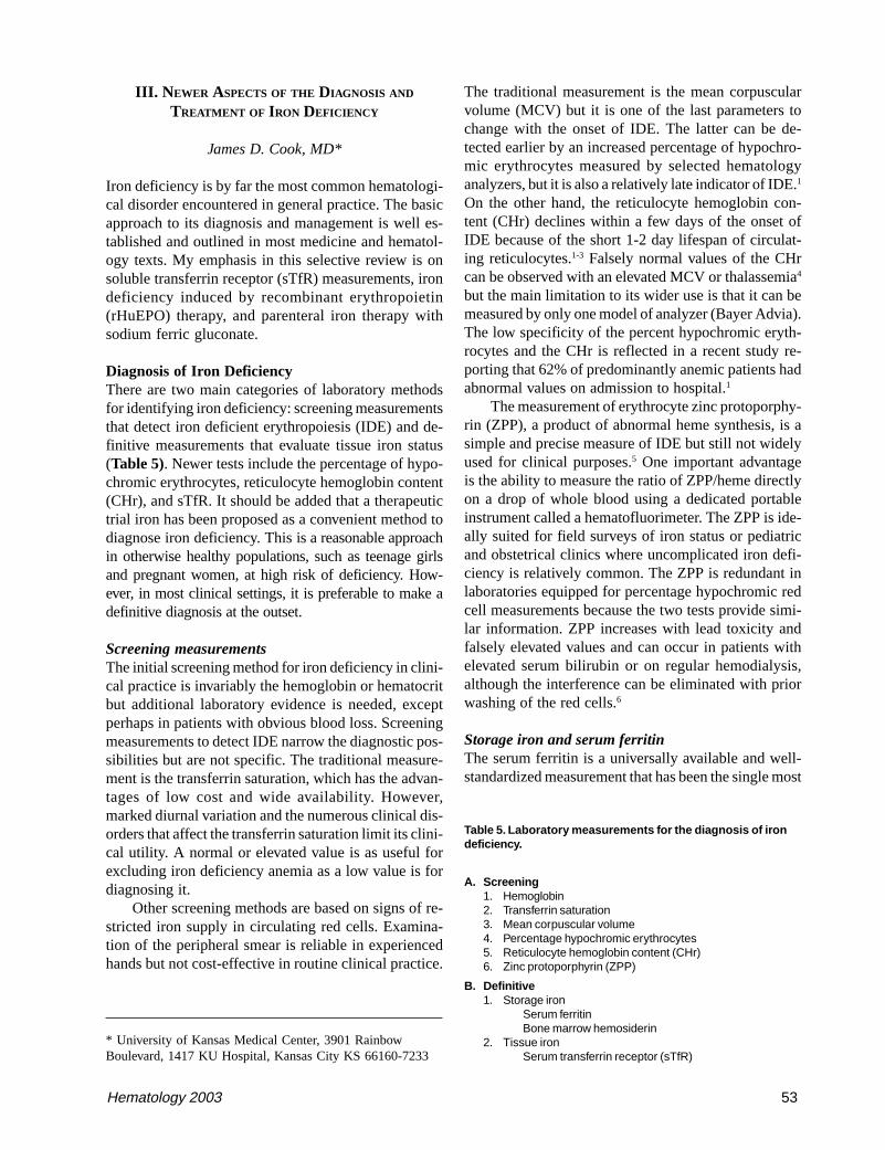
Diagnosis of Iron DeficiencyThere are two main categories of laboratory methodsfor identifying iron deficiency: screening measurementsthat detect iron deficient erythropoiesis (IDE) and de-finitive measurements that evaluate tissue iron status(Table 5). Newer tests include the percentage of hypo-chromic erythrocytes, reticulocyte hemoglobin content(CHr), and sTfR. It should be added that a therapeutictrial iron has been proposed as a convenient method todiagnose iron deficiency. This is a reasonable approachin otherwise healthy populations, such as teenage girlsand pregnant women, at high risk of deficiency. How-ever, in most clinical settings, it is preferable to make adefinitive diagnosis at the outset.
Screening measurementsThe initial screening method for iron deficiency in clini-cal practice is invariably the hemoglobin or hematocritbut additional laboratory evidence is needed, exceptperhaps in patients with obvious blood loss. Screeningmeasurements to detect IDE narrow the diagnostic pos-sibilities but are not specific. The traditional measure-ment is the transferrin saturation, which has the advan-tages of low cost and wide availability. However,marked diurnal variation and the numerous clinical dis-orders that affect the transferrin saturation limit its clini-cal utility. A normal or elevated value is as useful forexcluding iron deficiency anemia as a low value is fordiagnosing it.
Other screening methods are based on signs of re-stricted iron supply in circulating red cells. Examina-tion of the peripheral smear is reliable in experiencedhands but not cost-effective in routine clinical practice.
The traditional measurement is the mean corpuscularvolume (MCV) but it is one of the last parameters tochange with the onset of IDE. The latter can be de-tected earlier by an increased percentage of hypochro-mic erythrocytes measured by selected hematologyanalyzers, but it is also a relatively late indicator of IDE.1
On the other hand, the reticulocyte hemoglobin con-tent (CHr) declines within a few days of the onset ofIDE because of the short 1-2 day lifespan of circulat-ing reticulocytes.1-3 Falsely normal values of the CHrcan be observed with an elevated MCV or thalassemia4
but the main limitation to its wider use is that it can bemeasured by only one model of analyzer (Bayer Advia).The low specificity of the percent hypochromic eryth-rocytes and the CHr is reflected in a recent study re-porting that 62% of predominantly anemic patients hadabnormal values on admission to hospital.1
The measurement of erythrocyte zinc protoporphy-rin (ZPP), a product of abnormal heme synthesis, is asimple and precise measure of IDE but still not widelyused for clinical purposes.5 One important advantageis the ability to measure the ratio of ZPP/heme directlyon a drop of whole blood using a dedicated portableinstrument called a hematofluorimeter. The ZPP is ide-ally suited for field surveys of iron status or pediatricand obstetrical clinics where uncomplicated iron defi-ciency is relatively common. The ZPP is redundant inlaboratories equipped for percentage hypochromic redcell measurements because the two tests provide simi-lar information. ZPP increases with lead toxicity andfalsely elevated values and can occur in patients withelevated serum bilirubin or on regular hemodialysis,although the interference can be eliminated with priorwashing of the red cells.6
Storage iron and serum ferritinThe serum ferritin is a universally available and well-standardized measurement that has been the single most
Table 5. Laboratory measurements for the diagnosis of irondeficiency.
A. Screening1. Hemoglobin2. Transferrin saturation3. Mean corpuscular volume4. Percentage hypochromic erythrocytes5. Reticulocyte hemoglobin content (CHr)6. Zinc protoporphyrin (ZPP)
B. Definitive1. Storage iron
Serum ferritinBone marrow hemosiderin
2. Tissue ironSerum transferrin receptor (sTfR)
* University of Kansas Medical Center, 3901 RainbowBoulevard, 1417 KU Hospital, Kansas City KS 66160-7233
54 American Society of Hematology
important laboratory measure of iron status during thepast quarter century. Phlebotomy studies in normal sub-jects have demonstrated that 1 µg/L serum ferritin cor-responds to 8–10 mg or 120 µg storage iron/kg bodyweight,7 although a log transformation gives a moreaccurate estimate.8 Numerous studies have demon-strated its superiority over other iron-related measure-ments in identifying iron deficiency anemia. In 55 stud-ies culled from 1179 relevant citations, receiver-opera-tor characteristic curves in 2579 subjects gave a meanarea for the serum ferritin of 0.95 ± 0.1 (95% confi-dence limits) as compared with 0.77 for the ZPP, 0.76for the MCV, and 0.74 for the transferrin saturation.9
The well-known limitation of the serum ferritin is theelevation in values independent of iron status that oc-cur with acute or chronic inflammation, malignancy,liver disease, and alcoholism.
Many hematologists still rely on the assessmentof stainable iron on aspirated marrow smears or biopsyfor the definitive diagnosis of iron deficiency anemia.Although still widely regarded as the gold standard forthe diagnosis of iron deficiency, the reliability of themarrow iron stain is often suboptimal when used forroutine clinical purposes. In a recent study, 108 con-secutive bone marrow specimens from unselected he-matology patients reported to have absent iron werereviewed.10 One-third of the reports were incorrect dueeither to an inadequate specimen or detectable ironstores, and less than half of the patients with absentmarrow iron had clinical evidence that supported thediagnosis of iron deficiency anemia. In another recentreview of iron stains, high intra-observer variability inpathological diagnosis led the authors to conclude thatthe bone marrow is not a perfect gold standard.4 It shouldalso be noted that the bone marrow is no longer reli-able in diagnosing iron deficiency anemia afterparenteral iron therapy. While marrow iron stainingcontinues to play a critical role in validating newer labo-ratory measurements of iron status when performed andreviewed under standardized conditions in prospectivestudies by experienced investigators, bone marrow ex-aminations should seldom be performed solely to diag-nose iron deficiency because of the expense, discom-fort, and technical pitfalls with this approach.
Tissue iron and serum transferrin receptorTransferrin receptors are membrane glycoproteins thatserve as the gateway for circulating transferrin iron tothe interior of all body cells. In addition to erythroidprecursors that contain 80% of the total body receptormass, rapidly dividing cells and the placenta contain ahigh density of receptors. The synthesis of ferritin andtransferrin receptors is precisely regulated by a com-
mon mechanism involving the iron response protein anda nucleotide sequence termed the iron response element.
The sTfR is a soluble form of the cellular receptorlacking the first 100 amino acids and composed of theextracellular domain. Countless articles have been pub-lished on the clinical utility of the sTfR since it wasfirst discovered in 1986 by Kohgo and coworkers.11
Ferrokinetic studies have demonstrated that the sTfR isdirectly correlated with the total mass of erythroid pre-cursors over the complete spectrum of hematologicaldisorders ranging from marrow aplasia to thalassemiamajor.12 Its use for assessing erythropoiesis has beenreviewed recently.13 The only determinant of the sTfRother than the erythroid precursor mass is tissue irondeficiency, which increases the sTfR in proportion tothe severity of iron deficit.14 Several commercial as-says are now available, but wider application of sTfRmeasurements is limited by the divergent values re-ported with different assays,15 differences that couldprobably be eliminated by the development of an inter-national standard.
Isolated iron deficiencyIsolated or uncomplicated iron deficiency in the absenceof other diseases that influence measurements of ironstatus is seen most often with rapid growth or duringgestation, and in patients with excessive uterine or gas-trointestinal blood loss. The key laboratory measure-ment for its identification is the serum ferritin. A lowhemoglobin concentration in a patient with a serum fer-ritin < 30 µg/L is diagnostic of iron deficiency anemia.
One drawback of relying solely on a low hemoglo-bin and serum ferritin is that milder iron deficiencywithout anemia goes undetected. Individuals withbaseline hemoglobin values in the upper normal rangemust lose 20%–30% of their body iron before iron de-ficiency can be detected by anemia. A method for mea-suring body iron quantitatively using the log(sTfR/se-rum ferritin) ratio that permits detection of mild tissueiron deficiency has been described recently.16 Themethod estimates in mg/kg the surplus of storage ironin replete individuals or the tissue deficit in those withiron deficiency. Serial measurements in an individualremain constant over several months allowing those atrisk of recurrent iron deficiency to be monitored pro-spectively. The relevance of iron deficiency withoutanemia that can be assessed with this approach has beenunclear largely because of the difficulty in identifyingit. Greater improvement in exercise performance wasobserved in iron-depleted nonanemic females given ironsupplements as compared with unsupplemented con-trols despite no effect on hemoglobin values.17 Mild irondeficiency without anemia could explain chronic fa-
Hematology 2003 55
tigue in some individuals.18,19 It is also preferable to de-tect declining tissue iron levels in individuals with recur-rent iron deficiency before overt anemia develops.
Iron deficiency and chronic diseaseThe diagnosis of iron deficiency would be simple if itwere not for the many clinical disorders that influencethe internal iron cycle. The anemia of chronic diseaseis a common hematological disorder that is easier torecognize than it is to define. Because it alters screen-ing tests for iron status in the same manner as true irondeficiency, the distinction between the anemia ofchronic disease and iron deficiency anemia requires tis-sue-related iron measurements. To avoid the need forbone marrow examinations in patients in whom irondeficiency anemia is suspected, reliance is often placedon the serum ferritin concentration despite the well-known elevation with acute or chronic inflammation.The optimal cut-off values of the serum ferritin to dis-tinguish iron deficiency anemia from the anemia ofchronic disease was examined in a landmark study in259 anemic patients over 65 years of age.20 In 36% ofpatients with iron deficiency anemia based on bonemarrow examination, the serum ferritin was the onlytest of several that added useful diagnostic informa-tion. Only 2 of 49 patients with serum ferritin < 18 µg/L did not have iron deficiency anemia and only 8 of116 with a serum ferritin > 100 µg/L had iron deficiencyanemia. Between 18 and 100 µg/L, 40% had iron defi-ciency anemia although only 1 such patient had a se-rum ferritin > 45 µg/L. The authors later proposed se-rum ferritin values < 40 and < 70 µg/L to diagnose irondeficiency anemia in anemic patients without and withinflammation respectively.9 A serum ferritin < 50 µg/Lhas been proposed as the best cutoff to identify irondeficiency anemia in patients with liver disease.21
Because the sTfR concentration remains normal inpatients with the anemia of chronic disease,22 it is aninvaluable addition to the serum ferritin measurement.The sTfR cannot only distinguish iron deficiency ane-mia from the anemia of chronic disease but it can alsoidentify iron deficiency anemia when it occurs in pa-tients with the anemia of chronic disease. In 129 con-secutive anemic patients receiving a bone marrow forstainable iron, iron deficiency anemia was identified in48, the anemia of chronic disease in 64 and both disor-ders in 17.23 The sTfR was normal in all patients withthe anemia of chronic disease, elevated in 41 of 48 pa-tients with iron deficiency anemia and in 13 of 17 pa-tients with both the anemia of chronic disease and irondeficiency. The separation between the 3 groups wasfurther improved using the sTfR/log(serum ferritin)ratio; no patients with iron deficiency anemia over-
lapped those with anemia of chronic disease and all but1 patient with both the anemia of chronic disease andiron deficiency anemia had higher values than patientswith the anemia of chronic disease only. A recent studyhas indicated that even better discrimination can beobtained with the log(sTfR/serum ferritin)24 as describedabove for quantifying body iron. Use of the receptor/ferritin ratio can eliminate the need for bone marrowexamination to detect iron deficiency in patients withchronic inflammatory joint or bowel disease who areusually reluctant to undergo this unpleasant procedure.
Iron deficiency and rHuEPO therapyIron status in patients with chronic renal failure hasvaried widely over the past half century. Transfusionaliron overload was invariable until the introduction ofhemodialysis when iron deficiency emerged due to dia-lyzer blood loss. Vigorous parenteral iron therapy ledto reports of significant iron overload but with the intro-duction of rHuEPO therapy, iron deficiency again becamewidespread. Vigorous parenteral iron therapy has againraised concern about iatrogenic iron overload.25
The term functional iron deficiency has arisenmainly in the nephrology literature in reference to theIDE induced by rHuEPO therapy in dialysis patientswith residual iron stores.26 The diagnosis is usuallybased on one or more screening measurements (Table5). Aggressive parenteral iron therapy is commonlyadvised for its treatment on the assumption that func-tional iron deficiency is the major cause of resistanceto rHuEPO therapy. While this is undoubtedly true inmany patients, the laboratory features are also typicalof the anemia of chronic disease and there is sparseinformation about the role of inflammation in so-calledfunctional iron deficiency. The sTfR is not an optimalguide to the need for additional parenteral iron becauseof the enhancing effect of rHuEPO on erythropoiesisand consequently on the sTfR. More data is needed onthe extent to which inflammation contributes to func-tional iron deficiency and whether a rise in sTfR con-centration in patients on stable doses of rHuEPO canbe used as a guide to parenteral iron therapy.
In a dialysis patient without laboratory evidenceof inflammation, the amount of iron that should be givenduring the first 2–3 weeks of initiating rHuEPO therapycan be calculated from the anticipated increase in cir-culating hemoglobin (roughly 4 mg iron/kg body weightfor each 10 g/L hemoglobin rise) minus iron stores basedon 8 mg available iron per µg/L serum ferritin or 3–4mg/µg/L serum ferritin if the CRP is elevated. Afterachieving a hematological response, parenteral ironshould be continued to maintain a serum ferritin > 100µg/L if the CRP is normal and > 200–300 µg/L if el-
56 American Society of Hematology
evated. There is evidence that serum ferritin valuesabove 500–800 µg/L increase the risk of infections inhemodialysis patients26 and this risk could be evengreater in pancytopenic patients given rHuEPO therapywho are already at increased risk of infection.
Treatment of Iron Deficiency
Oral iron therapyIt is preferable to treat iron deficiency with oral ratherthan parenteral iron. One iron tablet taken daily with-out food is as effective as 3 tablets with meals, and thedisparity is even greater in patients with atrophic gas-tritis, chronic suppression of gastric acid secretion, andgastric stapling or bypass surgery. The major obstacleto successful oral therapy is the nausea and epigastricdiscomfort that occurs 30–60 minutes after taking iron,symptoms that are dose-related but often subside after2–3 days with continued treatment. Reducing the doseor taking a tablet at bedtime is usually helpful in reduc-ing side effects. Diarrhea or constipation are not dose-related and should be managed symptomatically. Com-mercial iron preparations promoted on the basis of fewerside effects are invariably less well absorbed. A brieffollow-up clinic visit 2–3 weeks after initiating oraltherapy can be helpful in tailoring an oral iron regimenin patients having troublesome side effects. Parenteraliron should not be used simply as a convenience for thepatient or physician.
Parenteral iron therapyThe main indications for parenteral iron are uncon-trolled blood loss, intolerance to oral iron, intestinalmalabsorption, and poor adherence to an oral regimen.Iron malabsorption can be detected by observing anincrease in serum iron of less than 100 µg% overbaseline in a fasting patient 1 or 2 hours after taking 60mg iron as ferrous sulfate.
Until recently, iron dextran has been the onlyparenteral iron preparation available in the UnitedStates. It is a low molecular weight dextran complexedwith ferric oxide and supplied as a dark brown solutioncontaining 50 mg iron per mL. Although intramuscularadministration is still recommended by current manu-facturers, intravenous (IV) administration is preferredby most physicians because of the ability to administerlarger doses. In hemodialysis patients in whom mul-tiple treatments can be given conveniently, 6-10 injec-tions of 100 mg iron can be administered on consecu-tive dialysis days.27 In other patients, a major advan-tage of iron dextran is the ability to administer rela-tively large doses of 500–2000 mg iron on a single oc-casion.28,29 After diluting the dose in 500 mL normal
saline, and premedicating with diphenylhydramine withor without steroids, the first 20–30 mL is given slowlyover 5 minutes as a test dose. If no allergic reactionoccurs within the first 15–30 minutes, the remainder ofthe dose is given over the next 3–4 hours.
The major drawback of iron dextran is a severeanaphylactic reaction that occurs within a few minutesof initiating the infusion and is sometimes fatal. At least30 deaths have been attributed to iron dextran use inthe US since 197630 and because anaphylaxis can oc-cur in those who have not reacted to iron dextran in thepast, a test dose is always required. Another drawbackof iron dextran is a less serious delayed reaction occur-ring 24–48 hours after the infusion. Characterized bymyalgia, arthralgia, headache, and malaise, delayedreactions occured in over 10% of patients given totaldose infusions.31 These symptoms usually respondpromptly to nonsteroidal anti-inflammatory drugs butthe reaction can be severe and prolonged in patientswith inflammatory joint disease.
Sodium ferric gluconate (SFG)This form of parenteral iron was approved in the US in1999 under the Food and Drug Administration’s prior-ity drug review, although it has been used in Europe formany years. SFG in sucrose is a stable macromolecu-lar complex supplied as a deep red solution in 5 mLampoules containing 62.5 mg elemental iron. The cur-rent product label advises administration of 5–10 mLas a direct IV push at a rate of 12.5 mg per min for 10minutes for a recommended dose of 125 mg iron. Notest dose is required. Unlike iron dextran, which canrequire several weeks for complete processing by mac-rophages, roughly 80% of SFG is delivered to transfer-rin within 24 hours. One report suggested that satura-tion of circulating transferrin can be exceeded follow-ing the infusion of SFG 32 but this was later explainedby the release iron from the ferric gluconate during theserum iron determination.33
The appeal of SFG administration is avoidance ofthe risk of the serious anaphylactic reactions associ-ated with iron dextran. In a review of data from theWorld Health Organization and pharmaceutical manu-facturers, no deaths were reported with 25 million in-fusions of SFG as compared with 31 deaths from abouthalf the number of administratons of iron dextran.30
Adverse reactions with SFG in a multicenter, random-ized, crossover, double-blind, placebo-controlled pro-spective study in 2534 hemodialysis patients was re-cently reported.34 Adverse events were similar betweenSFG and placebo groups, 12.3% versus 9.8%. Life-threatening events (immediate reactions requiring re-suscitation measures) and drug intolerance rates (any
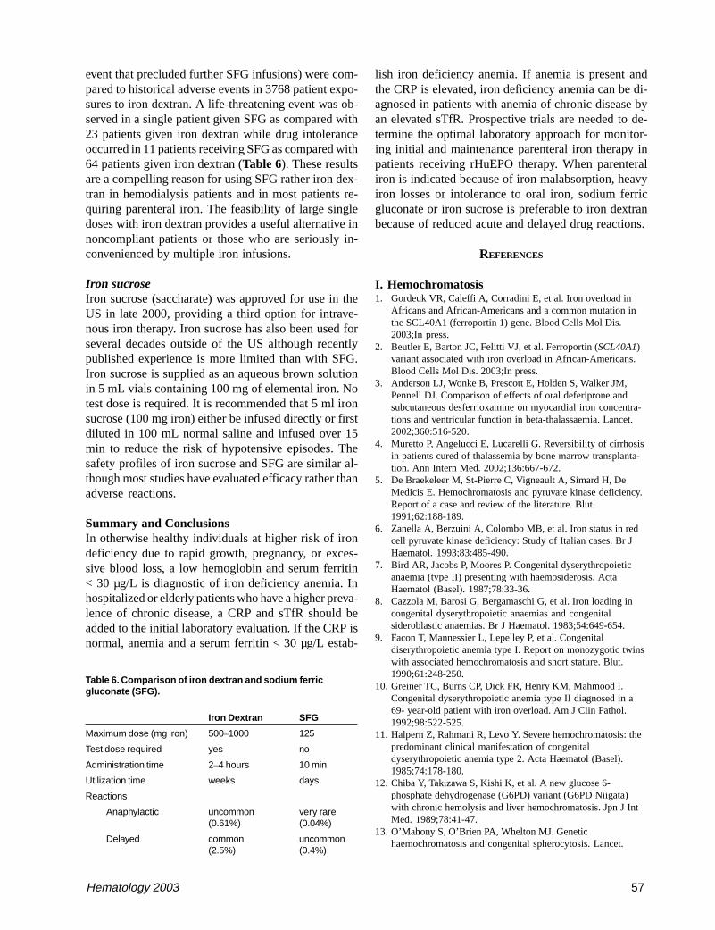
Hematology 2003 57
event that precluded further SFG infusions) were com-pared to historical adverse events in 3768 patient expo-sures to iron dextran. A life-threatening event was ob-served in a single patient given SFG as compared with23 patients given iron dextran while drug intoleranceoccurred in 11 patients receiving SFG as compared with64 patients given iron dextran (Table 6). These resultsare a compelling reason for using SFG rather iron dex-tran in hemodialysis patients and in most patients re-quiring parenteral iron. The feasibility of large singledoses with iron dextran provides a useful alternative innoncompliant patients or those who are seriously in-convenienced by multiple iron infusions.
Iron sucroseIron sucrose (saccharate) was approved for use in theUS in late 2000, providing a third option for intrave-nous iron therapy. Iron sucrose has also been used forseveral decades outside of the US although recentlypublished experience is more limited than with SFG.Iron sucrose is supplied as an aqueous brown solutionin 5 mL vials containing 100 mg of elemental iron. Notest dose is required. It is recommended that 5 ml ironsucrose (100 mg iron) either be infused directly or firstdiluted in 100 mL normal saline and infused over 15min to reduce the risk of hypotensive episodes. Thesafety profiles of iron sucrose and SFG are similar al-though most studies have evaluated efficacy rather thanadverse reactions.
Summary and ConclusionsIn otherwise healthy individuals at higher risk of irondeficiency due to rapid growth, pregnancy, or exces-sive blood loss, a low hemoglobin and serum ferritin< 30 µg/L is diagnostic of iron deficiency anemia. Inhospitalized or elderly patients who have a higher preva-lence of chronic disease, a CRP and sTfR should beadded to the initial laboratory evaluation. If the CRP isnormal, anemia and a serum ferritin < 30 µg/L estab-
lish iron deficiency anemia. If anemia is present andthe CRP is elevated, iron deficiency anemia can be di-agnosed in patients with anemia of chronic disease byan elevated sTfR. Prospective trials are needed to de-termine the optimal laboratory approach for monitor-ing initial and maintenance parenteral iron therapy inpatients receiving rHuEPO therapy. When parenteraliron is indicated because of iron malabsorption, heavyiron losses or intolerance to oral iron, sodium ferricgluconate or iron sucrose is preferable to iron dextranbecause of reduced acute and delayed drug reactions.
REFERENCES
I. Hemochromatosis1. Gordeuk VR, Caleffi A, Corradini E, et al. Iron overload in
Africans and African-Americans and a common mutation inthe SCL40A1 (ferroportin 1) gene. Blood Cells Mol Dis.2003;In press.
2. Beutler E, Barton JC, Felitti VJ, et al. Ferroportin (SCL40A1)variant associated with iron overload in African-Americans.Blood Cells Mol Dis. 2003;In press.
3. Anderson LJ, Wonke B, Prescott E, Holden S, Walker JM,Pennell DJ. Comparison of effects of oral deferiprone andsubcutaneous desferrioxamine on myocardial iron concentra-tions and ventricular function in beta-thalassaemia. Lancet.2002;360:516-520.
4. Muretto P, Angelucci E, Lucarelli G. Reversibility of cirrhosisin patients cured of thalassemia by bone marrow transplanta-tion. Ann Intern Med. 2002;136:667-672.
5. De Braekeleer M, St-Pierre C, Vigneault A, Simard H, DeMedicis E. Hemochromatosis and pyruvate kinase deficiency.Report of a case and review of the literature. Blut.1991;62:188-189.
6. Zanella A, Berzuini A, Colombo MB, et al. Iron status in redcell pyruvate kinase deficiency: Study of Italian cases. Br JHaematol. 1993;83:485-490.
7. Bird AR, Jacobs P, Moores P. Congenital dyserythropoieticanaemia (type II) presenting with haemosiderosis. ActaHaematol (Basel). 1987;78:33-36.
8. Cazzola M, Barosi G, Bergamaschi G, et al. Iron loading incongenital dyserythropoietic anaemias and congenitalsideroblastic anaemias. Br J Haematol. 1983;54:649-654.
9. Facon T, Mannessier L, Lepelley P, et al. Congenitaldiserythropoietic anemia type I. Report on monozygotic twinswith associated hemochromatosis and short stature. Blut.1990;61:248-250.
10. Greiner TC, Burns CP, Dick FR, Henry KM, Mahmood I.Congenital dyserythropoietic anemia type II diagnosed in a69- year-old patient with iron overload. Am J Clin Pathol.1992;98:522-525.
11. Halpern Z, Rahmani R, Levo Y. Severe hemochromatosis: thepredominant clinical manifestation of congenitaldyserythropoietic anemia type 2. Acta Haematol (Basel).1985;74:178-180.
12. Chiba Y, Takizawa S, Kishi K, et al. A new glucose 6-phosphate dehydrogenase (G6PD) variant (G6PD Niigata)with chronic hemolysis and liver hemochromatosis. Jpn J IntMed. 1989;78:41-47.
13. O’Mahony S, O’Brien PA, Whelton MJ. Genetichaemochromatosis and congenital spherocytosis. Lancet.
Table 6. Comparison of iron dextran and sodium ferricgluconate (SFG).
Iron Dextran SFG
Maximum dose (mg iron) 500–1000 125
Test dose required yes no
Administration time 2–4 hours 10 min
Utilization time weeks days
Reactions
Anaphylactic uncommon very rare(0.61%) (0.04%)
Delayed common uncommon(2.5%) (0.4%)
58 American Society of Hematology
1987;1:282.14. Brandenberg JB, Demarmels BF, Lutz HU, Wuillemin WA.
Hereditary spherocytosis and hemochromatosis. Ann Hematol.2002;81:202-209.
15. Cazzola M, Barosi G, Gobbi PG, Invernizzi R, Riccardi A,Ascari E. Natural history of idiopathic refractory sideroblasticanemia. Blood. 1988;71:305-312.
16. Deugnier Y, Jouanolle AM, Chaperon J, et al. Gender-specificphenotypic expression and screening strategies in C282Y-linked haemochromatosis: a study of 9396 French people. Br JHaematol. 2002;118:1170-1178.
17. Beutler E, Felitti VJ, Koziol JA, Ho NJ, Gelbart T. Penetranceof the 845G6A (C282Y) HFE hereditary haemochromatosismutation in the USA. Lancet. 2002;359:211-218.
18. Waalen J, Felitti V, Gelbart T, Ho NJ, Beutler E. Prevalence ofhemochromatosis-related symptoms in homozygotes for theC282Y mutation of the HFE gene. Mayo Clin Proc.2002;77:522-530.
19. Waalen J, Felitti VJ, Gelbart T, Ho NJ, Beutler E. Penetranceof hemochromatosis. Blood Cells Mol Dis. 2002;29:418-432.
20. Burt MJ, George PM, Upton JD, et al. The significance ofhaemochromatosis gene mutations in the general population:implications for screening. Gut. 1998;43:830-836.
21. Yang Q, McDonnell SM, Khoury MJ, Cono J, Parrish RG.Hemochromatosis-associated mortality in the United Statesfrom 1979 to 1992: an analysis of Multiple-Cause MortalityData. Ann Intern Med. 1998;129:946-953.
22. McCune CA, Al Jader LN, May A, Hayes SL, Jackson HA,Worwood M. Hereditary haemochromatosis: only 1% of adultHFE C282Y homozygotes in South Wales have a clinicaldiagnosis of iron overload. Hum Genet. 2002;111:538-543.
23. MacDonald RA. Hemochromatosis and cirrhosis in differentgeographic areas. Am J Med Sci. 1965;249:36-46.
24. MacSween RNM, Scott AR. Hepatic cirrhosis: A clinicopatho-logical review of 520 cases. J Clin Pathol. 1972;26: 936.
25. Lindmark B, Eriksson S. Regional differences in the idiopathichemochromatosis gene frequency in Sweden. Acta MedScand. 1985;218:299-304.
26. Adams P, Brissot P, Powell L. EASL International ConsensusConference on Haemochromatosis - Part II. Expert document.J Hepatol. 2000;33:487-496.
27. Beutler E, Ho N, Gelbart T. Low clinical penetrance ofhomozygotes for hemochromatosis detected in a healthscreening clinic. Blood. 2000;96:484a.
28. Allen KJ, Warner B, Delatycki MB. Clinicalhaemochromatosis in HFE carriers. Lancet. 2002;360:412-413.
29. Cox T, Rochette J, Camaschella C, Walker A, Robson K.Clinical haemochromatosis in HFE carriers. Lancet.2002;360:412.
30. Beutler E, Felitti VJ, Koziol JA, Gelbart T. Penetrance of the845G6A (C282Y) HFE hereditary haemochromatosismutation in the USA. Reply. Lancet. 2002;360:413-414.
31. Beutler E. The clinical penetrance of hereditary hemochroma-tosis. Hepatology. 2003;37:711.
32. Beutler E. Response to Ajioka and Kushner. Blood.2003;101:3354-3357.
33. Piippo K, Louhijua J, Tilvis R, Kontula K. You may live to theage of more than 100 years even if you are homozygous for ahaemochromatosis gene mutation. Eur J Clin Invest.2003;33:830-831.
34. Åsberg A, Hveem K, Kruger O, Bjerve KS. Persons withscreening-detected haemochromatosis: as healthy as thegeneral population? Scand J Gastroenterol. 2002;37:719-724.
35. Phatak PD, Ryan DH, Cappuccio J, et al. Prevalence and
penetrance of HFE mutations in 4865 unselected primary carepatients. Blood Cells Mol Dis. 2002;29:41-47.
36. Halsall DJ, McFarlane I, Luan J, Cox TM, Wareham NJ.Typical type 2 diabetes mellitus and HFE gene mutations: apopulation-based case-control study. Hum Mol Genet.2003;12:1361-1365.
37. Bulaj ZJ, Ajioka RS, Phillips JD, et al. Disease-relatedconditions in relatives of patients with hemochromatosis. NEngl J Med. 2000;343:1529-1535.
38. Olynyk JK, Cullen DJ, Aquilia S, Rossi E, Summerville L,Powell LW. A population-based study of the clinical expres-sion of the hemochromatosis gene. N Engl J Med.1999;341:718-724.
39. Åsberg A, Hveem K, Thorstensen K, et al. Screening forhemochromatosis: High prevalence and low morbidity in anunselected population of 65,238 persons. Scand JGastroenterol. 2001;36:1108-1115.
40. Beutler E. Genetic irony beyond haemochromatosis: Clinicaleffects of HLA-H mutations. Lancet. 1997;349:296-297.
41. Moczulski DK, Grzeszczak W, Gawlik B. Role of hemochro-matosis C282Y and H63D mutations in HFE gene in develop-ment of type 2 diabetes and diabetic nephropathy. DiabetesCare. 2001;24:1187-1191.
42. Kwan T, Leber B, Ahuja S, Carter R, Gerstein HC. Patientswith type 2 diabetes have a high frequency of the C282Ymutation of the hemochromatosis gene. Clinical & Investiga-tive Medicine. 1998;21:251-257.
43. Roest M, van der Schouw YT, de Valk B, et al. Heterozygosityfor a hereditary hemochromatosis gene is associated withcardiovascular death in women. Circulation. 1999;100:1268-1273.
44. Tuomainen TP, Kontula K, Nyyssonen K, Lakka TA, Helio T,Salonen JT. Increased risk of acute myocardial infarction incarriers of the hemochromatosis gene Cys282Tyr mutation - Aprospective cohort study in men in eastern Finland. Circula-tion. 1999;100:1274-1279.
45. Geier D, Hebert B, Potti A. Risk of primary non-hepatocellu-lar malignancies in hereditary hemochromatosis. AnticancerRes. 2002;22:3797-3799.
46. Shaheen NJ, Silverman LM, Keku T, et al. AssociationBetween Hemochromatosis (HFE) Gene Mutation CarrierStatus and the Risk of Colon Cancer. JNCI. 2003;95:154-159.
47. Bozzini C, Girelli D, Tinazzi E, et al. Biochemical and geneticmarkers of iron status and the risk of coronary artery disease:An angiography-based study. Clin Chem. 2002;48:622-628.
48. Waalen J, Felitti V, Gelbart T, Ho NJ, Beutler E. Prevalence ofcoronary heart disease associated with HFE mutations inadults attending a health appraisal center. Am J Med.2002;113:472-479.
49. Tannapfel A, Stolzel U, Kostler E, et al. C282Y and H63Dmutation of the hemochromatosis gene in German porphyriacutanea tarda patients. Virchows Archiv-An InternationalJournal of Pathology. 2001;439:1-5.
50. Bulaj ZJ, Phillips JD, Ajioka RS, et al. Hemochromatosisgenes and other factors contributing to the pathogenesis ofporphyria cutanea tarda. Blood. 2000;95:1565-1571.
51. Datz C, Haas T, Rinner H, Sandhofer F, Patsch W, PaulweberB. Heterozygosity for the C282Y mutation in the hemochro-matosis gene is associated with increased serum iron,transferrin saturation, and hemoglobin in young women: aprotective role against iron deficiency? Clin Chem.1998;44:2429-2432.
52. Beutler E, Felitti V, Gelbart T, Waalen J. Haematologicaleffects of the C282Y HFE mutation in homozygous andheterozygous states among subjects of northern and southern
Hematology 2003 59
European ancestry. Br J Haematol. 2003;120:887-893.53. Khoury MJ, McCabe LL, McCabe ERB. Genomic medicine -
Population screening in the age of genomic medicine. N EnglJ Med. 2003;348:50-58.
54. MacDonald RA. Primary hemochromatosis: Inherited oradquired? Prog Hematol. 1966;5:324-353.
55. Sheldon JH. Haemochromatosis. London: Oxford UniversityPress; 1935.
56. Fletcher LM, Powell LW. Hemochromatosis and alcoholicliver disease. Alcohol. 2003;30:131-136.
57. Beutler E, Felitti V, Ho N, Gelbart T. Relationship of body ironstores to levels of serum ferritin, serum iron, unsaturated ironbinding capacity and transferrin saturation in patients withiron storage disease. Acta Haematol (Basel). 2002;107:145-149.
58. Beaton M, Guyader D, Deugnier Y, Moirand R, Chakrabarti S,Adams P. Noninvasive prediction of cirrhosis in C282Y-linkedhemochromatosis. Hepatology. 2002;36:673-678.
59. Morrison ED, Brandhagen DJ, Phatak PD, et al. Serum FerritinLevel Predicts Advanced Hepatic Fibrosis among U.S. Patientswith Phenotypic Hemochromatosis. Ann Intern Med.2003;138:627-633.
60. Beutler E, Gelbart T, West C, et al. Mutation analysis inhereditary hemochromatosis. Blood Cells Mol Dis.1996;22:187-194.
61. Carella M, D’Ambrosio L, Totaro A, et al. Mutation analysisof the HLA-H gene in Italian hemochromatosis patients. Am JHum Genet. 1997;60:828-832.
62. Bezwoda WR, Disler PB, Lynch SR, et al. Patterns of foodiron absorption in iron-deficient white and Indian subjects andin venesected haemochromatotic patients. Br J Haematol.1976;33:425-436.
63. Bolan CD, Conry-Cantilena C, Mason G, Rouault TA, LeitmanSF. MCV as a guide to phlebotomy therapy for hemochroma-tosis. Transfusion. 2001;41:819-827.
64. Fracanzani AL, Conte D, Fraquelli M, et al. Increased cancerrisk in a cohort of 230 patients with hereditary hemochroma-tosis in comparison to matched control patients with non-iron-related chronic liver disease. Hepatology. 2001;33:647-651.
65. Brage A, Tome S, Figueruela B, Abdulkader I, Martinez J,Varo E. Hepatoma in a 40-year old male with hereditaryhemochromatosis in the absence of cirrhosis. Implications ofmolecular diagnosis. Rev Esp Enferm Dig. 2002;94:493-499.
66. Britto MRC, Thomas LA, Balaratnam N, Griffiths AP, DuanePD. Hepatocellular carcinoma arising in non-cirrhotic liver ingenetic haemochromatosis. Scand J Gastroenterol.2000;35:889-893.
67. Niederau C, Fischer R, Sonnenberg A, Stremmel W,Trampisch HJ, Strohmeyer G. Survival and causes of death incirrhotic and in noncirrhotic patients with primary hemochro-matosis. N Engl J Med. 1985;313:1256-1262.
68. Guyader D, Jacquelinet C, Moirand R, et al. Noninvasiveprediction of fibrosis in C282Y homozygous hemochromato-sis. Gastroenterology. 1998;115:929-936.
II. Iron Chelation Therapy1. Capellini N, Cohen A, Eleftheriou A, Piga A, Porter J, eds.
Guidelines for Clinical Management of Thalassemia. Nicosia,Cyprus: Thalassemia International Federation; 2000.
2. Porter JB. Practical management of iron overload. Br JHaematol. 2001;115:239-252.
3. Porter JB, Davis BA. Monitoring chelation therapy to achieveoptimal outcome in the treatment of thalassaemia. ClinHaematol. 2002;15:329-368.
4. Olivieri NF, Nathan DG, MacMillan JH, et al. Survival inmedically treated patients with homozygous β-thalassemia. N
Engl J Med. 1994;331:574-578.5. Telfer PT, Prescott E, Holden S, et al. Hepatic iron concentra-
tion combined with long-term monitoring of serum ferritin topredict complications of iron overload in thalassaemia major.Br J Haematol. 2000;110:971-977.
6. Fischer R, Longo F, Nielsen P, et al. Monitoring long-termefficacy of iron chelation therapy by deferiprone anddesferrioxamine in patients with β-thalassaemia major:application of SQUID biomagnetic liver susceptometry. Br JHaematol. 2003;121:938-948.
7. Angelucci E, Muretto P, Nicolucci A, et al. Effects of ironoverload and hepatitis C virus positivity in determiningprogression of liver fibrosis in thalassemia following bonemarrow transplantation. Blood. 2002;100:17-21.
8. Brittenham GM, Griffith PM, Nienhuis AW, et al. Efficacy ofdeferoxamine in preventing complications of iron overload inpatients with thalassemia major. N Engl J Med. 1994;331:567-573.
9. Anderson LJ, Wonke N, Prescott E, et al. Improved myocar-dial iron levels and ventricular function with oral deferipronecompared with subcutaneous desferrioxamine in thalassaemia.Lancet. 2002;360:516-520.
10. Jensen P D, Jensen FT, Christensen T, et al. Evaluation ofmyocardial iron by magnetic resonance imaging during ironchelation therapy with deferrioxamine: indication of closerelation between myocardial iron content and chelatable ironpool. Blood. 2003;101:4632-4639.
11. Anderson L, Holden S, Boris B, et al. Cardiovascular T2-star(T2) magnetic resonance for the early diagnosis of myocardialiron overload. Eur Heart J. 2001;22:2171-2179.
12. Anderson L, Bunce N, Davis B, et al. Reversal of sideroticcardiomyopathy: a prospective study with cardiac magneticresonance (CMR) [abstract]. Heart. 2001;85(suppl 1):33.
13. Breuer W, Ronson A, Slotki I, et al. The assessment of serumnon-transferrin-bound iron in chelation therapy and ironsupplementation. Blood. 2000;95:2975-2982.
14. Borgna-Pignatti C, Cohen A. Evaluation of a new method ofadministration of the iron chelating agent deferoxamine. JPediatr. 1997;130:86-88.
15. Araujo A, Kosaryon M, MacDowell A, et al. A novel deliverysystem for continuous desferrioxamine infusion in transfu-sional iron overload. Br J Haematol. 1996;93:835-837.
16. Davis BA, Porter JB. Long-term outcome of continuous 24-hour deferoxamine infusions via indwelling intravenouscatheters in high-risk beta-thalassemia. Blood. 2000;95:1229-1236.
17. Gabutti V, Piga A. Results of long-term iron chelating therapy.Acta Haematol. 1996;95:26-36.
18. Modell B, Khan M, Darlison M. Survival in beta thalassaemiamajor in the United Kingdom: data from the U.K.Thalassaemia Register. Lancet. 2000;355:2051-2052.
19. Aldouri MA, Wonke B, Hoffbrand AV, et al. High incidence ofcardiomyopathy in beta-thalassaemia patients receivingregular transfusion and iron chelation: reversal by intensifiedchelation. Acta Haematol. 1990;84:113-117.
20. Liu DY, Liu ZD, Hider RC. Oral iron chelators—developmentand application. Clin Haematol. 2002;15:369-384.
21. Barman Balfour JA, Foster RH. Deferiprone: a review of itsclinical potential in iron overload in beta thalassaemia majorand other transfusion-dependent diseases. Drugs. 1999;3:553-578.
22. Hoffbrand AV, Cohen A, Hershko C. Role of deferiprone inchelation therapy for transfusional iron overload. Blood.2003;102:17-24
23. Al-Refaie FN, Shephard LN, Nortey P, et al. Pharmacokinetics
60 American Society of Hematology
of the oral chelator deferiprone (L1) in patients with ironoverload. Br J Haematol. 1995;89:403-408.
24. Shalev O, Repka T, Goldfarb A, et al. Deferiprone (L1)chelates pathologic iron deposits from membranes of intactthalassaemic and sickle red blood cells both in vitro and invivo. Blood. 1995;86:2008-2013.
25. Pootrakul P, Pornpan S, Jongkal S, et al. Clinical trial ofdeferiprone iron chelation therapy in β-thalassaemia/haemoglobin E patients in Thailand. Br J Haematol.2003;122:305-310
26. Kontoghiorges GJ, Aldouri MA, Sheppard L, et al. 1-2-Dimethyl-3-hydroxypyrid-4-one, an orally active chelator fortreatment of iron overload. Lancet. 1987;i:1294-1295.
27. Agarwal MB, Gupte SS, Viswanathan C, et al. Long-termassessment of efficacy and safety of L1, an oral iron chelator,in transfusion dependent thalassaemia: Indian trial. Br JHaematol. 1992;82:460-466.
28. Collins AF, Fassos FF, Stobie S, et al. Iron-balance and dose-response studies of the oral iron chelator 1,2-dimethyl-3-hydroxypyrid-4-one (L1) in iron-loaded patients with sicklecell disease. Blood. 1994;83:2329-2333.
29. Olivieri NF, Brittenham GM, Matsui D, et al. Iron-chelationtherapy with oral deferiprone in patients with thalassemiamajor. N Engl J Med. 1995;332:918-922.
30. Grady RW, Berdoukas V, Rachmilewitz EA, et al. Combina-tions of desferrioxamine and deferiprone markedly enhanceiron excretion [abstract]. Blood. 2002;100:241a.
31. Wonke B, Wright C, Hoffbrand AV. Combined therapy withdeferiprone and desferrioxamine. Br J Haematol.1998;103:361-364.
32. Piga A, Gaglioti C, Fogliacco E, Tricta F. Comparative effectsof deferiprone and deferoxamine on survival and cardiacdisease in patients with thalassemia major: a retrospectiveanalysis. Haematologica. 2003;88:489-496.
33. Maggio A, D’Amico GD, Morabito A, et al. Deferiproneversus deferoxamine in patients with thalassemia major: arandomized clinical trial. Blood Cell Mol Dis. 2002;28:196-208.
34. Cohen AR, Galanella R, Piga A, DiPalma A, Vullo C, Tricta F.Safety profile of the oral iron chelator deferiprone: amulticenter study. Br J Haematol. 2000;108:305-312.
35. Cohen AR, Galanello R, Piga A, et al. Safety and effectivenessof long-term therapy with the oral iron chelator deferiprone.Blood. In press.
36. Ceci A, Baiardi P, Felisi M, et al. The safety and effectivenessof deferiprone in a large-scale, 3-year study in Italian patients.Br J Haematol. 2002;118:330-336.
37. Olivieri NF, Brittenham GM, McLaren CE, et al. Long-termsafety and effectiveness of iron-chelation therapy withdeferiprone for thalassemia major. N Engl J Med.1998;339:417-423.
38. Wanless A, Sweeney G, Dhillon AP, et al. Lack of progressivehepatic fibrosis during long-term therapy with deferiprone insubjects with transfusion-dependent beta-thalassemia. Blood.2002;100:1566-1569.
39. Balveer K, Pryor K, Wonke B. Combined oral and parenteraliron chelation in beta thalassaemia major. Med J Malaysia.2001;55:493-497.
40. Mourad FH, Hoffbrand AV, Sheikh-Taha M, et al. Comparisonbetween desferrioxamine and combined therapy withdesferrioxamine and deferiprone in iron overloadedthalassaemia patients. Br J Haematol. 2003;121:187-189.
41. Farmaki K, Anagnostopoulos G, Platis O, Gotsis E, Toulas P.Combined chelation therapy in patients with thalassemiamajor: a fast and effective method of reducing ferritin levelsand cardiological complications [abstract]. Hematol J.
2002;3(suppl 1):79.42. Alymara V, Bourantas DK, Chaidos A, et al. Combined iron
chelation therapy with desferrioxamine and deferiprone in β-thalassemic patients [abstract]. Hematol J. 2002;3(suppl 1):81.
43. Galanello R, Doneddu I, Dessi E, et al. Iron chelation inthalassemia major: combined treatment with deferiprone anddeferoxamine [abstract]. In: 11th International Conference onOral Chelation in the Treatment of Thalassemia and OtherDiseases. Catania; 2001:73.
44. Kattamis A, Kassou C, Ladis V, et al. Safety and efficacy ofcombining deferiprone and deferoxamine in iron chelationtherapy in patients with thalassemia [abstract]. Blood.2002;100:11.
45. Breuer W, Empers MJJ, Pootrakul P, et al. Deferoxamine-chelatable iron, a component of serum non-transfusion-boundiron, used for assessing chelation therapy. Blood.2001;97:792-798.
46. Aydinok Y, Nisli G, Kavakli K, et al. Sequential use ofdeferiprone and desferrioxamine in primary school childrenwith thalassaemia major in Turkey. Acta Haematol.1999;102:17-21.
47. Olivieri NF, Koren G, Matsui D, et al. Reduction of tissue ironstores and normalization of serum ferritin during treatmentwith the oral iron chelator L1 in thalassemia intermedia.Blood. 1992;79:2741-2748.
48. Nick HP, Acklin P, Fallen B, et al. ICL 670A: a new, potent,orally active iron chelator. In: Bergeron R, Badman D,Brittenham G, eds. Iron Chelators: New DevelopmentStrategies. Ponte Vedra, FL: Saratoga Publishers; 2000:311-331.
49. Nisbet-Brown E, Olivieri NF, Giardina PJ, et al. Effectivenessand safety of ICL 670 in iron loaded patients with thalas-semia: a randomised, double-blind, placebo-controlled, dose-escalation trial. Lancet. 2003;361:1597-1602.
50. Piga A, Galanello R, Cappellini MD, et al. Phase II study oforal chelator ICL 670 in thalassemia patients with transfu-sional iron overload: safety, pharmacokinetics (PK) andpharmacodynamics (PD) after 6 months of therapy [abstract].Blood. 2002;100:5.
III. Newer Aspects of the Diagnosis and Treatmentof Iron Deficiency
1. Thomas C, Thomas L. Biochemical markers and hematologicindices in the diagnosis of functional iron deficiency. ClinChem. 2002;48:1066-1076.
2. Brugnara C, Colella GM, Cremins J, et al. Effects of subcuta-neous recombinant human erythropoietin in normal subjects:Development of decreased reticulocyte hemoglobin contentand iron-deficient erythropoiesis. J Lab Clin Med.1994;123:660-667.
3. Brugnara C. Reticulocyte cellular indices: a new approach inthe diagnosis of anemias and monitoring of erythropoieticfunction. Crit Rev Clin Lab Sci. 2000;37:93-130.
4. Mast AE, Binder MA, Lu Q, Flax S, Dietzen DJ. Clinicalutility of the reticulocyte hemoglobin content in the diagnosisof iron deficiency. Blood. 2002;99:1489-1491.
5. Labbe RF, Vreman HJ, Stevenson DK. Zinc protoporphyrin: ametabolite with a mission. Clin Chem. 1999;45:2060-2072.
6. Garrett S, Worwood M. Zinc protoporphyrin and iron-deficienterythropoiesis. Acta Haematol. 1994;91:21-25.
7. Finch CA, Bellotti V, Stray S, et al. Plasma ferritin determina-tion as a diagnostic tool. West J Med. 1986;145:657-663.
8. Cook JD, Skikne BS, Lynch SR, Reusser ME. Estimates ofiron sufficiency in the US population. Blood. 1986;68:726-731.
9. Guyatt GH, Oxman AD, Ali M, Willan A, Mcilroy W,Patterson C. Laboratory diagnosis of iron-deficiency anemia:
Hematology 2003 61
an overview. J Gen Intern Med. 1992;7:145-153.10. Barron BA, Hoyer JD, Tefferi A. A bone marrow report of
absent stainable iron is not diagnostic of iron deficiency. AnnHematol. 2001;80:166-169.
11. Kohgo Y, Nishisato T, Kondo H, Tsushima N, Niitsu Y,Urushizaki I. Circulating transferrin receptor in human serum.Br J Haematol. 1986;64:277-281.
12. Huebers HA, Beguin Y, Pootrakul P, Einspahr D, Finch CA.Intact transferrin receptors in human plasma and their relationto erythropoiesis. Blood. 1990;75:102-107.
13. Beguin Y. Soluble transferrin receptor for the evaluation oferythropoiesis and iron status. Clin Chim Acta. 2003;329:9-22.
14. Skikne BS, Flowers CH, Cook JD. Serum transferrin receptor:a quantitative measure of tissue iron deficiency. Blood.1990;75:1870-1876.
15. Yeung GS, Kjarsgaard JC, Zlotkin SH. Disparity of serumtransferrin measurements among different assay methods. EurJ Clin Nutr. 1998;52:801-804.
16. Cook JD, Flowers CH, Skikne BS. The quantitative assessmentof body iron. Blood. 2003;101:3359-3363.
17. Brownlie T, Utermohlen V, Hinton PS, Giordano C, Haas JD.Marginal iron deficiency without anemia impairs aerobicadaptation among previously untrained women. Am J ClinNutr. 2002;75:734-742.
18. Brutsaert TD, Hernandez-Cordero S, Rivera J, Viola T, HughesG, Hass JD. Iron supplementation improves progressivefatigue resistance during dynamic knee extension exercise iniron-depleted, nonanemic women. Am J Clin Nutr.2003;77:441-448.
19. Beutler E, Larsh SE, Gurney JM. Iron therapy in chronicallyfatigued, nonanemic women: a double-blind study. Ann InternMed. 1960;52:378-394.
20. Guyatt GH, Patterson C, Ali M, et al. Diagnosis of iron-deficiency anemia in the elderly. Am J Med. 1990;88:205-209.
21. Intragumtornchai T, Rojnukkarin P, Swasdikul D, Israsena S.The role of serum ferritin in the diagnosis of iron deficiencyanaemia in patients with liver cirrhosis. J Intern Med.1998;243:233-241.
22. FergusonBJ, Skikne BS, Simpson KM, Baynes RD, Cook JD.Serum transferrin receptor distinguishes the anemia of chronicdisease from iron deficiency anemia. J Lab Clin Med.1992;119:385-590.
23. Punnonen K, Irjala K, Rajamaki A. Serum transferrin receptorand its ratio to serum ferritin in the diagnosis of irondeficiency. Blood. 1997;89:1052-1057.
24. Malope BI, MacPhail AP, Alberts M, Hiss DC. The ratio ofserum transferrin receptor and serum ferritin in the diagnosisof iron status. Br J Haematol. 2001;115:84-89.
25. Fishbane S. Intravenous iron therapy: reweighing risk andreward. Semin Dialysis. 1999;12:5-8.
26. Besarab A, Frinak S, Yee J. An indistinct balance: the safetyand efficacy of parenteral iron therapy. J Am Soc Nephrol.1999;10:2029-2043.
27. Drueke TB, Barany P, Cazzola M, et al. Management of irondeficiency in renal anemia: guidelines for the optimaltherapeutic approach in erythropoietin-treated patients. ClinNephrol. 1997;48:1-8.
28. Auerbach M, Witt D, Toler W, Fierstein M, Lerner RG, BallardH. Clinical use of the total dose intravenous infusion of irondextran. J Lab Clin Med. 1988;111:566-570.
29. Barton JC, Barton EH, Bertoli LF, Gothard CH, Sherrer JS.Intravenous iron dextran therapy in patients with irondeficiency and normal renal function who failed to respond toor did not tolerate oral iron supplementation. Am J Med.2000;109:27-32.
30. Faich G, Strobos J. Sodium ferric gluconate complex insucrose: safer intravenous iron therapy than iron dextrans. AmJ Kidney Dis. 1999;33:464-470.
31. Barton JC, Barton EH, Bertoli LF, Gothard CH, Sherrer JS.Intravenous iron dextran therapy in patients with irondeficiency and normal renal function who failed to respond toor did not tolerate oral iron supplementation. Am J Med.2000;109:27-32.
32. Zanen AL, Adriaasen HJ, van Brommel EF, Psthuma R, Th deJong GM. “Oversaturation” of transferrin after intravenousferric gluconate (Ferrlocet®) in haemodialysis patients.Nephrol Dial Transplant. 1996;11:820-824.
33. Seligman PA, Schleicher RB. Comparison of methods used tomeasure serum iron in the presence of iron gluconate or irondextran. Clin Chem. 1999;45:898-901.
34. Michael B, Coyne DW, Fishbane S, et al. Sodium ferricgluconate complex in hemodialysis patients: adverse reactionscompared to placebo and iron dextran. Kidney Int.2002;61:1830-1839.