Embed Size (px)
Citation preview

PRIORITY PUBLICATION
Iron Chelators Inhibit VCAM-1 Expression in Human DermalMicrovascular Endothelial Cells
Sang-Wahn Koo,n Katherine A. Casper,n Kristen B. Otto,n Amy K. Giran and Robert A. SwerlicknynDepartment of Dermatology, Emory University School of Medicine, yDepartment of Veteran A¡airs, Dermatology, Decatur Georgia, USA
Vascular cell adhesion molecule (VCAM)-1 expressionmay be coupled to redox-sensitive regulatory pathways,and iron may play a role in generation of reactive oxy-gen species that participate in these signaling pathways.To investigate the role of iron in TNFa-induced VCAM-1 gene expression, human dermal microvascular en-dothelial cells (HDMEC) were stimulated with TNFain the presence of iron chelators and examined for ex-pression of VCAM-1. The iron chelators dipyridyl (DP)and desferoxamine (DFO) inhibited VCAM-1 proteinand mRNA induction in a concentration- and time-dependent manner. The induction of VCAM-1 was notinhibited by nonmetal binding reactive oxygen species(ROS) scavengers, implying a direct e¡ect of ironin the expression of these adhesion molecules. Thee¡ect of iron was mediated at the level of gene tran-scription since pretreatment with DP abrogated theTNFa-mediated up-regulation of VCAM-1 hetero-geneous nuclear RNA. Pretreatment of HDMEC with
DP prior to TNFa treatment had no e¡ect on p65nuclear localization, DNA binding, or serine phosphory-lation. DP pretreatment inhibited TNFa- and IFNc-mediated interferon regulatory factor 1 (IRF-1) proteinexpression, although restoration of IRF-1 expressionfailed to reconstituteVCAM-1 expression. DP treatmentalso blocked VCAM-1 induction in human umbilicalvein endothelium and blocked induction of a hostof NF-kB activated genes in HDMEC includingICAM-1, IL-8, and tissue factor. IkBa, an NF-kB indu-cible and constitutively accessible gene not requiringchromatin remodeling for transcription, was nota¡ected by DP treatment. These data suggest that ironplays a critical role in TNFa mediated VCAM-1 induc-tion in HDMEC, and the target for iron e¡ects may beIRF-1, NF-kB, and potentially chromatin remodeling.Keywords: Endothelium/gene regulation/adhesion/iron. J InvestDermatol 120:871 ^879, 2003
Vascular cell adhesion molecule 1 (VCAM-1) is aninducible adhesion protein that mediates hetero-typic adhesive events within cells expressing thea4b1 (VLA-4) integrin receptor (Elices et al, 1990).VCAM-1 is minimally expressed in endothelial cells
(EC) in normal skin. However, VCAM-1 is induced in injuryor in a variety of cutaneous in£ammatory disorders includingpsoriasis, atopic dermatitis, delayed type hypersensitivity reac-tions, late phase reactions, and graft vs. host disease (Norris et al,1991; Norton and Sloane, 1991; Groves et al, 1993; Das et al, 1994;Onuma, 1994;Wakita et al, 1994; Litch¢eld et al, 1996). In experi-mental models of cutaneous in£ammation, blockade of theVCAM-1/VLA-4 binding pathway resulted in diminution of cuta-neous in£ammatory responses, further supporting an importantfunctional role of VCAM-1 (Silber et al, 1994).
The induction of VCAM-1 by pro-in£ammatory cytokinescan be blocked by a variety of agents with antioxidant activity(Marui et al, 1993). The mechanism of this e¡ect is not fully de-¢ned, although evidence points to involvement of redox- regula-tion of the activity of transcription factors such as NF-kB. Themodulation of redox-sensitive protein kinases and transcriptionfactors by reactive oxygen species (ROS) has previously beensuggested to be a central and early event in the induction of in-£ammatory reactions (Baeuerle and Henkel, 1994). Oxidativestress has also been linked to cutaneous in£ammatory disorderssuch as psoriasis, contact dermatitis and ultraviolet-induced skinchanges (Sharkey et al, 1991; Lontz et al, 1995; Brenneisen et al,1998). In addition, ROS released by in£ammatory cells and kera-tinocytes have been suggested to serve as second messengers inredox-sensitive signal transduction processes (Allen and Tresini,2000).Iron, like oxygen, is both physiologically essential and bio-
chemically dangerous (McCord, 1998). Within the body, iron istightly bound and regulated. Like other transition metals, ironhas the capacity to act as a catalyst generating reactive oxygenspecies that may damage biologic macromolecules. Certain stimulithat induce cell and tissue damage are known to release iron fromtheir normal protective carriers. For example, UV light inducedin£ammation and DNA damage may be dependent upon UV-mediated release of iron from carrier proteins such as ferritin(Pourzand et al, 1999). Like other transition metals, iron also hasthe capacity to act as a catalyst, generating reactive oxygen speciesthat in turn damage biologic macromolecules. Iron may exert
Reprint requests to: Dr Robert A. Swerlick, Department of Dermatol-ogy, Emory University School of Medicine, WMB 5014, Atlanta, GA30322, USA; E-mail: [email protected]
Manuscript received December 4, 2003; accepted for publication January24, 2003Abbreviations: DFO, desferoxamine; DMPO, 5 dimethyl-1-pyrroline
n-oxide; DP, 2,2-dipyridyl; EC, endothelial cell; HUVEC, human umbili-cal vein endothelial cells; HDMEC, human dermal microvascular endo-thelial cells; IL-8, interleukin-8; IRF-1, interferon regularory factor 1;PCR, polymerase chain reaction; ROS, reactive oxygen species; TEMPO,2,2,6,6-tetramethylpiperidine-1-oxyl; VCAM, vascular cell adhesionmolecule.
0022-202X/03/$15.00 . Copyright r 2003 by The Society for Investigative Dermatology, Inc.
871

biological e¡ects via generation of ROS such as the highly toxichydroxyl radical (HO � ), which may then act as an intracellularsecond messenger (Schreck et al, 1991). One potential target forhydroxyl radicals is activation of NF-kB (Schreck et al, 1991).This study was undertaken to address the question of whether
iron, via e¡ects on NF-kB activation, plays an important role inthe cytokine induction of VCAM-1 in dermal microvascular en-dothelial cells.We have determined that removal of iron inhibitsTNFa-mediated induction of VCAM-1 expression in dermal en-dothelial cells; this inhibition was not mediated by e¡ects on NF-kB nuclear translocation or DNA binding, but was associatedwith decreased VCAM-1 gene transcription.
MATERIALS AND METHODS
Cell culture Both primary cultures of human dermal microvascularendothelial cells (HDMEC), obtained from Emory Skin DiseasesResearch Center, and HDMEC immortalized with SV40 Large T (5A32cells) were utilized. Cells were isolated and passaged as describedpreviously (Ades et al, 1992; Swerlick et al, 1992a). Cells were grown in£asks coated with 0.1% gelatin in MCDB 131 media (Mediatech,Herndon,VA) supplemented with 10% (v/v) fetal bovine serum (Hyclone,Logan, Utah), 1% penicillin/streptomycin (Mediatech), 2 mM L-glutamine(Mediatech), 0.25 mg/ml cAMP and 1 mg/ml hydrocortisone (Sigma, St.Louis, MO) and maintained at 371C in a humidi¢ed atmosphere of 5% CO2.
Reagents and antibodies The iron chelators, 2,2-dipyridyl (DP), asoluble chelator of ferrous iron (Fe2þ ), and desferoxamine (DFO), whichbinds ferric iron (Fe3þ), were obtained from Sigma. 5, 5 dimethyl-1-pyrroline n-oxide (DMPO), a spin trapper without metal bind-ing capability, and 2,2,6,6-tetramethylpiperidine-1-oxyl (TEMPO), anitroxide spin trap that can maintain iron as Fe3þ, were also purchasedfrom Sigma. Recombinant human TNFa and IFNg were purchased fromR & D Systems, Inc. (Minneapolis, MN). Anti-IRF-1 antibody wasobtained from Santa Cruz Biotechnology Inc. (Santa Cruz, CA) andrabbit polyclonal antip65 and antip50 antibodies were purchased fromBiodesign International (Saco, ME). Anti-phospho-p65 (Ser 536) antibodywas purchased from Cell Signaling Technology, Inc (Beverly, MA). TheVCAM-1 P8B1 monoclonal antibody developed by E.A. Wayner andT. LeBien was obtained from the Developmental Studies HybridomaBank developed under the auspices of the NICHD and maintained byThe University of Iowa.
Cell surface ELISA ELISA using anti-VCAM-1monoclonal antibodiesP3C4 (generous gift of Dr. Elizabeth Wayner) or derived from the VII-6G10 hybridoma (ATCC, Rockville, MD), or using anti-ICAM-1monoclonal antibody 84H10 (generous gift from Dr. Stephen Shaw) wasperformed as previously described (Ades et al, 1992; Swerlick et al, 1992b).The cells were treated with DP at doses ranging from 125 to 1000 mM for0^24 h followed by 16 h of TNFa. Brie£y, the cells were incubated withprimary antibodies for one hour, ¢xed with 2% formaldehyde, andwashed with Hanks bu¡er (with calcium and magnesium). Secondaryantibody (goat antimouse HRP conjugate, Bio-Rad, Hercules, CA) wasincubated for one hour and then washed thoroughly with Hanks bu¡er(with calcium and magnesium). Color was developed using DAKO TMBsolution for 5^30 min, the reaction stopped, and then the plate read withan ELISA reader (Microplate Autoreader Bio-Tek Instruments EL 311 s)at O.D. 450 nm. Figures represent one representative experiment, and allwere repeated at least three times. Bars represent the mean OD of fourwells 7 SD.
Flow cytometric analysis Cell surface expression of VCAM-1 wasexamined in HDMEC by £ow cytometric analysis as previously described(Swerlick et al, 1992b). Brie£y, HDMEC were stained for VCAM-1expression before and after TNFa (1000 m/ml) for 16 h. Cells weretrypsinized, aliquotted, pelleted and stained with anti VCAM-1monoclonal antibody (P1B8, Hybridoma databank) followed by FITCconjugated goat antimouse IgG antibody (Caltag, Burlingame, CA). Meanchannel £uorescence was determined for each data point using a minimalof 10000 events. Cell viability was determined by simultaneous stainingwith propidium iodide.
Measurement of VCAM-1 mRNA and heterogeneous nuclearVCAM-1 RNA (hnRNA) expression HDMEC were treated with125^500 mM DP for 24 h followed by 200 U/ml TNFa for 24 h. TotalRNA was isolated with TriReagent (Sigma) from 5A32 HDMECaccording to manufacturer’s instructions. cDNA was prepared using the
Superscript Preampli¢cation System for 1st Strand cDNA Synthesis(Gibco/BRL, Carlsbad, CA) using the random primer protocol. Real-time polymerase chain reaction (PCR) was done using SYBR greentechnology with Perkin-Elmer 5700 Gene-Amp Detection System withVCAM-1 primers (forward sequence 50 -CAT GGA ATT CGA ACCCAA ACA-30 and reverse sequence 50 -GGC TGA CCA AGA CGG TTGTATC-30). Data was normalized with primers for a housekeeping gene.Relative quantities were determined by generating a standard curve fromdilutions of cDNA containing the message of interest. Data was thenexpressed as fold increase over control. Experiments were repeated at leastthree times, and data shown are a single representative study.VCAM-1 hnRNA expression was measured to assess the e¡ect of iron
chelation on the transcription of the VCAM-1 gene using a modi¢cationof the above technique (Elferink and Reiners, 1996). Prior to reversetranscription, total RNAwas treated with RNAse-free DNAse for 1 h atroom temperature to destroy any potential contaminating genomic DNA.DNAse was inactivated by heating at 751C for 20 min VCAM-1 primerswere selected that would amplify only nascent, unspliced hnRNA(forward sequence 50 -TAC AGT GTT ACT TCT TCT TCC ACA TTCAA-30 located at the intron between exon 6 and 7 and reverse sequence50 -GCC TCA TGA CTC ACT TTA ACC AATT-30 located at the intronbetween exon 6 and 7). Emory University Microchemical Facilitysynthesized all primers.
Electrophoretic mobility-shift assay (EMSA) Con£uent 5A32HDMEC were treated with 500 U/ml TNFa for 2 h after preincubationwith 500 mM DP for 24 h. Nuclear proteins were extracted and EMSAperformed as described previously (Gille et al, 1996). The VCAM-1oligonucleotide was synthesized to encompass the two NF-kB-like sitesof the human VCAM-1 promoter. The double stranded oligonucleotidewas 50 -end labeled using the forward reaction with T4 polynucleotidekinase (Gibco/BRL). The DNA binding reaction was performed underthe conditions described previously (Gille et al, 1996).The speci¢city of NF-kB binding complex was further resolved
using competition with a cold unlabeled VCAM-kB oligonucleotide,antibodies recognizing p50, p65 (Biodesign International, Saco, ME), orphosphoserine (Calbiochem, San Diego, CA). Competitor and antibodieswere added 20 min before adding the radiolabeled probe to the bindingreaction. Samples were subjected to electrophoresis on a native 4%polyacrylamide gel for 4 h at 120 V. Experiments were repeated at leastthree times, and data shown are a single representative study.
Western blot Cells were washed with PBS twice and whole cell extractswere prepared by lysing cells in 50 mM,Tris pH 8.0, 150 mM NaCl, 0.02%Na Azide, 1 mg/ml Aprotinin, 1% Triton X, 1 mM PMSF. NaF was addedfor the phosphoserine studies (Woetmann et al, 1999). The suspension wascentrifuged at 12,000 rpm for 15 min at 41C, and the protein in thesupernatant was quanti¢ed using the BioRad DC Protein Assay. Wholecell protein extract was resolved with SDS-PAGE using a 10%polyacrylamide gel and reducing conditions. After transfer to HybondECL nitrocellulose (Amersham Pharmacia Biotech), the protein wasstained with Ponceau S (Sigma) to verify uniform loading and transfer.Membranes were blocked with 5% BSA in TBS-T (50 mM Tris-Cl pH7.4, 150 mM NaCl, 0.1% Tween 20, pH 7.4) overnight and subsequentlyincubated with primary antibodies (IRF-1 Ab 1:500, anti-p65 Ab 1:2000,anti-phosphoserine 536 p65 Ab 1:1000) for 1h at room temperature. Themembrane was washed with TBS-T three times and incubated for 1 h atroom temperature with the appropriate horseradish peroxidase-conjugatedsecondary antibody. Subsequently, the membrane was washed three timeswithTBS-Tand analyzed by enhanced chemiluminescence (ECL Reagent,Amersham Pharmacia Biotech).
Immuno£uorescence microscopy All cells were grown overnight oneight-well chamber slides (Nalge Nunc International, Naperville, IL)before treatment. Designated wells were left untreated or treated with DPfor 24 h, DP followed by stimulation with TNFa 500 U/ml for 30 min, orTNFa alone. The cells were washed with PBS twice for 5 min each and¢xed with 3.7% para-formaldehyde for 20 min at room temperature. Thecells were washed again and permeabilized with 0.2% Triton X-100 for 10min at room temperature. The cells were washed and blocked with PBSwith 10% FBS at room temperature for at least 10 min. After blocking,the cells were incubated with anti-p65 rabbit antibody (Biodesign) at1:300 for 1 h at room temperature. The cells were washed three times withblocking solution before adding goat antirabbit FITC conjugated (Caltag,Burlingame, CA) secondary antibody to each well and incubating for 1 h atroom temperature in the dark. The cells were washed three times withPBS, mounting reagent added, and the slides visualized with £uorescentmicroscopy using a Leica DMR-E £uorescence microscope equipped witha Hammamatsu Orca camera. Images were captured and processed usingOpen Lab software (Improvision Inc., Lexington, MA).
872 KOO ETAL THE JOURNAL OF INVESTIGATIVE DERMATOLOGY
RESULTS
Iron chelators inhibit VCAM-1 induction in HDMECWhen endothelial cells were preincubated with 2,2-dipyridyl(DP), a ferrous iron chelator, and then stimulated with TNFa ata dose and for a duration which had been shown previously tomaximally induce VCAM-1 expression in HDMEC (Ades et al,1992; Swerlick et al, 1992b), VCAM-1 protein expression wasinhibited in a concentration- and time-dependent manner.Maximal inhibition was observed at doses as low as 500 mM(Fig 1A). Partial inhibition was observed when DP was addedconcurrently with TNFa stimulation, with increasing inhibitionobserved after 8 h (470%), and complete inhibition requiring atleast a 24-h preincubation (Fig 1B). Identical inhibition wasobserved when expression of induced VCAM-1 was examinedby £ow cytometric analysis (Fig 1C). No signi¢cant decreases incell viability were noted, even with treatment of cells up to 24 hwith doses of DP up to 4 mM when examined by £owcytometric analysis or trypan blue exclusion (data not shown).In order to address whether the DP-mediated inhibition of
VCAM-1 induction was mediated via iron chelation, we exam-ined the e¡ect of a di¡erent iron-speci¢c chelator, desferoxamine(DFO), on TNFa-mediated induction of VCAM-1. VCAM-1protein expression was again inhibited in a concentration-dependent fashion (Fig 2A) with 50% inhibition of TNFa-induced VCAM-1 expression seen at 500 mM, and maximalinhibition noted at 2000 mM. DFO is less cell-permeable thanDP, which probably accounts for the need for higher doses forcomparable e¡ects. When exogenous iron in the form of ferriccitrate (2000 mM) was combined with DFO prior to preincuba-tion, inhibition of TNFa-mediated VCAM-1 induction waspartially reversed (Fig 2B). In addition, when 5A32 HDMECwere treated with TEMPO, a nitroxide spin trap that maintainsiron as Fe3þ (Voest et al, 1993), and then stimulated with TNFa,VCAM-1 induction was also inhibited in a concentration-dependent fashion (Fig 2D). However, DMPO, a spin trapwithout metal binding capability (Rosen and Rauckman, 1984)did not inhibit TNFa induced VCAM-1 up-regulation (Fig 2C).These results further support a role for iron in expression ofVCAM-1.
Inhibition of VCAM-1 induction is not cell- or gene-speci¢c To determine whether the DP mediated inhibition wasgene or cell-speci¢c, we examined the e¡ect of DP pretreatmentonTNFa mediated VCAM-1 induction in human umbilical veinendothelial cells (HUVEC) and on TNFa mediated ICAM-1induction in HDMEC. DP inhibited VCAM-1 induction ofVCAM-1 in HUVEC in a dose dependent manner virtuallyidentical to that observed in HDMEC. Unlike the case forVCAM-1 expression, 8 h of pretreatment with DP (500 mM) hadvirtually no e¡ect on ICAM-1 cell surface expression. However,preincubation of cells with DP for 24 h resulted in markedinhibition of ICAM-1 protein expression (Fig 3).
Iron chelators inhibit VCAM-1 gene transcription In orderto examine whether inhibition of VCAM-1 expression may bemediated by changes in gene expression, we initially examinedwhether DP pretreatment prior to TNFa stimulation inhibitedTNFa-mediated increases in VCAM-1 mRNA expression. DPpretreatment inhibited up-regulation of VCAM-1 mRNA by490% (Fig 4A). Similarly, DFO almost completely inhibitedTNFa-mediated increases in VCAM-1 mRNA levels (Fig 4B).In contrast, the nonmetal binding spin trap DMPO had noe¡ect, consistent with its lack of e¡ect on TNFa inducedVCAM-1 protein expression (Fig 4B).In order to directly examine whether iron chelators inhibit
VCAM-1 expression by inhibition of VCAM-1 gene transcription,we utilized real time PCR to measure the expression of VCAM-1hnRNA after cytokine stimulation in the presence and absence ofthe iron chelator DP. PCR primer pairs were designed thatrecognized intronic sequences that are found only in unspliced
VCAM-1 hnRNA. Treatment of cells with TNFa resulted in amarked induction of hnRNA that was inhibited by pretreat-ment with actinomycin D or DP (Fig 5). These data providesupport for VCAM-1 gene transcription as being the target foriron chelator mediated inhibition of VCAM-1 expression.
Figure1. DP pretreatment blocks TNFa-mediated VCAM-1 induc-tion in 5A32 HDMEC in a dose and time-dependent manner.HDMEC were pretreated with DP before stimulation with TNFa andcell-surface VCAM-1 measured by ELISA. (A) 5A32 HDMEC were pre-treated with increasing doses of DP for 8 or 24 h as indicated followed by500 U/ml TNFa for 16 h. (B) 5A32 HDMEC were pretreated with 500 mMDP for times ranging from 0 to 24 h and then stimulated with 500 U/mlTNFa for 16 h. (C) Flow cytometric analysis of 5A32 HDMEC: Untreated(A), DP 1000 mM� 24 h (B), TNFa 1000 U/ml�16 h (C), and DP treated(1000 mM� 24 h) followed byTNFa (1000 U/ml�16 h).
IRON CHELATORS INHIBIT VCAM-1 EXPRESSION 873VOL. 120, NO. 5 MAY 2003

Iron chelators do not inhibit NF-kB translocation or DNAbinding Iron chelators have been shown to regulate thestability of transcription factors (Wang and Semenza, 1993; Linet al, 1997). Previous studies have also demonstrated that certainantioxidants may also block translocation of activated NF-kBcomplexes from the cytoplasm to the nucleus, thus inhibitingNF-kB mediated gene activation (Bowie et al, 1997). Wehypothesized that iron chelator-mediated inhibition of VCAM-1induction may be controlled via e¡ects on NF-kB stability orfunction. In order to test this hypothesis, we examined nuclearlocalization of NF-kB complexes after TNFa stimulation usingimmuno£uorescence microscopy. TNFa stimulation of HDMECresulted in prompt translocation of p65 from the cytoplasm tothe nucleus. However, DP pretreatment prior to TNFa hadno detectable e¡ect on this TNFa-mediated p65 translocation(Fig 6).It was possible that although DP pretreatment may not a¡ect
NF-kB translocation, DP may alter the ability of translocatedcomplexes to bind to relevant sequences in the VCAM-1promoter. In order to examine whether DP pretreatmentresulted in loss of NF-kB binding activity despite apparentlynormal translocation, nuclear extracts were examined for proteinbinding to the VCAM-1 NF-kB sequence using EMSA (Fig 7).DP treatment had no e¡ect on the binding of translocatedNF-kB complexes. After TNFa stimulation (lanes 3^8),complex formation was the same in both DP pretreated (lanes6^8) and untreated samples (lanes 3^5). New complex formation(bands A and B, lanes 3 and 6), which was shifted with additionof antip50 (band C, lanes 4 and 7) or antip65 (band D, lanes 5and 8) antibodies was also identical in each case. These datawould suggest that the DP e¡ect is mediated via targets otherthan NF-kB translocation or DNA binding.
Iron chelators inhibit TNFa-mediated IRF-1 induction Theprevious data suggested that the e¡ect of DP on VCAM-1 genetranscription may be mediated via an NF-kB independentmechanism. Another potential target is interferon regulatoryfactor 1 (IRF-1), a transcription factor that is also inducedby TNFa. In addition to the tandem NF-kB elements inthe VCAM-1 promoter, optimal transcriptional induction ofVCAM-1 depends to some degree upon binding of IRF-1 to adistinct and adjacent element in the VCAM-1 promoter (Neishet al, 1995; Lechleitner et al, 1998). In order to examine whetheriron chelation blocks IRF-1 expression, we examined IRF-1protein expression after cytokine stimulation with and withoutDP pretreatment. Treatment of HDMEC with either TNFaor IFNg induced expression of IRF-1 protein. Pretreatmentof 5A32 HDMEC with DP prior to either TNFa or IFNgtreatment completely inhibited the expression of IRF-1protein expression. This inhibition was dependent upon DPconcentration and preincubation time, roughly paralleling DPe¡ects on VCAM-1 induction. Thus, DP may inhibit VCAM-1induction via inhibition of IRF-1 induction (Fig 8A).In order to examine whether the DP mediated inhibition of
VCAM-1 induction was primarily mediated via e¡ects on IRF-1expression, we preinduced IRF-1 expression in 5A32 cells withIFNg. By doing so, we were able to induce persistent IRF-1expression without inducing VCAM-1 and IRF-1 expressionpersisted for up 8 h, even after the addition of DP. HDMECwere then treated with TNFa, with and without DPpretreatment (Fig 8B). We then utilized cell based ELISA todetermine whether preinduction of IRF-1 by IFNg couldrestore VCAM-1 induction by TNFa. However, induction ofIRF-1 with IFNg prior to DP treatment only partially restoredTNFa-mediated VCAM-1 induction (Fig 8C). These data
Figure 2. Iron chelation activity is required forVCAM-1 blockade. (A) 5A32 cells were pretreated with increasing concentrations of the iron chelatorDFO for 4 h prior to stimulation withTNFa (100 U/ml�16 h). Cell surfaceVCAM-1 expression was measured by ELISA. (B) DFO- mediated inhibition ofVCAM-1 induction was partially reversed when cells were pretreated with 200 mM DFO plus ferric citrate (2000 mM� 4 h) and stimulated withTNFa (500U/ml�16 h). 5A32 HDMEC were pretreated with the indicated concentrations of DMPO (C) and TEMPO (D) for 2 h and were stimulated with TNFa(500 U/ml�16 h). TNFa-mediatedVCAM-1 expression was blocked in a dose-dependent manner byTEMPO, a metal binding spin trapper that can main-tain iron as Fe3þ, but not by DMPO, a nonmetal binding spin trapper, providing further support for a role of iron in VCAM-1 expression in 5A32HDMEC.
874 KOO ETAL THE JOURNAL OF INVESTIGATIVE DERMATOLOGY

suggest that although inhibition of IRF-1 expression by DP mayplay a partial role in DP mediated-VCAM-1 inhibition additionaltargets for DP e¡ect needed to be de¢ned.
Iron chelators do not inhibit NF-kB phosphorylationPrevious studies have suggested that changes in NF-kB phos-phorylation, particularly at serine residues, may be critical forthe ability of NF-kB proteins, particularly p65, to transactivategenes without a¡ecting DNA binding (Wang and Baldwin,1998; Wang et al, 2000). To examine whether DP pretreatmentinhibited p65 phosphorylation, we utilized antibody in westernanalysis that speci¢cally recognized serine-phosphorylated p65.Under basal conditions, some p65 was constitutively phos-phorylated and TNFa stimulation resulted in a prompt increasein the level of phosphorylation. Pretreatment of HDMECwith DP had no e¡ect on constitutive phosphorylation or theincreases associated with TNFa treatment (Fig 9).
Iron chelators inhibit cytokine induction of NF-kB geneswith delayed kinetics, but not IkBa Despite blockingthe TNFa mediated induction of VCAM-1 and ICAM-1, DPappeared to have not discernable e¡ect on NF-kB translocation,binding, or phosphorylation that might a¡ect its transactivatingfunction. Alternative targets requiring iron are mechanismsregulating chromatin structure and availability of promotersof NF-kB responsive genes. Previous studies have shown thatgenes that are rapidly activated after cytokine stimulation, such
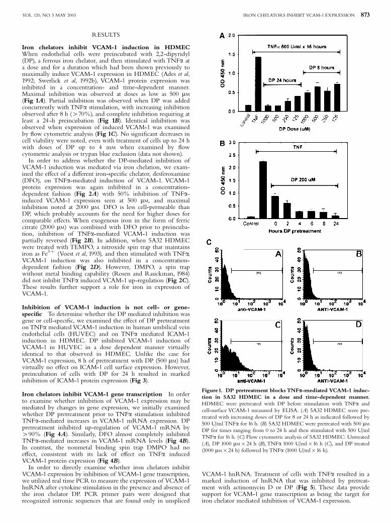
as IkBa, do not require chromatin modi¢cation for transcriptionfactor accessibility (Saccani et al, 2001; Saccani et al, 2002).In order to examine whether DP may a¡ect VCAM-1 expressionvia this target, we examined the TNFa induction of bothgenes with constitutively and immediately accessible (CIA) pro-moters (IkBa) and those with delayed inductions that presum-ably require stimulus-dependent modi¢cations in chromatinstructure to make NF-kB sites accessible (Saccani et al, 2001;Saccani et al, 2002). DP pretreatment blocked induction ofmRNA of two additional NF-kB regulated genes, interleukin-8(Fig 10) and tissue factor (data not shown), in HDMEC.In contrast, DP had no e¡ect on the TNFa mediated up-regulation of IkBa mRNA (Fig 10). These data are consistentwith DP targeting cytokine-mediated e¡ects on chromatinpackaging.
Figure 3. DP mediated inhibition is not cell- or gene speci¢c: (A)HUVEC were pretreated with DP (30^1000 mM) prior to stimulation withTNFa (1000 U/ml�16 h). Cells were assayed for VCAM-1 expression bycell based ELISA. (B) 5A32 HDMECwere pretreated with various concen-trations of DP for 8 or 24 h followed by 500 U/ml TNFa for 16 h. Cell-surface levels of ICAM-1were measured by ELISA. Preincubation of cellswith DP for 24 h prior to cytokine treatment signi¢cantly blocked TNFa-mediated ICAM-1 induction.
Figure 4. DP and DFO but not DMPO inhibits TNFa-mediatedVCAM-1 mRNA up-regulation. (A) 5A32 HDMEC were pretreatedwith 250 or 125 mM DP for 24 h and then stimulated with 200 U/ml TNFafor 24 h.VCAM-1mRNAwas measured by quantitative RT-PCR and dif-ferences represented as fold increase over baseline. (B) Con£uent mono-layers of 5A32 HDMEC were pretreated with 500 mM DP, 500 mM DFO,or 500 mM DMPO for 24 h and then stimulated with 200 U/ml TNFa for24 h. DP and DFO inhibit TNFa-mediated increases inVCAM-1mRNA,but DMPO did not.
Figure 5. DP inhibits TNFa-mediated VCAM-1 expression by inhi-bition of VCAM-1 gene transcription. 5A32 HDMEC were pretreatedwith 500 mM DP for 8 h or 2 mg/ml actinomycin D for 30 min prior tostimulation with 500 U/ml TNFa for 4 h. The fold di¡erences of VCAM-1 hnRNAwere determined by quantitative RT-PCR. Results are represen-tative of three experiments.
IRON CHELATORS INHIBIT VCAM-1 EXPRESSION 875VOL. 120, NO. 5 MAY 2003

DISCUSSION
Iron has been implicated as a key participant in a variety of in-£ammatory disorders and processes. Studies examining the e¡ectof iron chelators on the function of key transcription factors suchas NF-kB and HIF-1a have shown that chelation of iron a¡ectsproteasomal processing, resulting in inhibited degradation (Wang
Figure 6. DP does not a¡ect NF-kB translocation after TNFastimulation in 5A32 HDMEC cells. (A) Untreated cells; or (B) treatedwith 500 U/ml TNFa for 30 min; (C) 500 mM DP for 24 h; and (D) 500U/ml TNFa for 30 min after pretreatment with DP 500 mM for 24 h.Cellular localization of p65 was identi¢ed by immuno£uorescencemicroscopy.
Figure 7. DP does not a¡ect NF-kB binding to theVCAM-1 kB ele-ment afterTNFa stimulation. 5A32 HDMECwere stimulated with 500U/ml TNFa for 2 h with or without pretreatment with DP (500 mM) for 24h.Nuclear extractswere assessed for the bindingof NF-kB by EMSA.TNFatreatment results in appearance of complexes A and B, which appear incells regardless of whether pretreated with DP. In addition, both bands aresupershifted in treated cells by antip50 antibody (band C) or antip65 anti-body (band D) with or without DP pretreatment.
Figure 8. Pretreatment of 5A32 HDMEC cells with IFNc priorto DP pretreatment induces persistent IRF-1 expression but onlypartially restores TNFa-mediated VCAM-1 induction. (A) 5A32HDMECwere pretreated for 4, 8 and 24 hwith 1000 mM DP prior to TNFastimulation. Cell lysates were assayed for the presence of IRF-1 byWesternblotting. Untreated cells (lane 1), DP treated (500 mM� 24 h, lane 2),TNFatreated (500 U/ml� 2 h, lane 3) or 2 h of TNFa treatment preceded byincubations of DP (500 mM) for 4 h (lane 4), 8 h (lane 5), and 24 h (lane 6).(B) IFNg induces IRF-1 expression that persists after DP treatment. Un-treated cells (lane 1), IFNg (1000 U/ml) for 2 h (lane 2), IFNg (1000 U/ml)for 10 h (lane 3), IFNg (1000 U/ml)� 2 h followed by DP (1000 mM) for 8 h(lane 4), and IFNg (1000 U/ml) for 2 h followed by DP (1000 mM) for 24 h(lane 5). Cell lysates were prepared and assessed for the presence of IRF-1by Western blotting. (C) Prior induction of IRF-1 by IFNg does not re-store VCAM-1 induction inhibited by DP pretreatment as measuredby ELISA. Control cells (untreated), IFNg (1000 U/ml � 24 h), DP(500 mM� 24 h), IFN/DP (IFNg 1000 U/ml� 2 h followed by DP500 mM� 24 h), TNFa 500 U/ml�16 h), IFN/TNF (IFNg 1000 U/ml�2 h then TNFa 500 U/ml�16 h), DP/TNF (DP 200 mM� 6 h then TNFa500 U/ml�16 h), and IFN/DP/TNF (IFNg 1000 U/ml� 2 h, then DP500 mM� 6 h, thenTNFa 500 U/ml�16 h). Pretreatment of the cells withIFNg and induction of IRF-1 expression prior to DP pretreatment onlypartially restores TNFa-mediated VCAM-1 induction.
Figure 9. DP does not globally inhibit serine phosphorylation ofp65. 5A32 HDMEC cells were pretreated for 20 h with 500 mM of DPprior to stimulation with 500a U/ml TNFa for 30 min Cell lysates (A)untreated HDMEC (B) TNFa 500 U/ml� 30 min (C) DP 500 mM� 24 h(D) DP 500 mM� 24 h, then TNFa 500 U/ml� 30 min, were assessedfor the presence of phospho-Ser 536 p65 antibody (Cell SignalingTechnology) byWestern blotting. The same blots were then stripped andprobed with antip65 antibody.
876 KOO ETAL THE JOURNAL OF INVESTIGATIVE DERMATOLOGY
and Semenza, 1993; Bowie et al, 1997). Given the dependence ofVCAM-1 expression on proteasomal degradation of the IkBaregulatory subunit of NF-kB, it appeared to be reasonable to pre-dict that ironchelatorsmight inhibitVCAM-1inductionbyTNFa.Consistent with our hypothesis, the iron chelator DP almostcompletely inhibited TNFa-mediated induction of VCAM-1 atthe protein and mRNA level.Similar e¡ects upon TNFa-mediated induction of VCAM-1
were also observed in HDMEC with pretreatment using a di¡er-ent iron speci¢c chelator, DFO. Addition of exogenous iron inthe form of ferric citrate to DFO before stimulation with TNFapartially reversed the chelator e¡ect, supporting again iron bind-ing as the target of action of iron chelators. The electron spin trap5-dimethyl-1-pyrrolone-N-oxide (DMPO), which does not bindto iron, failed to inhibit TNFa mediated induction of VCAM-1.However, when HDMEC were treated with TEMPO, a nitrox-ide spin trap that maintains iron as Fe3þ, and then stimulatedwith TNFa, VCAM-1 induction was inhibited in a dose-depen-dent fashion. These data further support the role of iron inTNFa-mediated VCAM-1 up-regulation.Our data clearly point to VCAM-1 gene transcription as a
target for DP. DP pretreatment completely blockedTNFamedia-ted increases in VCAM-1 steady state mRNA expression, andhnRNA induction. However, the exact mechanism of this e¡ectis not completely de¢ned. Iron is required for activity of an intra-cellular prolyl hydroxylase that modi¢es HIF-1a, facilitating itsinteraction withVon Hippel Lindau protein and proteasomal de-gradation (Ivan et al, 2001). Similarly, DFO has also been shownto inhibit TNFa mediated IkBa degradation. We hypothesizedthat iron chelation may inhibit VCAM-1 gene activation byblocking TNFa-mediated IkBa degradation and NF-kB activa-tion. However, iron chelators had no e¡ect on either NF-kBtranslocation or the ability of translocated complexes to bindto VCAM-1 kB elements, even at doses su⁄cient to blockVCAM-1 protein and mRNA expression. Given the lack of ob-vious e¡ect of DP pretreatment on NF-kB binding to theVCAM-1 kB element, other potential targets for iron chelatorshad to be considered.VCAM-1 expression has also been shown to be sensitive to
antioxidants such as n-acetylcysteine (NAC) and pyrrolidine-dithiocarbamate (PDTC) (Marui et al, 1993). Agents such as
PDTC are also metal chelators, and it has been proposed thattheir potent e¡ects may be mediated via a combination of metalchelation and related antioxidant e¡ects (Saran and Bors, 1990). Itis possible that chelation of iron results in loss of metal dependentredox signaling. However, our data does not support this expla-nation since antioxidant mediated inhibition of VCAM-1 induc-tion has been shown to be mediated via inhibition of NF-kBactivation and translocation, and DP had no e¡ect whatsoeveron NF-kB activation, translocation, or DNA binding (Bowieet al, 1997).Alternatively, iron chelators may inhibit VCAM-1 induction
by blocking the induction of IRF-1 expression. Previous studieshave suggested that although IRF-1 augments the expression ofVCAM-1 transcription, it is not necessary for gene expression(Neish et al, 1995; Lechleitner et al, 1998). Our data are consistentwith these previous observations. Pretreatment of endothelialcells with DP completely blocked TNFa-mediated induction ofIRF-1 expression. However, prior induction of IRF-1 by treat-ment with IFNg only partially restored TNFa-mediatedVCAM-1 induction. Furthermore, DP pretreatment blocks TNFamediated up-regulation of ICAM-1 expression in HDMEC, aprocess that is NF-kB dependent and IRF-1 independent (Gilleet al, 1997; Paxton et al, 1997).It is possible that iron is required for NF-kB complexes to
transactivate theVCAM-1 gene without a¡ecting binding to sitesin the promoter as assessed by EMSA. Previous studies haveshown that anti-in£ammatory agents such as sulfasalazine inhibitthe p65 phosphorylation, and this phosphorylation is critical forp65-mediated gene transactivation (Egan et al, 1999). The targetfor phosphorylation may be one or more of a series of serine re-sidues. Multiple enzymes, which induce phosphorylation of spe-ci¢c serine residues, have been described (Wang and Baldwin,1998; Wang et al, 2000). Our EMSA studies do not demonstrateany global inhibition of NF-kB serine phosphorylation by DP.In addition, iron chelators do not block TNFa-mediated phos-phorylation of Ser536, a residue previously identi¢ed for its rolein p65 function (Sakurai et al, 1999). However, our studies cannotrule out an e¡ect on phosphorylation of other residues such asSer529 (Wang and Baldwin, 1998;Wang et al, 2000). Iron may stillbe critical for the phosphorylation of speci¢c serine residues inp65, and altered patterns of phosphorylation may a¡ect VCAM-1 gene transcription without a¡ecting translocation or DNAbinding.Paradoxically, although DP did not a¡ect the activation of NF-
kB or its ability to bind in vitro to oligonucleotides correspondingto VCAM-1 kB elements, it still almost completely blockedVCAM-1 gene transcription mediated byTNFa. It is possible thatiron blocked VCAM-1 induction in HDMEC via a mechanismcompletely independent of NF-kB. This is quite unlikely givenprevious studies of TNFa mediated VCAM-1 gene expression inthis context showing a strict dependence on NF-kB (Iademarcoet al, 1992; Neish et al, 1995; Gille et al, 1996). An alternativemechanism may be an iron dependent process regulating chro-matin modi¢cations and accessibility. The e¡ects of iron chelatorson TNFa mediated genes in HDMEC are consistent with ane¡ect on DNA packaging and histone modi¢cation.The di¡eren-tial e¡ects onVCAM-1 and IkBa could be explained on the basisof di¡erential requirements for chromatin remodeling as a pre-requisite for transcription (Saccani et al, 2001). Certain NF-kBdriven genes are not immediately accessible and require modi¢-cation of histones in addition to NF-kB translocation for expres-sion. The transcription of these genes tends to be delayed aftercytokine stimulation and NF-kB activation, presumably becausehistone modi¢cation requires additional time. In contrast, otherNF-kB driven genes such as IkBa are immediately accessibleand are rapidly induced after cytokine stimulation.We reasonedthat if iron was important for chromatin accessibility, iron chela-tors should not have an e¡ect on genes such as IkBa that wereconstitutively accessible while iron chelators should inhibitexpression of NF-kB inducible genes with delayed kinetics.Our observations that DP pretreatment had no e¡ect on IkBa
Figure10. DP blocks TNFa mediated increases in IL-8 mRNA butdoes not inhibit increases in IkBa mRNA: 5A32 HDMEC were sti-mulated with TNFa (1000 U/ml� 4 h) with and without pretreatmentwith DP (1000 mM� 24 h). Steady state levels of IL-8 and IkBa mRNAwere assessed by real-time quantitative PCR as described in material andmethods. Levels are expressed as fold-increase over unstimulated, untreatedcontrols.
IRON CHELATORS INHIBIT VCAM-1 EXPRESSION 877VOL. 120, NO. 5 MAY 2003

induction, while consistently blocking other NF-kB induciblegenes with delayed kinetics, support this hypothesis.Previous studies linking chromatin remodeling to iron also
provide additional indirect evidence for the role of iron. Iron isessential for expression of a number of genes critical for cellgrowth and proliferation, and removal of iron via the use of DFOhas e¡ects that are similar to the e¡ects of histone deacetylase(HDAC) (Kim et al, 2001) (Kramer et al, 2002). Treatment of cellswith DFO resulted in cell cycle arrest and down-regulation ofp21WAF and speci¢c cyclin dependent kinases. From these data,one can conclude that expression of p21WAF is iron dependent(Kramer et al, 2002). Similarly, HDAC inhibition resulted in geneexpression comparable to iron loading, with resultant expressionof p21WAF and cell proliferation (Kim et al, 2001). Consistent withour hypothesis is the converse of these observations. Histonedeacetylation has e¡ects that are essentially identical to ironchelation. It is reasonable to propose that they may operatevia a common mechanism and that mechanism likely involveschromatin accessibility.In conclusion, iron chelators inhibited VCAM-1 protein
and mRNA induction in a concentration- and time-dependentmanner via a mechanism that appears to be transcriptionallymediated. The target of iron chelator e¡ects was not NF-kBactivation, but appeared to be mediated through a combinationof inhibition of IRF-1 expression and e¡ects on the ability oftranslocated NF-kB complexes to transactivate the VCAM-1gene, potentially via iron dependent alterations in chromatinremodeling.
This work was supported by Post-doctoral Fellowship Program of Korea Science andEngineering Foundation (KOSEF) in 1998, NIH R01AR39632, Emory Skin Dis-ease Research Center (NIH P30 AR42687).We are grateful for the technical assis-tance of Mrs. Patricia Kowalczyk and Mr. Marvin Newton-West for their technicalassistance.
REFERENCES
Ades EW, Candal FJ, Swerlick RA, George VG, Summers S, Bosse DC, LawleyTJ:HMEC-1: Establishment of an immortalized human microvascular endothe-lial cell line. J Invest Dermatol 99:683^690, 1992
Allen RG, Tresini M: Oxidative stress and gene regulation. Free Radic Biol Med28:463^499, 2000
Baeuerle PA, Henkel T: Function and activation of NF-kappa B in the immune sys-tem. Annu Rev Immunol 12:141^179, 1994
Bowie AG, Moynagh PN, O’Neill LA: Lipid peroxidation is involved in the activa-tion of NF-kappaB by tumor necrosis factor but not interleukin-1 in thehuman endothelial cell line ECV304: Lack of involvement of H2O2 inNF-kappaB activation by either cytokine in both primary and transformedendothelial cells. J Biol Chem 272:25941^25950, 1997
Brenneisen P, Wenk J, Klotz LO, et al: Central role of Ferrous/Ferric iron in theultraviolet B irradiation-mediated signaling pathway leading to increasedinterstitial collagenase (matrix-degrading metalloprotease (MMP)-1) and stro-melysin-1 (MMP-3) mRNA levels in cultured human dermal ¢broblasts. J BiolChem 273:5279^5287, 1998
Das PK, de Boer OJ,Visser A,Verhagen CE, Bos JD, Pals ST: Di¡erential expressionof ICAM-1, E-selectin and VCAM-1 by endothelial cells in psoriasis and con-tact dermatitis. Acta DermVenereol 186 (Suppl):21^22, 1994
Egan LJ, Mays DC, Huntoon CJ, et al: Inhibition of interleukin-1-stimulatedNF-kappaB RelA/p65 phosphorylation by mesalamine is accompanied bydecreased transcriptional activity. J Biol Chem 274:26448^26453, 1999
Elferink CJ, Reiners JJ Jr: Quantitative RT-PCR on CYP1A1 heterogeneous nuclearRNA: A surrogate for the in vitro transcription run-on assay. Biotechniques20:470^477, 1996
Elices MJ, Osborn L, Takada Y, Crouse C, Luhowskyj S, Hemler ME, Lobb RR:VCAM-1 on activated endothelium interacts with the leukocyte integrinVLA-4 at a site distinct from the VLA-4/¢bronectin binding site. Cell60:577^584, 1990
Gille J, Paxton LL, Lawley TJ, Caughman SW, Swerlick RA: Retinoic acidinhibits the regulated expression of vascular cell adhesion molecule-1by cultured dermal microvascular endothelial cells. J Clin Invest 99:492^500,1997
Gille J, Swerlick RA, LawleyTJ, Caughman SW: Di¡erential regulation of vascularcell adhesion molecule-1 gene transcription by tumor necrosis factor alpha and
interleukin-1 alpha in dermal microvascular endothelial cells. Blood 87:211^217,1996
Groves RW, Ross EL, Barker JN, MacDonald DM:Vascular cell adhesion molecule-1:Expression in normal and diseased skin and regulation in vivo by interferongamma. J Am Acad Dermatol 29:67^72, 1993
Iademarco MF, McQuillan JJ, Rosen GD, Dean DC: Characterization of the pro-moter for vascular cell adhesion molecule-1 (VCAM-1). J Biol Chem 267:16323^16329, 1992
Ivan M, et al: HIFalpha targeted for VHL-mediated destruction by proline hydroxy-lation: Implications for O2 sensing. Science 292:464^468, 2001
Kim JS, Lee S, Lee T, Lee YW, Trepel JB: Transcriptional activation of p21 (WAF1/CIP1) by apicidin, a novel histone deacetylase inhibitor. Biochem Biophys ResCommun 281:866^871, 2001
Kramer JL, Baltathakis I, Alcantara OS, Boldt DH: Di¡erentiation of functionaldendritic cells and macrophages from human peripheral blood monocyte pre-cursors is dependent on expression of p21 (WAF1/CIP1) and requires iron. Br JHaematol 117:727^734, 2002
Lechleitner S, Gille J, Johnson DR, Petzelbauer P: Interferon enhances tumor necro-sis factor-induced vascular cell adhesion molecule 1 (CD106) expression in hu-man endothelial cells by an interferon-related factor 1-dependent pathway.J Exp Med 187:2023^2030, 1998
Lin M, Rippe RA, Niemela O, Brittenham G,Tsukamoto H: Role of iron in NF-kappa B activation and cytokine gene expression by rat hepatic macrophages.AmJ Physiol 272:G1355^G1364, 1997
Litch¢eld TM, Smith CH, Atkinson BA, Norris PG, Elliott P, Haskard DO, Lee TH:Eosinophil in¢ltration into human skin is antigen-dependent in the late-phasereaction. Br J Dermatol 134:997^1004, 1996
Lontz W, Sirsjo A, Liu W, Lindberg M, Rollman O, Torma H: Increased mRNAexpression of manganese superoxide dismutase in psoriasis skin lesions and incultured human keratinocytes exposed to IL-1 beta and TNF-alpha. Free RadicBiol Med 18:349^355, 1995
Marui N, O¡ermann MK, Swerlick R, et al: Vascular cell adhesion molecule-1(VCAM-1) gene transcription and expression are regulated through an antiox-idant-sensitive mechanism in human vascular endothelial cells. J Clin Invest92:1866^74, 1993
McCord JM: Iron, free radicals, and oxidative injury. Semin Hematol 35:5^12,1998
Neish AS, Read MA,Thanos D, Pine R, Maniatis T, Collins T: Endothelial interfer-on regulatory factor 1 cooperates with NF-kappa B as a transcriptional activa-tor of vascular cell adhesion molecule 1. Mol Cell Biol 15:2558^2569, 1995
Norris P, Poston RN,Thomas DS,Thornhill M, Hawk J, Haskard DO: The expres-sion of endothelial leukocyte adhesion molecule-1 (ELAM-1), intercellularadhesion molecule-1 (ICAM-1), and vascular cell adhesion molecule-1 (VCAM-1)in experimental cutaneous in£ammation: A comparison of ultraviolet Berythema and delayed hypersensitivity. J Invest Dermatol 96:763^770, 1991
Norton J, Sloane JP, al-Sa¡ar N, Haskard DO: Vessel associated adhesion moleculesin normal skin and acute graft-versus-host disease. J Clin Pathol 44:586^591, 1991
Onuma S: Immunohistochemical studies of in¢ltrating cells in early and chroniclesions of psoriasis. J Dermatol 21:223^232, 1994
Paxton LL, Li LJ, SecorV, Du¡ JL, Naik SM, Shibagaki N, Caughman SW: Flankingsequences for the human intercellular adhesion molecule-1 NF-kappaBresponse element are necessary for tumor necrosis factor alpha-induced geneexpression. J Biol Chem 272:15928^15935, 1997
Pourzand C, Watkin RD, Brown JE, Tyrrell RM: Ultraviolet A radiation inducesimmediate release of iron in human primary skin ¢broblasts: The role of ferri-tin. Proc Natl Acad Sci USA 96:6751^6756, 1999
Rosen GM, Rauckman EJ: Spin trapping of superoxide and hydroxyl radicals. MethEnzymol 105:198^209, 1984
Saccani S, Pantano S, Natoli G: p38-Dependent marking of in£ammatory genes forincreased NF-kappa B recruitment. Nat Immunol 3:69^75, 2002
Saccani S, Pantano S, Natoli G: Two waves of nuclear factor kappa B: Recruitmentto target promoters. J Exp Med 193:1351^1359, 2001
Sakurai H, Chiba H, Miyoshi H, Sugita T,Toriumi W: IkappaB kinases phosphory-late NF-kappaB p65 subunit on serine 536 in the transactivation domain. J BiolChem 274:30353^30356, 1999
Saran M, Bors W: Radical reactions in vivo: An overview. Radiat Environ Biophys29:249^262, 1990
Schreck R, Rieber P, Baeuerle PA: Reactive oxygen intermediates as apparentlywidely used messengers in the activation of the NF-kappa B transcription fac-tor and HIV-1. Embo J 10:2247^2258, 1991
Sharkey P, Eedy DJ, Burrows D, McCaigue MD, Bell AL: A possible role for super-oxide production in the pathogenesis of contact dermatitis. Acta DermVenereol71:156^159, 1991
Silber A, Newman W, Reimann KA, Hendricks E, Walsh D, Ringler DJ: Kineticexpression of endothelial adhesion molecules and relationship to leukocyte re-cruitment in two cutaneous models of in£ammation. Lab Invest 70:163^175,1994
Swerlick RA, Lee KH, Li LJ, Sepp NT, Caughman SW, Lawley TJ: Regulation ofvascular cell adhesion molecule 1 on human dermal microvascular endothelialcells. J Immunol 149:698^705, 1992b
Swerlick RA, Lee KH,WickTM, LawleyTJ: Human dermal microvascular endothe-lial but not human umbilical vein endothelial cells express CD36 in vivo andin vitro. J Immunol 148:78^83, 1992a
878 KOO ETAL THE JOURNAL OF INVESTIGATIVE DERMATOLOGY

Voest EE, van Faassen E, Marx JJ: An electron paramagnetic resonance study of theantioxidant properties of the nitroxide free radical TEMPO. Free Radic Biol Med15:589^595, 1993
Wakita H, Sakamoto T, Tokura Y, Takigawa M: E-selectin and vascular celladhesion molecule-1 as critical adhesion molecules for in¢ltration ofT lymphocytes and eosinophils in atopic dermatitis. J Cutan Pathol 21:33^39,1994
Wang D, Baldwin AS Jr: Activation of nuclear factor-kappaB-dependent transcrip-tion by tumor necrosis factor-alpha is mediated through phosphorylation ofRelA/p65 on serine 529. J Biol Chem 273:29411^29416, 1998
Wang GL, Semenza GL: Desferrioxamine induces erythropoietin gene expressionand hypoxia-inducible factor 1 DNA-binding activity: Implications for modelsof hypoxia signal transduction. Blood 82:3610^3615, 1993
Wang D, Westerheide SD, Hanson JL, Baldwin AS Jr: Tumor necrosis factoralpha-induced phosphorylation of RelA/p65 on Ser529 is controlled by caseinkinase II. J Biol Chem 275:32592^32597, 2000
Woetmann A, Nielsen M, Christensen ST, et al: Inhibition of protein phosphatase2A induces serine/threonine phosphorylation, subcellular redistribution,and functional inhibition of STAT3. Proc Natl Acad Sci USA 96:10620^10625,1999
IRON CHELATORS INHIBIT VCAM-1 EXPRESSION 879VOL. 120, NO. 5 MAY 2003