Embed Size (px)
Citation preview
a. Biomaterials, Biomechanics and Tissue Engineering Group, Department of Materials Science and Metallurgical Engineering, Technical University of Catalonia, Av. Diagonal 647, Barcelona 08028, Spain.
b. Centre for Research in Nanoengineering, Technical University of Catalonia, Pascual i Vila 15, Barcelona 08028, Spain.
† Electronic Supplementary Information (ESI) available: characterisation of co- doped magnesium/carbonate hydroxyapatite nanoparticles and videos of tomographies showing NP internalization in MG63 by soft X-ray imaging . ‡
These authors contributed equally to this work. *Corresponding author. Email: [email protected]
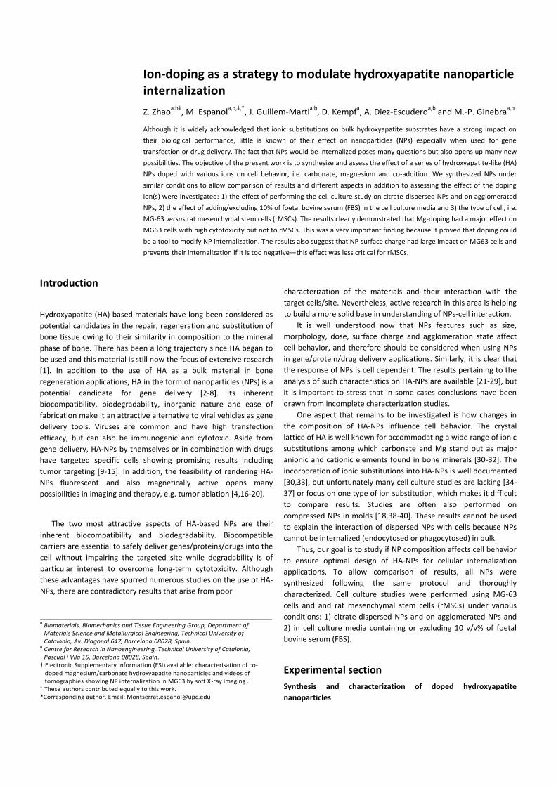
Ion-doping as a strategy to modulate hydroxyapatite nanoparticle internalization
Z. Zhaoa,b‡
, M. Espanola,b,‡,*
, J. Guillem-Martia,b
, D. Kempfa, A. Diez-Escudero
a,b and M.-P. Ginebra
a,b
Although it is widely acknowledged that ionic substitutions on bulk hydroxyapatite substrates have a strong impact on
their biological performance, little is known of their effect on nanoparticles (NPs) especially when used for gene
transfection or drug delivery. The fact that NPs would be internalized poses many questions but also opens up many new
possibilities. The objective of the present work is to synthesize and assess the effect of a series of hydroxyapatite-like (HA)
NPs doped with various ions on cell behavior, i.e. carbonate, magnesium and co-addition. We synthesized NPs under
similar conditions to allow comparison of results and different aspects in addition to assessing the effect of the doping
ion(s) were investigated: 1) the effect of performing the cell culture study on citrate-dispersed NPs and on agglomerated
NPs, 2) the effect of adding/excluding 10% of foetal bovine serum (FBS) in the cell culture media and 3) the type of cell, i.e.
MG-63 versus rat mesenchymal stem cells (rMSCs). The results clearly demonstrated that Mg-doping had a major effect on
MG63 cells with high cytotoxicity but not to rMSCs. This was a very important finding because it proved that doping could
be a tool to modify NP internalization. The results also suggest that NP surface charge had large impact on MG63 cells and
prevents their internalization if it is too negative—this effect was less critical for rMSCs.
Introduction
Hydroxyapatite (HA) based materials have long been considered as
potential candidates in the repair, regeneration and substitution of
bone tissue owing to their similarity in composition to the mineral
phase of bone. There has been a long trajectory since HA began to
be used and this material is still now the focus of extensive research
[1]. In addition to the use of HA as a bulk material in bone
regeneration applications, HA in the form of nanoparticles (NPs) is a
potential candidate for gene delivery [2-8]. Its inherent
biocompatibility, biodegradability, inorganic nature and ease of
fabrication make it an attractive alternative to viral vehicles as gene
delivery tools. Viruses are common and have high transfection
efficacy, but can also be immunogenic and cytotoxic. Aside from
gene delivery, HA-NPs by themselves or in combination with drugs
have targeted specific cells showing promising results including
tumor targeting [9-15]. In addition, the feasibility of rendering HA-
NPs fluorescent and also magnetically active opens many
possibilities in imaging and therapy, e.g. tumor ablation [4,16-20].
The two most attractive aspects of HA-based NPs are their
inherent biocompatibility and biodegradability. Biocompatible
carriers are essential to safely deliver genes/proteins/drugs into the
cell without impairing the targeted site while degradability is of
particular interest to overcome long-term cytotoxicity. Although
these advantages have spurred numerous studies on the use of HA-
NPs, there are contradictory results that arise from poor
characterization of the materials and their interaction with the
target cells/site. Nevertheless, active research in this area is helping
to build a more solid base in understanding of NPs-cell interaction.
It is well understood now that NPs features such as size,
morphology, dose, surface charge and agglomeration state affect
cell behavior, and therefore should be considered when using NPs
in gene/protein/drug delivery applications. Similarly, it is clear that
the response of NPs is cell dependent. The results pertaining to the
analysis of such characteristics on HA-NPs are available [21-29], but
it is important to stress that in some cases conclusions have been
drawn from incomplete characterization studies.
One aspect that remains to be investigated is how changes in
the composition of HA-NPs influence cell behavior. The crystal
lattice of HA is well known for accommodating a wide range of ionic
substitutions among which carbonate and Mg stand out as major
anionic and cationic elements found in bone minerals [30-32]. The
incorporation of ionic substitutions into HA-NPs is well documented
[30,33], but unfortunately many cell culture studies are lacking [34-
37] or focus on one type of ion substitution, which makes it difficult
to compare results. Studies are often also performed on
compressed NPs in molds [18,38-40]. These results cannot be used
to explain the interaction of dispersed NPs with cells because NPs
cannot be internalized (endocytosed or phagocytosed) in bulk.
Thus, our goal is to study if NP composition affects cell behavior
to ensure optimal design of HA-NPs for cellular internalization
applications. To allow comparison of results, all NPs were
synthesized following the same protocol and thoroughly
characterized. Cell culture studies were performed using MG-63
cells and and rat mesenchymal stem cells (rMSCs) under various
conditions: 1) citrate-dispersed NPs and on agglomerated NPs and
2) in cell culture media containing or excluding 10 v/v% of foetal
bovine serum (FBS).
Experimental section
Synthesis and characterization of doped hydroxyapatite
nanoparticles
All HA-NPs were synthesized by neutralization of Ca(OH)2 with
H3PO4 (Eq. 1) at 40 oC in air. The temperature was controlled by
means of a thermal bath (Huber Kältemaschinenbau GmbH,
Germany), and the reaction was performed on thermojacketed
vessels connected to the bath. The pH was continuously monitored
throughout the reaction.
10 Ca(OH)2 + 6 H3PO4 Ca10(PO4)6(OH)2 + 18 H2O Eq. (1)
The synthesis of the non-doped NPs was carried out as follows:
100 mL of 0.334 mol L-1
Ca(OH)2 (Fluka, 96 wt% pure) was first
prepared and then a 0.2 M H3PO4 (Panreac, 85 wt% pure) solution
was added dropwise into the system at the constant rate of 1
mL/min under constant stirring. The reaction was stopped when the
pH reached 8. Next, the suspension was left stirring for 20-30 min
and was then transferred into a glass bottle where it was left to
mature overnight at room temperature. The next day, the
suspension was rinsed with MilliQ water to constant conductivity
following cycles of 5 min centrifugation at 800 g (Beckman Allegra
21 Benchtop Centrifuge)/re-suspension. Afterwards, the product
was frozen at -80 oC and lyophilized (Telstar Cryodos). Once
lyophilized, the powder was kept in a desiccator.
To prepare carbonate-doped HA-NPs, different amounts of
sodium hydrogen carbonate (NaHCO3, Sigma-Aldrich, ReagentPlus®,
≥99.5 wt% pure) were added to the Ca(OH)2 suspension prior to
addition of the phosphoric acid. The amount of CO32-
added was
calculated so as to have 5, 10 and 20 wt% of carbonate. Similarly,
the synthesis of the Mg-doped NPs was achieved by mixing the
calcium hydroxide suspension with MgCl2·6H2O (Panreac, 99 wt%
pure) to get 5, 10 and 20 wt% of Mg2+
. Co-substitution of carbonate
and Mg was accomplished with a slight modification in the protocol
[41]. The Ca(OH)2 powder was suspended in 80 mL (instead of 100
mL) and the desired amount of Mg salt was dissolved into it. The
carbonate salt was dissolved separately in 20 mL of water and
added dropwise (1 mL min-1
) into the basic suspension prior to the
addition of the phosphoric acid. For this particular reaction, the
addition of acid was stopped at pH 7. All other parameters were
held constant. Maturation, rinsing and drying of the NPs was
performed as stated above. The different NPs were designated as
follows:
HA: non-doped NPs,
5C, 10C, 20C: carbonate-doped HA-NPs (5, 10 and 20
wt% respectively)
5M, 10M: magnesium-doped HA-NPs (5 and 10 wt%
respectively)
4M/7C: 4 wt% magnesium and 7 wt% carbonate co-
doped HA-NPs.
Characterization of the NPs was performed with multiple
techniques. The phase composition was determined with X-ray
powder diffraction (XRD) using a D8 Advance Diffractometer
(Bruker, Karlsruhe, Germany) with Cu Kα radiation at 40 kV and 40
mA. The XRD spectra were recorded from 10–80 o with a step size
of 0.02 and a counting time of 1 s. The crystallite size of
hydroxyapatite nanoparticles was calculated through the Scherrer
formula as follows:
𝑿𝒉𝒌𝒍 = 𝑲 · 𝝀 𝜷𝟏/𝟐⁄ · 𝒄𝒐𝒔𝜽 Eq. (2)
where Xhkl is the crystallite size (nm), λ is the wavelength of
monochromatic X-ray beam (nm) (0.15418 nm for CuKα radiation),
β1/2 is the full width at half maximum for the diffraction peak under
consideration (rad), θ is the diffraction angle (in degrees), and K is a
constant varying with crystal habit and here set to 0.9.
Fourier transform infrared spectroscopy (FTIR) in the ATR mode
(Attenuated Total Reflectance mode, Nicolet 6700 spectrometer,
Thermo Scientific) was used to check the typical functional groups
present in apatite and, in particular, the presence of carbonate
bands in the powders. All spectra were obtained by averaging 32
scans collected from 500–4000 cm-1
with a spectral resolution of 4
cm-1
.
Elemental carbon determination analysis (Thermo EA 1108
CHNS-O Thermo Scientific, Milan, Italy) was performed on 10 mg of
sample previously dehydrated at 120 oC to measure the amount of
carbonate in the powders. The sample was pyrolyzed and
combusted using vanadium oxide (V2O5) as an oxidant at 1000 o
C in
an oxygen atmosphere and the resulting gaseous products were
chromatographically separated and quantified to yield the
concentration of carbon.
Quantitative inductively coupled plasma–optical emission
spectrometry analysis (ICP-OES, Perkin Elmer Optima 3200RL) was
used to determine the overall content of Ca, P, Na and Mg. Samples
were prepared by dissolving 100 mg of powder in 5 mL of 10 wt%
HNO3 (Panreac, 69 wt% pure) that was then 10 or 20-fold diluted.
The specific surface area (SSA) of the NPs was determined from
the nitrogen adsorption data in the relative pressure range (P/P0)
from 0.05 to 0.35 with the Brunauer-Emmett-Teller (BET) method
with a ASAP2020 physisorption analyzer (Micromeritics, Norcross,
GA, USA). The sample was outgassed at 120 oC before analysis.
The morphology of the NPs was assessed by transmission
electron microscopy (TEM, JEOL 1010). Samples for TEM
examination were prepared by soaking a 300 mesh carbon-coated
copper grid in the solution of interest, blotting to remove excess
liquid and air-dried.
The apparent solubility of the NPs was evaluated by monitoring
the conductivity changes (Crison MM41) of a 0.1 mol L-1
acetate
buffer solution (25 oC, pH=5.5) containing 1 mg mL
-1 of NPs with
time.
Nanoparticles dispersion
NPs dispersion was achieved by mixing 0.1 g of the NPs with 10 mL
of 1 wt% sodium citrate solution (C6H5Na3O7 · x H2O, Sigma-Aldrich,
ReagentPlus®, purity ≥99%). To improve dispersion, the suspension
was sonicated with a high frequency ultrasound probe sonicator
(Branson Digital, Model 250W) to separate the agglomerates and
facilitate citrate adsorption. The following settings/conditions were
applied: 3 mm diameter tip, 40% amplitude (~50 Watts) and 2
minutes of sonication (with cycles of 15 sec sonication followed by
10 sec pause to prevent excessive heating) in an ice bath.
Suspensions without citrate were also prepared following the same
protocol but by replacing the 10 mL of 1 wt% sodium citrate
solution with 10 mL of MilliQ water (18.2 MΩ cm-1
).
The amount of citrate adsorbed on the NPs was quantified with
HPLC (high performance liquid chromatography, Alliance 2695,
Waters) using a resin-based column adequate for the analysis of
organic acids. Prior to measurement, the NP suspensions were
filtered with a 0.22 µm filter syringe to remove the NPs, and the NP-
free solution free was measured. A calibration curve was prepared
using known concentrations of trisodium citrate. The samples were
injected into the column (Aminex HPX-87H, BioRad) using a mobile
phase consisting of 10 mM sulphuric acid at a flow rate of 0.8
mL/min at 60 oC. An ultraviolet-visible detector (Jasco UV-1970) at
210 nm was the detector to monitor the the C=O double bond in
citrate. The amount adsorbed was determined by difference
between the initial citrate solution and the citrate remaining after
NPs addition.
The NPs surface charge was determined by measuring the zeta
potential of the NPs in milliQ water at a concentration of 250 μg mL-
1 of NPs (Zetasizer Nano-ZS from Malvern Instruments Ltd., UK).
Prior to measurement, the stock suspensions of 1 wt% NPs
dispersed in either 1 wt% citrate or water were first diluted in cell
culture media (DMEM, Dulbecco’s Modified Eagle medium)
containing or excluding 10 v/v% FBS (Foetal Bovine serum) to
achieve a final concentration of 1000 μg mL-1
of NPs. The mixture
was then allowed to interact for a couple of hours, and the NPs
were next centrifuged, rinsed with milliQ water and resuspended
with milliQ water to make the final concentration 250 μg mL-1
prior
to measurement.
To note, all cell culture tests performed on citrate dispersed NPs
were performed without removing the unbound citrate from the
suspension to prevent any desorption from the NPs upon citrate
depletion.
Cell culture studies Human osteoblast-like MG-63 cell line and rat mesenchymal stem
cells were cultured in Dulbecco’s Modified Eagle medium (DMEM)
or advanced DMEM, respectively, containing 10 v/v% foetal bovine
serum (FBS), penicillin/streptomycin (50 U mL-1
and 50 µg mL-1
,
respectively), 2 mmol L-1
L-glutamine and 20 mmol L-1
HEPES buffer
at 37 oC in a humidified atmosphere at 5% CO2. When cells attained
confluence, they were detached using 0.25% Trypsin and seeded
onto standard polystyrene tissue culture plates (96 wells) at a cell
density of 1 × 10⁴ cells well
-1. The seeded cells were incubated
overnight to allow cell adhesion prior to adding the NPs.
Exposure of cells to doped and non-doped NPs Cells were exposed to all NPs (dispersed in citrate or in water) in
two different scenarios: in cell culture media supplemented with 10
v/v% FBS and without FBS. The appropriate volume of NPs from the
stock (1 wt% NPs in either 1 wt% of sodium citrate or water) was
added to the corresponding cell culture media to make a final
concentration of 100 μg mL-1
. Before adding the suspensions to the
cells, the mixture was vortexed to homogenize it. The incubation
period was 24 h. For selected compositions (i.e., HA and 10M) the
same experiment was performed using transwells by placing the
NPs on the transwells to prevent direct contact of the NPs with the
cells but allowing any ionic exchange through the transwell
membrane (6.5 mm Transwell with 0.4 µm pore polyester
membrane insert for 24 well plates from Sigma). The number of
cells seeded on the 24 well plates was 2.5 × 10⁴ cells well
-1, and the
concentration of NPs was 100 μg mL-1
.
To study the effect of NP dose on cytotoxicity, the amount of 10
M NPs in cell culture media was increased with the following
concentrations: 100, 250, 500, and 1000 μg mL-1
. Non-doped HA
was also included as a control.
Cytotoxicity test For cytotoxicity evaluation of various NPs, the cell culture medium
was removed after 24 h of incubation time. The viable cells
attached on the surface of polystyrene tissue culture plates were
lysed with 100 μL of mammalian protein extraction reagent (M-PER,
Thermo Scientific Inc., USA). Upon lysis, the cells released lactate
dehydrogenase (LDH) that was measured using a commercially
available kit (Cytotoxicity Detection KitPLUS, Roche, USA) following
the manufacturer’s instructions. The absorbance at 492 nm was
quantified on a micro spectrophotometer (PowerWave XS, Bio-Tek
Instruments, USA), and the percentage of viability was calculated by
the following equation:
𝐯𝐢𝐚𝐛𝐢𝐥𝐢𝐭𝐲% =𝐞𝐱𝐩. 𝒗𝒂𝒍𝒖𝒆 − 𝒏𝒆𝒈𝒂𝒕𝒊𝒗𝒆 𝒄𝒐𝒏𝒕𝒓𝒐𝒍
𝒑𝒐𝒔𝒊𝒕𝒊𝒗𝒆 𝒄𝒐𝒏𝒕𝒓𝒐𝒍 − 𝒏𝒆𝒈𝒂𝒕𝒊𝒗𝒆 𝒄𝒐𝒏𝒕𝒓𝒐𝒍× 𝟏𝟎𝟎 𝐄𝐪. (𝟑)
Here, the ‘positive control’ was the absorbance value of cells
incubated in cell culture medium under the same conditions of the
experimental value but without NPs and the ‘negative control’
corresponded to the absorbance of the well without NPs and
without cells. The studies done in triplicate were expressed as mean
± standard error of the mean. The Kruskal-Wallis non-parametric
test and the Mann–Whitney test with Bonferroni correction were
used to determine statistical significance (p-value < 0.05) between
the means of the different groups.
Table 1 Compilation of various characteristics of the NPs.
CO32-
Mg2+
Na+ Ca/P (Ca+Mg)/P SSA Crystallite size (002)
wt % wt % wt % Atomic atomic m2/g Nm
HA 2.46 ± 0.03 0.27 ± 0.01 0 1.76 ± 0.03 1.78 ± 0.03 93.55 ± 0.32 28.2
5C 6.63 ± 0.42 0.26 ± 0.01 0.23 ± 0.07 1.85 ± 0.04 1.89 ± 0.06 87.04 ± 0.34 26.0
10C 10.1 ± 0.20 0.26 ± 0.01 0.35 ± 0.07 1.92 ± 0.03 1.94 ± 0.02 74.61 ± 0.32 25.5
20C 11.4 ± 0.40 0.26 ± 0.01 0.54 2.07 ± 0.03 2.10 ± 0.03 48.42 ± 0.17 23.4
5M n.d. 1.23 ± 0.01 0 1.68 ± 0.06 1.77 ± 0.04 103.32 ± 0.43 27.4
10M n.d. 2.32 ± 0.25 0 1.57 ± 0.02 1.77 ± 0.01 85.19 ± 0.41 24.8
4M7C 7.8 1.51 ± 0.01 0.05 ± 0.01 1.77 ± 0.03 1.91 ± 0.03 59.83 ± 0.29 25.1
Measures performed on two independent batches for every NP-type except for the SSA where only one batch was measured.
Cellular uptake of NPs
TEM and soft X-ray imaging were used to visualize MG63
cellular uptake of HA and 10 M NPs dispersed in milliQ water.
For TEM imaging cells were exposed for 24 h to the NPs in
cell culture media excluding FBS. The cultured cells were then
fixed with 2.5 wt% glutaraldehyde in 0.1 mol L-1
phosphate buffer
for 1 h 30 min at room temperature and both the media and
the film formed at the bottom of the well were detached and
centrifuged to form a pellet. The pellet was then rinsed prior
to adding 1 wt% osmium tetroxide and dehydrated in an
ascending series of acetone solution up to 100 % and
infiltrated with EPON12 resin. Upon resin polymerization (48 h,
60 oC) blocks were sectioned using an ultramicrotome
(Ultracut UCT, Leica Microsystems GmbH, Vienna, Austria).
The sections were stained with 2 wt% uranyl acetate and
imaged with an optical microscope (Leica DM2000 LED,
Leica Microsystems GmbH, Vienna, Austria) and transmission
electron microscopy (Tecnai Spirit FEI, Eindhoven, The
Netherlands).
For soft X-ray imaging, Quantifoil R2/2 Au G200F1 finder
grids were pre-coated with fibronectin (Sigma, F2006) prior to
MG63 seeding. After overnight cell attachment, the cell
culture media was replaced by fresh media containing NPs but
without FBS. NPs were allowed to interact with cells for 2-3 h.
Afterwards the grids were gently rinsed with PBS and
immediately plunged freeze to vitrify cells and stored in liquid
nitrogen tanks till observation. Cryo transmission soft X-ray
imaging was made at the MISTRAL beamline in the
Synchrotron facilities from Alba (Cerdanyola, Spain).
Tomographic series were collected at 520 eV irradiating the
cell for a few seconds (2-10 sec) per section at a spatial
resolution down to 30 nm.
Results and discussion
Characterization of carbonate-doped nanoparticles
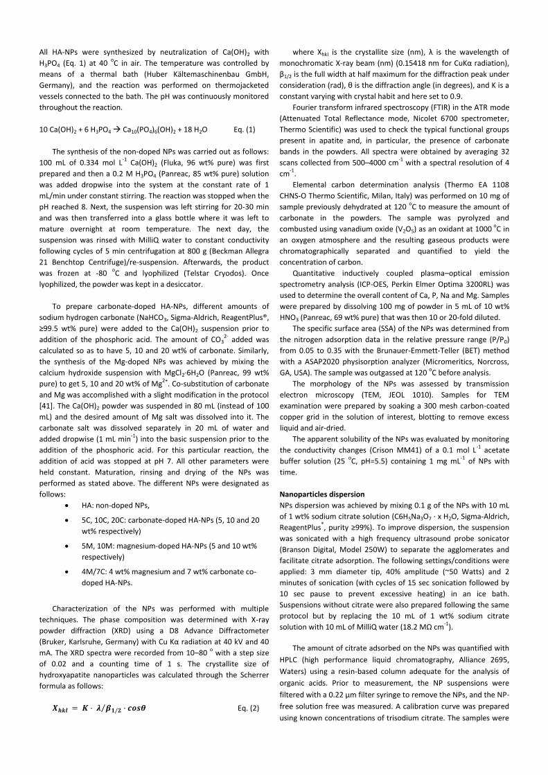
Figure 1 shows the XRD and FTIR patterns of the NPs
synthesized with various concentrations of carbonate, i.e. 0, 5,
10 and 20 wt%. The XRD results (Figure 1A) showed that all
synthesized NPs were phase pure with no peaks other than
those assigned to HA (ICDD No. 9-432). Moreover, the broad
peaks in all samples showed the poor crystallinity of the NPs.
This was also reflected in the small crystallite sizes (size of the
coherently scattering domains) calculated for all NPs
regardless of composition while considering the diffraction
peak corresponding to the (002) reflection (Table 1). A detailed
examination of the XRD data for the different NPs revealed
additional characteristics. Carbonation caused a marked
decrease in the intensity of the (00x) reflections with a shift
towards lower 2θ angles. The first of these two features was
indicative of a change in the crystal morphology. The later
suggested incorporation of carbonate into the apatite lattice
replacing the phosphate groups (B-type substitution) [42]. The
FTIR results (Figure 1B) further confirmed the B-type
substitution, which is typical of hydroxyapatites precipitated at
low temperature via the presence of specific bands—the
asymmetric stretch vibration, ν3, at 1430 and 1450 cm-1
and
the out-of-plane bend vibration, ν2, close to 870 cm-1
[43]. An
additional feature observed from both XRD and FTIR was the
loss in the resolution of the peaks and bands that were
explained by the crystal distortion provoked by carbonate
incorporation.
Figure 1 (a) X-Ray diffraction data for the different NPs: non-doped (HA) and NPs doped with various carbonate contents, (b) detail of the peak shift corresponding to the (002) reflection with carbonate content and, (c) FTIR spectra of the various compositions.
Despite the fact that during synthesis specific amounts of
carbonate salt were added, the exact amount incorporated in
the NPs differed from the nominal value (Table 1). The 2.46
wt% of carbonate observed in NPs prepared without adding
any carbonate salt is caused by the traces of carbonate present
in the Ca(OH)2 powder and also by the dissolution of carbon
dioxide in the solution reaction during precipitation.
There is a limited increase in carbonate uptake by the NPs
when adding 20 wt% of NaHCO3 versus 10 wt%. For the 10
wt% reaction, 10 wt% is incorporated but for the 20 wt% only
11 wt% of carbonate is incorporated. This shows that
carbonate saturation occurred in the apatite. The ICP-MS
results (Table 1) showed that the incorporation of carbonate
was accompanied by Na incorporation. This co-substitution is
very common as it helps balancing the charges in the exchange
of PO43-
by CO32-
[37].
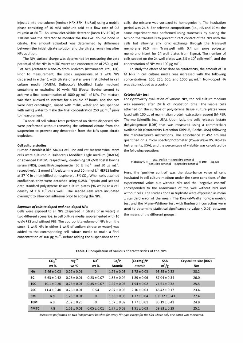
The incorporation of carbonate in the apatite lattice had a
marked effect on the morphology of the NPs (Figure 2a). The
NPs changed from a needle-like shape to a more isotropic
shape with increasing carbonate content similar to other
reports [44]. Such a change in shape was already hinted at the
XRD data by the decrease in intensity of the (00x) reflections
for the higher carbonate-containing NPs. Indeed, it is known
that needle-like HA crystals exhibit preferential growth along
the c axis, which translates into a selective increase in the
(00x) diffraction peaks. Thus, it is not surprising to find that
more isotropic crystals should result in an “intensity” decrease
because such peaks are observed with an increase in
carbonation (Figure 1). Not only does the shape become more
isotropic but also the SSA decreases with carbonation (Table
1). This decrease in SSA can be explained from two different
perspectives. On one hand, the morphological changes reveal
a clear increase in NP width for a similar length. This is
especially true for 20C NPs when compared to non-doped HA
NPs. This thickening of the NPs is one of the arguments to
explain the reduction in their SSA. However, it has also been
reported that freeze-drying of NPs can induce aggregation
[45]. In this regards, the decrease in crystallinity with
carbonation could particularly favour crystal fusion during the
drying process and thus reduce their SSA. The incorporation of
carbonate in the structure of HA was further proved from the
increase in their solubility as indicated by the increase in
conductivity (Figure 2b).
Figure 2 (a) TEM micrographs of the non-doped (HA) and
carbonate-doped NPs and (b) apparent solubility curves
determined through conductivity measurements for the
various NPs.
Characterization of magnesium-doped nanoparticles
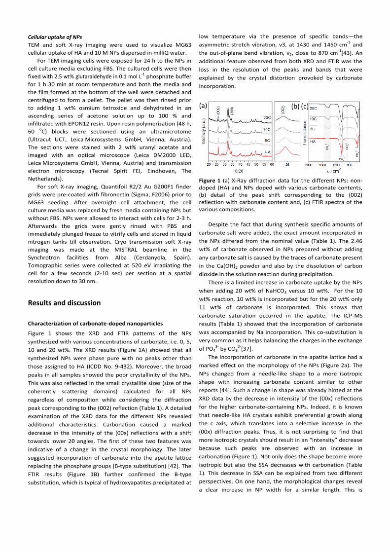
XRD and FTIR results pertaining to the synthesis of Mg-doped
NPs are summarized in Figure 3. Similar to what was achieved
for the carbonate-doped NPs, Mg containing NPs are also
phase-pure because no peaks of foreign phases were detected
by XRD. The addition of Mg salt during synthesis led to slightly
broader peaks in the XRD pattern, which accounted for a
decrease in the crystallinity of the NPs with increasing Mg
content. The slight shift of the (002) diffraction peak (Fig. 3b)
for Mg-substituted samples is due to contraction of the cell
lattice parameters of HA caused by the smaller ionic radius of
the Mg cation compared to calcium [46]. XRD thus proves that
at least some of the Mg cations have indeed been
incorporated into the HAP structure. Similarly to what was
observed for the carbonate-doped NPs, not all Mg that was
added in the synthesis reaction became incorporated into the
crystal structure. Substitution of Mg for Ca in the structure of
HA occurs over a limited composition range (up to about 10
at.% [30]).
Figure 3 (a) X-Ray diffraction data for the different NPs: non-doped (HA) and NPs doped with various Mg contents. (b) Detail of the peak shift corresponding to the (002) reflection with Mg content and, (c) FTIR spectra of the various compositions.
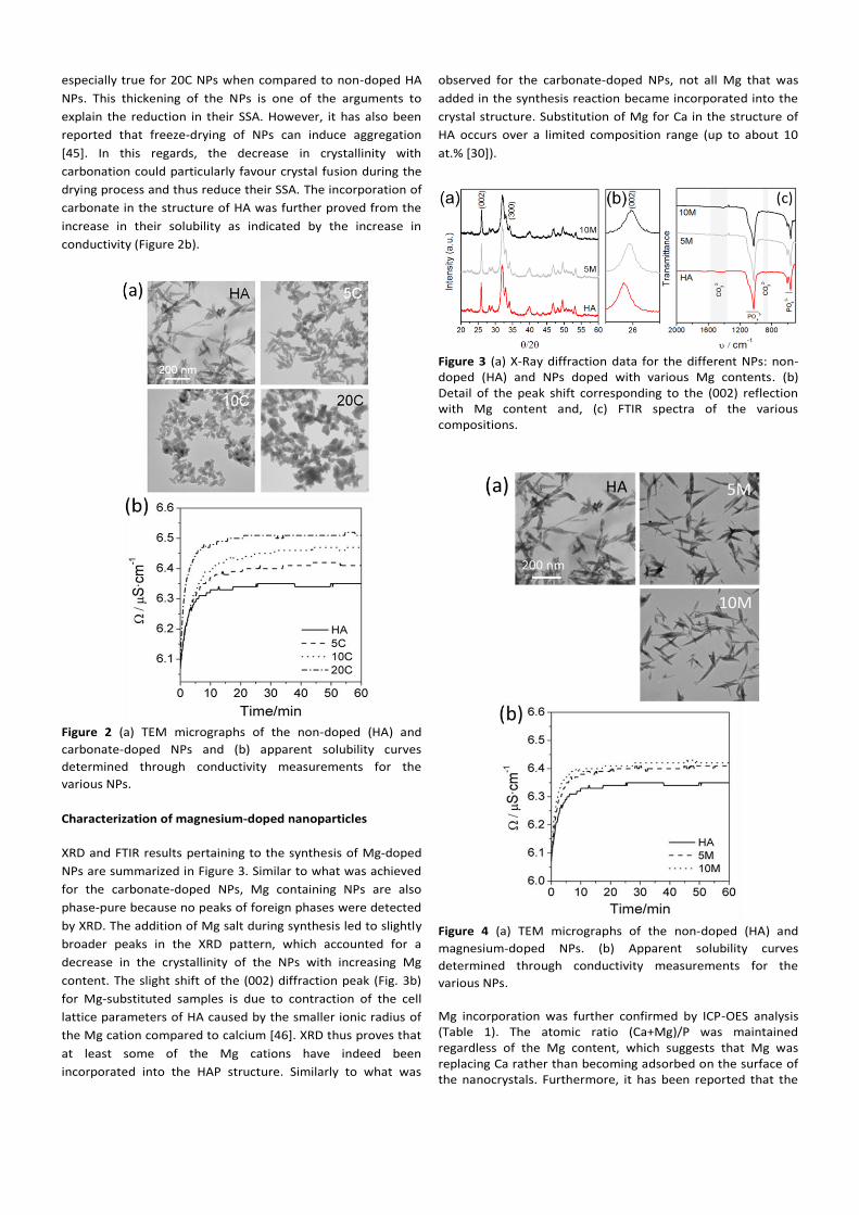
Figure 4 (a) TEM micrographs of the non-doped (HA) and
magnesium-doped NPs. (b) Apparent solubility curves
determined through conductivity measurements for the
various NPs.
Mg incorporation was further confirmed by ICP-OES analysis (Table 1). The atomic ratio (Ca+Mg)/P was maintained regardless of the Mg content, which suggests that Mg was replacing Ca rather than becoming adsorbed on the surface of the nanocrystals. Furthermore, it has been reported that the
incorporation of low Mg contents in the HA structure preserves the (Ca+Mg)/P stoichiometry [46]. Mg incorporation slightly influenced the FTIR spectra—there was a loss in the resolution of the bands (Figure 3c). Carbonate was also present in the Mg-doped NPs as can be detected from the FTIR analyses. The level of carbonate was similar to that found in the “non-doped” HA. As explained earlier, carbonation was due to the presence of carbonate in the Ca(OH)2 reagent and also from atmospheric carbon dioxide. Despite the incorporation of Mg inside the crystal lattice, the morphology of the Mg-doped NPs was not altered as shown by the identical size and needle-like shape to the non-doped ones (Figure 4). This was probably due to the low content of Mg incorporated. The solubility of the Mg-doped NPs slightly increased versus non-doped HA NPs—there was no significant differences between 5 M and 10 M (Table 1). This increase in solubility is consistent with the distortion that Mg causes when enters in the crystal lattice of HA (owing to the mismatch between ion sizes) and the decrease in crystallinity. Similar SSA values were also obtained for the Mg-doped and non-doped HA (Table 1).
Characterization of magnesium/ carbonate-doped
nanoparticles
Co-doping with both Mg and carbonate led to NPs sharing
characteristics of both substitutions. The presence of Mg and
carbonate was verified by ICP and elemental carbon
determination, respectively. Co-doping slightly facilitated the
introduction of Mg into the HA crystal lattice as observed from
the 1.52 wt% content that was superior to that found for non-
carbonated Mg-NPs. This could be explained from the more
open and distorted crystal lattice that results from carbonate
incorporation.
One uncertainty that often exists in the synthesis of doped
hydroxyapatite NPs is whether ions get incorporated into the
crystal structure or they simply adsorb on the surface of the
NPs in the so-called hydrated layer [47]. The simultaneous
incorporation of carbonate and magnesium makes it very
difficult to determine this. The fact that ion-substitution is
often associated to a loss in crystallinity prevents accurate
evaluation of crystal lattice parameters by conventional XRD.
Dispersion behavior and surface charge of the various ion
doped NPs The high surface energy inherent to NPs inevitably causes
aggregation. The addition of tri-sodium citrate with a high
affinity for HA is a common strategy to prevent aggregation by
conferring the NPs with a net negative charge. The interaction
of citrate ions with apatites occurs via surface ionic exchanges
of citrate for phosphate and the concomitant re-structuring of
the hydrated layer surrounding the NPs [48,49]. Although
other dispersing molecules could have been used, citrate was
selected because of its biocompatibility and presence in bone
(citrate accounts for 5.5 wt% of the organic matter in bone)
[50]. Quantification of the citrate content in the NPs showed
that they contained between 2.6-4.6 wt% (Table 2). As
expected, the presence of citrate in the NP dispersion in cell
culture media formed a colloidal suspension. Without citrate,
the samples slowly settled out of solution.
Table 2 Surface charge and citrate content in the NPs under various conditions.
ζ (H20) ζ (FBS/H20) ζ (citr)* ζ (FBS/citr.) Citr. ads.
mV Mv mV mV %
HA +4.0 ± 0.1 -20.1 ± 0.3 -13.3 ± 0.5 -23.3 ± 1.5 3.99 ± 0.05
5C +1.4 ± 0.1 -19.8 ± 1.0 -14.2 ± 0.6 -22.4 ± 0.6 4.62 ± 0.53
10C +1.3 ± 0.1 -19.2 ± 0.7 -13.6 ± 0.3 -21.9 ± 0.6 4.15 ± 0.02
5M +1.0 ± 0.1 -17.9 ± 0.3 -15.5 ± 0.4 -23.9 ± 0.3 2.93 ± 0.61
10M -1.7 ± 0.1 -17.1 ± 0.2 -13.3 ± 0.4 -19.8 ± 0.5 2.58 ± 0.47
4M7C -1.7 ± 0.1 -20.3 ± 0.3 -11.2 ± 0.5 -20.3 ± 0.4 3.24 ± 0.15
*Values slightly underestimated due to the rinsing step applied prior to measurement. Rinsing might have partially desorbed citrate from the NPs surface.
The surface charge of the NPs pre-incubated in various conditions
and subsequently centrifuged and resuspended in water prior to
measurement (removal of salts was required to minimize damage
of the electrodes) is summarized in Table 2. Pre-incubation of
citrate-dispersed and non-dispersed NPs was done in cell culture
media (DMEM) including/excluding 10 v/v% of FBS. As can be seen,
the surface charge of the bare NPs (without citrate and without
FBS) is low in terms of absolute value. When FBS, citrate or a
mixture of both was added, the surface charge clearly became
negative due to the adsorption of proteins and citrate on the
surface of the NPs [51]. No significant differences in surface charge
were detected among the different NP compositions regardless of
the pre-incubation conditions. Of note, values for 20C NPs are not
included in Table 2 because we did not succeed in adequately
dispersing the NPs in citrate. We believe that the bigger dimensions
of the NPs compromised their colloidal stability despite the
presence of citrate. Thus, we did not pursue this category further.
Cytotoxicity of ion doped NPs
The main goal of the present work was to investigate the cytotoxic
behaviour of ion-doped HA-NPs to explore how the presence of
foreign ions could affect cellular internalization. Unfortunately,
doping, besides incorporating the foreign ion/s can cause
morphological and physicochemical changes in the structure of the
NPs as demonstrated in the previous sections. This has to be
considered in the interpretation of results.
Cytotoxicity on MG63 cells Figure 5 shows cell viability after incubating MG63 cells with
100 µg mL-1
NPs for 24 h. Four different conditions were
explored. We investigated the effect of well dispersed (+NaCit)
versus as-prepared (-NaCit) NPs as well as the effect of adding
or excluding 10 v/v% FBS (+FBS or –FBS respectively) during
cell culture. Both the adsorption of dispersant and proteins
from FBS on the surface of the NPs can substantially change
the surface properties of the NPs that in turn affects cell
internalization [52]. Thus, it was interesting to study in an
environment free of dispersant and proteins the real effect of
the bare NPs even though if this would cause NPs
agglomeration. The studies performed with the bare material
provide unique information even if agglomeration/aggregation
presumably affect the reproducibility of the experiments and
hampers the targeting efficiency of the nanoparticles for cells
and influences the degree of uptake and toxicity [53,54].
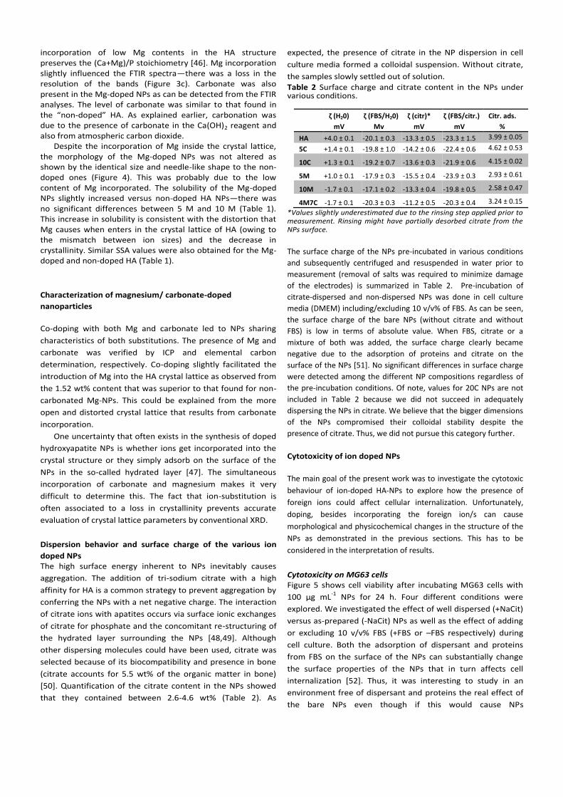
Results in Figure 5 clearly showed that for MG63 cells, NPs
dispersed in sodium citrate—regardless of the presence of FBS
in the cell culture media—were not cytotoxic as shown by the
high percentage of viable cells. Experiments without
dispersant showed NP sedimentation that formed a layer on
top of the cells after 24 h of cell culture. This occurred
regardless of the presence of FBS (Figure 5). Interestingly,
under these circumstances, when NPs were cultured in the
presence of FBS, viability was preserved for all NPs
compositions. However, when the experiments were carried
out on the bare NPs, 10M and 4M7C NPs were cytotoxic.
Figure 5 Viability of MG63 cells exposed to 100 μg mL
-1 of the
various NPs formulations for 24 h. (a) Cells exposed to NPs
dispersed in citrate in the presence or absence of 10 v/v% FBS
and (b) cells exposed to NPs dispersed in water in the presence
or absence of 10 v/v% FBS. Letters indicate significant
differences among NPs cultured with FBS: a- indicates
differences with HA, b- differences with 5C, c- differences with
10C, d- differences with 5M and e- differences with 10M.
Letters with (‘) indicate significant differences among NPs
cultured without FBS (P<0.05, N=3). The * indicates significant
differences for the same type of NPs cultured with and without
FBS (P<0.05, N=3).
Experiments performed using different batches of NPs
consistently confirmed the results. The experiments indicate
that a specific content of Mg (10M and 4M7C were 2.3 and 1.5
wt%, respectively) was needed to trigger such a response. The
fact that 5M with Mg content close to that of 4M7C did not
show the same behaviour could be related to the lower
solubility of this NP. Unfortunately the synthesis of Mg-doped
NPs with higher doping contents did not result in a phase-pure
compound impeding corroboration of the cytotoxic effect
using HA-NPs with even higher contents of Mg.
It is interesting to discuss all cell culture results and
consider the specific features that surrounded each study and
each NP. Because 10M and 4M7C NPs induced cytotoxicity in a
specific scenario (in absence of sodium citrate and FBS), it
seems reasonable to think that these particular NPs in all other
scenarios (with FBS and/or citrate) were probably non
cytotoxic simply because they could not be internalized (or
they were less internalized). A very straightforward
explanation to this could be the surface charge. The high
affinity of citrate and proteins for hydroxyapatite is very well
known [55-56]. Upon adsorption, both give the surface a
negative charge (Table 1) that could then prevent/minimize
internalization due to electrostatic repulsion with the cell
membrane [26]. This occurs regardless of whether the NPs are
dispersed (citrate) or not (without citrate but with FBS). It is
not clear whether the lack of cytotoxicity in the remaining bare
NPs (HA, 5C, 10C and 5M cultured without citrate and FBS) was
due to a lack of internalization or to a non-toxic response upon
internalization.
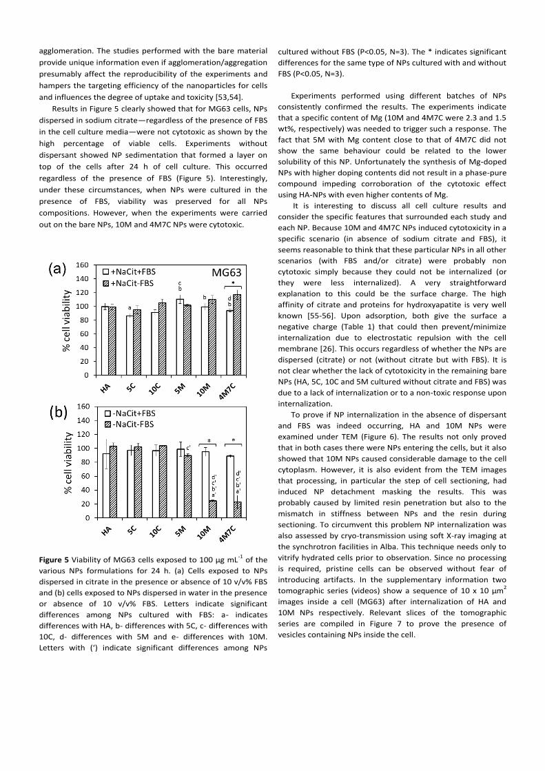
To prove if NP internalization in the absence of dispersant
and FBS was indeed occurring, HA and 10M NPs were
examined under TEM (Figure 6). The results not only proved
that in both cases there were NPs entering the cells, but it also
showed that 10M NPs caused considerable damage to the cell
cytoplasm. However, it is also evident from the TEM images
that processing, in particular the step of cell sectioning, had
induced NP detachment masking the results. This was
probably caused by limited resin penetration but also to the
mismatch in stiffness between NPs and the resin during
sectioning. To circumvent this problem NP internalization was
also assessed by cryo-transmission using soft X-ray imaging at
the synchrotron facilities in Alba. This technique needs only to
vitrify hydrated cells prior to observation. Since no processing
is required, pristine cells can be observed without fear of
introducing artifacts. In the supplementary information two
tomographic series (videos) show a sequence of 10 x 10 µm2
images inside a cell (MG63) after internalization of HA and
10M NPs respectively. Relevant slices of the tomographic
series are compiled in Figure 7 to prove the presence of
vesicles containing NPs inside the cell.
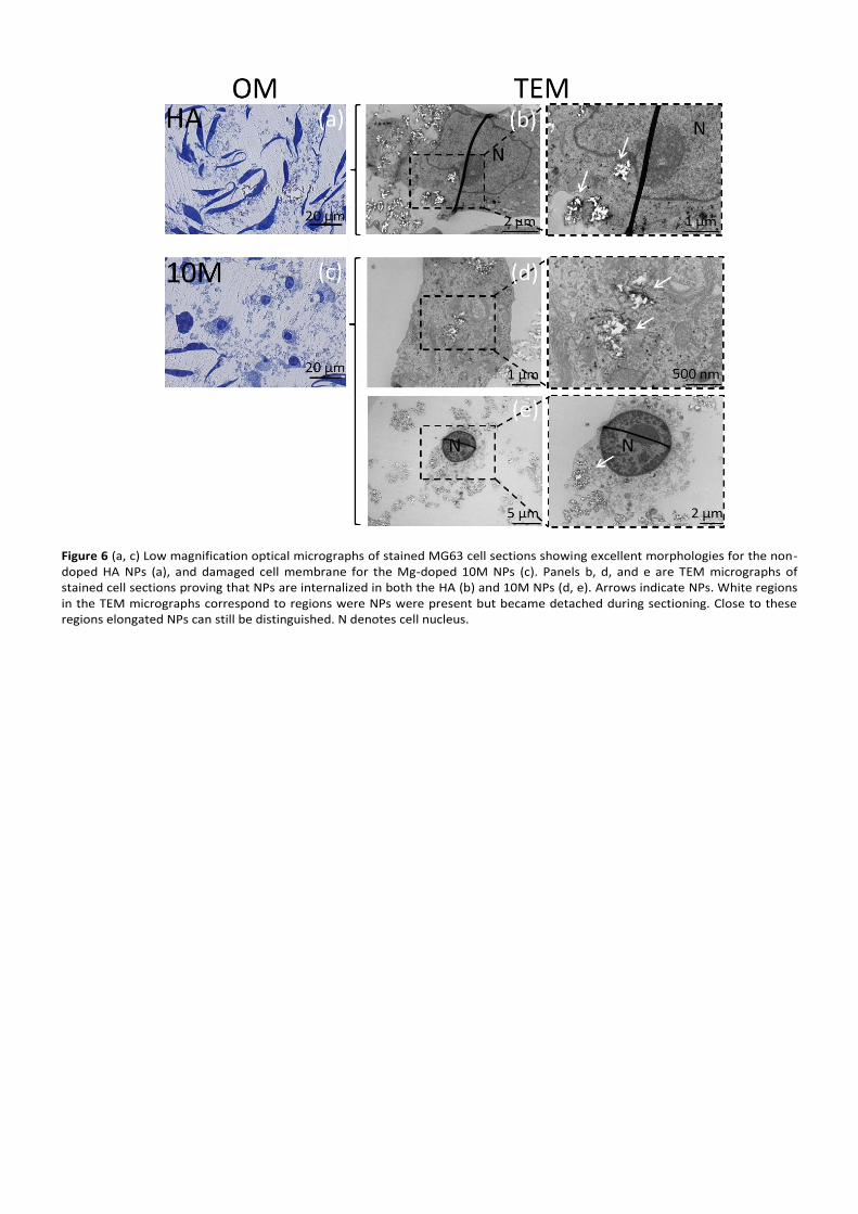
Figure 6 (a, c) Low magnification optical micrographs of stained MG63 cell sections showing excellent morphologies for the non-doped HA NPs (a), and damaged cell membrane for the Mg-doped 10M NPs (c). Panels b, d, and e are TEM micrographs of stained cell sections proving that NPs are internalized in both the HA (b) and 10M NPs (d, e). Arrows indicate NPs. White regions in the TEM micrographs correspond to regions were NPs were present but became detached during sectioning. Close to these regions elongated NPs can still be distinguished. N denotes cell nucleus.
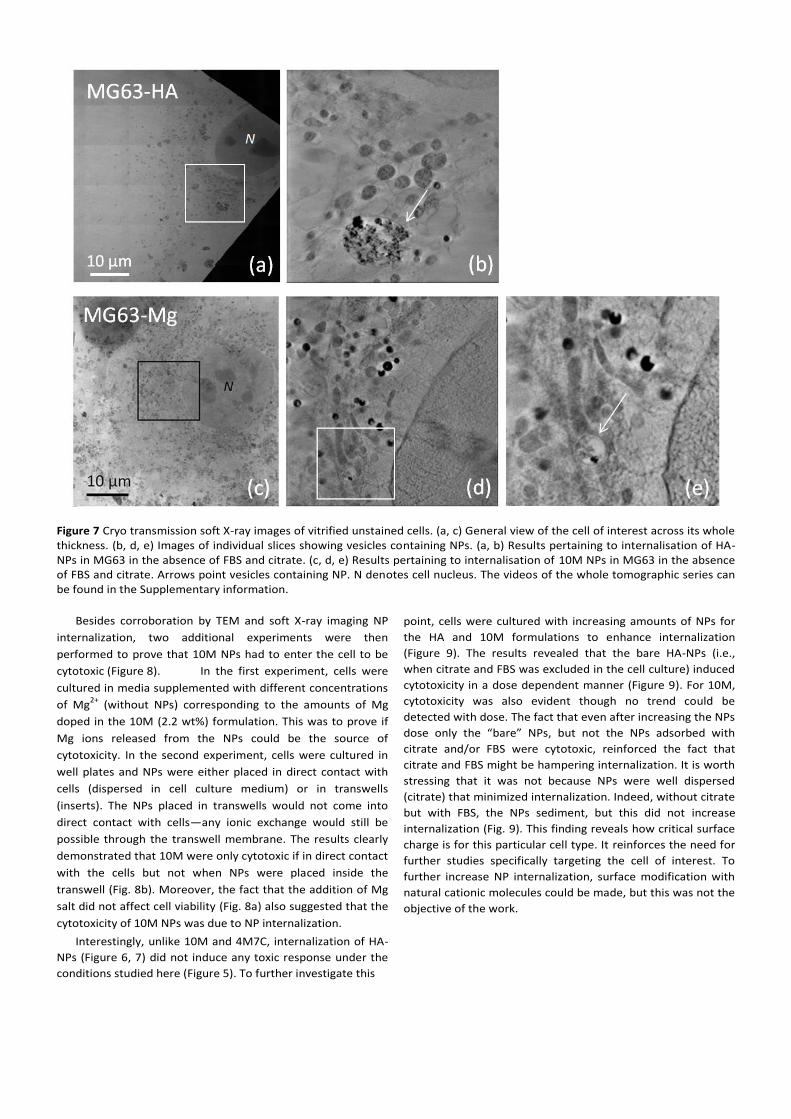
Figure 7 Cryo transmission soft X-ray images of vitrified unstained cells. (a, c) General view of the cell of interest across its whole thickness. (b, d, e) Images of individual slices showing vesicles containing NPs. (a, b) Results pertaining to internalisation of HA-NPs in MG63 in the absence of FBS and citrate. (c, d, e) Results pertaining to internalisation of 10M NPs in MG63 in the absence of FBS and citrate. Arrows point vesicles containing NP. N denotes cell nucleus. The videos of the whole tomographic series can be found in the Supplementary information.
Besides corroboration by TEM and soft X-ray imaging NP
internalization, two additional experiments were then
performed to prove that 10M NPs had to enter the cell to be
cytotoxic (Figure 8). In the first experiment, cells were
cultured in media supplemented with different concentrations
of Mg2+
(without NPs) corresponding to the amounts of Mg
doped in the 10M (2.2 wt%) formulation. This was to prove if
Mg ions released from the NPs could be the source of
cytotoxicity. In the second experiment, cells were cultured in
well plates and NPs were either placed in direct contact with
cells (dispersed in cell culture medium) or in transwells
(inserts). The NPs placed in transwells would not come into
direct contact with cells—any ionic exchange would still be
possible through the transwell membrane. The results clearly
demonstrated that 10M were only cytotoxic if in direct contact
with the cells but not when NPs were placed inside the
transwell (Fig. 8b). Moreover, the fact that the addition of Mg
salt did not affect cell viability (Fig. 8a) also suggested that the
cytotoxicity of 10M NPs was due to NP internalization.
Interestingly, unlike 10M and 4M7C, internalization of HA-
NPs (Figure 6, 7) did not induce any toxic response under the
conditions studied here (Figure 5). To further investigate this
point, cells were cultured with increasing amounts of NPs for
the HA and 10M formulations to enhance internalization
(Figure 9). The results revealed that the bare HA-NPs (i.e.,
when citrate and FBS was excluded in the cell culture) induced
cytotoxicity in a dose dependent manner (Figure 9). For 10M,
cytotoxicity was also evident though no trend could be
detected with dose. The fact that even after increasing the NPs
dose only the “bare” NPs, but not the NPs adsorbed with
citrate and/or FBS were cytotoxic, reinforced the fact that
citrate and FBS might be hampering internalization. It is worth
stressing that it was not because NPs were well dispersed
(citrate) that minimized internalization. Indeed, without citrate
but with FBS, the NPs sediment, but this did not increase
internalization (Fig. 9). This finding reveals how critical surface
charge is for this particular cell type. It reinforces the need for
further studies specifically targeting the cell of interest. To
further increase NP internalization, surface modification with
natural cationic molecules could be made, but this was not the
objective of the work.
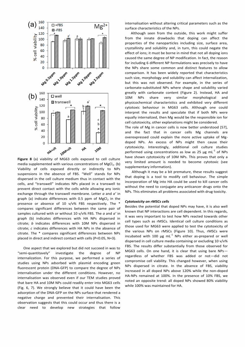
Figure 8 (a) viability of MG63 cells exposed to cell culture
media supplemented with various concentrations of MgCl2. (b)
Viability of cells exposed directly or indirectly to NPs
suspensions in the absence of FBS. “Well” stands for NPs
dispersed in the cell culture medium thus in contact with the
cells, and “transwell” indicates NPs placed in a transwell to
prevent direct contact with the cells while allowing any ionic
exchange through the transwell membrane. Letter a and a’ in
graph (a) indicate differences with 0.5 ppm of MgCl2 in the
presence or absence of 10 v/v% FBS respectively. The *
compares significant differences between the same pair of
samples cultured with or without 10 v/v% FBS. The a and a’ in
graph (b) indicates differences with HA NPs dispersed in
citrate; b indicates differences with 10M NPs dispersed in
citrate; c indicates differences with HA NPs in the absence of
citrate. The * compares significant differences between NPs
placed in direct and indirect contact with cells (P<0.05, N=3).
One aspect that we explored but did not succeed in was to
“semi-quantitavely” investigate the degree of NP
internalization. For this purpose, we performed a series of
studies using NPs adsorbed with plasmid encoding green
fluorescent protein (DNA-GFP) to compare the degree of NPs
internalisation under the different conditions. However, no
internalisation was observed even if our TEM studies proved
that bare HA and 10M NPs could readily enter into MG63 cells
(Fig. 6, 7). We strongly believe that it could have been the
adsorption of the DNA-GFP on the NPs surface that rendered a
negative charge and prevented their internalisation. This
observation suggests that this could occur and thus there is a
clear need to develop new strategies that follow
internalisation without altering critical parameters such as the
surface characteristics of the NPs.
Although seen from the outside, this work might suffer
from the innate drawbacks that doping can affect the
properties of the nanoparticles including size, surface area,
crystallinity and solubility and, in turn, this could negate the
effect of ions; it must be borne in mind that not all doping ions
caused the same degree of NP modification. In fact, the reason
for including 6 different NP formulations was precisely to have
the NPs share some common and distinct features to allow
comparison. It has been widely reported that characteristics
such size, morphology and solubility can affect internalization,
but this was not observed. For example, in the series of
carbonate-substituted NPs where shape and solubility varied
greatly with carbonate content (Figure 2). Instead, HA and
10M NPs share very similar morphological and
physicochemical characteristics and exhibited very different
cytotoxic behaviour in MG63 cells. Although one could
interpret the results and speculate that if both NPs were
equally internalized, then Mg would be the responsible ion for
cell cytotoxicity, other explanations might be considered.
The role of Mg in cancer cells is now better understood [57],
and the fact that in cancer cells Mg channels are
overexpressed could explain the more active uptake of Mg-
doped NPs. An excess of NPs might then cause their
cytotoxicity. Interestingly, additional cell culture studies
performed using concentrations as low as 25 µg mL-1
of NPs
have shown cytotoxicity of 10M NPs. This proves that only a
very limited amount is needed to become cytotoxic (see
supplementary information).
Although it may be a bit premature, these results suggest
that doping is a tool to modify cell behaviour. The simple
incorporation of Mg into HA could be used to kill cancer cells
without the need to conjugate any anticancer drugs onto the
NPs. This eliminates all problems associated with drug toxicity.
Cytotoxicity on rMSCs cells
Besides the potential that doped NPs may have, it is also well
known that NP interactions are cell dependent. In this regards,
it was very important to test how NPs reacted towards other
cell types such as rMSCs. Identical cell culture conditions as
those used for MG63 were applied to test the cytotoxicity of
the various NPs on rMSCs (Figure 10). Thus, rMSCs were
incubated with 100 μg mL-1
NPs either as-prepared or well
dispersed in cell culture media containing or excluding 10 v/v%
FBS. The results differ substantially from those observed for
MG63 cells. On one hand, it is clear that using bare NPs—
regardless of whether FBS was added or not—did not
compromise cell viability. This changed however, when using
NPs dispersed in citrate. In the absence of FBS, viability
increased in all doped NPs above 120% while the non-doped
HA-NPs remained at 100%. In the presence of 10% FBS, we
noted an opposite trend: all doped NPs showed 80% viability
while 100% was maintained for HA.
Figure 9 Dose-dependent cytotoxicity of HA (a) and Mg-doped HA (10M) (b) in the presence or absence of 10 v/v% FBS. NPs dispersed in
citrate and in water (control) were tested. To discard any effect of citrate in the media, cytotoxicity was also performed by supplementing
the cell culture media with the same concentration of citrate that was added for the NPs (c). Letters in the graphs indicate significant
differences between NPs doses cultured with FBS: a- indicates differences with 100 µg mL-1
, b- differences with 250 µg mL-1
, c- differences
with 500 µg mL-1
and, d- differences with 1000 µg mL-1
. Letters with (‘) indicate significant differences between NPs doses cultured without
FBS (P<0.05, N=3). Independent analyses were done for the samples containing citrate (capital letters) versus those without citrate (lower
case letters). The * compares significant differences between the same pair of NPs cultured with or without FBS (P<0.05, N=3).
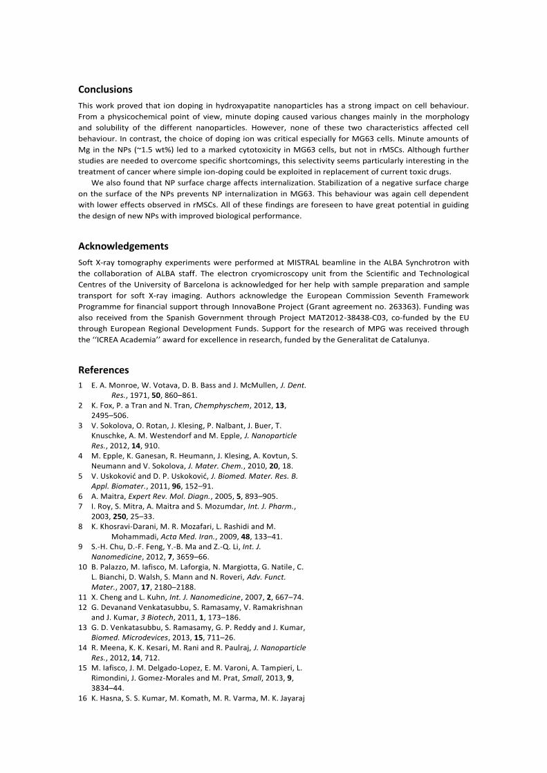
Studies performed with increasing NP dose on HA and 10M
NPs showed that viability slightly decreased for all cases in a
dose dependent manner. This could be an indicator for a
higher degree of internalization (Figure 11). In sum (Figure 10-
11), it is interesting to see that unlike MG63 cells, the negative
charge that surrounded the NPs upon citrate adsorption did
not prevent NPs internalization in rMSCs.
With regards to the effect of the various formulations of
doped NPs on rMSCs, there were no significant differences
that could be attributed to the presence of the different ions
(Figure 10). These findings clearly contrasted with the results
obtained for MG63 cells where 10M and 4M7C NPs reacted
differently from the rest (Figure 5). Although Mg and
carbonate apparently did not play any major role on rMSCs,
this cannot prove that this cell type is not sensitive to NP
composition. Other ions might be more critical than the ones
selected here. Nevertheless it is worth mentioning that the
dose dependent cytotoxicity of HA and 10M formulations did
reveal a more pronounced cytotoxic behaviour for HA than for
10M at higher NPs doses.
This contrasting behaviour of MG63 and rMSCs when
treated with NPs has been reported by other authors with
similar non-doped HA-NPs. It is not new that HA-NPs can be
cytotoxic to cancerous cells but not to normal cells [58]. In
fact, the dose dependent assay shown here (Fig. 9) suggests
that even the non-doped NPs can become cytotoxic at high NP
doses (500 µg mL-1
). This anti-tumour activity is generally
explained by the higher metabolic activity of cancerous cells
versus non-cancerous cells, which causes a higher degree of
NP internalization. Moreover, for specific cancerous cells (such
as human gastric cancer cells, human cervical adenocarcinoma
epithelial cells and human hepatoma cells) higher levels of
apoptosis has been shown due to selective NP accumulation in
the cell nuclei with no translocation detected in normal cells
(e.g. human normal liver cells) [59]. Once in the nuclei, the NPs
strongly interact with DNA or DNA-related proteins and disrupt
cell functions and trigger apoptosis. However, it is not
compulsory for the NPs to enter the nucleus because during
cell mitosis the nucleus disassembles and re-forms—this
favours NP internalisation.
Figure 10 Viability of rMSCs cells exposed to 100 μg mL-1
of the
various NPs for 24 h. (a) Cells exposed to NPs dispersed in
citrate in the presence or absence of 10 v/v% FBS, and (b) cells
exposed to NPs dispersed in water in the presence or absence
of 10 v/v% FBS. Letters indicate significant differences among
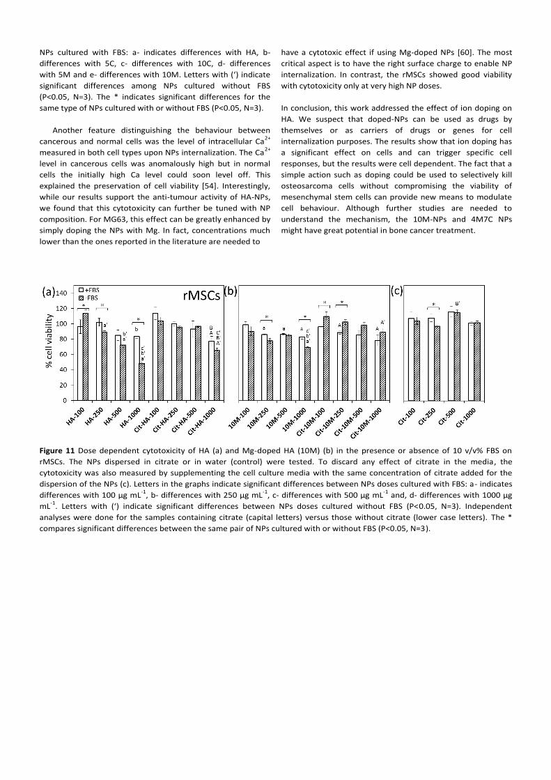
NPs cultured with FBS: a- indicates differences with HA, b-
differences with 5C, c- differences with 10C, d- differences
with 5M and e- differences with 10M. Letters with (‘) indicate
significant differences among NPs cultured without FBS
(P<0.05, N=3). The * indicates significant differences for the
same type of NPs cultured with or without FBS (P<0.05, N=3).
Another feature distinguishing the behaviour between
cancerous and normal cells was the level of intracellular Ca2+
measured in both cell types upon NPs internalization. The Ca2+
level in cancerous cells was anomalously high but in normal
cells the initially high Ca level could soon level off. This
explained the preservation of cell viability [54]. Interestingly,
while our results support the anti-tumour activity of HA-NPs,
we found that this cytotoxicity can further be tuned with NP
composition. For MG63, this effect can be greatly enhanced by
simply doping the NPs with Mg. In fact, concentrations much
lower than the ones reported in the literature are needed to
have a cytotoxic effect if using Mg-doped NPs [60]. The most
critical aspect is to have the right surface charge to enable NP
internalization. In contrast, the rMSCs showed good viability
with cytotoxicity only at very high NP doses.
In conclusion, this work addressed the effect of ion doping on
HA. We suspect that doped-NPs can be used as drugs by
themselves or as carriers of drugs or genes for cell
internalization purposes. The results show that ion doping has
a significant effect on cells and can trigger specific cell
responses, but the results were cell dependent. The fact that a
simple action such as doping could be used to selectively kill
osteosarcoma cells without compromising the viability of
mesenchymal stem cells can provide new means to modulate
cell behaviour. Although further studies are needed to
understand the mechanism, the 10M-NPs and 4M7C NPs
might have great potential in bone cancer treatment.
Figure 11 Dose dependent cytotoxicity of HA (a) and Mg-doped HA (10M) (b) in the presence or absence of 10 v/v% FBS on
rMSCs. The NPs dispersed in citrate or in water (control) were tested. To discard any effect of citrate in the media, the
cytotoxicity was also measured by supplementing the cell culture media with the same concentration of citrate added for the
dispersion of the NPs (c). Letters in the graphs indicate significant differences between NPs doses cultured with FBS: a- indicates
differences with 100 µg mL-1
, b- differences with 250 µg mL-1
, c- differences with 500 µg mL-1
and, d- differences with 1000 µg
mL-1
. Letters with (‘) indicate significant differences between NPs doses cultured without FBS (P<0.05, N=3). Independent
analyses were done for the samples containing citrate (capital letters) versus those without citrate (lower case letters). The *
compares significant differences between the same pair of NPs cultured with or without FBS (P<0.05, N=3).
Conclusions
This work proved that ion doping in hydroxyapatite nanoparticles has a strong impact on cell behaviour.
From a physicochemical point of view, minute doping caused various changes mainly in the morphology
and solubility of the different nanoparticles. However, none of these two characteristics affected cell
behaviour. In contrast, the choice of doping ion was critical especially for MG63 cells. Minute amounts of
Mg in the NPs (~1.5 wt%) led to a marked cytotoxicity in MG63 cells, but not in rMSCs. Although further
studies are needed to overcome specific shortcomings, this selectivity seems particularly interesting in the
treatment of cancer where simple ion-doping could be exploited in replacement of current toxic drugs.
We also found that NP surface charge affects internalization. Stabilization of a negative surface charge
on the surface of the NPs prevents NP internalization in MG63. This behaviour was again cell dependent
with lower effects observed in rMSCs. All of these findings are foreseen to have great potential in guiding
the design of new NPs with improved biological performance.
Acknowledgements
Soft X-ray tomography experiments were performed at MISTRAL beamline in the ALBA Synchrotron with
the collaboration of ALBA staff. The electron cryomicroscopy unit from the Scientific and Technological
Centres of the University of Barcelona is acknowledged for her help with sample preparation and sample
transport for soft X-ray imaging. Authors acknowledge the European Commission Seventh Framework
Programme for financial support through InnovaBone Project (Grant agreement no. 263363). Funding was
also received from the Spanish Government through Project MAT2012-38438-C03, co-funded by the EU
through European Regional Development Funds. Support for the research of MPG was received through
the ‘‘ICREA Academia’’ award for excellence in research, funded by the Generalitat de Catalunya.
References
1 E. A. Monroe, W. Votava, D. B. Bass and J. McMullen, J. Dent. Res., 1971, 50, 860–861. 2 K. Fox, P. a Tran and N. Tran, Chemphyschem, 2012, 13, 2495–506. 3 V. Sokolova, O. Rotan, J. Klesing, P. Nalbant, J. Buer, T. Knuschke, A. M. Westendorf and M. Epple, J. Nanoparticle Res., 2012, 14, 910. 4 M. Epple, K. Ganesan, R. Heumann, J. Klesing, A. Kovtun, S. Neumann and V. Sokolova, J. Mater. Chem., 2010, 20, 18. 5 V. Uskoković and D. P. Uskoković, J. Biomed. Mater. Res. B. Appl. Biomater., 2011, 96, 152–91. 6 A. Maitra, Expert Rev. Mol. Diagn., 2005, 5, 893–905. 7 I. Roy, S. Mitra, A. Maitra and S. Mozumdar, Int. J. Pharm., 2003, 250, 25–33. 8 K. Khosravi-Darani, M. R. Mozafari, L. Rashidi and M. Mohammadi, Acta Med. Iran., 2009, 48, 133–41. 9 S.-H. Chu, D.-F. Feng, Y.-B. Ma and Z.-Q. Li, Int. J. Nanomedicine, 2012, 7, 3659–66. 10 B. Palazzo, M. Iafisco, M. Laforgia, N. Margiotta, G. Natile, C. L. Bianchi, D. Walsh, S. Mann and N. Roveri, Adv. Funct. Mater., 2007, 17, 2180–2188. 11 X. Cheng and L. Kuhn, Int. J. Nanomedicine, 2007, 2, 667–74. 12 G. Devanand Venkatasubbu, S. Ramasamy, V. Ramakrishnan and J. Kumar, 3 Biotech, 2011, 1, 173–186. 13 G. D. Venkatasubbu, S. Ramasamy, G. P. Reddy and J. Kumar, Biomed. Microdevices, 2013, 15, 711–26. 14 R. Meena, K. K. Kesari, M. Rani and R. Paulraj, J. Nanoparticle Res., 2012, 14, 712. 15 M. Iafisco, J. M. Delgado-Lopez, E. M. Varoni, A. Tampieri, L. Rimondini, J. Gomez-Morales and M. Prat, Small, 2013, 9, 3834–44. 16 K. Hasna, S. S. Kumar, M. Komath, M. R. Varma, M. K. Jayaraj
and K. R. Kumar, Phys. Chem. Chem. Phys., 2013, 15, 8106– 11. 17 S. Panseri, C. Cunha, T. D’Alessandro, M. Sandri, G. Giavaresi, M. Marcacci, C. T. Hung and A. Tampieri, J. Nanobiotechnology, 2012, 10, 32. 18 A. Tampieri, T. D’Alessandro, M. Sandri, S. Sprio, E. Landi, L. Bertinetti, S. Panseri, G. Pepponi, J. Goettlicher, M. Bañobre- López and J. Rivas, Acta Biomater., 2012, 8, 843–51. 19 A. Doat, F. Pellé, N. Gardant and a. Lebugle, J. Solid State Chem., 2004, 177, 1179–1187. 20 E. I. Altinoglu, T. J. Russin, J. M. Kaiser, B. M. Barth, P. C. Eklund, M. Kester and J. H. Adair, ACS Nano, 2008, 2, 2075– 2084. 21 K. H. Müller, M. Motskin, A. J. Philpott, A. F. Routh, C. M. Shanahan, M. J. Duer and J. N. Skepper, Biomaterials, 2013, 35, 1074–1088. 22 Z. Shi, X. Huang, Y. Cai, R. Tang and D. Yang, Acta Biomater., 2009, 5, 338–45. 23 T. Ding, Y. Xue, H. Lu, Z. Huang and J. Sun, IEEE Trans. Nanobioscience, 2012, 11, 336–40. 24 X. Zhao, S. Ng, B. C. Heng, J. Guo, L. Ma, T. T. Y. Tan, K. W. Ng and S. C. J. Loo, Arch. Toxicol., 2013, 87, 1037–52. 25 Y. Cai, Y. Liu, W. Yan, Q. Hu, J. Tao, M. Zhang, Z. Shi and R. Tang, J. Mater. Chem., 2007, 17, 3780. 26 L. Chen, J. M. Mccrate, J. C.-M. Lee and H. Li, Nanotechnology, 2011, 22, 1–20. 27 V. Sokolova, D. Kozlova, T. Knuschke, J. Buer, A. M. Westendorf and M. Epple, Acta Biomater., 2013, 9, 7527– 7535. 28 Z. Xu, C. Liu, J. Wei and J. Sun, J. Appl. Toxicol., 2012, 32, 429–35. 29 F. Qing, Z. Wang, Y. Hong, M. Liu, B. Guo, H. Luo and X. Zhang, J. Mater. Sci. Mater. Med., 2012, 23, 2245–2251. 30 E. Boanini, M. Gazzano and a Bigi, Acta Biomater., 2010, 6, 1882–94. 31 C. Rey, C. Combes, C. Drouet and M. J. Glimcher, Osteoporos. Int., 2009, 20, 1013–1021. 32 B. Wopenka and J. D. Pasteris, Mater. Sci. Eng. C, 2005, 25, 131–143. 33 J. H. Shepherd, D. V Shepherd and S. M. Best, J. Mater. Sci. Mater. Med., 2012, 23, 2335–47. 34 V. Aina, G. Lusvardi, B. Annaz, I. R. Gibson, F. E. Imrie, G. Malavasi, L. Menabue, G. Cerrato and G. Martra, J. Mater. Sci. Mater. Med., 2012, 23, 2867–79. 35 S. Dasgupta, S. S. Banerjee, A. Bandyopadhyay and S. Bose, Langmuir, 2010, 26, 4958–64. 36 N. Y. Mostafa, H. M. Hassan and O. H. Abd Elkader, J. Am. Ceram. Soc., 2011, 94, 1584–1590. 37 M. S. Sader, K. Lewis, G. a. Soares and R. Z. LeGeros, Mater. Res., 2013, 16, 779–784. 38 C. Capuccini, P. Torricelli, E. Boanini, M. Gazzano, R. Giardino and a Bigi, J. Biomed. Mater. Res. A, 2009, 89, 594–600. 39 S. C. Cox, P. Jamshidi, L. M. Grover and K. K. Mallick, Mater. Sci. Eng. C. Mater. Biol. Appl., 2014, 35, 106–14. 40 Y. Li, C. T. Nam and C. P. Ooi, J. Phys. Conf. Ser., 2009, 187, 012024. 41 E. Landi, A. Tampieri, M. Mattioli-Belmonte, G. Celotti, M. Sandri, A. Gigante, P. Fava and G. Biagini, J. Eur. Ceram. Soc., 2006, 26, 2593–2601.