Embed Size (px)
DESCRIPTION
Professor Sefton, University of Toronto, Ion Channels and Active Transport
Citation preview

1
Cell Membranes: Ion channels and Active Transport
Sadava et al, Chapter 6.4; Active Processes of Membrane Transport
Protein mediated transport pmechanisms
Facilitated diffusion (see previous lecture)
A. Ion channelsA. Ion channelsB. Co-transportC. Primary active
transport

2
• Move ions / small molecules down chemical / electricalgradient • single-file motion• non-gated: open most of the time;• gated: open in response to signal
A. Ion channels
Structure of the K+ channel from Streptomyces lividans (KcsA channel)* KcsA channel is formed by four identical subunits, each subunit containing two alpha-helicesconnected by an approximately 30 amino acids long loop
http://www.ks.uiuc.edu/Research/kvchannel/
5.11 The Potassium Channel
• Custom fit for K+ ions [to bind to O on helices]; others don’t fit [e.g., Na+ is too small]

3
5.10 A Gated Channel Protein Opens in Response to a Stimulus
e.g., a ligand-gated channel
e.g., voltage gated channels open at a threshold voltage
5.13 Three Types of Proteins for facilitated diffusion or co-transport
B. Coupled transport
• not ATP dependent active transport (although Sadava terms these active transport)
uniporter symporterantiporter

4
Indicates direction of concentration
gradient
High
• Move ions / small molecules across membrane• uniporters: transport single type of molecule down gradient: facilitated diffusion• antiporters/symporters: couple transport of one species down its concentration gradient with movement of another against its gradient: co-transporter
Low
gradient with movement of another against its gradient: co transporter• NB: these do NOT use ATP• rely on electrochemical gradient to provide energy for transport• movement of co-transported ion is down its gradient
• this favourable process provides the energy to drive the unfavourable transport of other species against its concentration gradient
Key: movement of both species is obligatory - Coupled Transport
Indicates direction of concentration
gradient
Low
High
Low
Symport: transported molecule and co-transported ion move in same directionAntiport: transported molecule and co-transported ion move in opposite direction
e.g. Na+/H+ antiporter : exports H+ (unfavourable) against favourable import of Na+
Recall: GLUT1 (carrier for glucose) was an example of a uniporter = facilitated diffusion( g ) p p

5
5.15 Coupled transport of glucose and sodium
• Passive diffusion of Na+ drives glucose against the concentration gradient (a symport) [eg in intestinal cells]
• Free energy cost of glucose transport is paid by free energy of sodium transport
• Symporter changes conformation to permit transporter when Na+ and glucose bind
• Gradient of Na+ is established by Na/K pump
Membrane Transport
Cellular pH regulation
Cellular metabolism
Anaerobic:Glucose metabolism lactic acidGlucose metabolism lactic acid
Aerobic: CO2 H2CO3
Dissociation of these weak acids H+
Need to remove these excess protons from cells• Na+NCO3
-/Cl- antiporter• imports Na+, HCO3
- (down gradient)• exports Cl- (against gradient)
CO2 + OH-HCO3-
Cytosolic pH7.2 - 7.4
• Na+ / H+ antiporter• imports Na+ (down gradient)• exports H+ (against gradient)
• Cl- / HCO3- antiporter
• imports Cl- (down gradient)• exports HCO3
- (against gradient)
Raise pH
Lower pH
This whole system works like a series of coupled feedback loops to maintain
the right pH inside the cell.
3 different transporters working together

6
C. Active Transport
• Move ions / small molecules against chemical / electrical gradient (active transport)• Conversion of ATP -> ADP releases energy to facilitate the transport processe.g., Na+/K+ or Ca++
Toyoshima et al. 2000, Nature 405, 647-655
Intracellular vs extracellularTypical ion concentrations in mammalian cytoplasm and blood.
Concentration in Concentration in
Ion cytosol(millimolar)
Concentration in blood (millimolar)
Potassium 139 4
Sodium 12 145
Chloride 4 116
Bicarbonate 12 29
Amino acids in t i
138 9proteins
138 9
Magnesium 0.8 1.5
wikipedia

7
5.14 Primary Active Transport: The Sodium–Potassium Pump
• Uses ATP– moves both Na and K against their gradients [a type of antiport]
• Na+ is high outside cells and K+ is high inside cells
Calcium pump
Muscle contraction is due toMuscle contraction is due to Ca2+ released from the Ca2+
stores in the cell.
Ca2+ -ATPase transports Ca2+
back during the relaxation phase of the muscle contraction cycle.
The protein performs this jobThe protein performs this job efficiently at the expense of ATP hydrolysis, which leads to Ca2+
concentrations 103-104 times higher on the inside than on the outside of the membrane.
Toyoshima et al. 2000, Nature 405, 647-655

8
Several types of ATP pumps
• 2 catalytic ATP binding sites• one region is phosphorylated
• proton pumps• V: from cytosolic -> exoplasmic
• proton pumps• F: from exoplasmic -> cytosolic
• 4 core domains• two transmembrane• two cytosolic ATP binding domains
Membrane TransportKey points:• most molecules cannot pass through membrane
• exceptions: O2, CO2, small hydrophobic molecules
• transport mediated by• ATP-pumps• channels channels• transporters (uni-, anti-, sym-)
• facilitated transport:• assists movement down concentration gradient
• co-transport:• coupled movement down concentration gradient by movement of second substrate againstgradient
• active transport:• ATP hydrolysis fuels transport against concentration gradient
• protein-catalyzed transport >> diffusion
9
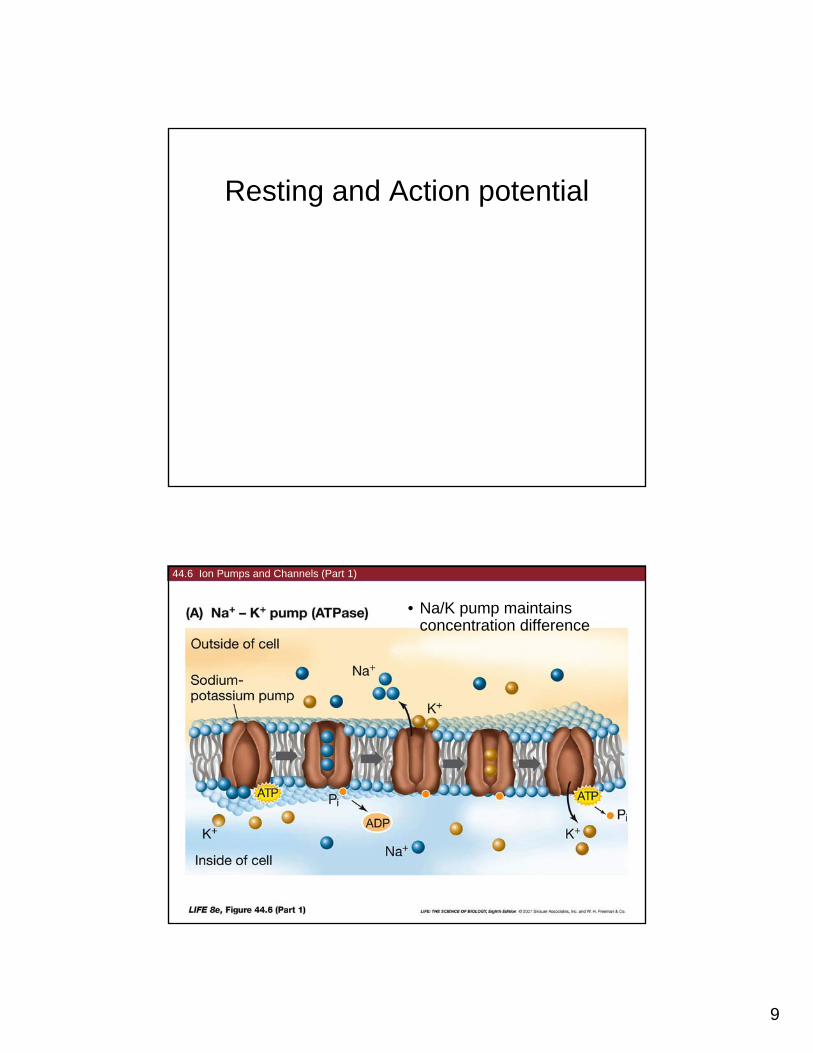
Resting and Action potential
44.6 Ion Pumps and Channels (Part 1)
• Na/K pump maintains concentration difference

10
44.6 Ion Pumps and Channels (Part 2)
• Open channels permit K+ out of cell and Na+ into cell; voltage gated channels permit tuning of rates
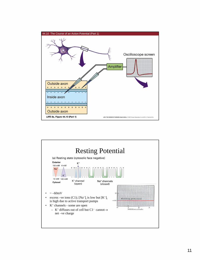
44.5 Measuring the Resting Potential (Part 2)
11
44.10 The Course of an Action Potential (Part 1)
Resting Potential
• ~ –60mV• excess –ve ions (C1); [Na+]i is low but [K+]i( ); [ ]i [ ]i
is high due to active transport pumps• K+ channels - some are open
– K+ diffuses out of cell but C1– cannotnet –ve charge

12
Depolarization
Open Na channels (less –ve charge); potential will go +ve, if enough channels open)
Action potential

13
Action Potential
• sudden change in potential lasts 1-2 ms• sudden change in potential lasts 1-2 ms
• Na+ channels open rapid reversal of potentialfrom - 60 to +50 mV - creates a spike
• Na+ channels close; K+ channels stay open a bit longer to enable E to return to –60
N + h l i ti t d ’t d f• Na+ channels inactivated - can’t respond for a short time -- membrane is refractory
• Na+ channels are voltage gated (open as a f(V)) -threshold effect
44.10 The Course of an Action Potential (Part 2)
2: Some Na channels open depolarizing membrane to threshold

14
Nerve transmission
• Sequences of action potentialsSequences of action potentials
• action potential
– all or nothing (no information)
– no amplitude differences
• information encoded in frequency
– stimulus to sensory receptor y paltered frequency of action potential sequence
• capability of reliable transmission
over long distances (metres)
44.11 Action Potentials Travel along Axons (Part 2)
Depolarizing current spreads down axon
Eventually potential reaches threshold and action potential is generated downstream

15
44.13 Chemical Synaptic Transmission Begins with the Arrival of an Action Potential
Action potential drives release of neurotransmitter from vesiclesvesicles
Neurotransmitters bind to receptor regenerating elevated Na+
44.14 The Acetylcholine Receptor Is a Chemically Gated Channel
Nerve gas inhibits enzyme

16
Acetylcholine wakes the brain (cerebral cortex –site of consciousness)consciousness) [reinforced by other neuro-transmitters (serotonin, dopamine, etc)]
GABA shuts
Over time, ATP is used, adenosine builds up and triggers activity in site 6 (VLPO)
down the arousal system to enable sleep
VLPO activity influenced by the brain’s clock (7) which is influenced by signals from the retina and melatonin from pineal gland (8) (circadian control)

17
Insomnia
Chapter summary
• Ion channels are membrane proteins that allow rapid facilitated diffusion of ionsthrough membranes. These can be gated channels, where the channel can be opened or closed by certain conditions of chemicals. The opening or closing of channels can set up an electrochemical gradient with unequal charged species on different sides of a membrane.
6.4 How do substances cross membranes against a concentration gradient?
• Active transport requires the use of chemical energy to move substances across a membrane against a concentration gradient. [Active] transport proteins may be uniports symports or antiportsuniports, symports, or antiports.
• In primary active transport, energy from the hydrolysis of ATP is used to move ionsinto or out of cells against their concentration gradients. The sodium-potassium pump is an important example.
• Secondary active transport couples the passive movement of one substance with its concentration gradient to the movement of another substance against its concentration gradient. Energy from ATP is used indirectly to establish the concentration gradient that results in the movement of the first substance