Embed Size (px)
Citation preview
CELL MEMBRANE, ION CHANNELS, PASSIVE AND ACTIVE PROPERTIES
OF THE NEURON
Prof. Maja Valić, MD, PhDDepartment of Neuroscience
University of Split School of Medicine
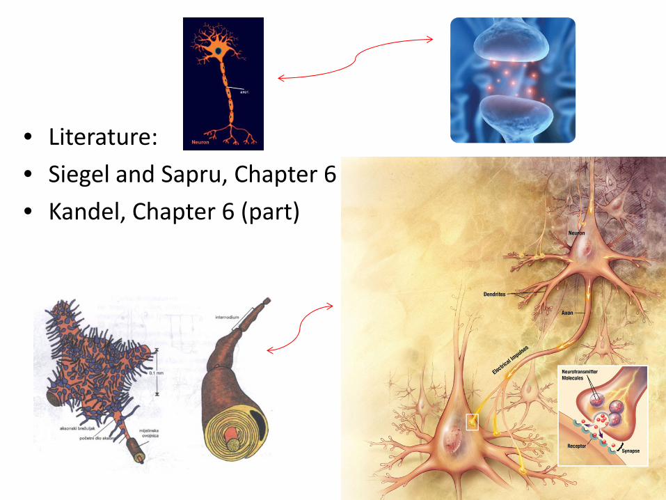
• Literature:• Siegel and Sapru, Chapter 6• Kandel, Chapter 6 (part)
• The most important role of the neuronal membrane is synaptic transmission!
Synaptic transmission
• Exocytosis of the neurotransmitter• Receptors• Ion channel• Changes in the excitability of the neuron• Resistance of the cell membrane• Capacity (Phospholipid bilayer)• Conductance (Ion channel)
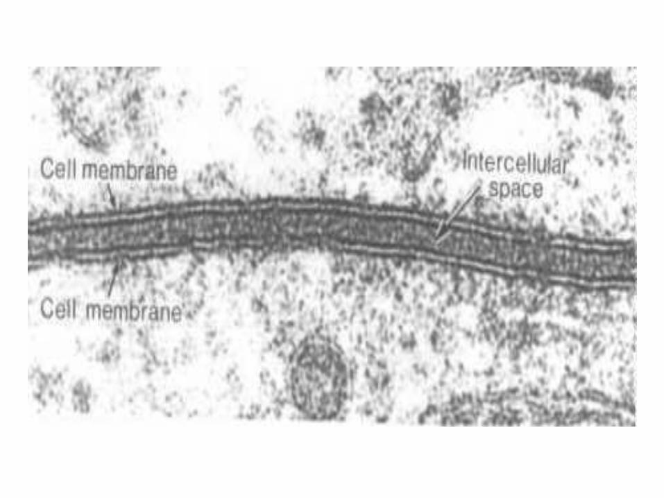
The role of the cell membrane:
• Protective• Maintance of the shape• Regulation of the transport• Intercellular signalization• Cellular recognition • Gap junction – neuronal communication
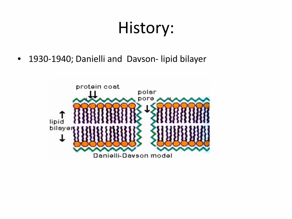
History:
• 1930‐1940; Danielli and Davson‐ lipid bilayer
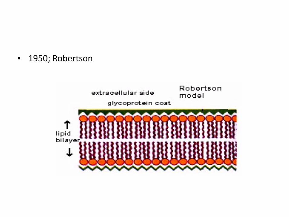
• 1950; Robertson

• 1966; Lenard and Singer‐membran protein, α helix• freeze fracture
Structure of the neuronal membrane:
• Lipid bilayer (phospholipids, glycolipids and cholesterol)
• proteins (transmembrane i peripheral)• glycocalyx (glicolipids, proteoglicans, glicoproteins)
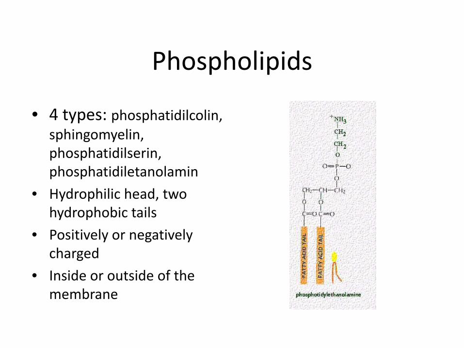
Phospholipids
• 4 types: phosphatidilcolin, sphingomyelin, phosphatidilserin, phosphatidiletanolamin
• Hydrophilic head, two hydrophobic tails
• Positively or negatively charged
• Inside or outside of the membrane
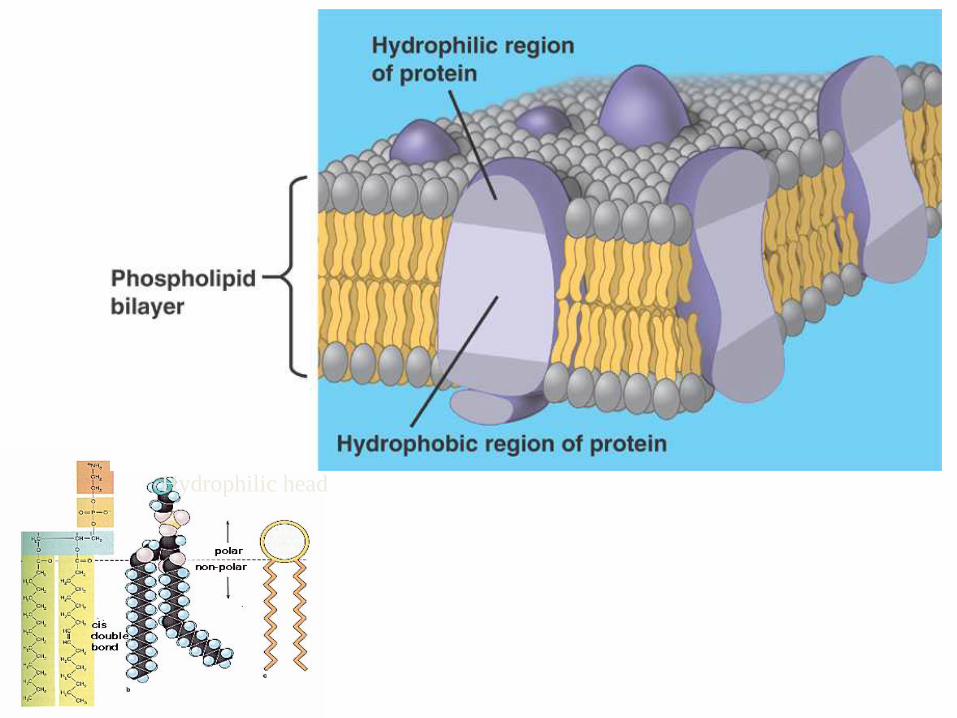
Hydrophilic head
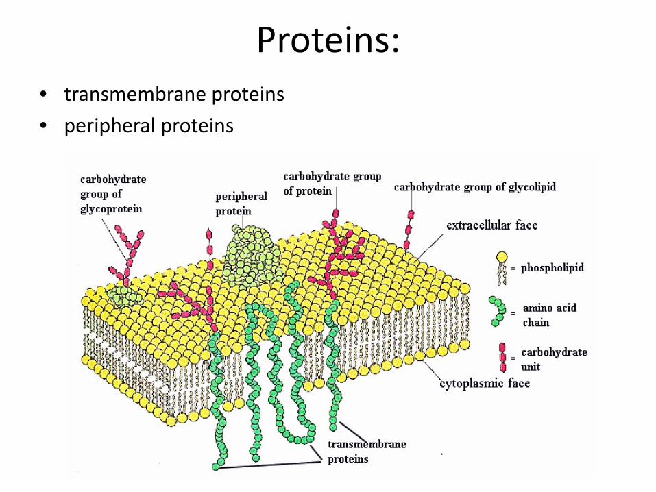
Proteins:• transmembrane proteins• peripheral proteins
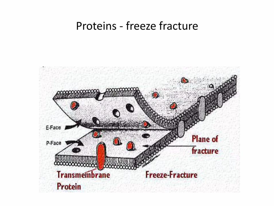
Proteins ‐ freeze fracture
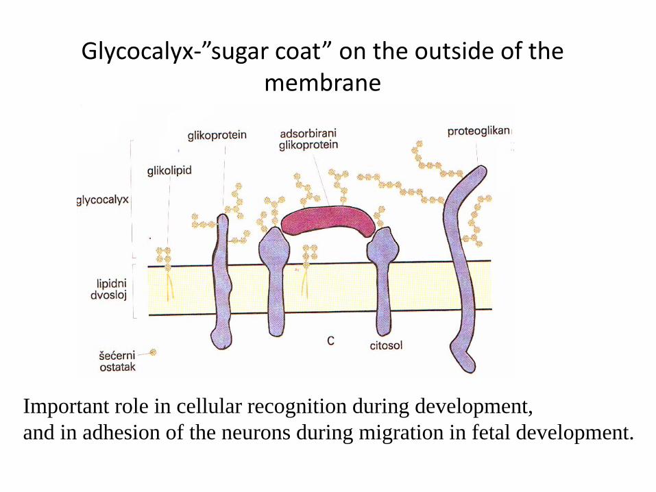
Glycocalyx‐”sugar coat” on the outside of themembrane
Important role in cellular recognition during development, and in adhesion of the neurons during migration in fetal development.
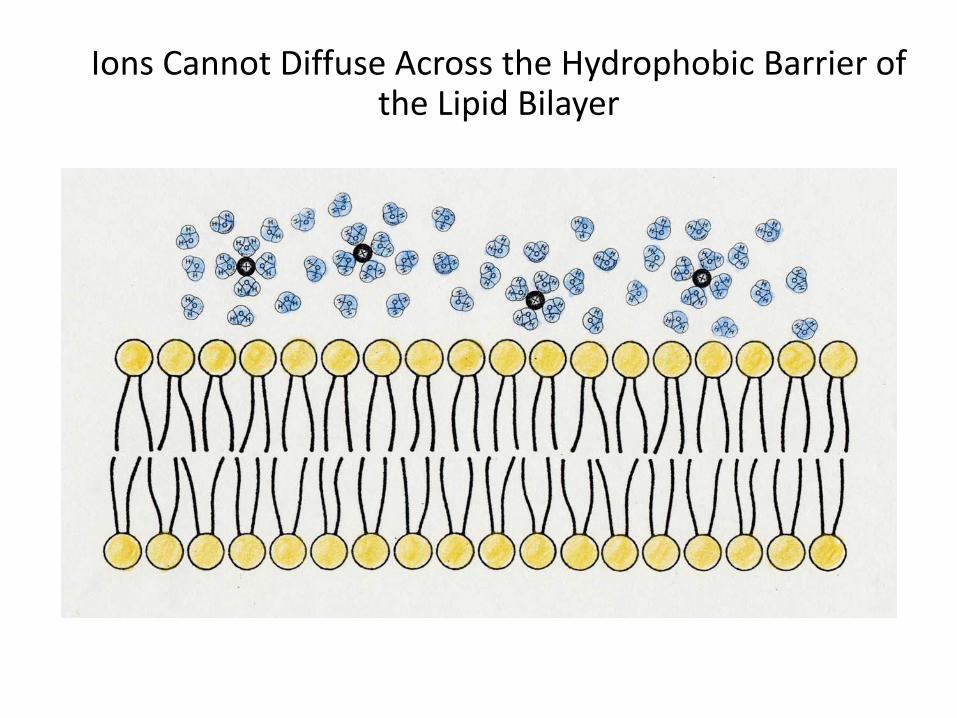
Ions Cannot Diffuse Across the Hydrophobic Barrier of the Lipid Bilayer
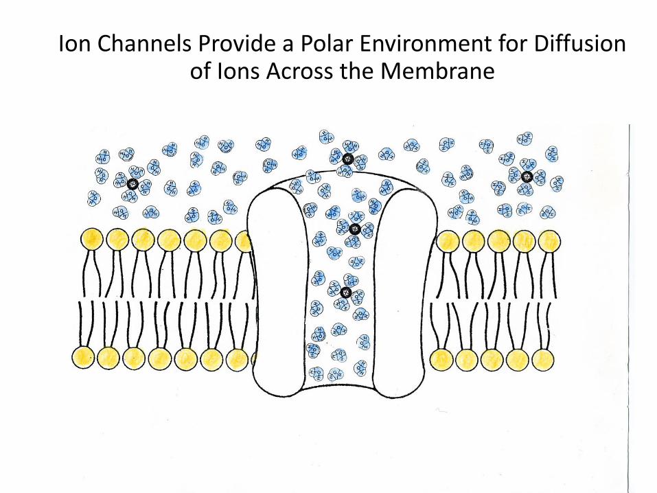
Ion Channels Provide a Polar Environment for Diffusion of Ions Across the Membrane
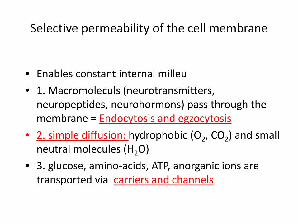
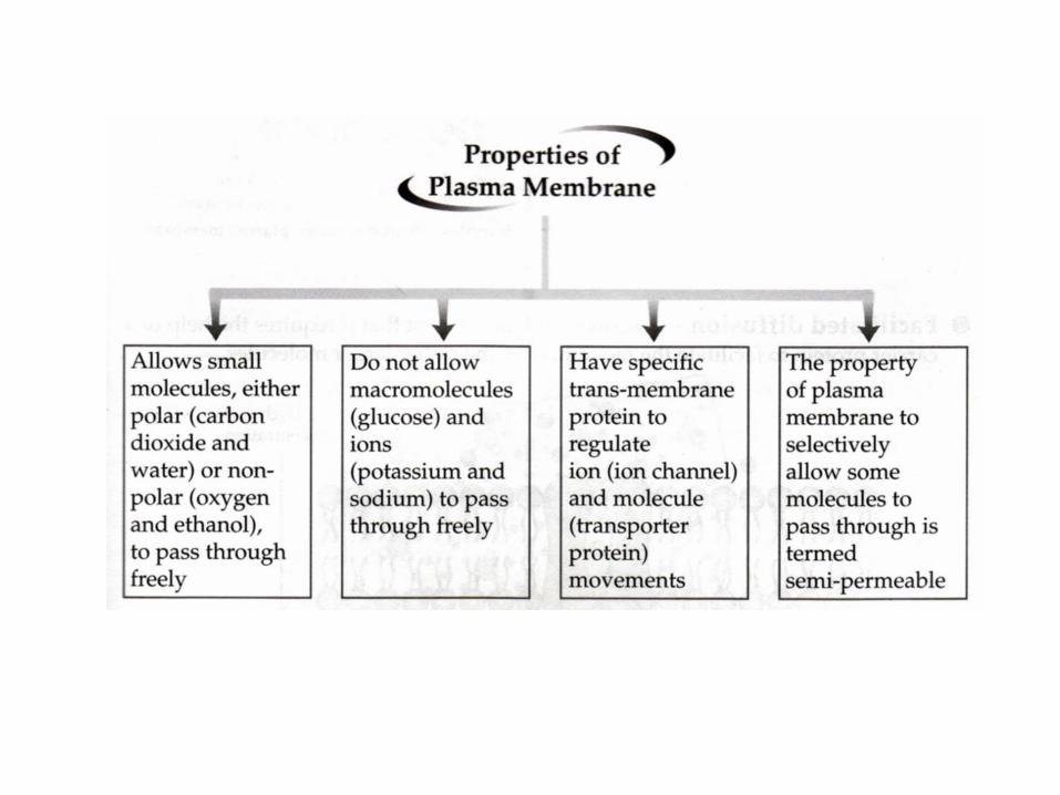
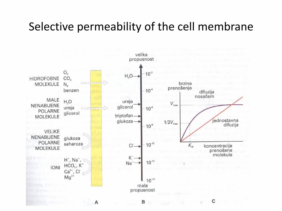
Selective permeability of the cell membrane
• Enables constant internal milleu• 1. Macromoleculs (neurotransmitters, neuropeptides, neurohormons) pass through themembrane = Endocytosis and egzocytosis
• 2. simple diffusion: hydrophobic (O2, CO2) and smallneutral molecules (H2O)
• 3. glucose, amino‐acids, ATP, anorganic ions are transported via carriers and channels
Selective permeability of the cell membrane
A) PASSIVE TRANSPORT
• diffusion: down the concentration gradient
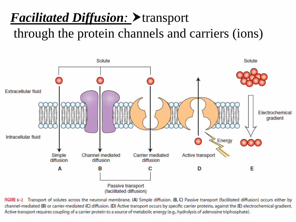
Facilitated Diffusion: transportthrough the protein channels and carriers (ions)
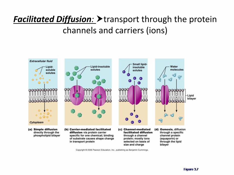
Facilitated Diffusion: transport through the protein channels and carriers (ions)
Ion channels
• They form hydrophilic pores through themembrane.
• Transport is faster then via carriers. • The flow of ions through the channels does not require metabolic energy; the flow is passive.
• The electrochemical driving force across the membrane, but not the channel itself, determines the direction and eventual equilibrium of this flow.
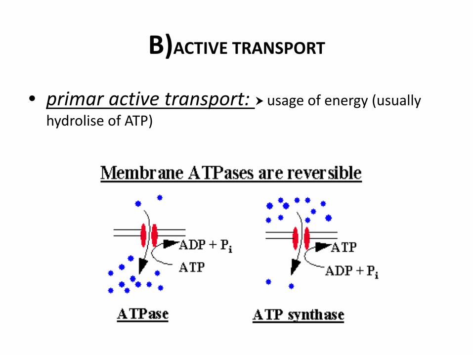
B)ACTIVE TRANSPORT
• primar active transport: usage of energy (usually hydrolise of ATP)
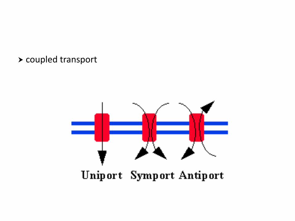
coupled transport
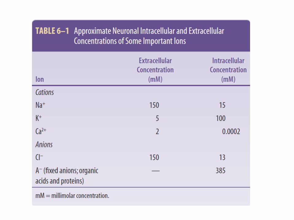
Ion ballance
• Na+
• K+
• Ca2+
• Cl‐
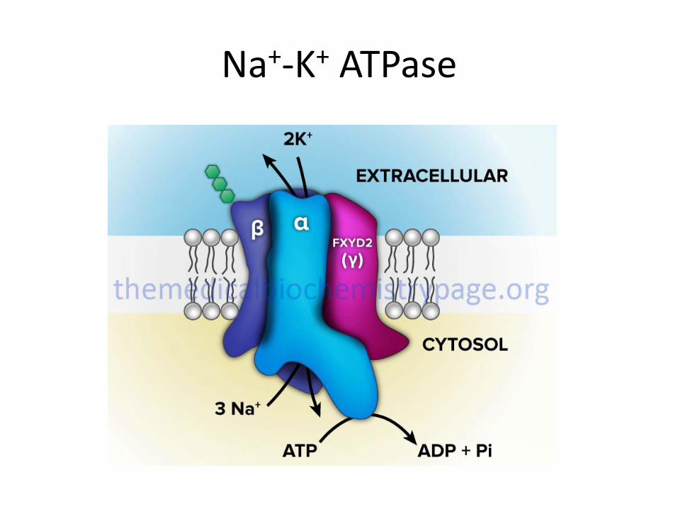
Na+‐K+ ATPase
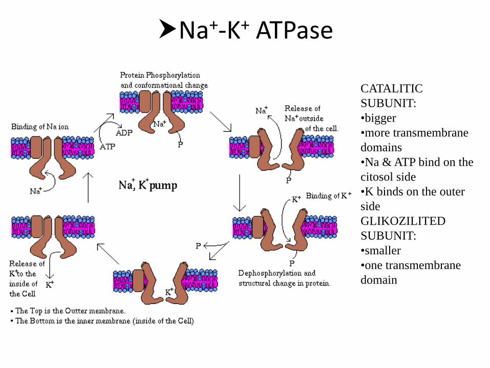
Na+‐K+ ATPase
CATALITIC SUBUNIT: •bigger•more transmembrane domains•Na & ATP bind on the citosol side•K binds on the outer sideGLIKOZILITED SUBUNIT:•smaller•one transmembrane domain
Na+‐K+ ATP‐ase
• Works on the principle of antiport • ATP is allways hydrolised• Na+ & ATP have to be in the cell, and K+ outside of the cell
• ouabain inhibits Na‐K ATP‐ase binding itself on the K+ binding spot
• One molecule of ATP: 3 Na+ get out, and 2 K+ get in.
Na+‐K+ ATP‐ase
• Contributes to the negative potential inside the neuron
• Contributes to the osmotic balance• Stabilizes cell volume
Concentracion of Ca2+
• Ca2+ inside the cell 10‐7 M• Ca2+ outside the cell 10‐3 M• concentracion gradient towards inside• Role of:• Ca2+ ATP‐ase• Ca2+ ‐ Na+ transporter
Maintanence of pH value
• Inside the neuron pH=7,2• Na+ ‐ H+ pump (gets H+ out the cell)• Cl‐ ‐ HCO3
‐pump (HCl gets out, NaHCO3 gets in)
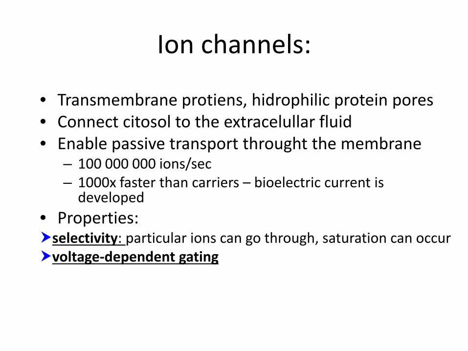
Ion channels:
• Transmembrane protiens, hidrophilic protein pores• Connect citosol to the extracelullar fluid• Enable passive transport throught the membrane
– 100 000 000 ions/sec– 1000x faster than carriers – bioelectric current is developed
• Properties:selectivity: particular ions can go through, saturation can occurvoltage‐dependent gating
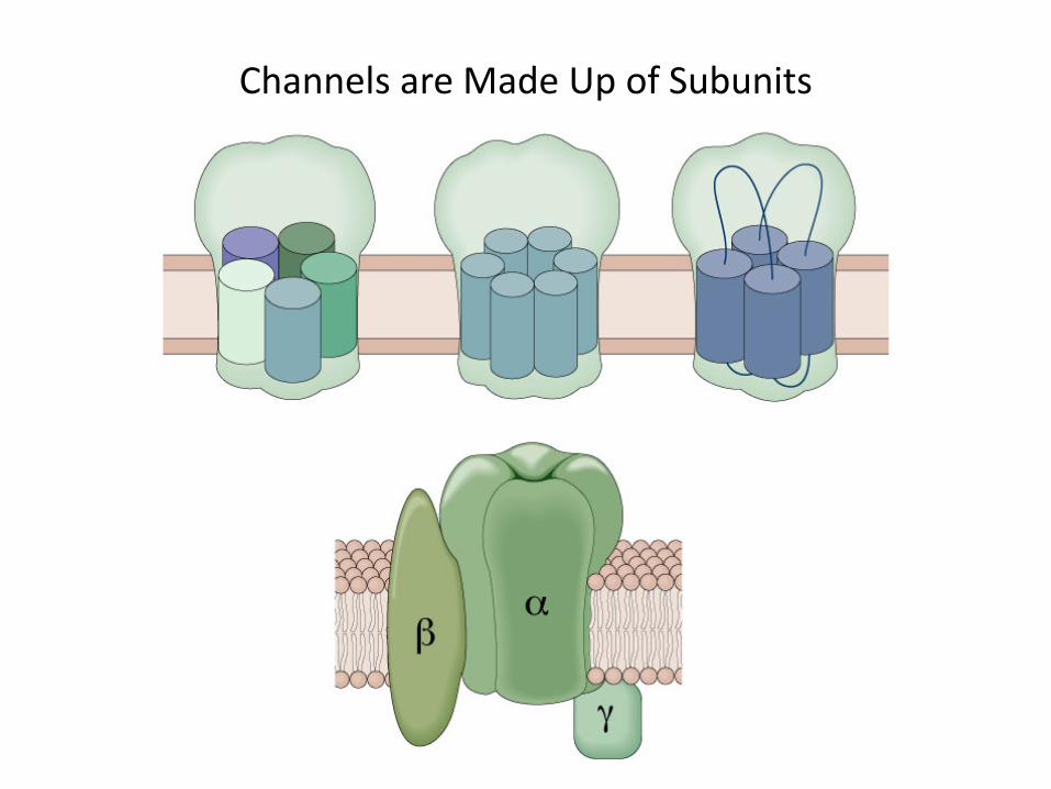
Channels are Made Up of Subunits
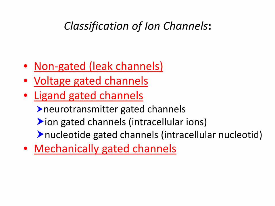
Classification of Ion Channels:
• Non‐gated (leak channels)• Voltage gated channels• Ligand gated channels
neurotransmitter gated channelsion gated channels (intracellular ions)nucleotide gated channels (intracellular nucleotid)
• Mechanically gated channels
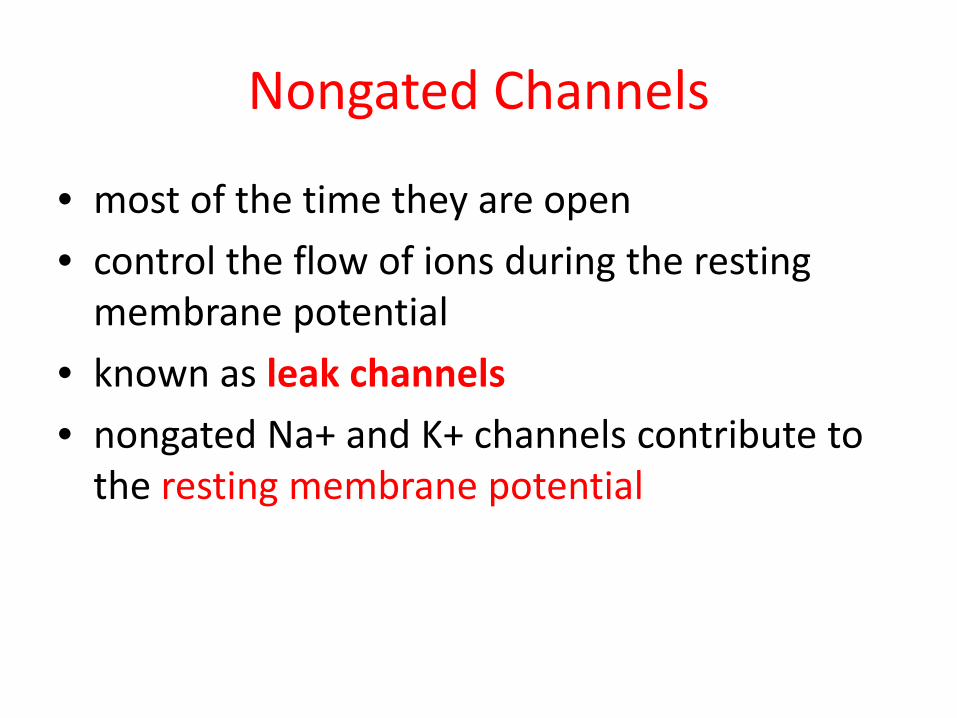
Nongated Channels
• most of the time they are open• control the flow of ions during the restingmembrane potential
• known as leak channels• nongated Na+ and K+ channels contribute to the resting membrane potential
Gated Channels
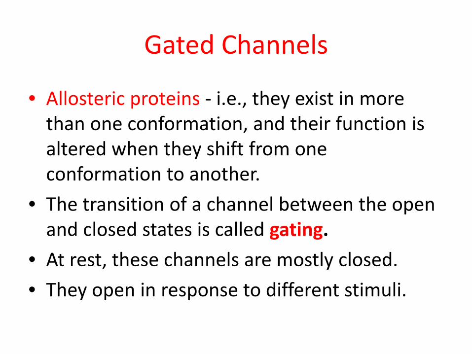
• Allosteric proteins ‐ i.e., they exist in more than one conformation, and their function is altered when they shift from oneconformation to another.
• The transition of a channel between the open and closed states is called gating.
• At rest, these channels are mostly closed.• They open in response to different stimuli.
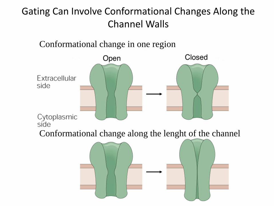
Gating Can Involve Conformational Changes Along the Channel Walls
Conformational change in one region
Conformational change along the lenght of the channel
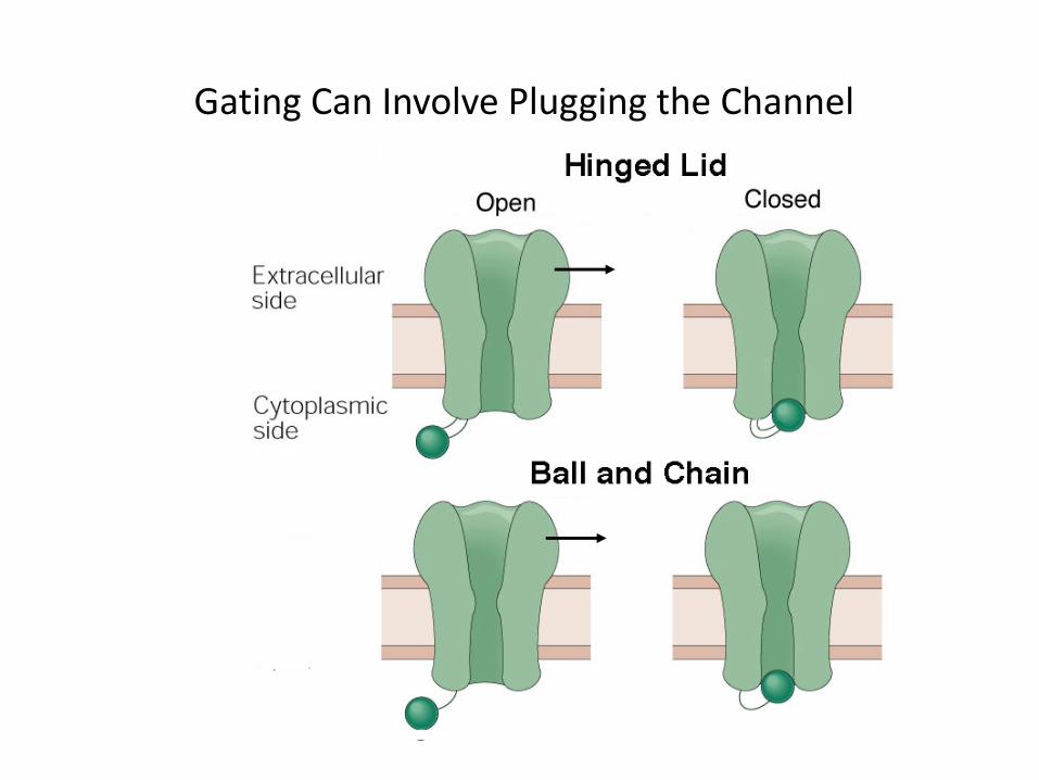
Gating Can Involve Plugging the Channel
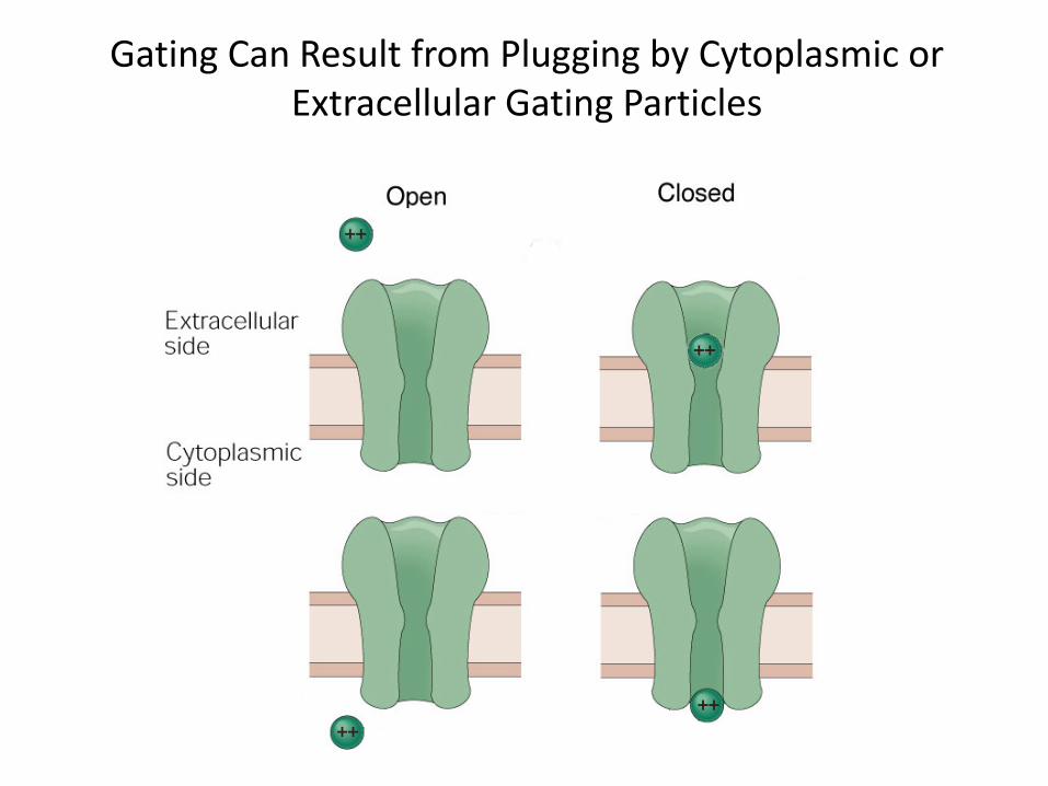
Gating Can Result from Plugging by Cytoplasmic or Extracellular Gating Particles
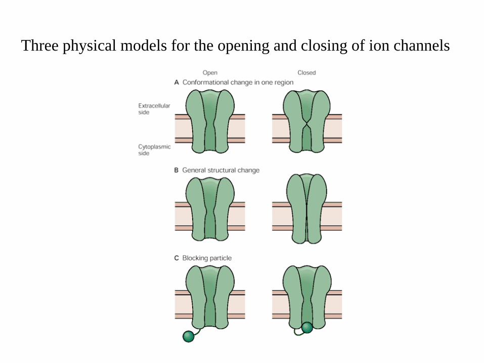
Three physical models for the opening and closing of ion channels
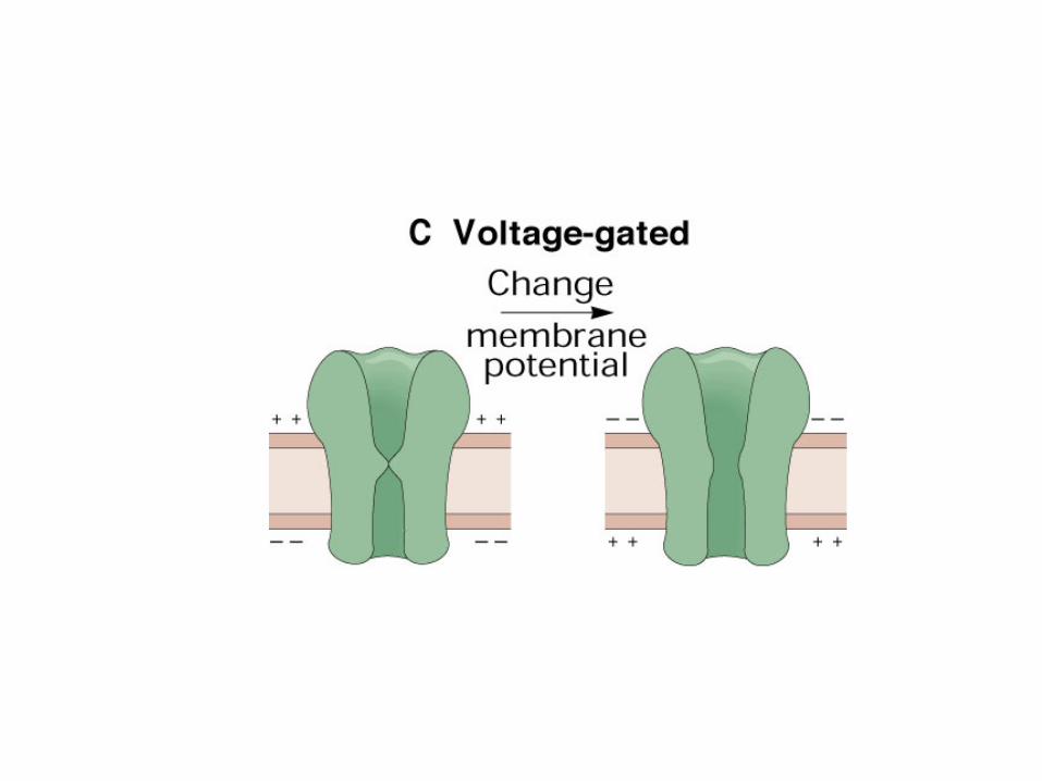
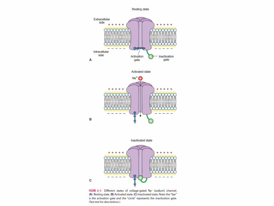
Voltage‐gated channels
• ‐ are opened or closed by a change in themembrane potential
• exist in three states:• (1) resting state, in which the channel is closed but can be activated;
• (2) active state, in which the channel is open;• (3) refractory state, in which the channel is inactivated
• voltage‐gated Na+ channel• single long polypeptide that has four domains• Channel is more permeable to Na+ than to K+
• The S4 segment undergoes a conformationalchange when the membrane potential changes
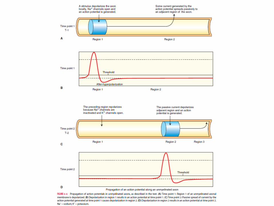
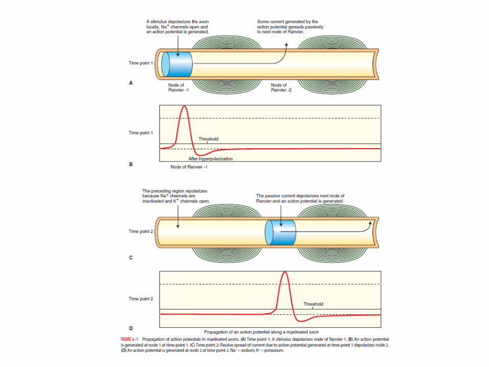
• When the membrane is depolarized beyond the threshold potential, a sufficient number of voltage‐gated Na+ channels open
• action potentials are generated

• There are some cases in which Na+ permeability is blocked.
• Tetrodotoxin (TTX) ‐ a toxin isolated from theovaries of Japanese puffer fish
• binds to the sodium channel on the outside and blocks the sodium permeability pore
• Neurons are not able to generate actionpotentials after the application of TTX
• Lidocaine also blocks these channels
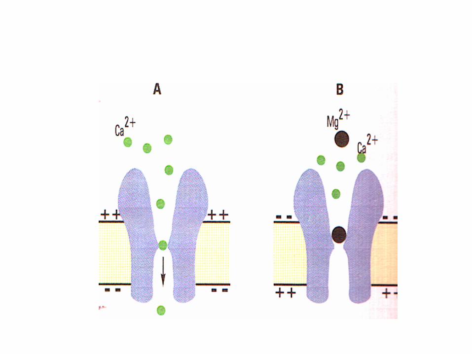
• voltage‐gated Ca2+ channel• Ca2+ ions enter the postsynaptic neurons through these channels
• Ca2+ ions activate enzymes• Depolarization of presynaptic nerve terminals results in entry of Ca2+ ions into the terminal viathese channels
• An increase in the levels of intracellular Ca2+ results in the release of transmitters frompresynaptic nerve terminals.
• voltage‐gated K+ channels• K+ channels are generally blocked by chemicals, such as tetraethylammonium or 4‐aminopyridine
• The opening of voltage‐ gated K+ channels is also caused by depolarization of the neuronal membrane
• Because these voltage‐gated K+ channels open with a delay (about 1 msec) after the membranedepolarization, they are called delayed rectifier K+channels.
• Repolarization of the neuron• after‐hyperpolarization or undershoot• TEA – blocks these channels
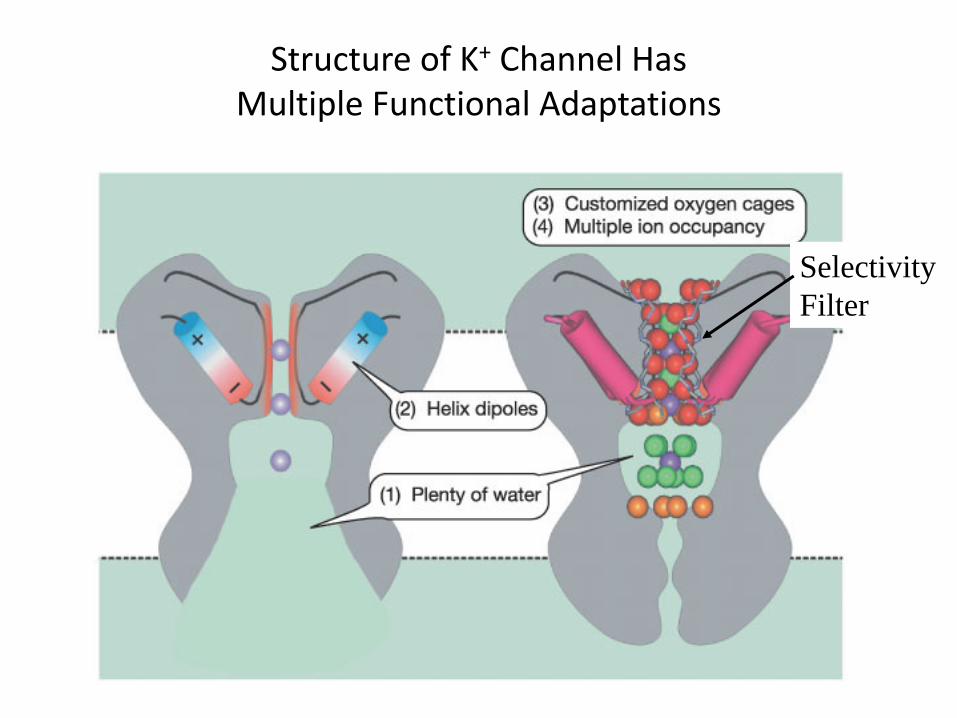
Structure of K+ Channel HasMultiple Functional Adaptations
SelectivityFilter
Ligand‐gated channels
• are opened by noncovalent binding of chemical substances with their receptors on the neuronal membrane
• transmitters or hormones present in the extracellular fluid (acetylcholine, γ‐aminobutyric acid [GABA], or glycine)
• an intracellular second messenger (e.g., cyclic adenosine monophosphate, which is activated by a transmitter such as norepinephrine
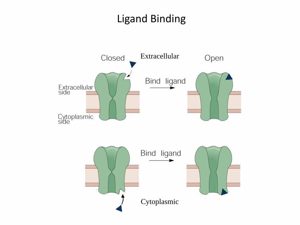
Ligand Binding
Extracellular
Cytoplasmic
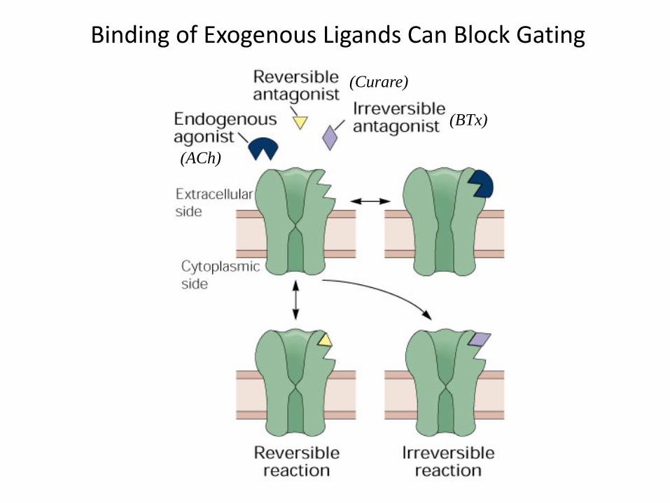
Binding of Exogenous Ligands Can Block Gating
(ACh)
(Curare)
(BTx)
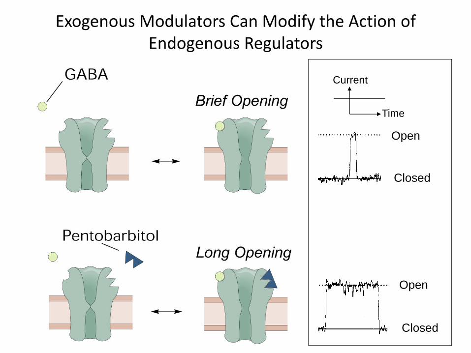
Exogenous Modulators Can Modify the Action of Endogenous Regulators
Open
Closed
Open
Closed
Current
Time
• Directly gated ligand channel:• five protein subunits• Recognition site for the chemical substance is part of the ion channel
• Ionotropic receptor• A neurotransmitter binds to an ionotropic receptor and brings about a conformational change that results in the opening of the ion channel
• usually bring about fast synaptic responses that last for only a few milliseconds
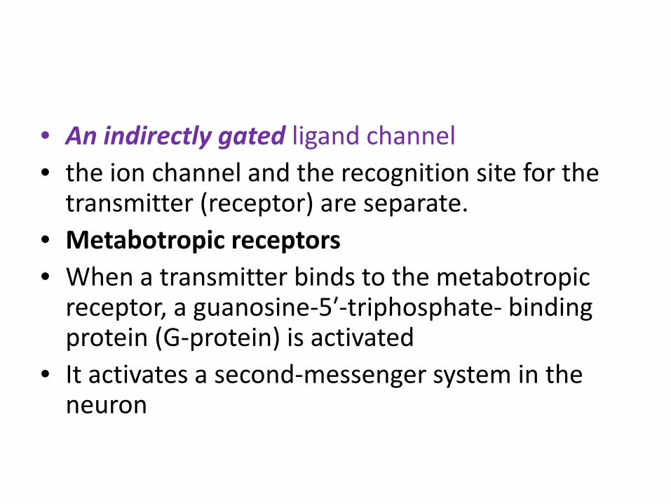
• An indirectly gated ligand channel• the ion channel and the recognition site for the transmitter (receptor) are separate.
• Metabotropic receptors• When a transmitter binds to the metabotropicreceptor, a guanosine‐5′‐triphosphate‐ binding protein (G‐protein) is activated
• It activates a second‐messenger system in the neuron
• The second messenger can either act directly on the ion channel to open it, or
• It can activate an enzyme that, opens the channel by phosphorylating the channelprotein in the presence of a protein kinase
• Elicits slow, long‐lasting synaptic actions
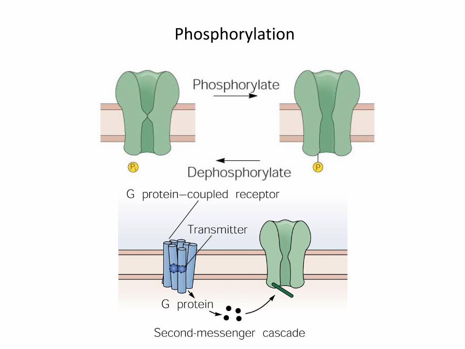
Phosphorylation
Mechanically gated channels
• open by a mechanical stimulus• include the channels involved in producinggenerator potentials of stretch and touch receptors
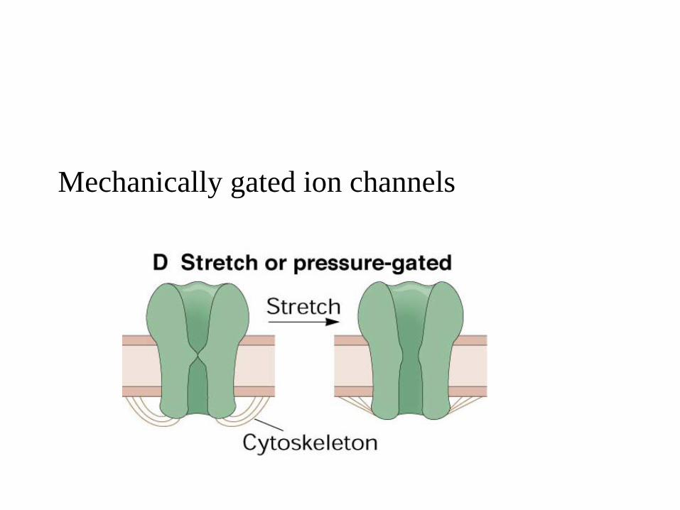
Mechanically gated ion channels
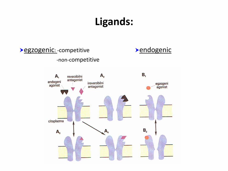
Ligands:
egzogenic: ‐competitive endogenic‐non‐competitive
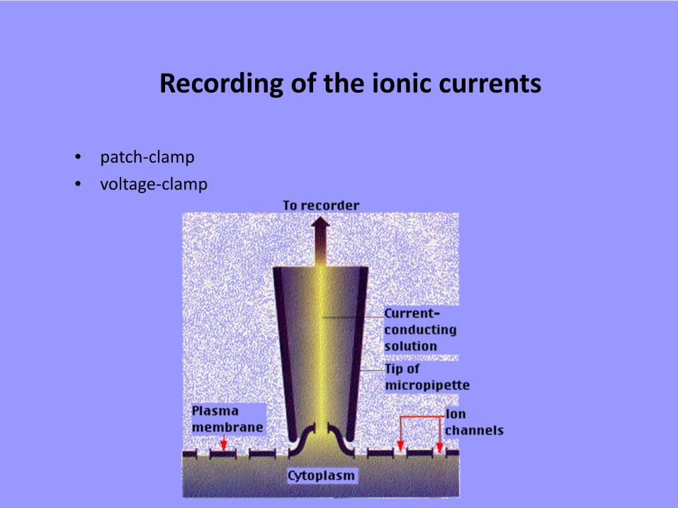
Recording of the ionic currents
• patch‐clamp• voltage‐clamp
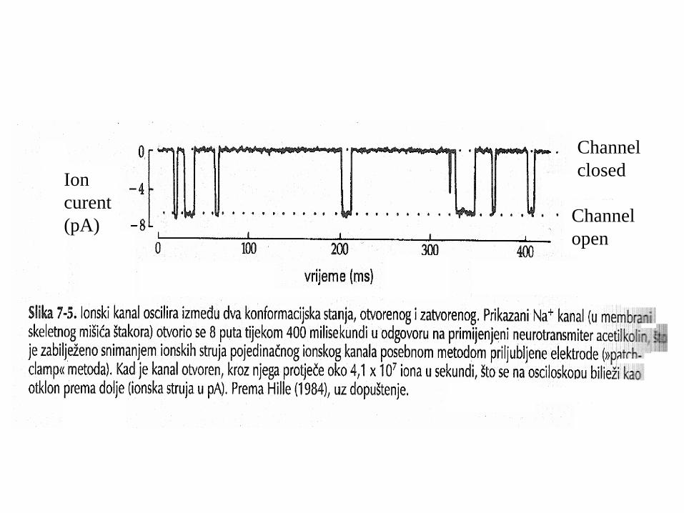
Channel closed
Channel open
Ion curent(pA)
CLINICAL CONSIDERATIONS• Lambert‐Eaton (Eaton‐Lambert) Syndrome• associated with small‐cell carcinoma of the lung• cells expressing voltage‐gated Ca2+ channels• An antibody is produced in the body against these Ca2+
channels,• its presence results in a loss of voltage‐gated Ca2+ channels
in the presynaptic terminals• less Ca2+ enters the presynaptic terminal during
depolarization and, consequently, there is a reduction inthe release of the transmitter (Ach)
• results in muscle weakness, dry mouth, constipation, reduced sweating, orthostatic hypotension (dizziness while standing or walking), and impotence
• Multiple Sclerosis• an autoimmune disease with infl ammatory features that affect the CNS
• demyelination occurs in the axons of the CNS• Antibodies attack myelin, which then swells anddetaches.
• A scar (sclerosis) develops on the nerve fibers, which delays or blocks nerve impulses
• nerve fibers degenerate• commonly affects young people, especially women, in the 20‐year‐old to 40‐year‐old age group.
• Symptoms:• numbness in one or more limbs, typically on one side. • tingling or pain in parts of the body. • Visual pathways are affected, resulting in vision
disturbances• disturbances in speech• weakness in one or more limbs on one side• tremor, lack of coordination, wide steps, and an unsteady
gait• Neurological examination: hyper‐reflexia and Babinski’s
sign• Magnetic resonance imaging shows scarring in the CNS.
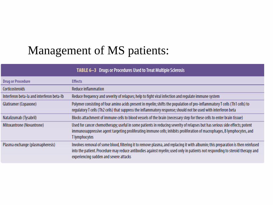
Management of MS patients:
• Cystic Fibrosis• an inherited disease that affects primarily the respiratory system
• Ion channel through which Cl− ions are transported into the cells is affected
• movement of water into the tissues is controlled to maintain the fluidity of mucus is changed
• Guillain‐Barré Syndrome• demyelination of axons in the peripheral nerves occurs
Summary
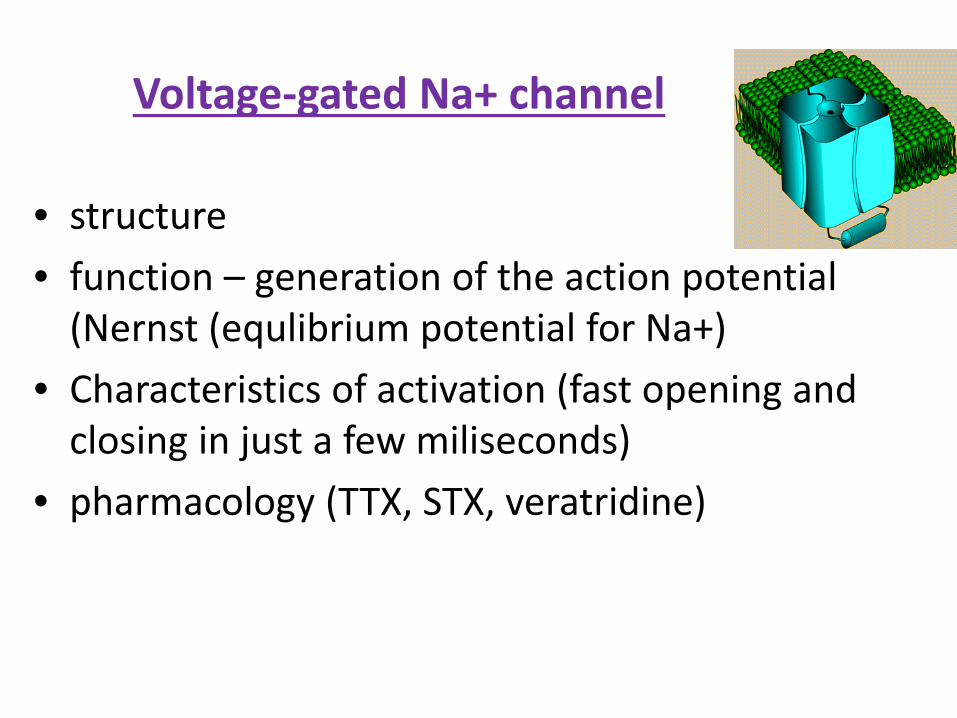
Voltage‐gated Na+ channel
• structure• function – generation of the action potential (Nernst (equlibrium potential for Na+)
• Characteristics of activation (fast opening and closing in just a few miliseconds)
• pharmacology (TTX, STX, veratridine)
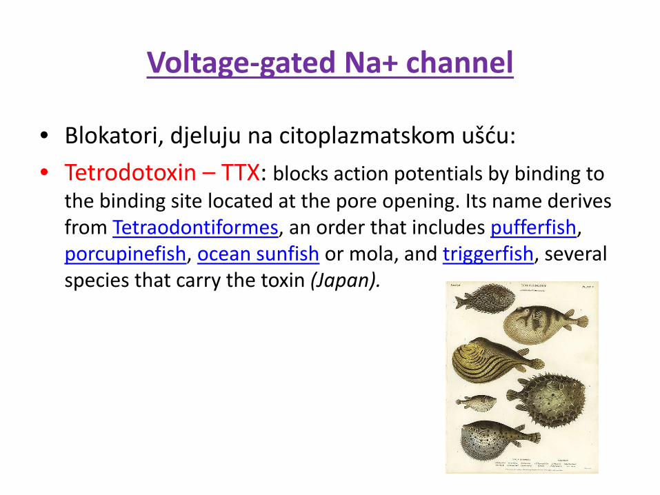
Voltage‐gated Na+ channel
• Blokatori, djeluju na citoplazmatskom ušću:• Tetrodotoxin – TTX: blocks action potentials by binding to
the binding site located at the pore opening. Its name derives from Tetraodontiformes, an order that includes pufferfish, porcupinefish, ocean sunfish or mola, and triggerfish, several species that carry the toxin (Japan).
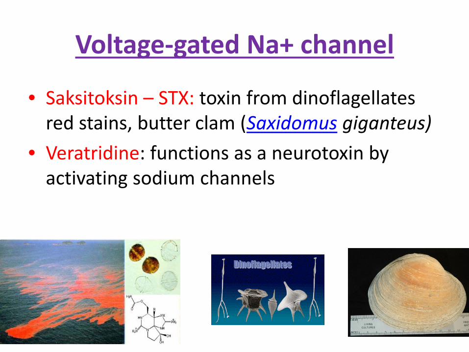
Voltage‐gated Na+ channel
• Saksitoksin – STX: toxin from dinoflagellatesred stains, butter clam (Saxidomus giganteus)
• Veratridine: functions as a neurotoxin by activating sodium channels
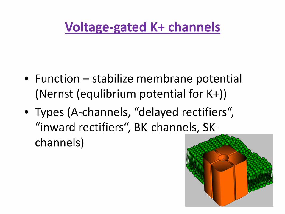
Voltage‐gated K+ channels
• Function – stabilize membrane potential(Nernst (equlibrium potential for K+))
• Types (A‐channels, “delayed rectifiers“, “inward rectifiers“, BK‐channels, SK‐channels)
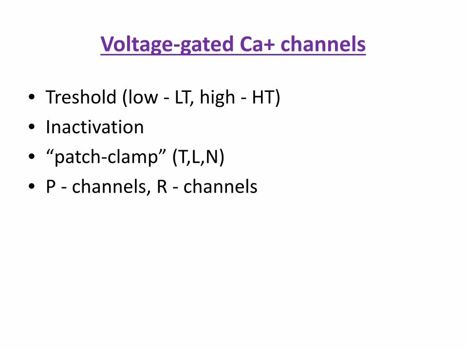
Voltage‐gated Ca+ channels
• Treshold (low ‐ LT, high ‐ HT)• Inactivation• “patch‐clamp” (T,L,N)• P ‐ channels, R ‐ channels
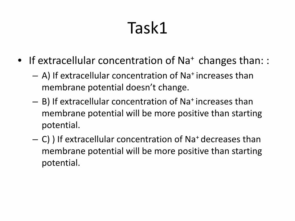
Task1
• If extracellular concentration of Na+ changes than: :– A) If extracellular concentration of Na+ increases thanmembrane potential doesn’t change.
– B) If extracellular concentration of Na+ increases thanmembrane potential will be more positive than startingpotential.
– C) ) If extracellular concentration of Na+ decreases thanmembrane potential will be more positive than startingpotential.
Task 2
• What are the factors to determine restingpotential: – ______________– ______________– ______________
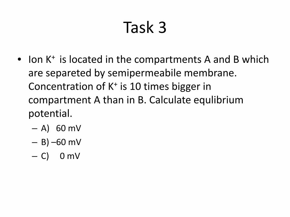
Task 3
• Ion K+ is located in the compartments A and B which are separeted by semipermeabile membrane. Concentration of K+ is 10 times bigger in compartment A than in B. Calculate equlibriumpotential. – A) 60 mV– B) –60 mV– C) 0 mV
Nernst equation
• EA‐EB = ‐ 60 mV/z * log((XB/XA))
Carriers• na sebe vežu specifičnu molekulu• podliježu konformacijskim promjenama• omogućuju prenošenje te molekule kroz membranu• nosač ima specifično vezno mjesto• proces prenošenja nalikuje reakciji enzim‐supstrat• konstanta vezanja supstrata (Km) = koncentracija tvari kad je
brzina prenošenja pola od maksimalne• Vmax – prenošenje najvećom brzinom kad suspstrat zauzme
sva vezna mjesta nosača
Nosači
• Vezanje supstrata može se blokirati• kompeticijski inhibitori – sprečavaju vezanje supstrata
• nekompeticijski inhibitori – alosterička modulacija