Embed Size (px)
Citation preview
Myopathies with myotonia or periodic weakness
Björn Falck
Deprtment of clinical neurophysiology
University hospital
Uppsala, Sweden
Ion channel disorders in neurology
MyopathiesMyotoniasOther episodic disorders
Neuromuscular tranmissionMyasthenic syndrome
Peripheral nervesNeuromyotonia
Central nervous systemEpilepsies – idiopathicAtaxias
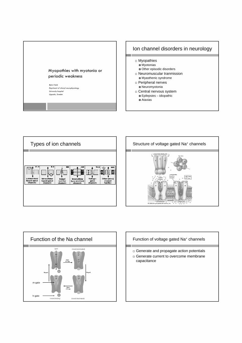
Types of ion channels Structure of voltage gated Na+ channels
Function of the Na channel
m-gate
h-gate
Function of voltage gated Na+ channels
Generate and propagate action potentialsGenerate current to overcome membrane capacitance
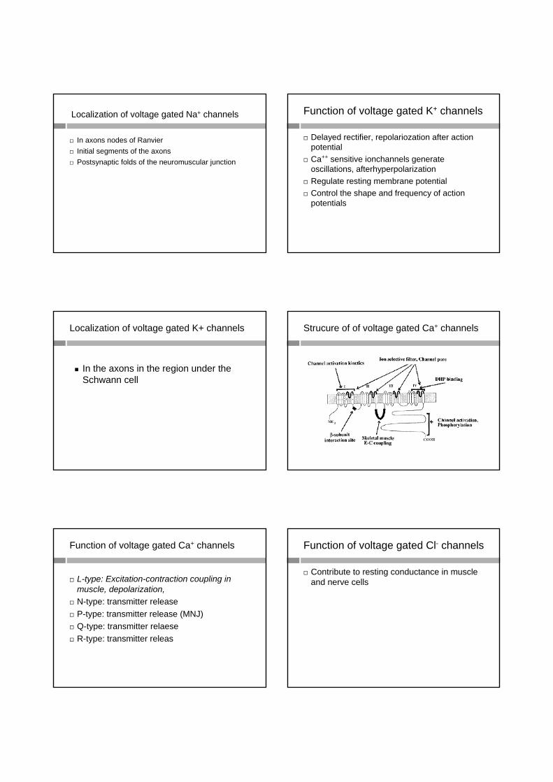
Localization of voltage gated Na+ channels
In axons nodes of RanvierInitial segments of the axonsPostsynaptic folds of the neuromuscular junction
Function of voltage gated K+ channels
Delayed rectifier, repolariozation after action potentialCa++ sensitive ionchannels generate oscillations, afterhyperpolarizationRegulate resting membrane potentialControl the shape and frequency of action potentials
Localization of voltage gated K+ channels
In the axons in the region under the Schwann cell
Strucure of of voltage gated Ca+ channels
Function of voltage gated Ca+ channels
L-type: Excitation-contraction coupling in muscle, depolarization,N-type: transmitter releaseP-type: transmitter release (MNJ)Q-type: transmitter relaeseR-type: transmitter releas
Function of voltage gated Cl- channels
Contribute to resting conductance in muscle and nerve cells
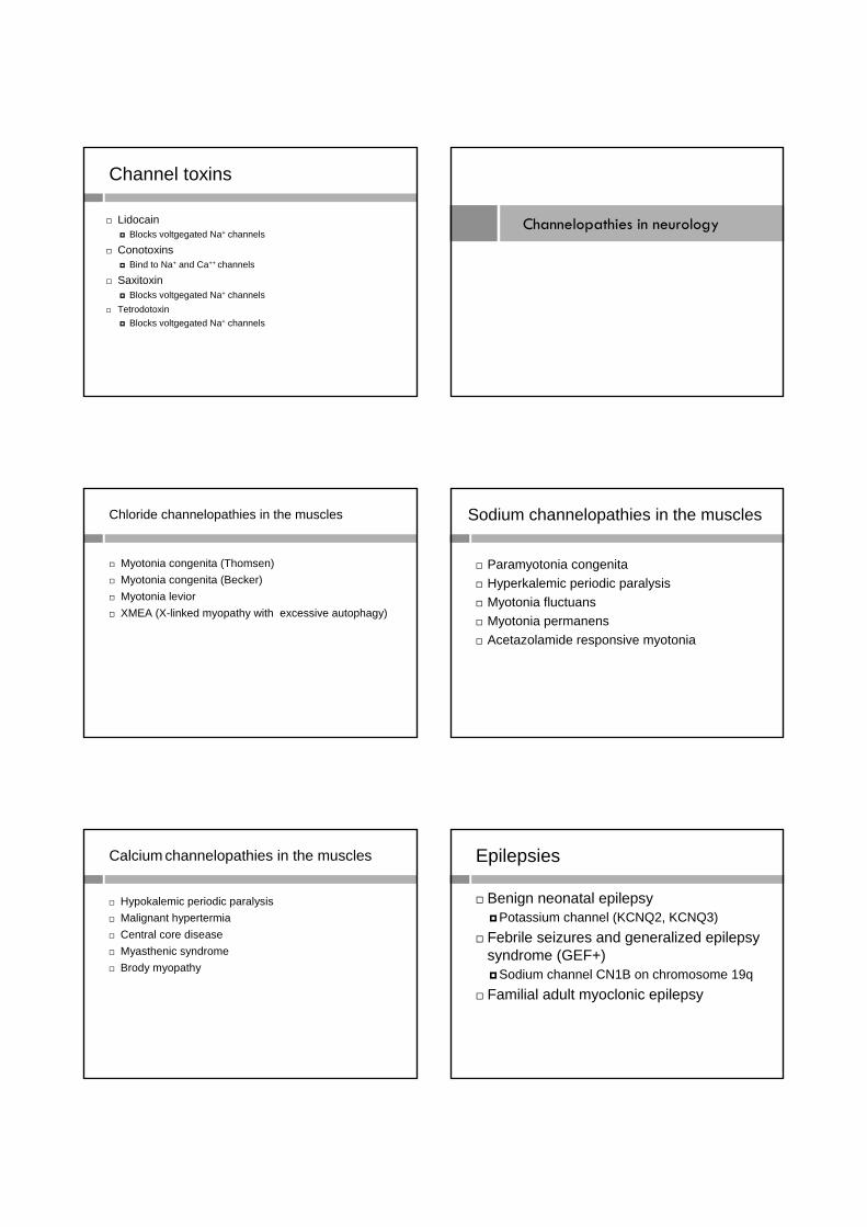
Channel toxins
LidocainBlocks voltgegated Na+ channels
ConotoxinsBind to Na+ and Ca++ channels
SaxitoxinBlocks voltgegated Na+ channels
TetrodotoxinBlocks voltgegated Na+ channels
Channelopathies in neurology
Chloride channelopathies in the muscles
Myotonia congenita (Thomsen)Myotonia congenita (Becker) Myotonia leviorXMEA (X-linked myopathy with excessive autophagy)
Sodium channelopathies in the muscles
Paramyotonia congenitaHyperkalemic periodic paralysisMyotonia fluctuansMyotonia permanensAcetazolamide responsive myotonia
Calcium channelopathies in the muscles
Hypokalemic periodic paralysisMalignant hypertermiaCentral core diseaseMyasthenic syndromeBrody myopathy
Epilepsies
Benign neonatal epilepsyPotassium channel (KCNQ2, KCNQ3)
Febrile seizures and generalized epilepsy syndrome (GEF+)
Sodium channel CN1B on chromosome 19qFamilial adult myoclonic epilepsy
Ataxias
Episodic ataxia type 2Ca++ channel
Episodic ataxia/Myokymia syndromeK+ channel
Progressive ataxia SCA6
Potassium channelopathies in the nerves
Neuromyotonia
Myotonic disorders
Myotonia - clinical definition
Uncontrolled temporary stiffness of muscle due to transient hyperexcitability of muscle fiber membrane
Myotonia after rest Myotonia, effect of warm-up
AANEM definition: myotonic discharges
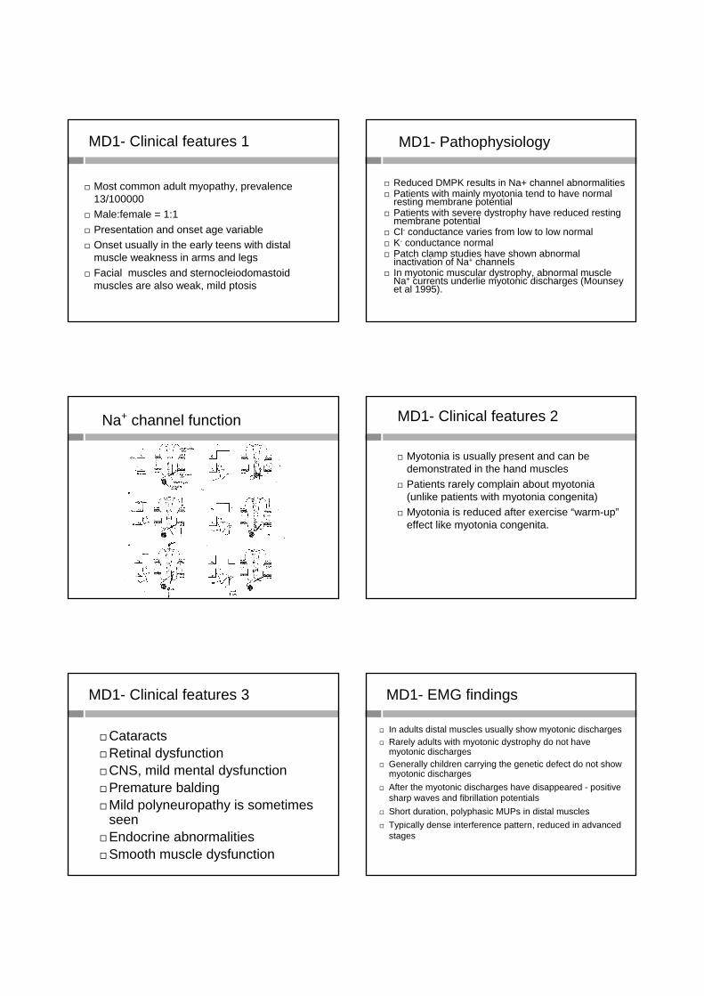
Repetitive discharge at rates of 20 to 80 Hz are of two different types: (1) biphasic (positive negative) spike potentials less than 5 ms in duration resembling fibrillation potentials, (2) positive waves of 5 to 20 ms duration resembling positive sharp waves. Both potential forms are recorded after needle insertion, after voluntary muscle contraction or after muscle percussion, and aredue to independent, repetitive discharges of single muscle fibers. The amplitude and frequency of the potentials must both wax and wane to be identified as myotonic discharges. This change produces a characteristic musical sound due to corresponding change in pitch, which has been likened to the sound of a “dive bomber”.
Muscle and Nerve 1987; vol 10
Myotonic discharges
Clinical features of myotonia
Sustained involuntary muscle contraction following strong muscle contractionEffect of exercise
Improvement (myotonia congenita, myotonic dystrophy)Aggravation (paramyotonia, myotonia fluctuans)
Effect of temperatureAggravation by cooling in paramyotonia congenita
Variation of symptoms over timeCl channelopathies usually stableNa channelopathies vary
Clinical classification
Progressive myopathy and myotoniaMyotonic dystrophy type 1( MD1) and type 2 (MD2)
Main symptom myotoniaMyotonia congenita (Thomsen and Becker)Myotonia fluctuans
Myotonia and episodic weaknessParamyotonia congenitaHyperkalemic periodic paralyis
Periodic paralysisHypokalemic periodic paralysis
Myotonic dystrophy type 1 (MD1)
MD1- Genetics
Autosomal dominant inheritanceGene location 19q13.2-q13.3Gene product myotonin protein kinaseDMPKExpanded CGT trinucleotide repeat in DMPK in normal subjects the length is up to 37 repeats in patients with myotonic dystrophy up to 2000 repeats can be foundSeverity is related with the number of repeats Reduced amount of DMPK may lead to cell apoptosis
MD1- Clinical features 1
Most common adult myopathy, prevalence 13/100000Male:female = 1:1Presentation and onset age variable Onset usually in the early teens with distal muscle weakness in arms and legsFacial muscles and sternocleiodomastoidmuscles are also weak, mild ptosis
MD1- Pathophysiology
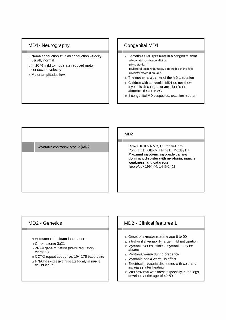
Reduced DMPK results in Na+ channel abnormalitiesPatients with mainly myotonia tend to have normal resting membrane potentialPatients with severe dystrophy have reduced resting membrane potentialCl- conductance varies from low to low normalK- conductance normalPatch clamp studies have shown abnormal inactivation of Na+ channels In myotonic muscular dystrophy, abnormal muscle Na+ currents underlie myotonic discharges (Mounseyet al 1995).
Na+ channel function MD1- Clinical features 2
Myotonia is usually present and can be demonstrated in the hand musclesPatients rarely complain about myotonia(unlike patients with myotonia congenita)Myotonia is reduced after exercise “warm-up”effect like myotonia congenita.
MD1- Clinical features 3
CataractsRetinal dysfunctionCNS, mild mental dysfunctionPremature baldingMild polyneuropathy is sometimes seenEndocrine abnormalitiesSmooth muscle dysfunction
MD1- EMG findings
In adults distal muscles usually show myotonic dischargesRarely adults with myotonic dystrophy do not have myotonic dischargesGenerally children carrying the genetic defect do not show myotonic dischargesAfter the myotonic discharges have disappeared - positive sharp waves and fibrillation potentialsShort duration, polyphasic MUPs in distal musclesTypically dense interference pattern, reduced in advanced stages
MD1- Neurography
Nerve conduction studies conduction velocity usually normal In 10 % mild to moderate reduced motor conduction velocityMotor amplitudes low
Congenital MD1
Sometimes MD1presents in a congenital formNeonatal respiratory distresHypotoniaBilateral facial weakness, deformities of the footMental retardation, and
The mother is a carrier of the MD 1mutationChildren with congenital MD1 do not show myotonic discharges or any significant abnormalities on EMGIf congenital MD suspected, examine mother
Myotonic dystrophy type 2 (MD2)
MD2
Ricker K, Koch MC, Lehmann-Horn F, Pongratz D, Otto M, Heine R, Moxley RT Proximal myotonic myopathy: a new dominant disorder with myotonia, muscle weakness, and cataracts.Neurology 1994;44: 1448-1452
MD2 - Genetics
Autosomal dominant inheritanceChromosome 3q21ZNF9 gene mutation (sterol regulatory element)CCTG repeat sequence, 104-176 base pairsRNA has exessive repeats focaly in muclecell nucleus
MD2 - Clinical features 1
Onset of symptoms at the age 8 to 60Intrafamilial variability large, mild anticipationMyotonia varies, clinical myotonia may be absentMyotonia worse during pregancyMyotonia has a warm-up effectElectrical myotonia decreases with cold and increases after heatingMild proximal weakness especially in the legs, develops at the age of 40-50
MD2 - Clinical features 2
Muscle pain is common, often disablingCataracts> 20years in 100%Diabetes in 20%Caridac arrthmiasHearing loss in 20%Clinical course is benignMild elevation of CKMuscle biopsy shows non-specific myopathic findings
MD2 - Pathophysiology
Not clearly understoodNormal resting membrane potentialNormal Cl- conductanceNeurophysiological basis for the myotonia is different from other channelopathies
MD2 - EMG findings
Myotonic discharges, especially in distal musclesMyotonic discharges may increase if the muscle temperature increasesMUP analysis normal or mild myopathicabnormalities, especially in proximal muscles
MD2 - Other findings
NeurographySensory neurography normalMotor CV normal, amplitudes low
Repetitive nerve stimulationUsually no decrement
Myotonia congenita (Thomsen)
Thomsen - Genetics
Autosomal dominant inheritanceMutation of the muscle chloride channel gene CLCN-1 on chromosome 7q35Gene product: muscle Cl - channel, ClC-1 Prevalence 4/100000 (Becker 1957)Penetrance 90%
Thomsen - Pathophysiology 1
The mutation interferes with Cl- ion channel tetramere formationCl- conductance stabilizes membrane potential, at rest Cl- ion movement across the membrane accounts for 70% of the membrane conductance The chloride conductance in the muscle fiber membrane is reduced resulting in accumulation of K+ ions in the t-tubules
Thomsen - Pathophysiology 2
Abnormal Na+ channel reopenings have been demonstratedThe membrane depolarization is prolonged giving rise to spontaneous repeated action potentialsOther factors contribute
Small changes in Na+ equilibrium may alter myotoniaNa + channel modification due to intracellular mechanisms may cause short term alterations
Thomsen - Clinical features
Onset of symptoms in infancy or early childhoodPersists throughout lifePresentation varies within familiesMyotonia is usually mild“Warm-up”Muscle strength normal, muscles appear hypertrophicMyotonia may fluctuate over timeMyotonia may be aggravated by β2 agonist drugs (fenoterol) and also β blocking agentsSometimes diuretic drugs aggravate myotoniaDepolarizing muscle relaxants may cause spasms
Thomsen - EMG
Myotonic discharges, particularly distally Warming up effect - less myotonia after a period of maximal contractionMotor unit potential analysis normal
Thomsen - Repetitive nerve stimulation
Decrementing response, especially at high stimulation frequencies (30 Hz) without intratetanic facilitationFollowing exercise the amplitude is decreased, reverse to facilitationCooling has no effect (in contrast to paramyotonia congenita)
Myotonia congenita (Becker)
Myotonia congenita (Becker) - Genetics
Autosomal recessive inheritanceMutation of the gene CLCN-1 gene on chromosome 7q35> 20 point mutations identifiedMost patients are compound heterozygotesGene product: muscle Cl – channelPrevalence 2-4/100 000 (Becker 1957)Prevalence probably much higher!! In Northern Finland 7/100 000. (Baumann et 1998)
Becker - Pathophysiology
Not as clear as for the dominant formInward rectification permits Cl- efflux but nor influxNo physiological mechanism identified for some mutations
Becker - Clinical features
Myotonia manifests at 7-14 years of agePersists throughout lifeMyotonia more severe than Thomsen’sNormal muscle strength, short period of exercise decreases strength, returns to normal with exerciseLeg and gluteal muscles are usually hypertrophicWeakness is worse in arms, myotonia worse in legsLordosis is commonNeck, shoulder and arm muscles appear atrophic“Warm-up”
Becker - EMG
Myotonic discharges, particularly distally MUP analysis may show mild myopathicabnormalitiesWarming up effect - less myotonia after a period of repeated contractions
Becker - Repetitive nerve stimulation
Decrementing response, especially at high stimulation frequencies (30 Hz)Following exercise the amplitude is decreased, reverse to facilitationCooling does not affect this in contrast to paramyotonia congenita
Becker - Carriers
Male carriers have myotonic discharges on EMGFemale carriers do not have myotonicdischarges on EMG
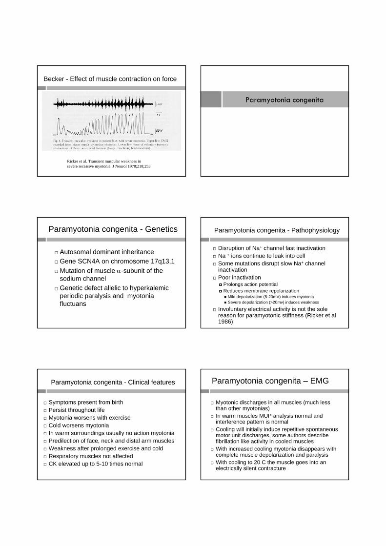
Becker - Effect of muscle contraction on force
Ricker et al. Transient muscular weakness in severe recessive myotonia. J Neurol 1978;218;253
Paramyotonia congenita
Paramyotonia congenita - Genetics
Autosomal dominant inheritanceGene SCN4A on chromosome 17q13,1Mutation of muscle α-subunit of the sodium channelGenetic defect allelic to hyperkalemicperiodic paralysis and myotoniafluctuans
Paramyotonia congenita - Pathophysiology
Disruption of Na+ channel fast inactivationNa + ions continue to leak into cellSome mutations disrupt slow Na+ channel inactivationPoor inactivation
Prolongs action potentialReduces membrane repolarization
Mild depolarization (5-20mV) induces myotoniaSevere depolarization (>20mv) induces weakness
Involuntary electrical activity is not the sole reason for paramyotonic stiffness (Ricker et al 1986)
Paramyotonia congenita - Clinical features
Symptoms present from birthPersist throughout lifeMyotonia worsens with exerciseCold worsens myotoniaIn warm surroundings usually no action myotoniaPredilection of face, neck and distal arm musclesWeakness after prolonged exercise and coldRespiratory muscles not affectedCK elevated up to 5-10 times normal
Paramyotonia congenita – EMG
Myotonic discharges in all muscles (much less than other myotonias)In warm muscles MUP analysis normal and interference pattern is normalCooling will initially induce repetitive spontaneous motor unit discharges, some authors describe fibrillation like activity in cooled musclesWith increased cooling myotonia disappears with complete muscle depolarization and paralysisWith cooling to 20 C the muscle goes into an electrically silent contracture
Paramyotonia congenita – Neurography
NeurographyMCS amplitude is reduced in cooled muscles
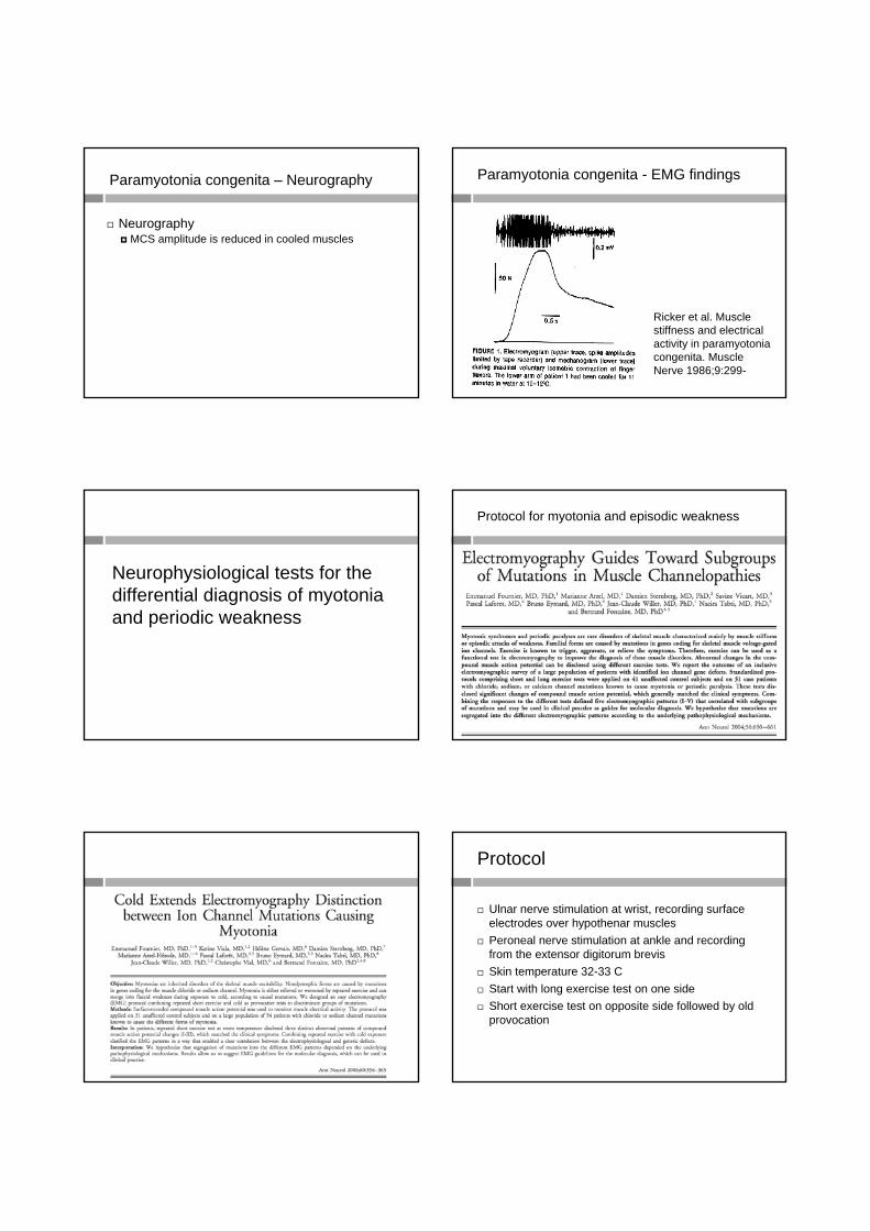
Paramyotonia congenita - EMG findings
Ricker et al. Muscle stiffness and electrical activity in paramyotoniacongenita. Muscle Nerve 1986;9:299-
Neurophysiological tests for the differential diagnosis of myotonia and periodic weakness
Protocol for myotonia and episodic weakness
Protocol
Ulnar nerve stimulation at wrist, recording surface electrodes over hypothenar musclesPeroneal nerve stimulation at ankle and recording from the extensor digitorum brevisSkin temperature 32-33 CStart with long exercise test on one sideShort exercise test on opposite side followed by old provocation
Short exercise test protocol
Repeat three times
M wave recorded at 10 second intervals for one minute
Strong isometric contraction 10-12 sec
Rep stim 10 stimuli at 3 Hz
60 second rest
Baseline M wave at rest
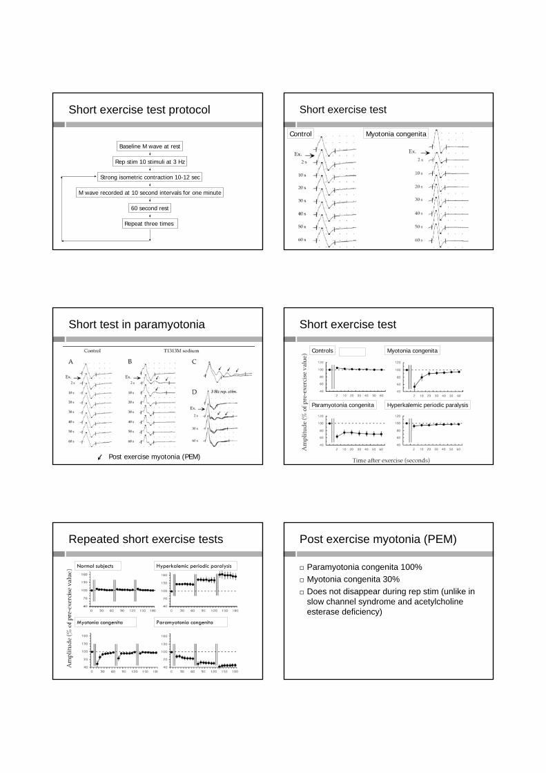
Short exercise test
Control Myotonia congenita
Short test in paramyotonia
Post exercise myotonia (PEM)
Short exercise test
Hyperkalemic periodic paralysisParamyotonia congenita
Myotonia congenitaControls
Repeated short exercise tests
Normal subjects Hyperkalemic periodic paralysis
Myotonia congenita Paramyotonia congenita
Post exercise myotonia (PEM)
Paramyotonia congenita 100%Myotonia congenita 30%Does not disappear during rep stim (unlike in slow channel syndrome and acetylcholine esterase deficiency)
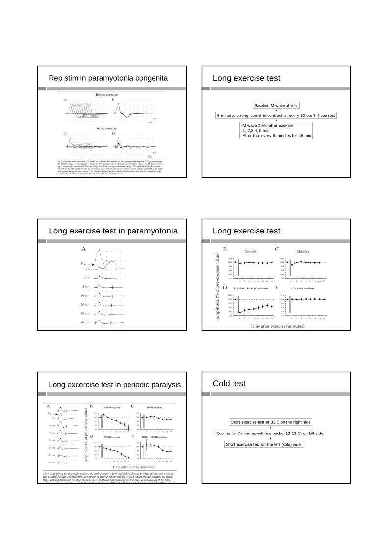
Rep stim in paramyotonia congenita Long exercise test
5 minutes strong isometric contraction every 30 sec 3-4 sec rest
-M wave 2 sec after exercise-1, 2,3,4, 5 min-After that every 5 minutes for 45 min
Baseline M wave at rest
Long exercise test in paramyotonia Long exercise test
Long excercise test in periodic paralysis Cold test
Short exercise test at 33 C on the right side
Cooling for 7 minutes with ice packs (12-13 C) on left side
Short exercise test on the left (cold) side
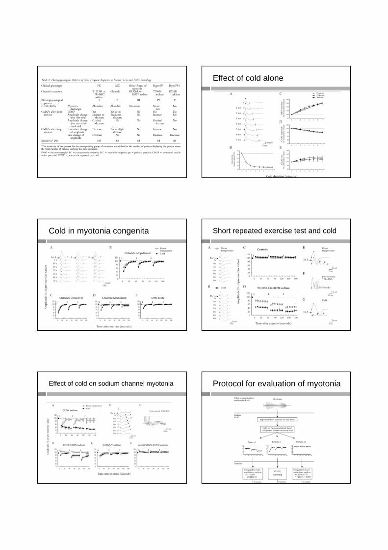
Effect of cold alone
Cold in myotonia congenita Short repeated exercise test and cold
Effect of cold on sodium channel myotonia Protocol for evaluation of myotonia
Protocol continued
Repetitive nerve stimulation (RNS) in myotonic disorders
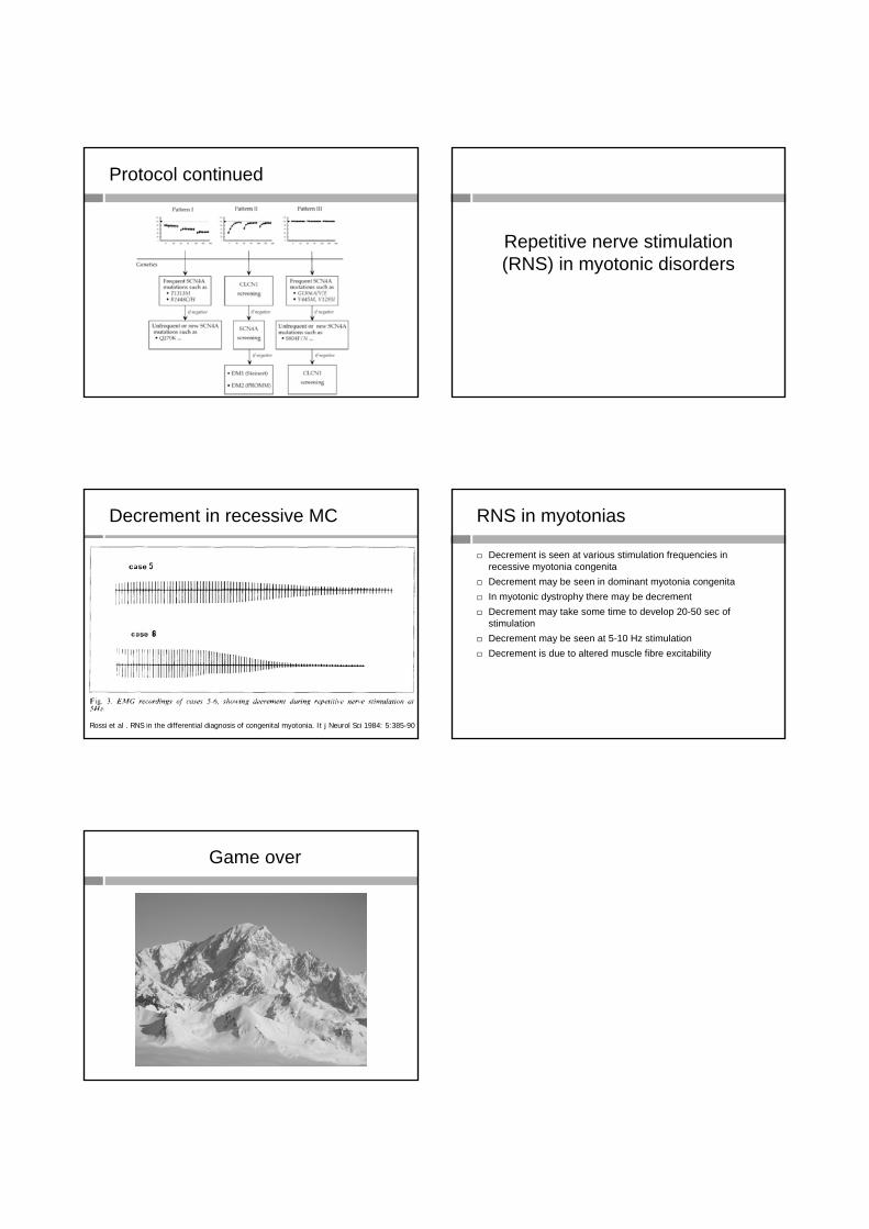
Decrement in recessive MC
Rossi et al . RNS in the differential diagnosis of congenital myotonia. It j Neurol Sci 1984: 5:385-90
RNS in myotonias
Decrement is seen at various stimulation frequencies in recessive myotonia congenitaDecrement may be seen in dominant myotonia congenitaIn myotonic dystrophy there may be decrementDecrement may take some time to develop 20-50 sec of stimulationDecrement may be seen at 5-10 Hz stimulationDecrement is due to altered muscle fibre excitability
Game over