Embed Size (px)
Citation preview
RESEARCH Open Access
Involvement of peripheral ionotropic glutamatereceptors in orofacial thermal hyperalgesia in ratsKuniya Honda1,2, Noboru Noma3,4, Masamichi Shinoda1,9, Makiko Miyamoto1,5, Ayano Katagiri6, Daiju Kita1,Ming-Gang Liu1, Barry J Sessle7, Masafumi Yasuda8 and Koichi Iwata1,9,10*
Abstract
Background: The purpose of the present study was to elucidate the mechanisms that may underlie thesensitization of trigeminal spinal subnucleus caudalis (Vc) and upper cervical spinal cord (C1-C2) neurons to heat orcold stimulation of the orofacial region following glutamate (Glu) injection.
Results: Glu application to the tongue or whisker pad skin caused an enhancement of head-withdrawal reflex andextracellular signal-regulated kinase (ERK) phosphorylation in Vc-C2 neurons. Head-withdrawal reflex and ERKphosphorylation were also enhanced following cold stimulation of the tongue but not whisker pad skin in Glu-injected rats, and the head-withdrawal reflex and ERK phosphorylation were enhanced following heat stimulationof the tongue or whisker pad skin. The enhanced head-withdrawal reflex and ERK phosphorylation after heatstimulation of the tongue or whisker pad skin, and those following cold stimulation of the tongue but not whiskerpad skin were suppressed following ionotropic glutamate receptor antagonists administration into the tongue orwhisker pad skin. Furthermore, intrathecal administration of MEK1/2 inhibitor PD98059 caused significantsuppression of enhanced head-withdrawal reflex in Glu-injected rats, heat head-withdrawal reflex in the rats withGlu injection into the tongue or whisker pad skin and cold head-withdrawal reflex in the rats with Glu injectioninto the tongue.
Conclusions: The present findings suggest that peripheral Glu receptor mechanisms may contribute to coldhyperalgesia in the tongue but not in the facial skin, and also contribute to heat hyperalgesia in the tongue andfacial skin, and that the mitogen-activated protein kinase cascade in Vc-C2 neurons may be involved in these Glu-evoked hyperalgesic effects.
Keywords: MAP kinase, trigeminal spinal subnucleus caudalis, ionotropic glutamate receptor, sensitization
BackgroundIt is well known from human psychophysical studiesthat thermal and mechanical sensitivity of the tongue isdifferent from that of the facial skin [1-4]. Cold andheat sensory thresholds are significantly higher in thetongue compared to the facial skin. The pain thresholdis also higher in tongue compared to the facial skin. Pre-vious histological studies have also reported that cuta-neous tissues are covered by orthokeratinized tissues,whereas mucosal membranes are covered by parakerati-nized tissues, and mucosal surfaces are highly
moisturized [5]. Furthermore, a larger number of smallsalivary glands are distributed in the intraoral mucosabut not in the facial skin [6]. These human psychophysi-cal and histological data strongly suggest that thermaland mechanical sensory mechanisms are differentbetween intraoral tissues and the facial skin.It is also known that tissue inflammation or injury of
intraoral tissues causes severe pain, such as burningpain, referred pain or chronic pain [7-9]. Following per-ipheral tissue inflammation or injury, a variety of inflam-matory mediators, neuropeptides or adenosinetriphosphate (ATP) is released from the inflamed orinjured tissue [10,11]. These molecules bind specificreceptors in the primary afferent neurons, resulting insensitization of primary afferent fibers. It has also been
* Correspondence: [email protected] of Physiology, Nihon University School of Dentistry, 1-8-13Kandasurugadai, Chiyoda-ku, Tokyo 101-8310, JapanFull list of author information is available at the end of the article
Honda et al. Molecular Pain 2011, 7:75http://www.molecularpain.com/content/7/1/75 MOLECULAR PAIN
© 2011 Honda et al; licensee BioMed Central Ltd. This is an Open Access article distributed under the terms of the Creative CommonsAttribution License (http://creativecommons.org/licenses/by/2.0), which permits unrestricted use, distribution, and reproduction inany medium, provided the original work is properly cited.
reported that glutamate (Glu) is another candidate toactivate primary afferent nociceptors following itsrelease from inflamed or injured tissues [12-17]. Theelevated concentration of Glu has also been detected inhuman tissues in association with chronic non-inflam-matory pain conditions and may contribute to chronicdeep tissue pain in humans [18,19]. It has also beenreported that N-methyl-d-aspartate (NMDA) receptorantagonist ketamine injection into the temporomandibu-lar joint (TMJ) causes significant attenuation of the Glu-induced TMJ pain in human subjects, suggesting thatthe ionotropic glutamate receptor is involved in Glu-induced TMJ pain [20]. These findings also suggest thatGlu is released from the peripheral tissues after tissueinflammation or injury and binds to Glu receptor a-amino-3-hydroxy-5-methyl-4-isoxazolepropionic acid(AMPA) or NMDA receptor subtypes. This mechanismmay be involved in the enhancement of primary afferentexcitability. Some previous animal studies have revealedthat the injection of Glu into the tongue [21], jaw mus-cles or TMJ sensitizes small-diameter primary afferentneurons innervating deep orofacial tissues and inducesnociceptive processes in the central nervous system[22-25]. These findings raise the possibility that Glumay also be released peripherally after orofacial inflam-mation or injury and may be involved in enhancing theactivity of primary afferents innervating orofacial tissuessuch as the tongue and facial skin. However, whetherperipheral Glu receptors are involved in orofacial ther-mal hyperalgesia has not been investigated.Extracellular signal-regulated kinase (ERK) is known
as one of the mitogen-activated protein kinases(MAPKs) [26-28]. ERK in dorsal root ganglion (DRG)and spinal dorsal horn (DH) neurons is phosphorylatedwithin 10 min following peripheral noxious stimuli andthe number of phosphorylated ERK-immunoreactive(pERK-IR) cells increases in the DRG and DH as nox-ious stimulus intensity increases [29,30]. Recently, it hasbeen reported that ERK is phosphorylated in many neu-rons in trigeminal spinal subnucleus caudalis (Vc) andupper cervical spinal cord (C1-C2) dorsal horn within 5min following noxious stimulation of orofacial tissues[31,32]. These findings suggest that the activation ofneurons following noxious orofacial stimulation isreflected in the phosphorylation of ERK in Vc and C1-C2 neurons, and also indicate that the ERK phosphory-lation in Vc and C1-C2 neurons is a reliable marker ofnociceptive neurons activated by orofacial noxiousstimuli.Thus, the aim of this study was to clarify whether per-
ipheral Glu receptors may be involved in the centralsensitization of Vc and C1-C2 neurons activated by nox-ious heat or cold stimulation of these orofacial tissuesby using immunohistochemical technique to detect ERK
phosphorylation in Vc and C1-C2 neurons followingthermal stimulation of the tongue or whisker pad skin.
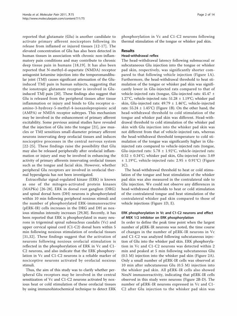
ResultsHead-withdrawal reflexThe head-withdrawal latency following submucosal orsubcutaneous Glu injection into the tongue or whiskerpad skin, respectively, was significantly shorter com-pared to that following vehicle injection (Figure 1A).Furthermore, the head-withdrawal threshold to heat sti-mulation of the tongue or whisker pad skin was signifi-cantly lower in Glu-injected rats compared to that ofvehicle-injected rats (tongue, Glu-injected rats: 45.47 ±1.27°C, vehicle-injected rats: 51.28 ± 1.19°C; whisker padskin, Glu-injected rats: 49.79 ± 1.46°C, vehicle-injectedrats: 55.24 ± 1.45°C) (Figure 1B). On the other hand, thehead-withdrawal threshold to cold stimulation of thetongue and whisker pad skin was different. Head-with-drawal threshold to cold stimulation of the whisker padskin with Glu injection into the whisker pad skin wasnot different from that of vehicle-injected rats, whereasthe head-withdrawal threshold temperature to cold sti-mulation of the tongue was significantly higher in Glu-injected rats compared to vehicle-injected rats (tongue,Glu-injected rats: 5.78 ± 1.92°C, vehicle-injected rats:0.52 ± 0.34°C; whisker pad skin, Glu-injected rats: 3.49± 1.19°C, vehicle-injected rats: 2.95 ± 0.91°C) (Figure1C).The head-withdrawal threshold to heat or cold stimu-
lation of the tongue and heat stimulation of the whiskerpad skin was also measured in the contralateral side toGlu injection. We could not observe any differences inhead-withdrawal thresholds to heat or cold stimulationof the contralateral tongue and heat stimulation of thecontralateral whisker pad skin compared to those ofvehicle injections (Figure 1D, E).
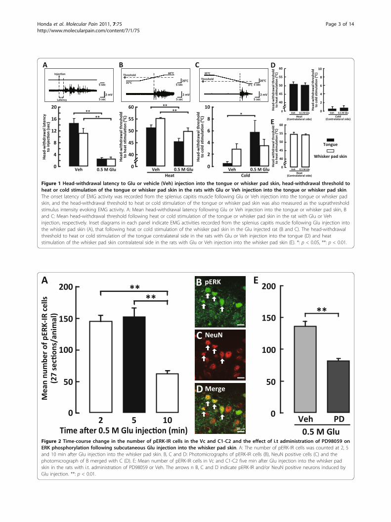
ERK phosphorylation in Vc and C1-C2 neurons and effectof MEK 1/2 inhibitor on ERK phosphorylationIn order to define the peak time point when the largestnumber of pERK-IR neurons was noted, the time courseof changes in the number of pERK-IR neurons in Vcand C1-C2 was analyzed following subcutaneous injec-tion of Glu into the whisker pad skin. ERK phosphoryla-tion in Vc and C1-C2 neurons was detected within 2min and peaked at 5 min following subcutaneous Glu(0.5 M) injection into the whisker pad skin (Figure 2A).Only a small number of pERK-IR cells was observed at10 min after subcutaneous Glu (0.5 M) injection intothe whisker pad skin. All pERK-IR cells also showedNeuN immunoreactivity, indicating that pERK-IR cellsobserved in this study were neurons (Figure 2B-D). Thenumber of pERK-IR neurons expressed in Vc and C1-C2 after Glu injection to the whisker pad skin was
Honda et al. Molecular Pain 2011, 7:75http://www.molecularpain.com/content/7/1/75
Page 2 of 14
20
16
12
8
4
0Veh 0.5 M Glu
60
55
50
45
40
0Veh 0.5 M Glu
Heat
Hea
d-w
ithd
raw
al la
tenc
y
o
A B
****
10
8
6
4
2
0Veh 0.5 M Glu
Cold
o
C
*****
20oC 20oC
60oC
35oC
35oC
0oC
Latency
D
Veh 0.5 M Glu
Heat
60
55
50
45
40
0
o
Tongue
10
8
6
4
2
0
o
Veh 0.5 M Glu
Cold
E 60
55
50
45
40
0
o
Veh 0.5 M Glu
Heat
Figure 1 Head-withdrawal latency to Glu or vehicle (Veh) injection into the tongue or whisker pad skin, head-withdrawal threshold toheat or cold stimulation of the tongue or whisker pad skin in the rats with Glu or Veh injection into the tongue or whisker pad skin.The onset latency of EMG activity was recorded from the splenius capitis muscle following Glu or Veh injection into the tongue or whisker padskin, and the head-withdrawal threshold to heat or cold stimulation of the tongue or whisker pad skin was also measured as the suprathresholdstimulus intensity evoking EMG activity. A: Mean head-withdrawal latency following Glu or Veh injection into the tongue or whisker pad skin, Band C: Mean head-withdrawal threshold following heat or cold stimulation of the tongue or whisker pad skin in the rat with Glu or Vehinjection, respectively. Inset diagrams in each panel indicate EMG activities recorded from the splenius capitis muscle following Glu injection intothe whisker pad skin (A), that following heat or cold stimulation of the whisker pad skin in the Glu injected rat (B and C). The head-withdrawalthreshold to heat or cold stimulation of the tongue contralateral side in the rats with Glu or Veh injection into the tongue (D) and heatstimulation of the whisker pad skin contralateral side in the rats with Glu or Veh injection into the whisker pad skin (E). *: p < 0.05, **: p < 0.01.
****
Mea
n nu
mbe
r of
pER
K-IR
cel
ls
200
100
02 5 10
A
C NeuN
B pERK
D Merge
20 m
Veh PD
**
E200
100
0
150
50
150
50
Figure 2 Time-course change in the number of pERK-IR cells in the Vc and C1-C2 and the effect of i.t administration of PD98059 onERK phosphorylation following subcutaneous Glu injection into the whisker pad skin. A: The number of pERK-IR cells was counted at 2, 5and 10 min after Glu injection into the whisker pad skin. B, C and D: Photomicrographs of pERK-IR cells (B), NeuN positive cells (C) and thephotomicrograph of B merged with C (D). E: Mean number of pERK-IR cells in Vc and C1-C2 five min after Glu injection into the whisker padskin in the rats with i.t. administration of PD98059 or Veh. The arrows n B, C and D indicate pERK-IR and/or NeuN positive neurons induced byGlu injection. **: p < 0.01.
Honda et al. Molecular Pain 2011, 7:75http://www.molecularpain.com/content/7/1/75
Page 3 of 14
significantly smaller in MEK 1/2 inhibitor PD98059-injected rats compared to that of vehicle-injected rats(Figure 2E).A large number of pERK-IR neurons was restricted to
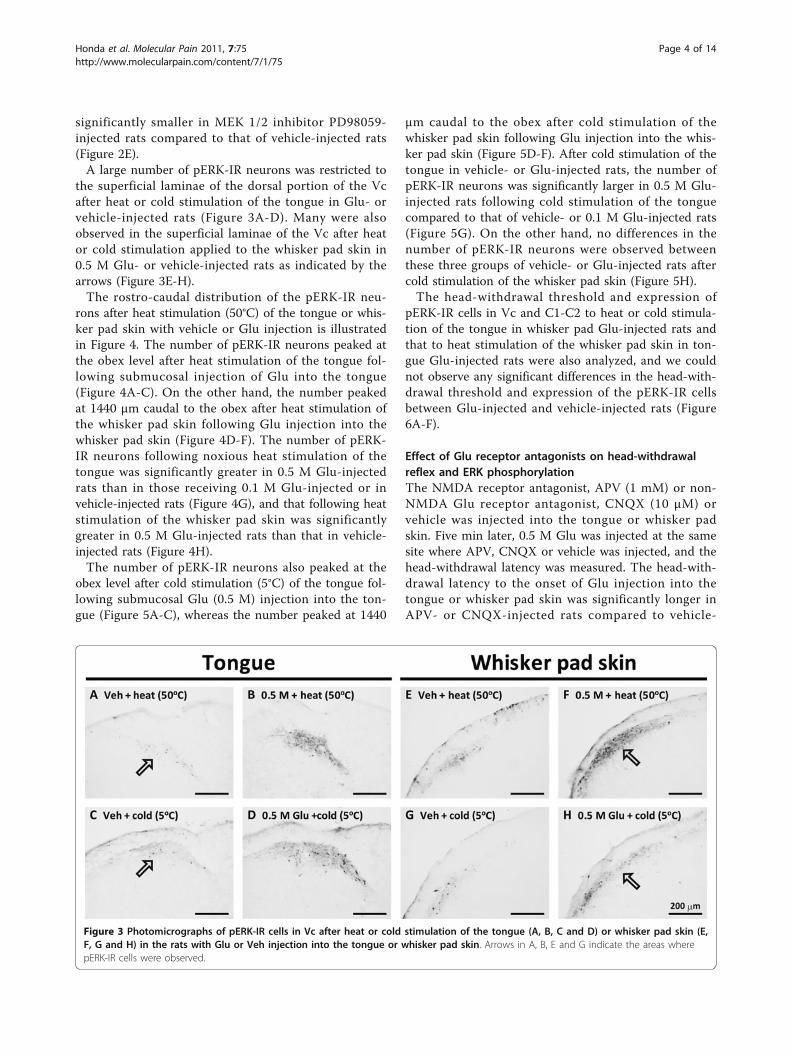
the superficial laminae of the dorsal portion of the Vcafter heat or cold stimulation of the tongue in Glu- orvehicle-injected rats (Figure 3A-D). Many were alsoobserved in the superficial laminae of the Vc after heator cold stimulation applied to the whisker pad skin in0.5 M Glu- or vehicle-injected rats as indicated by thearrows (Figure 3E-H).The rostro-caudal distribution of the pERK-IR neu-
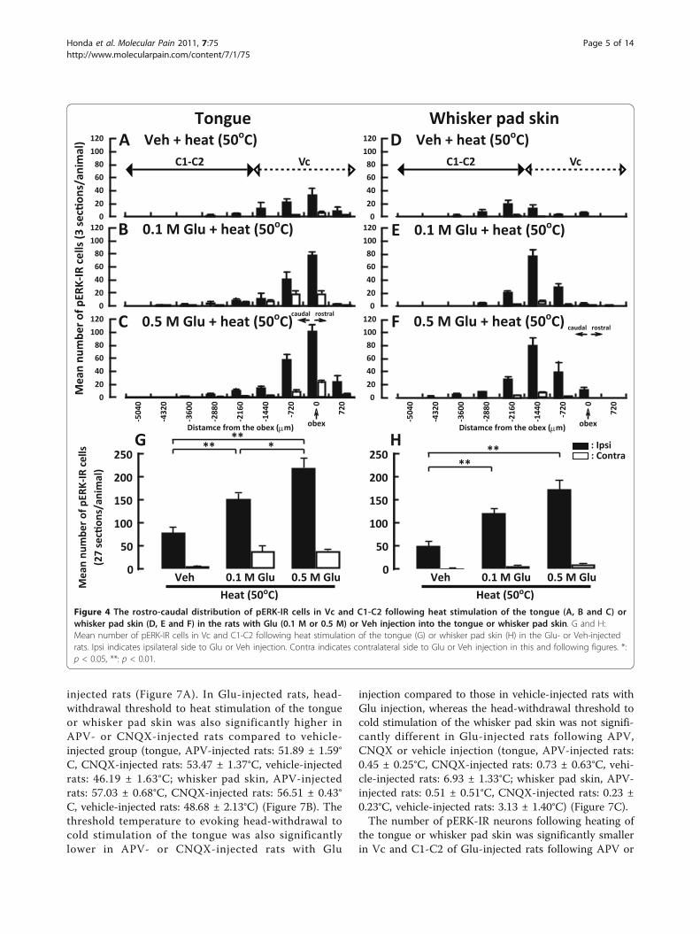
rons after heat stimulation (50°C) of the tongue or whis-ker pad skin with vehicle or Glu injection is illustratedin Figure 4. The number of pERK-IR neurons peaked atthe obex level after heat stimulation of the tongue fol-lowing submucosal injection of Glu into the tongue(Figure 4A-C). On the other hand, the number peakedat 1440 μm caudal to the obex after heat stimulation ofthe whisker pad skin following Glu injection into thewhisker pad skin (Figure 4D-F). The number of pERK-IR neurons following noxious heat stimulation of thetongue was significantly greater in 0.5 M Glu-injectedrats than in those receiving 0.1 M Glu-injected or invehicle-injected rats (Figure 4G), and that following heatstimulation of the whisker pad skin was significantlygreater in 0.5 M Glu-injected rats than that in vehicle-injected rats (Figure 4H).The number of pERK-IR neurons also peaked at the
obex level after cold stimulation (5°C) of the tongue fol-lowing submucosal Glu (0.5 M) injection into the ton-gue (Figure 5A-C), whereas the number peaked at 1440
μm caudal to the obex after cold stimulation of thewhisker pad skin following Glu injection into the whis-ker pad skin (Figure 5D-F). After cold stimulation of thetongue in vehicle- or Glu-injected rats, the number ofpERK-IR neurons was significantly larger in 0.5 M Glu-injected rats following cold stimulation of the tonguecompared to that of vehicle- or 0.1 M Glu-injected rats(Figure 5G). On the other hand, no differences in thenumber of pERK-IR neurons were observed betweenthese three groups of vehicle- or Glu-injected rats aftercold stimulation of the whisker pad skin (Figure 5H).The head-withdrawal threshold and expression of
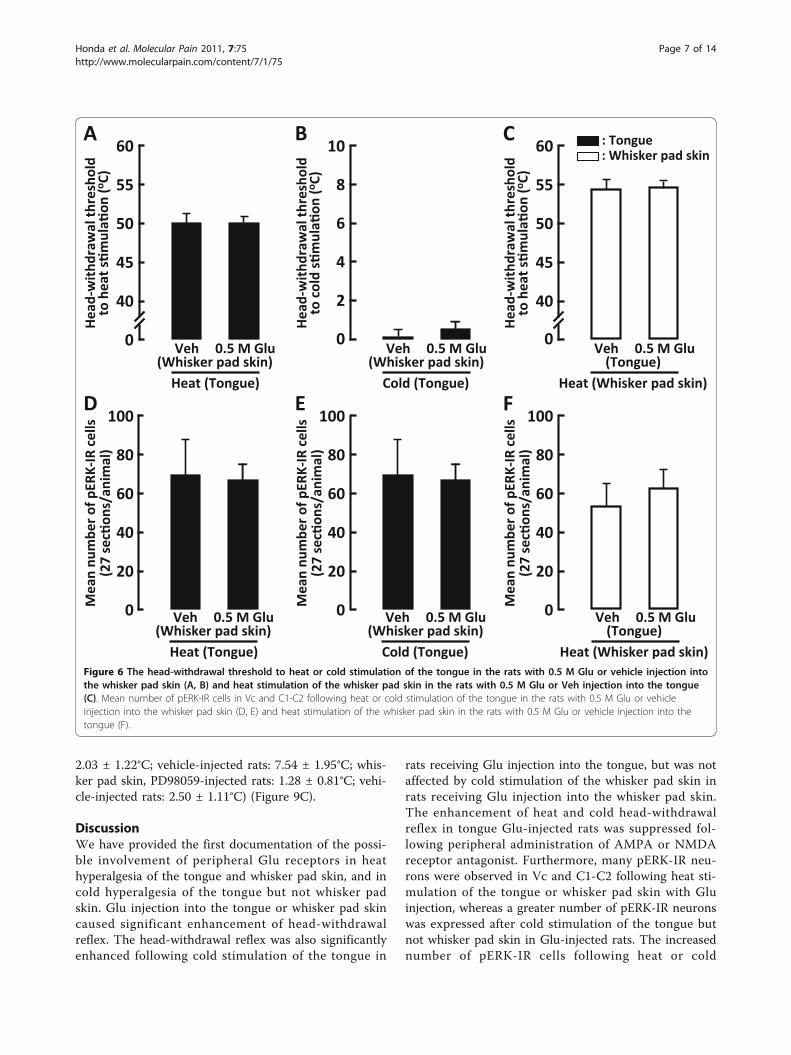
pERK-IR cells in Vc and C1-C2 to heat or cold stimula-tion of the tongue in whisker pad Glu-injected rats andthat to heat stimulation of the whisker pad skin in ton-gue Glu-injected rats were also analyzed, and we couldnot observe any significant differences in the head-with-drawal threshold and expression of the pERK-IR cellsbetween Glu-injected and vehicle-injected rats (Figure6A-F).
Effect of Glu receptor antagonists on head-withdrawalreflex and ERK phosphorylationThe NMDA receptor antagonist, APV (1 mM) or non-NMDA Glu receptor antagonist, CNQX (10 μM) orvehicle was injected into the tongue or whisker padskin. Five min later, 0.5 M Glu was injected at the samesite where APV, CNQX or vehicle was injected, and thehead-withdrawal latency was measured. The head-with-drawal latency to the onset of Glu injection into thetongue or whisker pad skin was significantly longer inAPV- or CNQX-injected rats compared to vehicle-
Figure 3 Photomicrographs of pERK-IR cells in Vc after heat or cold stimulation of the tongue (A, B, C and D) or whisker pad skin (E,F, G and H) in the rats with Glu or Veh injection into the tongue or whisker pad skin. Arrows in A, B, E and G indicate the areas wherepERK-IR cells were observed.
Honda et al. Molecular Pain 2011, 7:75http://www.molecularpain.com/content/7/1/75
Page 4 of 14
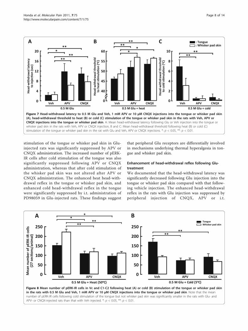
injected rats (Figure 7A). In Glu-injected rats, head-withdrawal threshold to heat stimulation of the tongueor whisker pad skin was also significantly higher inAPV- or CNQX-injected rats compared to vehicle-injected group (tongue, APV-injected rats: 51.89 ± 1.59°C, CNQX-injected rats: 53.47 ± 1.37°C, vehicle-injectedrats: 46.19 ± 1.63°C; whisker pad skin, APV-injectedrats: 57.03 ± 0.68°C, CNQX-injected rats: 56.51 ± 0.43°C, vehicle-injected rats: 48.68 ± 2.13°C) (Figure 7B). Thethreshold temperature to evoking head-withdrawal tocold stimulation of the tongue was also significantlylower in APV- or CNQX-injected rats with Glu
injection compared to those in vehicle-injected rats withGlu injection, whereas the head-withdrawal threshold tocold stimulation of the whisker pad skin was not signifi-cantly different in Glu-injected rats following APV,CNQX or vehicle injection (tongue, APV-injected rats:0.45 ± 0.25°C, CNQX-injected rats: 0.73 ± 0.63°C, vehi-cle-injected rats: 6.93 ± 1.33°C; whisker pad skin, APV-injected rats: 0.51 ± 0.51°C, CNQX-injected rats: 0.23 ±0.23°C, vehicle-injected rats: 3.13 ± 1.40°C) (Figure 7C).The number of pERK-IR neurons following heating of
the tongue or whisker pad skin was significantly smallerin Vc and C1-C2 of Glu-injected rats following APV or
200
150
0
250
100
50
Veh 0.1 M Glu 0.5 M Glu
Heat (50oC)
: Ipsi: Contra
120
100
80
60
40
20
0120
100
80
60
40
20
0
120
100
80
60
40
20
0
-504
0
-432
0
-360
0
-288
0
-216
0
-144
0
-720 0
720
Distamce from the obex ( m) obex
rostralcaudal
G ***
**
A Veh + heat (50oC)
B 0.1 M Glu + heat (50oC)
C 0.5 M Glu + heat (50oC)
Mea
n nu
mbe
r of
pER
K-IR
cel
ls
200
150
0
250
100
50
Veh 0.1 M Glu 0.5 M Glu
Heat (50oC)
120
100
80
60
40
20
0120
100
80
60
40
20
0
120
100
80
60
40
20
0-5
040
-432
0
-360
0
-288
0
-216
0
-144
0
-720 0
720
Distamce from the obex ( m) obex
H**
**
D Veh + heat (50oC)
E 0.1 M Glu + heat (50oC)
F 0.5 M Glu + heat (50oC)
VcC1-C2
Tongue Whisker pad skin
rostralcaudal
VcC1-C2
Figure 4 The rostro-caudal distribution of pERK-IR cells in Vc and C1-C2 following heat stimulation of the tongue (A, B and C) orwhisker pad skin (D, E and F) in the rats with Glu (0.1 M or 0.5 M) or Veh injection into the tongue or whisker pad skin. G and H:Mean number of pERK-IR cells in Vc and C1-C2 following heat stimulation of the tongue (G) or whisker pad skin (H) in the Glu- or Veh-injectedrats. Ipsi indicates ipsilateral side to Glu or Veh injection. Contra indicates contralateral side to Glu or Veh injection in this and following figures. *:p < 0.05, **: p < 0.01.
Honda et al. Molecular Pain 2011, 7:75http://www.molecularpain.com/content/7/1/75
Page 5 of 14

CNQX injection compared to that following vehicleinjection (Figure 8A). The number of pERK-IR neuronsfollowing cold stimulation of the tongue was signifi-cantly smaller in Glu-injected rats after APV or CNQXinjection compared to those after vehicle injection (Fig-ure 8B), but was not significantly different between APVand CNQX-injected and vehicle-injected rats followingcold stimulation of the whisker pad skin (Figure 8B).
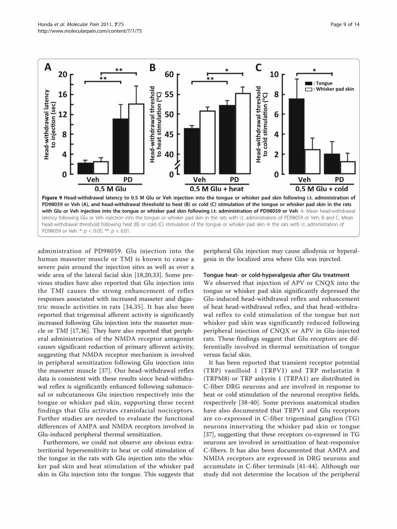
Effect of MEK1/2 inhibitor on head-withdrawal reflexWe also studied the effect of the i.t. administration ofPD98059 on head-withdrawal reflex in Glu-injected rats.The head-withdrawal latency after Glu injection into thetongue or whisker pad skin was significantly longer in
PD98059-injected rats compared to vehicle-injected rats(Figure 9A). The head-withdrawal threshold to heat sti-mulation of the tongue or whisker pad skin was signifi-cantly higher in PD98059-injected rats compared tovehicle-injected rats (tongue, PD98059-injected rats:52.22 ± 1.28°C, vehicle-injected rats: 46.44 ± 0.83°C;whisker pad skin, PD98059-injected rats: 55.24 ± 1.46°C,vehicle-injected rats: 50.82 ± 0.98°C) (Figure 9B). Thehead-withdrawal threshold to cold stimulation of thetongue was also significantly lower in PD98059-injectedrats compared with that of vehicle-injected rats, whereasthat to cold stimulation of the whisker pad skin was notsignificantly different between PD98059-injected ratsand vehicle-injected (tongue, PD98059-injected rats:
200
150
0
250
100
50
Veh 0.1 M Glu 0.5 M Glu
Cold (5oC)
120
100
80
60
40
20
0120
100
80
60
40
20
0
120
100
80
60
40
20
0
-504
0
-432
0
-360
0
-288
0
-216
0
-144
0
-720 0
720
Distamce from the obex ( m) obex
A Veh + cold (5oC)
B 0.1 M Glu + cold (5oC)
C 0.5 M Glu + cold (5oC)
G
***
Mea
n nu
mbe
r of
pER
K-IR
cel
ls
200
150
0
250
100
50
Veh 0.1 M Glu 0.5 M Glu
Cold (5oC)
120
100
80
60
40
20
0120
100
80
60
40
20
0
120
100
80
60
40
20
0
-504
0
-432
0
-360
0
-288
0
-216
0
-144
0
-720 0
720
Distamce from the obex ( m) obex
D Veh + cold (5oC)
E 0.1 M Glu + cold (5oC)
F 0.5 M Glu + cold (5oC)
H
VcC1-C2
: Ipsi: Contra
Tongue Whisker pad skin
VcC1-C2
rostralcaudal rostralcaudal
Figure 5 The rostro-caudal distribution (A, B, C, D, E and F) and mean number (G and H) of pERK-IR cells in Vc and C1-C2 followingcold stimulation of the tongue (A, B, C and G) or whisker pad skin (D, E, F and H) in the rats with Glu (0.1 M or 0.5 M) or vehicleinjection into the tongue or whisker pad skin. Note that significantly larger number of pERK-IR cells was observed in tongue Glu-injected ratscompared with that in whisker Glu-injected rats. *: p < 0.05, **: p < 0.01.
Honda et al. Molecular Pain 2011, 7:75http://www.molecularpain.com/content/7/1/75
Page 6 of 14
2.03 ± 1.22°C; vehicle-injected rats: 7.54 ± 1.95°C; whis-ker pad skin, PD98059-injected rats: 1.28 ± 0.81°C; vehi-cle-injected rats: 2.50 ± 1.11°C) (Figure 9C).
DiscussionWe have provided the first documentation of the possi-ble involvement of peripheral Glu receptors in heathyperalgesia of the tongue and whisker pad skin, and incold hyperalgesia of the tongue but not whisker padskin. Glu injection into the tongue or whisker pad skincaused significant enhancement of head-withdrawalreflex. The head-withdrawal reflex was also significantlyenhanced following cold stimulation of the tongue in
rats receiving Glu injection into the tongue, but was notaffected by cold stimulation of the whisker pad skin inrats receiving Glu injection into the whisker pad skin.The enhancement of heat and cold head-withdrawalreflex in tongue Glu-injected rats was suppressed fol-lowing peripheral administration of AMPA or NMDAreceptor antagonist. Furthermore, many pERK-IR neu-rons were observed in Vc and C1-C2 following heat sti-mulation of the tongue or whisker pad skin with Gluinjection, whereas a greater number of pERK-IR neuronswas expressed after cold stimulation of the tongue butnot whisker pad skin in Glu-injected rats. The increasednumber of pERK-IR cells following heat or cold
: Whisker pad skin: Tongue
Hea
d-w
ithdr
awal
thre
shol
do C
)
A10
8
6
4
2
0
Hea
d-w
ithdr
awal
thre
shol
do C
)
B60
55
50
45
40
0
Hea
d-w
ithdr
awal
thre
shol
do C
)
C
D100
80
60
40
20
100
80
60
40
20
F
60
55
50
45
40
0 Veh 0.5 M Glu Veh 0.5 M Glu
Veh 0.5 M Glu
Veh 0.5 M Glu
Veh 0.5 M Glu
100
80
60
40
20
0 Veh 0.5 M Glu 0 0
Figure 6 The head-withdrawal threshold to heat or cold stimulation of the tongue in the rats with 0.5 M Glu or vehicle injection intothe whisker pad skin (A, B) and heat stimulation of the whisker pad skin in the rats with 0.5 M Glu or Veh injection into the tongue(C). Mean number of pERK-IR cells in Vc and C1-C2 following heat or cold stimulation of the tongue in the rats with 0.5 M Glu or vehicleinjection into the whisker pad skin (D, E) and heat stimulation of the whisker pad skin in the rats with 0.5 M Glu or vehicle injection into thetongue (F).
Honda et al. Molecular Pain 2011, 7:75http://www.molecularpain.com/content/7/1/75
Page 7 of 14
stimulation of the tongue or whisker pad skin in Glu-injected rats was significantly suppressed by APV orCNQX administration. The increased number of pERK-IR cells after cold stimulation of the tongue was alsosignificantly suppressed following APV or CNQXadministration, whereas that after cold stimulation ofthe whisker pad skin was not altered after APV orCNQX administration. The enhanced heat head-with-drawal reflex in the tongue or whisker pad skin, andenhanced cold head-withdrawal reflex in the tonguewere significantly suppressed by i.t. administration ofPD98059 in Glu-injected rats. These findings suggest
that peripheral Glu receptors are differentially involvedin mechanisms underlying thermal hyperalgesia in ton-gue and whisker pad skin.
Enhancement of head-withdrawal reflex following Glu-treatmentWe documented that the head-withdrawal latency wassignificantly decreased following Glu injection into thetongue or whisker pad skin compared with that follow-ing vehicle injection. The enhanced head-withdrawalreflex in the rats with Glu injection was suppressed byperipheral injection of CNQX, APV or i.t.
60
55
50
45
40
0Veh APV CNQX
0.5 M Glu + heat
20
16
12
8
4
0Veh APV CNQX
: Whisker pad skin: Tongue
Hea
d-w
ithd
raw
al la
tenc
y
Hea
d-w
ithd
raw
al t
hres
hold
o
A B ****
** 10
8
6
4
2
0Veh APV CNQX
0.5 M Glu + cold
Hea
d-w
ithd
raw
al t
hres
hold
o
C
****
0.5 M Glu
****
***
Figure 7 Head-withdrawal latency to 0.5 M Glu and Veh, 1 mM APV or 10 μM CNQX injections into the tongue or whisker pad skin(A), head-withdrawal threshold to heat (B) or cold (C) stimulation of the tongue or whisker pad skin in the rats with Veh, APV orCNQX injections into the tongue or whisker pad skin. A: Mean head-withdrawal latency following Glu or Veh injection into the tongue orwhisker pad skin in the rats with Veh, APV or CNQX injection, B and C: Mean head-withdrawal threshold following heat (B) or cold (C)stimulation of the tongue or whisker pad skin in the rat with Glu and Veh, APV or CNQX injections. *: p < 0.05, **: p < 0.01.
250
200
150
100
50
0Mea
n nu
mbe
r of
pER
K-IR
cel
ls 250
200
150
100
50
0Veh APV CNQX
0.5 M Glu + Heat (50o
Veh APV CNQX
0.5 M Glu + Cold (5o
: Tongue
A B**
***
*
****
Figure 8 Mean number of pERK-IR cells in Vc and C1-C2 following heat (A) or cold (B) stimulation of the tongue or whisker pad skinin the rats with 0.5 M Glu and Veh, 1 mM APV or 10 μM CNQX injections into the tongue or whisker pad skin. Note that the meannumber of pERK-IR cells following cold stimulation of the tongue but not whisker pad skin was significantly smaller in the rats with Glu- andAPV- or CNQX-injected rats than that with Veh injected. *: p < 0.05, **: p < 0.01.
Honda et al. Molecular Pain 2011, 7:75http://www.molecularpain.com/content/7/1/75
Page 8 of 14
administration of PD98059. Glu injection into thehuman masseter muscle or TMJ is known to cause asevere pain around the injection sites as well as over awide area of the lateral facial skin [18,20,33]. Some pre-vious studies have also reported that Glu injection intothe TMJ causes the strong enhancement of reflexresponses associated with increased masseter and digas-tric muscle activities in rats [34,35]. It has also beenreported that trigeminal afferent activity is significantlyincreased following Glu injection into the masseter mus-cle or TMJ [17,36]. They have also reported that periph-eral administration of the NMDA receptor antagonistcauses significant reduction of primary afferent activity,suggesting that NMDA receptor mechanism is involvedin peripheral sensitization following Glu injection intothe masseter muscle [37]. Our head-withdrawal reflexdata is consistent with these results since head-withdra-wal reflex is significantly enhanced following submuco-sal or subcutaneous Glu injection respectively into thetongue or whisker pad skin, supporting these recentfindings that Glu activates craniofacial nociceptors.Further studies are needed to evaluate the functionaldifferences of AMPA and NMDA receptors involved inGlu-induced peripheral thermal sensitization.Furthermore, we could not observe any obvious extra-
territorial hypersensitivity to heat or cold stimulation ofthe tongue in the rats with Glu injection into the whis-ker pad skin and heat stimulation of the whisker padskin in Glu injection into the tongue. This suggests that
peripheral Glu injection may cause allodynia or hyperal-gesia in the localized area where Glu was injected.
Tongue heat- or cold-hyperalgesia after Glu treatmentWe observed that injection of APV or CNQX into thetongue or whisker pad skin significantly depressed theGlu-induced head-withdrawal reflex and enhancementof heat head-withdrawal reflex, and that head-withdra-wal reflex to cold stimulation of the tongue but notwhisker pad skin was significantly reduced followingperipheral injection of CNQX or APV in Glu-injectedrats. These findings suggest that Glu receptors are dif-ferentially involved in thermal sensitization of tongueversus facial skin.It has been reported that transient receptor potential
(TRP) vanilloid 1 (TRPV1) and TRP melastatin 8(TRPM8) or TRP ankyrin 1 (TRPA1) are distributed inC-fiber DRG neurons and are involved in response toheat or cold stimulation of the neuronal receptive fields,respectively [38-40]. Some previous anatomical studieshave also documented that TRPV1 and Glu receptorsare co-expressed in C-fiber trigeminal ganglion (TG)neurons innervating the whisker pad skin or tongue[37], suggesting that these receptors co-expressed in TGneurons are involved in sensitization of heat-responsiveC-fibers. It has also been documented that AMPA andNMDA receptors are expressed in DRG neurons andaccumulate in C-fiber terminals [41-44]. Although ourstudy did not determine the location of the peripheral
20
16
12
8
4
0Veh PD
60
55
50
45
40
0Veh PD
0.5 M Glu + heat
: Whisker pad skin: Tongue
Hea
d-w
ithd
raw
al la
tenc
y
Hea
d-w
ithd
raw
al t
hres
hold
o
A B ***
10
8
6
4
2
0Veh PD
0.5 M Glu + cold
Hea
d-w
ithd
raw
al t
hres
hold
o
C *****
0.5 M GluFigure 9 Head-withdrawal latency to 0.5 M Glu or Veh injection into the tongue or whisker pad skin following i.t. administration ofPD98059 or Veh (A), and head-withdrawal threshold to heat (B) or cold (C) stimulation of the tongue or whisker pad skin in the ratswith Glu or Veh injection into the tongue or whisker pad skin following i.t. administration of PD98059 or Veh. A: Mean head-withdrawallatency following Glu or Veh injection into the tongue or whisker pad skin in the rats with i.t. administration of PD98059 or Veh, B and C: Meanhead-withdrawal threshold following heat (B) or cold (C) stimulation of the tongue or whisker pad skin in the rats with i.t. administration ofPD98059 or Veh. *: p < 0.05, **: p < 0.01.
Honda et al. Molecular Pain 2011, 7:75http://www.molecularpain.com/content/7/1/75
Page 9 of 14

ionotropic glutamate receptors, which have been shownto occur on afferent terminals as well as on other per-ipheral tissue cells [45,46], nonetheless the above-men-tioned findings along with our own data suggest thatperipheral Glu receptors are differentially involved insensitization of heat and cold receptors in tongue versuswhisker pad skin. Together with the previous primaryafferent data, our results also suggest the following twopossible mechanisms: One mechanism is that cold andheat receptors are co-expressed with Glu receptors inC-fiber TG neurons innervating the tongue, whereas TGneurons with heat and Glu receptors innervate the facialskin, resulting in the enhancement of cold and heatresponses of Vc and C1-C2 neurons after Glu injectioninto the tongue, and the enhancement of heat responseof those neurons after Glu injection into the facial skin,and the other mechanism is that these receptors areexpressed in different primary afferent neurons innervat-ing the tongue or facial skin and that converge onto Vcand C1-C2 neurons. In the case of the whisker pad skin,our data suggest that cold and Glu receptors are not co-expressed in TG neurons, and that they are expressed indifferent primary afferent neurons contacting on differ-ent Vc and C1-C2 neurons. Further support for the dif-ferential involvement of Glu and cold receptors ontongue versus facial skin comes from our ERK data.
Involvement of ERK phosphorylation in tonguehyperalgesiaA large number of pERK-IR neurons was expressed inthe Vc and C1-C2 after Glu injection into the tongue orwhisker pad skin. The number of pERK-IR neurons fol-lowing heat stimulation of the tongue or whisker padskin was significantly greater in tongue or whisker Glu-injected rats compared with that in vehicle-injected rats.The ERK phosphorylation in Vc and C1-C2 neuronswas significantly enhanced following cold stimulation ofthe tongue in tongue Glu-injected rats, but were notaffected by cold stimulation of the whisker pad skin inwhisker Glu-injected rats. The ERK phosphorylation fol-lowing heat stimulation of the tongue or whisker padskin and cold stimulation of the tongue was significantlysuppressed following subcutaneous or submucosal injec-tion of APV or CNQX. We also observed that i.t.administration of MEK1/2 inhibitor PD98059 causedsignificant inhibition of the head-withdrawal reflex afterGlu injection itself into the tongue or whisker pad skin,and also inhibited the heat head-withdrawal reflex andERK phosphorylation in Glu-injected rats. Cold head-withdrawal reflex after Glu injection into the tongue butnot whisker pad skin was also significantly suppressedby i.t. PD98059 administration.It is well known that ERK is phosphorylated in DRG
and TG neurons after noxious stimulation of the hind
paw via Ca2+ influx [47], suggesting that i.t. PD98059administration likely blocks Ca2+ influx in Vc and C1-C2 neurons and TG primary afferent terminals whichwould contribute to the block of heat-evoked head-with-drawal reflex with Glu injection into the tongue or whis-ker pad skin, and to the blockade of the cold-evokedhead-withdrawal reflex in tongue Glu-injected rats.We could not observe any differences in the suppres-
sive effect on head-withdrawal reflex to heat stimulationof the tongue and whisker pad skin and ERK phosphor-ylation between CNQX and APV administration in Glu-injected rats. On the other hand, the effect of CNQXand APV on cold head-withdrawal reflex in Glu-injectedrats was different between the tongue and whisker padskin. It is likely that AMPA and NMDA receptor sub-types are similarly involved in Glu-induced ERK phos-phorylation in Vc and C1-C2 neurons following heatingthe tongue and whisker pad skin, but are differentiallyinvolve in Glu-induced ERK phosphorylation followingnoxious cold stimulation of the tongue and whisker padskin, consistent with our head-withdrawal reflexfindings.
ConclusionsThe present findings revealed that Glu application to thetongue or whisker pad skin caused an enhancement ofhead-withdrawal reflex and ERK phosphorylation in Vcand C1-C2 neurons to heat stimulation; head-withdra-wal reflex and ERK phosphorylation were also enhancedfollowing cold stimulation of the tongue but not whiskerpad skin in Glu-injected rats. These findings suggestthat peripheral Glu receptor mechanisms may contri-bute to cold hyperalgesia in the tongue but not in thefacial skin, and also contribute to heat hyperalgesia inthe tongue and facial skin, and that the mitogen-acti-vated protein kinase cascade in Vc and C1-C2 neuronsmay be involved in these Glu-evoked hyperalgesiceffects.
MethodsAnimalsA total of 408 male Sprague-Dawley rats weighting 200-250 g were used. This study was approved by the Ani-mal Experimentation Committee at the Nihon Univer-sity. All surgeries and animal care were conducted inaccordance with the National Institutes of Health Guidefor the Care and Use of Laboratory Animals and theguidelines for Institutional Animal Care, and the guide-lines of the International Association for the Study ofPain [48].
Head-withdrawal reflex testingRats were lightly anesthetized with 2% isoflurane(Mylan, Canonsburg, PA) with 100% O2. As previously
Honda et al. Molecular Pain 2011, 7:75http://www.molecularpain.com/content/7/1/75
Page 10 of 14
described [49], bipolar enamel-coated silver wire electro-des (Narishige, Tokyo, Japan) were placed in the leftsplenius capitis muscle for electromyographic (EMG)recordings to detect the time point of head movement(inter-electrode distance: 5-6 mm). Glu (Sigma-Aldrich,St. Louis, MI) was dissolved in isotonic saline to pro-duce a 0.5 M Glu solution. Isotonic saline was used asthe vehicle control. Five min after 0.5 M Glu (10 μl) orvehicle (10 μl) was injected with 27 gage fine needleinto center of the left whisker pad skin or lateral edgeof the tongue subcutaneously (depth: 2 mm), heat (1°C/sec, cut-off temperature: 60°C) or cold (1°C/sec, cut-offtemperature: 0°C) stimulation was applied to the sameskin or tongue site received Glu or vehicle injection (n= 6 in each group). The thermal stimuli were deliveredby a contact thermal probe (5 mm in diameter, adaptingtemperature: 35°C, Intercross, Tokyo, Japan) in the ipsi-lateral or contralateral side to the Glu or vehicle injec-tion. The head-withdrawal latency to Glu or vehicleinjection into the whisker pad skin or tongue was calcu-lated by measuring the onset latency of splenius capitismuscle EMG activity after Glu or vehicle injection. Thehead-withdrawal threshold to heat or cold stimulation 5min after Glu injection was determined as the stimulusintensity when small EMG activity could be recordedafter thermal stimulation as illustrated in the inset ofFigure 1A. Measurement of the head-withdrawal latencyto Glu injection or vehicle and head-withdrawal thresh-old following thermal stimulation in Glu-injected ratswas conducted only once in each rat by the same inves-tigator to ensure standardization.The head-withdrawal threshold to heat or cold stimu-
lation of the tongue in whisker pad Glu-injected ratsand heat stimulation of the whisker pad skin in tongueGlu-injected rats were also measured (n = 5 in eachgroup).Although we injected the very small amount of Glu
into the whisker pad or tongue, we cannot rule out thepossibility of Glu entering in other body areas throughthe systemic circulation.
pERK immunohistochemistryTo determine the time course and peak time of ERKphosphorylation, rats were anesthetized with sodiumpentobarbital (50 mg/kg, i.p., Kyoritsu Seiyaku, Tokyo,Japan) and were perfused through the aorta with saline(500 ml) followed by 4% paraformaldehyde (Wako,Osaka, Japan) in 0.1 M phosphate buffer (PB, pH 7.4,500 ml) at 1, 5 or 10 min after 0.5 M Glu was injectedinto the whisker pad skin with a fine needle (n = 5 ineach time point). The Vc-C2 area was removed andpost-fixed in 4% paraformaldehyde for 1 day at 4°C. Thetissues were then transferred to 20% sucrose (Wako) insaline for overnight for cryoprotection. Thirty μm thick
sections were cut with a freezing microtome (LeicaMicrosystems Japan, Tokyo, Japan). Every 4th sectionwas collected in phosphate-buffered saline (PBS, pH7.4). Free-floating tissue sections were dipped in 10%normal goat serum (NGS, Millipore, Billerica, MA) inPBS for 12 hours at 4°C, and then incubated in rabbitanti-Phospho-p44/42 MAP Kinase (Thr202/Tyr204)antibody (1 : 1000, Cell Signaling, Danvers, MA) for 72hours at 4°C. Next, the sections were incubated in bioti-nylated goat anti-rabbit IgG (1 : 600; Vector Labora-tories, Burlingame, CA) for 2 hours at roomtemperature. After washing in PBS, the sections wereincubated in peroxidase-conjugated avidin-biotin com-plex (1 : 100; Vector Laboratories) for 2 hours at roomtemperature. They were then washed in 0.05 M tris buf-fer (TB, pH 7.4), and dipped in 0.035% 3,3’-diaminoben-zidine-tetra HCl (Sigma-Aldrich), 0.2% nickelammonium sulfate (Wako) and 0.05% hydrogen perox-ide in 0.05 M TB. The sections were then washed inPBS, and serially mounted on gelatin-coated slides. Theslides were dipped in a series from 50 to 100% alcoholsafter dehydrated and then cover slipped.Next, in order to evaluate the expression of pERK-IR
neurons following thermal stimulation, at 5 min after0.1 M or 0.5 M Glu or vehicle was injected into thewhisker pad skin or the tongue, heat (50°C, duration: 60sec, interval: 10 sec, train: 5) or cold (5°C, duration: 60sec, interval: 10 sec, train: 5) stimulation was applied tothe whisker pad skin or the tongue at the site of Glu orvehicle injection through a contact thermal probe (n = 5in each group). Five min after stimulation, rats wereperfused and pERK immunohistochemistry was carriedout, as described above. To study the effect of Glu injec-tion on pERK-IR cell expression, rats were also perfused5 min after Glu or vehicle injection into the whiskerpad skin and tongue, and Vc and C1-C2 sections wereprocessed for pERK immunohistochemistry (n = 5 ineach group).The pERK-IR neurons were counted by Neurolucida
Explorer (MicroBrightField, Williston, VT). Every 720μm sections and 2 sections before and after 720 μmwere collected and the mean number of pERK-IR neu-rons from these 3 sections was neurons calculated, andthe graphs of rostro-caudal distribution of pERK-IR neu-rons were prepared. The mean number of pERK-IR neu-rons in all the sections (27 sections/rat) was alsocalculated for each animal and the mean number ofpERK-IR neurons in each group was compared.Double immunofluorescence histochemistry was also
used to determine if the pERK-IR neurons expressed ornot a neuronal label. Five min after 0.5 M Glu injectioninto the whisker pad skin, heat stimuli (50°C) wereapplied at the site of the Glu injection (n = 5). Five minafter heat stimulation, rats were perfused through the
Honda et al. Molecular Pain 2011, 7:75http://www.molecularpain.com/content/7/1/75
Page 11 of 14
aorta with saline (500 ml) followed by 4% paraformalde-hyde in 0.1 M PB (pH 7.4, 500 ml). The tissues werethen transferred to 20% sucrose (Wako) in saline forovernight for cryoprotection. Thirty μm thick sectionswere cut and processed for double-labeling immunohis-tochemistry for pERK and the neuronal label NeuN.Free-floating tissue sections were dipped in 10% NGS inPBS for 12 hours at 4°C, and then incubated in rabbitanti-Phospho-p44/42 MAPK Antibody (1 : 300) for 72hours at 4°C. Next, the sections were mouse anti-NeuNAntibody (1 : 1000, Chemicon, Temecula, CA) for 1hour at room temperature and secondary antibodies(FITC- and rhodamine-, 1 : 100; Jackson ImmunoResearch, West Grove, PA) conjugated for 1 hour atroom temperature in a dark room. Then the sectionswere washed in PBS 3 times for 5 min. The sectionswere mounted on slides and cover slipped in Perma-Fluor (Sigma-Aldrich). pERK-IR neurons and NeuNimmunoreactive neurons were observed by confocalmicroscope (Carl Zeiss, Tokyo, Japan).The expression of pERK-IR cells in Vc and C1-C2 to
heat or cold stimulation of the tongue in whisker padGlu-injected rats and heat stimulation of the whiskerpad skin in tongue Glu-injected rats were also analyzed(n = 5 in each group).
The effect of injection of Glu receptor antagonist APV orCNQXRats were anesthetized with 2% isoflurane with 100% O2
(for behavioral tests) or sodium pentobarbital (50 mg/kg, i.p., for immunohistochemistry). The NMDA recep-tor antagonist APV (Research Biochemicals Interna-tional, Natick, MA), and the AMPA receptor antagonistCNQX (Research Biochemicals International) were dis-solved in isotonic saline to produce a 1 mM APV and a10 μM CNQX solution respectively, in accordance withprevious studies [43,50]; isotonic saline was used as thevehicle. Five min after 10 μl APV, CNQX or vehicle wasinjected into ipsilateral whisker pad skin or tongue witha fine needle (27 gauge), and 5 min after that 0.5 M Gluwas injected at the site of antagonist injection. At 5 minafter Glu injection, behavioral tests were performed onipsilateral side (n = 6 in each group), and anothergroups of animals were perfused for subsequent exami-nation of pERK-IR in Vc and C1-C2 neurons (n = 5 ineach group), as described above.
The effect of intrathecal (i.t.) injection of MAPK Kinase(MEK) 1/2 inhibitor PD98059In order to block the ERK phosphorylation in neuronsand TG primary afferent terminals in the Vc and C1-C2, MEK1/2 inhibitor PD98059 (CalBiochem, SanDiego, CA) was i.t. administrated. PD98059 was dis-solved in 20% dimethyl sulfoxide (DMSO) at a
concentration of 1 μg/μl (3.7 mM) as stock solution,and then further diluted to 0.1 μg/μl in 10% DMSO fori.t. injection, in accordance with previous studies[32,51]. A solution of 10% DMSO (in isotonic saline)was used as vehicle control. Rats were anesthetized withsodium pentobarbital (50 mg/kg, i.p.). The distancefrom C4-C5 to C1-C2 was measured in pilot studiesand the length of a p10 polyethylene tube was adjustedfor this length (n = 2). The polyethylene tube wasinserted into the subdural space at the C4-C5 level andthe tip of the tube was located near the C1-C2 level.After the experiments, vertebrae were removed and thetip of the tube was confirmed at C1-C2. An osmoticpressure pump was connected to the tube and placedsubcutaneously on the back. PD98059 or vehicle wasinjected at a flow rate of 1.0 μl/hour for 7 days with theosmotic pressure pump. One week after a pump filledwith PD98059 or vehicle pump placement, behavioraltests were performed (n = 6 in each group) and pERK-IR neurons was explored immunohistochemically (n = 5in each group), as described above.
Statistical analysisResults were presented as mean ± SEM. Statistical ana-lysis was performed using one-way analysis of variance(ANOVA) followed by Newman-Keuls test, Student’s t-test or Welch’s t-test as appropriate by using statisticalprogram (SigmaStat 3.5, SYSTAT, Chicago, IL). Differ-ences were considered significant at p < 0.05.
List of abbreviationsATP: adenosine triphosphate; Glu: glutamate; TMJ: temporomandibular joint;AMPA: α-amino-3-hydroxy-5-methyl-4-isoxazolepropionic acid; NMDA: N-methyl-d-aspartate; ERK: extracellular signal-regulated kinase; MAPKs:mitogen-activated protein kinases; DRG: dorsal root ganglion; DH: spinaldorsal horn; pERK-IR: phosphorylated ERK-immunoreactive; Vc: trigeminalspinal subnucleus caudalis; C1-C2: upper cervical spinal cord; EMG:electromyographic; PB: phosphate buffer; PBS: phosphate-buffered saline;NGS: normal goat serum; TB: tris buffer; APV: D(-)-2-amino-5-phosphonopentanoic acid; CNQX: 6-cyano-7-nitroquinoxa-line 2,3-dione;DMSO: dimethyl sulfoxide; ANOVA: analysis of variance.
AcknowledgementsThis study was supported in part by Research grants from Sato and UemuraFunds from Nihon University School of Dentistry, and Grant from DentalResearch Center, Nihon University School of Dentistry; Nihon Universitymultidisciplinary research grant and Individual Research Grant; a Grant fromthe Ministry of Education, Culture, Sports, Science and Technology topromote multi-disciplinary research projects; grants from the Ministry ofEducation, Culture, Sports, Science, Technology to promote multidisciplinaryresearch projects “Brain Mechanisms for Cognition, Memory and Behavior”and “Translational Research Network on Orofacial Neurological Disorders” atNihon University, and Japan-Canada (CIHR) Joint Health Research Program,and NIH grant DE015420.
Author details1Department of Physiology, Nihon University School of Dentistry, 1-8-13Kandasurugadai, Chiyoda-ku, Tokyo 101-8310, Japan. 2Department of Oraland Maxillofacial Surgery, Nihon University School of Dentistry, 1-8-13Kandasurugadai, Chiyoda-ku, Tokyo 101-8310, Japan. 3Department of OralDiagnosis, Nihon University School of Dentistry, 1-8-13 Kandasurugadai,
Honda et al. Molecular Pain 2011, 7:75http://www.molecularpain.com/content/7/1/75
Page 12 of 14
Chiyoda-ku, Tokyo 101-8310, Japan. 4Division of Oral Health Science, DentalResearch Center, Nihon University School of Dentistry, 1-8-13Kandasurugadai, Chiyoda-ku, Tokyo 101-8310, Japan. 5Department ofAnesthesiology, Nihon University School of Dentistry, 1-8-13 Kandasurugadai,Chiyoda-ku, Tokyo 101-8310, Japan. 6Department of Psychosomatic Dentistry,Tokyo Medical and Dental University Graduate School, 1-5-45 Yushima,Bunkyo-ku, Tokyo 113-8510, Japan. 7Department of Oral Physiology, Facultyof Dentistry, University of Toronto, 124 Edward Street, Toronto, Ontario M5G1G6, Canada. 8Department of Pedodontics, Nihon University School ofDentistry, 1-8-13 Kandasurugadai, Chiyoda-ku, Tokyo 101-8310, Japan.9Division of Functional Morphology, Dental Research Center, NihonUniversity School of Dentistry, 1-8-13 Kandasurugadai, Chiyoda-ku, Tokyo101-8310, Japan. 10Division of Applied System Neuroscience AdvancedMedical Research Center, Nihon University Graduate School of MedicalScience, 30-1 Ohyaguchi-Kamimachi, Itabashi-ku, Tokyo 173-8610, Japan.
Authors’ contributionsAll authors read and approved the final manuscript. KH, NN and MS carriedout the experiments and data analysis, and KH and NN were equallycontributed for conducting this experiment. MM, AK, DK and ML helped theexperiments, data analysis. BJS provided data interpretation and helped tofinalize the manuscript. KI conceptualized the hypothesis, designed andsupervised the experiments, directed the data analysis, and finalized themanuscript.
Competing interestsThe authors declare that they have no competing interests.
Received: 7 June 2011 Accepted: 28 September 2011Published: 28 September 2011
References1. Komiyama O, De Laat A: Tactile and pain thresholds in the intra- and
extra-oral regions of symptom-free subjects. Pain 2005, 115:308-315.2. Manrique S, Zald DH: Individual differences in oral thermosensation.
Physiol Behav 2006, 88:417-424.3. Green BG, Gelhard B: Perception of temperature on oral and facial skin.
Somatosens Res 1987, 4:191-200.4. Komiyama O, Kawara M, De Laat A: Ethnic differences regarding tactile
and pain thresholds in the trigeminal region. J Pain 2007, 8:363-369.5. Gartner LP: Oral anatomy and tissue types. Semin Dermatol 1994, 13:68-73.6. Fawcett DW: Bloom and Fawcett, a textbook of histology.Edited by:
Fawcett DW, Raviola E. New York, London: Chapman , 12 1994:525-567.7. Raber-Durlacher JE, Elad S, Barasch A: Oral mucositis. Oral Oncol 2010,
46:452-456.8. Lopez-Jornet P, Camacho-Alonso F, Andujar-Mateos P, Sanchez-Siles M,
Gomez-Garcia F: Burning mouth syndrome: an update. Med Oral PatolOral Cir Bucal 2010, 15:e562-568.
9. McGuire DB, Yeager KA, Dudley WN, Peterson DE, Owen DC, Lin LS,Wingard JR: Acute oral pain and mucositis in bone marrow transplantand leukemia patients: data from a pilot study. Cancer Nurs 1998,21:385-393.
10. Shinoda M, Feng B, Gebhart GF: Peripheral and central P2X receptorcontributions to colon mechanosensitivity and hypersensitivity in themouse. Gastroenterology 2009, 137:2096-2104.
11. Gold MS, Gebhart GF: Nociceptor sensitization in pain pathogenesis. NatMed 2010, 16:1248-1257.
12. Omote K, Kawamata T, Kawamata M, Namiki A: Formalin-induced releaseof excitatory amino acids in the skin of the rat hindpaw. Brain Res 1998,787:161-164.
13. deGroot J, Zhou S, Carlton SM: Peripheral glutamate release in thehindpaw following low and high intensity sciatic stimulation. Neuroreport2000, 11:497-502.
14. Jin YH, Nishioka H, Wakabayashi K, Fujita T, Yonehara N: Effect of morphineon the release of excitatory amino acids in the rat hind instep: Pain ismodulated by the interaction between the peripheral opioid andglutamate systems. Neuroscience 2006, 138:1329-1339.
15. Castrillon EE, Ernberg M, Cairns BE, Wang K, Sessle BJ, Arendt-Nielsen L,Svensson P: Interstitial glutamate concentration is elevated in themasseter muscle of myofascial temporomandibular disorder patients. JOrofac Pain 2010, 24:350-360.
16. Cairns BE, Hu JW, Arendt-Nielsen L, Sessle BJ, Svensson P: Sex-relateddifferences in human pain and rat afferent discharge evoked byinjection of glutamate into the masseter muscle. J Neurophysiol 2001,86:782-791.
17. Cairns BE, Svensson P, Wang K, Hupfeld S, Graven-Nielsen T, Sessle BJ,Berde CB, Arendt-Nielsen L: Activation of peripheral NMDA receptorscontributes to human pain and rat afferent discharges evoked byinjection of glutamate into the masseter muscle. J Neurophysiol 2003,90:2098-2105.
18. Svensson P, Cairns BE, Wang K, Hu JW, Graven-Nielsen T, Arendt-Nielsen L,Sessle BJ: Glutamate-evoked pain and mechanical allodynia in thehuman masseter muscle. Pain 2003, 101:221-227.
19. Ge HY, Madeleine P, Arendt-Nielsen L: Gender differences in painmodulation evoked by repeated injections of glutamate into the humantrapezius muscle. Pain 2005, 113:134-140.
20. Alstergren P, Ernberg M, Nilsson M, Hajati AK, Sessle BJ, Kopp S: Glutamate-induced temporomandibular joint pain in healthy individuals is partiallymediated by peripheral NMDA receptors. J Orofac Pain 2010, 24:172-180.
21. Adachi K, Murray GM, Lee JC, Sessle BJ: Noxious lingual stimulationinfluences the excitability of the face primary motor cerebral cortex(face MI) in the rat. J Neurophysiol 2008, 100:1234-1244.
22. Lam DK, Sessle BJ, Hu JW: Glutamate and capsaicin effects on trigeminalnociception II: activation and central sensitization in brainstem neuronswith deep craniofacial afferent input. Brain Res 2009, 1253:48-59.
23. Cairns BE, Sessle BJ, Hu JW: Temporomandibular-evoked jaw musclereflex: role of brain stem NMDA and non-NMDA receptors. Neuroreport2001, 12:1875-1878.
24. Cairns BE, Sessle BJ, Hu JW: Characteristics of glutamate-evokedtemporomandibular joint afferent activity in the rat. J Neurophysiol 2001,85:2446-2454.
25. Lam DK, Sessle BJ, Hu JW: Glutamate and capsaicin effects on trigeminalnociception I: Activation and peripheral sensitization of deepcraniofacial nociceptive afferents. Brain Res 2009, 1251:130-139.
26. Cano E, Mahadevan LC: Parallel signal processing among mammalianMAPKs. Trends Biochem Sci 1995, 20:117-122.
27. Seger R, Krebs EG: The MAPK signaling cascade. FASEB J 1995, 9:726-735.28. Ji RR, Gereau RWt, Malcangio M, Strichartz GR: MAP kinase and pain. Brain
Res Rev 2009, 60:135-148.29. Dai Y, Iwata K, Fukuoka T, Kondo E, Tokunaga A, Yamanaka H, Tachibana T,
Liu Y, Noguchi K: Phosphorylation of extracellular signal-regulated kinasein primary afferent neurons by noxious stimuli and its involvement inperipheral sensitization. J Neurosci 2002, 22:7737-7745.
30. Liu Y, Obata K, Yamanaka H, Dai Y, Fukuoka T, Tokunaga A, Noguchi K:Activation of extracellular signal-regulated protein kinase in dorsal hornneurons in the rat neuropathic intermittent claudication model. Pain2004, 109:64-72.
31. Noma N, Tsuboi Y, Kondo M, Matsumoto M, Sessle BJ, Kitagawa J, Saito K,Iwata K: Organization of pERK-immunoreactive cells in trigeminal spinalnucleus caudalis and upper cervical cord following capsaicin injectioninto oral and craniofacial regions in rats. J Comp Neurol 2008,507:1428-1440.
32. Honda K, Kitagawa J, Sessle BJ, Kondo M, Tsuboi Y, Yonehara Y, Iwata K:Mechanisms involved in an increment of multimodal excitability ofmedullary and upper cervical dorsal horn neurons following cutaneouscapsaicin treatment. Mol Pain 2008, 4:59.
33. Arendt-Nielsen L, Svensson P, Sessle BJ, Cairns BE, Wang K: Interactionsbetween glutamate and capsaicin in inducing muscle pain andsensitization in humans. Eur J Pain 2008, 12:661-670.
34. Fischer L, Torres-Chavez KE, Clemente-Napimoga JT, Jorge D, Arsati F, deArruda Veiga MC, Tambeli CH: The influence of sex and ovarianhormones on temporomandibular joint nociception in rats. J Pain 2008,9:630-638.
35. Lam DK, Sessle BJ, Cairns BE, Hu JW: Peripheral NMDA receptormodulation of jaw muscle electromyographic activity induced bycapsaicin injection into the temporomandibular joint of rats. Brain Res2005, 1046:68-76.
36. Cairns BE, Gambarota G, Svensson P, Arendt-Nielsen L, Berde CB:Glutamate-induced sensitization of rat masseter muscle fibers.Neuroscience 2002, 109:389-399.
37. Dong XD, Mann MK, Kumar U, Svensson P, Arendt-Nielsen L, Hu JW,Sessle BJ, Cairns BE: Sex-related differences in NMDA-evoked rat masseter
Honda et al. Molecular Pain 2011, 7:75http://www.molecularpain.com/content/7/1/75
Page 13 of 14
muscle afferent discharge result from estrogen-mediated modulation ofperipheral NMDA receptor activity. Neuroscience 2007, 146:822-832.
38. Kobayashi K, Fukuoka T, Obata K, Yamanaka H, Dai Y, Tokunaga A,Noguchi K: Distinct expression of TRPM8, TRPA1, and TRPV1 mRNAs inrat primary afferent neurons with adelta/c-fibers and colocalization withtrk receptors. J Comp Neurol 2005, 493:596-606.
39. Obata K, Katsura H, Mizushima T, Yamanaka H, Kobayashi K, Dai Y,Fukuoka T, Tokunaga A, Tominaga M, Noguchi K: TRPA1 induced insensory neurons contributes to cold hyperalgesia after inflammationand nerve injury. J Clin Invest 2005, 115:2393-2401.
40. Dhaka A, Viswanath V, Patapoutian A: Trp ion channels and temperaturesensation. Annu Rev Neurosci 2006, 29:135-161.
41. Sato K, Kiyama H, Park HT, Tohyama M: AMPA, KA and NMDA receptorsare expressed in the rat DRG neurones. Neuroreport 1993, 4:1263-1265.
42. Willcockson H, Valtschanoff J: AMPA and NMDA glutamate receptors arefound in both peptidergic and non-peptidergic primary afferent neuronsin the rat. Cell Tissue Res 2008, 334:17-23.
43. Bleakman D, Alt A, Nisenbaum ES: Glutamate receptors and pain. SeminCell Dev Biol 2006, 17:592-604.
44. Marvizon JC, McRoberts JA, Ennes HS, Song B, Wang X, Jinton L,Corneliussen B, Mayer EA: Two N-methyl-D-aspartate receptors in ratdorsal root ganglia with different subunit composition and localization. JComp Neurol 2002, 446:325-341.
45. Carlton SM: Peripheral NMDA receptors revisited - Hope floats. Pain 2009,146:1-2.
46. Carlton SM: Peripheral excitatory amino acids. Curr Opin Pharmacol 2001,1:52-56.
47. Xia Z, Dudek H, Miranti CK, Greenberg ME: Calcium influx via the NMDAreceptor induces immediate early gene transcription by a MAP kinase/ERK-dependent mechanism. J Neurosci 1996, 16:5425-5436.
48. Zimmermann M: Ethical guidelines for investigations of experimentalpain in conscious animals. Pain 1983, 16:109-110.
49. Okumura M, Iwata K, Yasuda K, Inoue K, Shinoda M, Honda K, Shibuta K,Yasuda M, Kondo E: Alternation of gene expression in trigeminalganglion neurons following complete Freund’s adjuvant or capsaicininjection into the rat face. J Mol Neurosci 2010, 42:200-209.
50. Davidson EM, Coggeshall RE, Carlton SM: Peripheral NMDA and non-NMDA glutamate receptors contribute to nociceptive behaviors in therat formalin test. Neuroreport 1997, 8:941-946.
51. Kobayashi A, Shinoda M, Sessle BJ, Honda K, Imamura Y, Hitomi S, Tsuboi Y,Okada-Ogawa A, Iwata K: Mechanisms involved in extraterritorial facialpain following cervical spinal nerve injury in rats. Mol Pain 2011, 7:12.
doi:10.1186/1744-8069-7-75Cite this article as: Honda et al.: Involvement of peripheral ionotropicglutamate receptors in orofacial thermal hyperalgesia in rats. MolecularPain 2011 7:75.
Submit your next manuscript to BioMed Centraland take full advantage of:
• Convenient online submission
• Thorough peer review
• No space constraints or color figure charges
• Immediate publication on acceptance
• Inclusion in PubMed, CAS, Scopus and Google Scholar
• Research which is freely available for redistribution
Submit your manuscript at www.biomedcentral.com/submit
Honda et al. Molecular Pain 2011, 7:75http://www.molecularpain.com/content/7/1/75
Page 14 of 14