Embed Size (px)
Citation preview
Involvement of nuclear factor-kappa B, Bax and Bcl-2 in induction of cellcycle arrest and apoptosis by apigenin in human prostate carcinoma cells
Sanjay Gupta*,1, Farrukh Afaq1,2 and Hasan Mukhtar1,2
1Department of Dermatology, Case Western Reserve University & The Research Institute of University Hospitals of Cleveland,11100 Euclid Avenue, Cleveland, Ohio 44106, USA
Apigenin, a common dietary ¯avonoid abundantly presentin fruits and vegetables, may have the potential forprevention and therapy for prostate cancer. Here, wereport for the ®rst time that apigenin inhibits the growth ofandrogen-responsive human prostate carcinoma LNCaPcells and provide molecular understanding of this e�ect.The cell growth inhibition achieved by apigenin treatmentresulted in a signi®cant decrease in AR protein expressionalong with a decrease in intracellular and secreted forms ofPSA. These e�ects were also observed in DHT-stimulatedcells. Further, apigenin treatment of LNCaP cells resultedin G1 arrest in cell cycle progression which was associatedwith a marked decrease in the protein expression of cyclinD1, D2 and E and their activating partner cdk2, 4 and 6with concomitant induction of WAF1/p21 and KIP1/p27.The induction of WAF1/p21 appears to be transcription-ally upregulated and is p53 dependent. In addition,apigenin inhibited the hyperphosphorylation of the pRbprotein in these cells. Apigenin treatment also resulted ininduction of apoptosis as determined by DNA fragmenta-tion, PARP cleavage, ¯uorescence microscopy and ¯owcytometry. These e�ects were found to correlate with ashift in Bax/Bcl-2 ratio more towards apoptosis. Apigenintreatment also resulted in down-modulation of theconstitutive expression of NF-kB/p65. Taken together,these ®ndings suggest that apigenin has strong potentialfor development as an agent for prevention againstprostate cancer.Oncogene (2002) 21, 3727 ± 3738. DOI: 10.1038/sj/onc/1205474
Keywords: apigenin; prostate cancer; apoptosis; cell-cycle arrest; chemoprevention; nuclear factor-kappa B(NF-kB)
Introduction
For prevention and treatment of prostate cancer,development of novel agents present in fruits and
vegetables consumed by humans is a desirable goal.Apigenin (4',5,7,-Trihydroxy¯avone; Figure 1), one ofthe most common ¯avonoids, is widely distributed inmany fruits and vegetables, including parsley, onions,orange, tea, chamomile, wheat sprouts and in someseasonings (Birt et al., 1998; Duthie and Crozier, 2000).Apigenin has been shown to possess anti-in¯ammatory,and anti-carcinogenic e�ects for skin and free radicalscavenging properties in many in vitro systems (Kim etal., 1998). Studies have shown that apigenin possessesgrowth inhibitory properties against many humancancer cell lines, including breast (Yin et al., 2001),colon (Wang et al., 2000), skin (Caltagirone et al.,2000), thyroid (Yin et al., 1999), leukemia cells(Takahashi et al., 1998), and solid malignant tumorcells (Fotsis et al., 1998). Apigenin has been found tobe anti-mutagenic against nitropyrene-induced geno-toxicity in Chinese hamster ovary cells (Kuo et al.,1992). Apigenin has been reported to inhibit mitogenactivated protein kinase and the downstream onco-genes in v-Ha-ras-transformed NIH3T3 cells (Kuo etal., 1994). Studies have also shown that apigenin is astrong inhibitor of ornithine decarboxylase activity, anenzyme playing a major role in tumor promotion (Weiet al., 1990). Furthermore, apigenin has been shown toincrease gap junctions in rat epithelial cells, a propertythat may also contribute to its chemopreventive abilityby maintaining cell ± cell communication (Chaumontetet al., 1994). Apigenin under in vivo conditions hasbeen shown to inhibit tumor necrosis factor-inducedintercellular adhesion molecule-1 upregulation (Paneset al., 1996). Exposure of apigenin prior to carcino-genic insult has been shown to a�ord protective e�ectin murine skin model systems (Wei et al., 1990; Birt etal., 1997). More recently, apigenein has been shown toinduce a reversible G2/M arrest in both epidermal cellsand ®broblast by inhibition of p34cdc2 kinase activity(Lepley et al., 1996; Lepley and Pelling, 1997). A recentstudy has shown that apigenin is a potent inhibitor ofthe activation of nuclear transcription factor NF-kB,which has been implicated to play a key role in theregulation of cell growth, cell-cycle regulation, andapoptosis (Liang et al., 1999).
Our recent data, demonstrating that apigenin causesselective cell cycle arrest and apoptosis of severalhuman prostate carcinoma cells but not of normal cells(Gupta et al., 2001), o�ers promise for the use of
Oncogene (2002) 21, 3727 ± 3738ã 2002 Nature Publishing Group All rights reserved 0950 ± 9232/02 $25.00
www.nature.com/onc
*Correspondence: S Gupta; E-mail: [email protected] address: Dermatology Division, University of Wisconsin,One South Park, Madison, Wisconsin, WI 53715, USAReceived 15 January 2002; revised 7 March 2002; accepted 12March 2002

apigenin for prevention and therapy of prostate cancer.Extending these studies, here, for the ®rst time, weshow that apigenin decreases intracellular and secretedlevels of PSA in androgen-responsive human prostatecarcinoma LNCaP cells under both serum- andandrogen-stimulated conditions, with concomitantinhibition of AR and cell growth via a G1 arrestduring cell cycle progression. The G1 arrest byapigenin leads to the stabilization of p53 and down-modulation of pRb phosphorylation. These events wereassociated with alterations in the levels of Bax and Bcl-2 shifting the Bax : Bcl-2 ratio more towards apoptosisalong with inhibition of NF-kB/p65.
Results
Apigenin decreases serum- and DHT-stimulated AR andPSA expression in LNCaP cells
Evidence suggests that androgens are also involved inthe development and progression of prostate cancerwhere AR is the essential mediator for androgen action(Lamb et al., 2001; Wang et al., 1997). We therefore,®rst determined the e�ect of apigenin on the ARprotein expression. Treatment of LNCaP cells withapigenin resulted in a signi®cant decrease in the ARprotein expression at 20 mM concentrations (Figure 2a).In the time-dependent study however, a modestdecrease in the AR protein expression was observedup to 72 h of treatment with 10 mM apigenin. In DHT-stimulated cells a slight decrease in the AR proteinexpression was observed at the highest concentration of100 nM (Figure 2b).
Recent studies have shown that modulation in ARleads to alteration in androgen-responsive genes (Nazand Herness, 2001). One of the androgen-responsivegene is prostate-speci®c antigen (PSA), which isspeci®cally expressed, in prostatic tissue and at presentis the most sensitive biomarker and screening tool forprostate cancer in humans (Stamey et al., 1987). TheLNCaP cells grown in 10% FBS exhibit high proteinlevels of intracellular PSA as evidenced by a 34-KDband. The dose-dependent e�ect of apigenin on
LNCaP cells exhibited a signi®cant decrease in PSAprotein levels by 46, 61, 70, and 73% at 1, 5, 10 and20 mM concentration (Figure 2c). Similarly, cells grownin 10% FBS with 10 mM concentration of apigeninexhibited a signi®cant decrease in PSA protein levels by59, 72 and 78% observed at 24, 48 and 72 h,respectively (Figure 2c).
Because promoter of PSA gene contains functionalandrogen-responsive elements (Wang et al., 1997), andDHT up-regulates AR protein expression as well asPSA production in LNCaP cells, we next examined thee�ect of apigenin on PSA levels. The cells were grownin charcoal-stripped serum with varying concentrationsof DHT. Incubation of LNCaP cells with increasingconcentrations of DHT lead to an increase in proteinexpression of PSA, which was signi®cantly reduced bytreatment with 10 mM concentration of apigenin(Figure 2d).
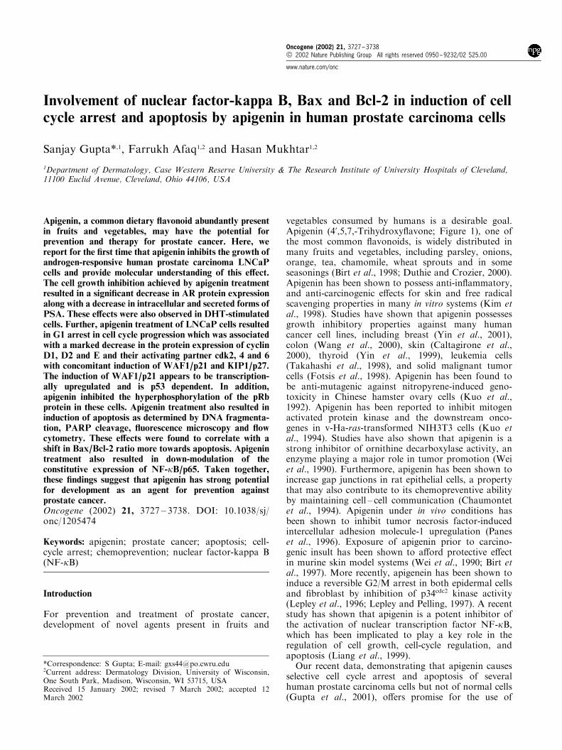
We next examined the e�ect of apigenin on secretedlevels of PSA in LNCaP cells grown in 10% FBS.Treatment of cells with apigenin resulted in asigni®cant decrease in the secreted levels of PSA by41, 53, 60 and 66% at 1, 5, 10 and 20 mMconcentration (Figure 3a). Similarly, cells grown in10% FBS and treated with 10 mM concentration ofapigenin exhibited a signi®cant decrease in 24, 48, 68%at 24, 48 and 72 h (Figure 3a). The DHT-stimulatedcells leads to an increase in intracellular levels of PSA(Figure 3b) and secreted PSA levels in the culturemedium (data not shown), which was signi®cantlyreduced by treatment with 10 mM concentration ofapigenin (Figure 3b). Taken together, the inhibitorye�ects of apigenin on FBS- and DHT-stimulatedLNCaP cell growth were consistent with a decreasein AR protein expression and both intracellular andsecreted PSA levels.
Apigenin induces cell growth inhibition in LNCaP cells
The MTT assay demonstrated that apigenin (1 ± 80 mM)treatment, resulted in a dose-dependent inhibition ofLNCaP cell growth, as compared to untreated controls(Figure 4). Apigenin treatment also resulted in time-dependent inhibition of cell growth. This e�ect wasmore pronounced at 48 and 72 h post-treatment(Figure 4). The inhibition of cell growth could be aresult of the induction of apoptosis that may bemediated by cell cycle growth arrest. We, therefore,hypothesized that apigenin-mediated cell growth in-hibitory e�ects may be due to perturbation in cell cyclethat may possibly lead to programmed cell death.
Apigenin induces G1 cell cycle arrest and alterations in G1cell cycle regulatory proteins in LNCaP cells
Treatment of LNCaP cells with apigenin resulted in adose-dependent inhibition of cell growth as comparedwith their untreated controls at all the time pointsobserved. We considered the possibility that this mayinvolve an arrest of cells at speci®c check point(s) inthe cell cycle. We therefore assessed the e�ect of
Figure 1 Structure of apigenin, a common ¯avonoid present infruits and vegetables. Apigenin consists of a benzene ring (a)fused with a six-member ring that in position 2 carries a phenylring (b) as a substituent
Apigenin induced cell-cycle regulation and apoptosisS Gupta et al
3728
Oncogene

apigenin on cell cycle perturbations. The cells weresynchronized by serum deprivation for 36 h and laterincubated with 10% FBS with varying concentrationsof apigenin for 24 h. Compared with the vehicle-treated controls, apigenin treatment resulted in anappreciable arrest of cells in G0 ±G1 phase of cell cycleafter 24 h of the treatment. The treatment caused anarrest of 75% cells in G0 ±G1 phase of the cell cycle at1 mM concentration that further increased to 80% at 5-mM, 84% at 10 mM and 89% at the highest dose of20 mM in these cells (Figure 5). This increase in G0 ±G1 cell population was accompanied with a concomi-tant decrease of cell number in S phase and G2-Mphase of the cell cycle (Figure 5).
Next, we assessed the protein expression of thecyclins and cdks, which are known to be operative inG1-phase to investigate if their function is altered.Treatment of LNCaP cells with apigenin resulted in adose-dependent decrease in the protein expression ofcyclins D1, D2 and E (Figure 6a). The decrease incyclin D1 protein expression was more pronouncedthan that of cyclin D2 and cyclin E. Similar time-dependent inhibition was observed in cyclins D1, D2and E after treatment of cells with 10 mM concentrationof apigenin for 24, 48 and 72 h (Figure 6a).
The e�ect of apigenin was also observed in theprotein levels of cdks. Treatment of LNCaP cells withapigenin resulted in a dose-dependent decrease in cdk2,4 and 6 (Figure 6b). The decrease in cdk2 proteinexpression was more pronounced than that of cdk4and cdk6. Similar time-dependent inhibition was
observed in cdk2, 4 and 6 after treatment of cells with10 mM concentration of apigenin for 24, 48 and 72 h(Figure 6b). The decrease in the protein expression ofcdk6 was much more pronounced at 48 and 72 h thanat 24 h post-apigenin treatment (Figure 6b).
We next assessed the e�ect of apigenin treatment onthe induction of WAF1/p21, which is known toregulate the entry of cells at G1?S transitioncheckpoint. Western blot analysis revealed thatapigenin treatment results in appreciable induction ofWAF1/p21 even at the lowest concentration of 1 mMwith a signi®cant increase at the high concentration of20 mM. Similarly, 10 mM apigenin treatment resulted ina signi®cant induction of WAF1/p21 at 24, 48, and72 h after the treatment, when compared with the basallevels (Figure 6c). We also estimated the protein levelsof KIP1/p27 after treatment with apigenin. As shownin Figure 6c, treatment of apigenin resulted in a dose-dependent induction of KIP1/p27 and was maximumat the highest concentration of 20 mM. Further,Western blot analysis revealed that apigenin treatmentresulted in a signi®cant induction of KIP1/p27 at 24and 48 h with diminished protein levels at 72 h afterthe treatment, compared with the basal levels (Figure6c).
Apigenin stabilizes p53 protein expression and inhibits Rbphosphorylation in LNCaP cells
Employing Western blot analysis, we next assessed theprotein expression of p53 that is also known to play an
Figure 2 E�ect of apigenin on protein expression of AR and intracellular PSA in LNCaP cells. (a,c) Dose- and time-dependente�ects. The cells were treated with vehicle (DMSO) only or speci®ed concentrations of apigenin for 24 h and then harvested. Fortime-dependent studies, the cells were treated with vehicle only or apigenin (10 mM) for speci®ed times and then harvested. (b,d)Studies with DHT-stimulated cells. Cells were grown in 10% FBS, charcoal stripped serum (c-FBS) with varying concentrations ofDHT with or without apigenin (10 mM) for 24 h. The details are described under Materials and methods. The values below the®gures represent change in protein expression of the bands normalized to b-actin
Oncogene
Apigenin induced cell-cycle regulation and apoptosisS Gupta et al
3729
important role in cell cycle regulation. Since LNCaPcells harbor wild-type p53, we studied the e�ect ofapigenin on the protein expression of p53 in these cells.As shown in Figure 7, apigenin treatment resulted in adose-dependent accumulation in p53 protein expressionin LNCaP cells, which was more pronounced atconcentration of 10 and 20 mM. The time-dependenttreatment with 10 mM concentration of apigeninresulted in the stabilization of p53 protein, which wasobserved up to 72 h post-treatment compared to thebasal levels (Figure 7).
Another key regulator of the G1?S phase transitionin the cell cycle is the retinoblastoma (pRb) tumorsuppressor protein (Adams, 2001). The protein productof the retinoblastoma gene, pRb is a nuclearphosphoprotein that controls the cells during the G1phase of the cell cycle by repressing transcription ofgenes required for the G1?S phase transition (Ahmadet al., 1999). Studies have demonstrated the pRb isphosphorylated in a cell-cycle dependent manner andthat progression of cell through G1?S transitionrequires inactivation of pRb by phosphorylation(Adams, 2001; Ahmad et al., 1999). As shown inFigure 7, apigenin treatment resulted in a signi®cantdose-dependent decrease in the protein expression ofhyper-phosphorylated form of ppRb and this wasaccompanied with a concomitant increase in the hypo-phosphorylated form of pRb. Similar observationswere observed with time-dependent study where thecells were treated with 10 mM concentration of apigeninfor 24, 48 and 72 h. In subsequent experiment,employing a phospho-speci®c antibody we evaluatedthe e�ect of apigenin treatment on serine 780phosphorylation of pRb. As shown in Figure 7,apigenin treatment resulted in dose- as well as time-dependent decrease in serine-780 phosphorylation ofpRb in these cells.
Apigenin induces apoptosis in LNCaP cells
In the next series of experiments we assessed the e�ectof apigenin treatment on apoptosis in LNCaP cells.The cells were treated with varying concentration ofapigenin (1 ± 20 mM). As shown in Figure 8a, comparedwith vehicle-treated control, apigenin treatment (10 and20 mM for 48 h) resulted in the formation of DNAfragments in LNCaP cells. Similarly, treatment with10 mM concentration of apigenin resulted in theformation of DNA ladder at 48, and 72 h post-treatment. Additionally, PARP cleavage analysisshowed that the full size PARP (116 KD) proteinwas cleaved to yield an 85 KD fragment aftertreatment of cells with apigenin at 10 and 20 mMconcentrations (Figure 8b). The time-dependent e�ectof apigenin treatment was almost similar to that ofDNA fragmentation where 10 mM concentration ofapigenin resulted in the cleavage of PARP at 48 and72 h post-treatment (Figure 8b). The induction ofapoptosis by apigenin was also evident from themorphology of cells as assessed by ¯uorescencemicroscopy after labeling the cells with annexin V
Figure 3 E�ect of apigenin on secreted levels of PSA in LNCaPcells. (a) Dose- and time-dependent e�ects. The cells were treatedwith vehicle (DMSO) only or speci®ed concentrations of apigeninfor 24 h and then harvested. For time-dependent studies, the cellswere treated with vehicle only or apigenin (10 mM) for speci®edtimes and then harvested. (b) Studies with DHT-stimulated cells.Cells were grown in 10% FBS, charcoal stripped serum (c-FBS)with varying concentrations of DHT with or without apigenin(10 mM) for 24 h. The PSA levels were determined by enzymelinked immunoabsorbent assay (ELISA). The details are describedunder Materials and methods. The data represents the mean+s.e.of three experiments each conducted in duplicate
Figure 4 E�ect of apigenin on the viability of LNCaP cells. Thecells were treated with speci®ed concentrations of apigenin for 24,48 and 72 h, and cell viability was determined by MTT assay. Thevalues are represented as the percentage cell inhibition wherevehicle-treated cells were regarded as 100%. The details aredescribed under Materials and methods. The data represents themean+s.e. of two experiments each conducted in triplicate
Apigenin induced cell-cycle regulation and apoptosisS Gupta et al
3730
Oncogene

(Figure 8c). We used this method because it identi®esthe apoptotic (green ¯uorescence) as well as necrotic(red ¯uorescence) cells. As shown by data in Figure 8c,apigenin treatment resulted in a dose-dependentapoptosis in LNCaP cells. These data indicated thatapigenin treatment also resulted in necrosis of thesecells. This is possibly the apoptosis induced byapigenin, which is preceded by secondary necrosis inthese cells.
We next quanti®ed the extent of apoptosis by ¯owcytometric analysis of the LNCaP cells labeled withdeoxyuridine triphosphate and propidium iodide. Thecells were treated with 1 ± 20 mM of apigenin for 24 h.As shown by data in Figure 9, apigenin treatmentresulted in 10.1, 13.8, and 17.3% of apoptotic cells at5, 10, and 20 mM (Figure 9). While the induction ofapoptosis was almost negligible (3.2% compared to1.5% of control) at the lowest dose (1 mM), the highestdose (20 mM) resulted in a signi®cant increase inapoptosis as determined by ¯ow cytometry.
Apigenin alters Bax and Bcl-2 protein expression inLNCaP cells
Since Bax and Bcl-2 play a crucial role in apoptosis, wenext studied the dose-and time-dependent e�ects ofapigenin on the constitutive protein levels of Bax andBcl-2 in LNCaP cells. The Western blot analysisexhibited a signi®cant increase in the protein expressionof Bax at 10 and 20 mM concentration of apigenin(Figure 10a). The time-dependent treatment with10 mM concentration of apigenin resulted in the
stabilization of Bax protein, which was observed upto 48 h post-treatment compared to the basal levels(Figure 10a). In sharp contrast, the protein expressionof Bcl-2 was signi®cantly decreased by apigenintreatment in a dose-dependent fashion (Figure 10a).Similar decrease in Bcl-2 protein expression wasobserved during time-dependent study with 10 mMconcentration of apigenin for 24, 48 and 72 h (Figure10a). A signi®cant dose- and time-dependent shift inthe ratio of Bax to Bcl-2 was observed after apigenintreatment indicating the induction of apoptotic process(Figure 10b).
Apigenin inhibits constitutive-NF-kB expression inLNCaP cells
In the next series of experiment we investigated thee�ect of apigenin on the constitutive expression of NF-kB/p65 and IkBa in LNCaP cells. Number of studiessuggests the role of NF-kB in cell survival by inhibitingapoptosis (Liang et al., 1999; Ahmad et al., 2000).Employing immunoblot analysis, we investigated thee�ect of apigenin (1 ± 20 mM concentration for 24 h) onthe constitutively expressed NF-kB/p65 in the nuclearfractions obtained from the cells. As shown byimmunoblot analysis (Figure 11a), apigenin treatmentresulted in a dose- and time-dependent inhibition ofNF-kB/p65 protein expression in these cells. Almostsimilar results were observed in the cytosolic fractionsof cells treated with apigenin (data not shown). Theinhibition of NF-kB in the nuclear fraction correlatedwith the increase in the protein expression of IkBa in
Figure 5 E�ect of apigenin on the DNA cell cycle analysis in synchronized LNCaP cells. The cells were synchronized in G0 phaseby depleting the nutrients for 36 h (referred as control) and replating at sub con¯uent densities into complete medium containingvehicle or apigenin (1, 5, 10 and 20 mM concentration) for 24 h, stained with PI (50 mg/ml) and analysed by ¯ow cytometry.Percentage of cells in G0 ±G1, S and G2-M phase were calculated using cell-®t computer software and are represented in the rightside of the histograms. The details are described under Materials and methods
Oncogene
Apigenin induced cell-cycle regulation and apoptosisS Gupta et al
3731

the cytosol both in dose- and time-dependent fashion(Figure 11a). These results were further con®rmed by amore sensitive method that can detect NF-kB andrecognize an epitope on p65 by ELISA (Figure 11b).
Discussion
Flavonoids are naturally occurring plant polyphenolsfound in abundance in diets rich in fruits, vegetablesand plant-derived beverages (Birt et al., 1998; Duthieand Crozier, 2000; Middleton, 1984). In particular,apigenin, the most common ¯avonoid present in plant
sources has been shown to possess cancer chemopre-ventive e�ects in cell culture system as well as in rodentskin carcinogenesis models (Yin et al., 1999, 2001;Wang et al., 2000; Caltagirone et al., 2000; Takahashiet al., 1998; Fotsis et al., 1998; Kuo et al., 1992, 1994;Wei et al., 1990; Chaumontet et al., 1994; Panes et al.,1996; Birt et al., 1997; Lepley et al., 1996; Lepley andPelling, 1997; Liang et al., 1999). Recently, we haveshown the selective growth inhibitory and apoptoticresponse of apigenin in human prostate carcinoma butnot in normal cells (Gupta et al., 2001). Extendingthese studies here we have provided the molecular basisby which apigenin may exert its cancer chemopreven-tive e�ects on androgen-responsive human prostatecarcinoma LNCaP cells. LNCaP cells are unique invitro model to study human prostate cancer as theypossess an aneuploid male karyotype and secrete PSA(Lee et al., 1995). As most of the molecularmechanisms for the development of prostate cancerinvolve modulation in the functions of androgenreceptor and its signaling pathways we studied thee�ect of apigenin in inhibiting the AR proteinexpression and its subsequent e�ect on PSA produc-tion. Our results indicate that apigenin signi®cantlydecreases serum-induced AR protein expression inthese cells.
Expression of PSA protein has been shown tocorrelate with ¯uctuating androgen levels during maledevelopment (Lamb et al., 2001). PSA belongs to themember of the kallikerin family, is a serine proteasesynthesized by prostate epithelial cells (Naz andHerness, 2001). PSA is currently the most acceptedmarker for assessment of prostate cancer progressionin humans and is being detected in the serum ofpatients with prostate diseases including prostatitis,benign prostatic hypertrophy and prostate cancer(Stamey et al., 1987). Studies have demonstrated thatthe expression of PSA glycoprotein and mRNA inLNCaP cells are regulated by androgens via the
Figure 6 E�ect of apigenin on protein expression of (a) CyclinsD1, D2 and E. (b) Cdk2, Cdk4, and Cdk6. (c) WAF1/p21 andKIP1/p27 in LNCaP cells. The cells were treated with vehicle(DMSO) only or speci®ed concentrations of apigenin for 24 h andthen harvested. For time-dependent studies, the cells were treatedwith vehicle only or apigenin (10 mM) for speci®ed times and thenharvested. The details are described under Materials and methods.The values below the ®gures represent change in proteinexpression of the bands normalized to b-actin
Figure 7 E�ect of apigenin on protein expression of p53, pRband serine phosphorylation of pRb in LNCaP cells. The upperband (slower migration) represents the hyper-phosphorylatedform of pRb, and the lower band (faster migration) representshypo-phosphorylated form of pRb. The cells were treated withvehicle (DMSO) only or speci®ed concentrations of apigenin for24 h and then harvested. For time-dependent studies, the cellswere treated with vehicle only or apigenin (10 mM) for speci®edtimes and then harvested. The details are described underMaterials and methods. The values below the ®gures representchange in protein expression of the bands normalized to b-actin
Apigenin induced cell-cycle regulation and apoptosisS Gupta et al
3732
Oncogene

androgen receptor (Lee et al., 1995; Henttu et al.,1990). Further, PSA has been reported to cleave theseminal vesicle protein that may augment the activa-tion of growth factors leading to tumor progression(Wang et al., 1997; Henttu et al., 1990). Our studiesdemonstrate a signi®cant decrease in intracellular aswell as secreted levels of PSA by apigenin, suggestingthat this may be an important strategy to preventprostate cancer progression.
The cell growth and proliferation of mammaliancells are mediated via cell cycle progression. In recentyears, studies have shown an association between cellcycle regulation and cancer, and inhibition of the cellcycle has become an appreciated target for manage-ment of cancer (Pucci et al., 2000; Sandhu andSlingerland, 2000). Our studies indicate that apigenin
exerts strong growth inhibitory e�ects on humanprostate carcinoma cells by perturbation in G1?Sphase of the cell cycle. The passage through the cellcycle, in eukaryotes is orchestrated by the function ofa family of protein kinase complexes. Each complex iscomposed minimally of a catalytic subunit, the cdk,and its essential activating partner, the cyclins (Pestellet al., 1999). Cyclins D and E are involved duringG1?S phase of the cell cycle. In controlled cellgrowth, association of cyclins D and E with cdk2, 4or 6 leads to phosphorylation of Rb; hyperpho-sphorylated Rb leads to its release from E2F. Thefree E2F then activates c-myc, resulting in progressionof the cell cycle and cellular proliferation. Evidencesindicate that overexpression of cyclin D is associatedwith various cancers and tumor-derived cell lines
Figure 8 E�ect of apigenin on the induction of apoptosis in LNCaP cells. (a) DNA fragmentation induced by apigenin in LNCaPcells. For dose-dependent studies, the cells were treated with vehicle (DMSO) or with the speci®ed concentrations of apigenin for48 h. For time-dependent studies the cells were treated with vehicle only or apigenin (10 mM) for speci®ed times and then harvested.DNA was isolated and subjected to agarose gel electrophoresis, followed by visualization of bands and polaroid photography. (b)Immunoblot analysis of PARP cleavage induced by apigenin in LNCaP cells. (c) Fluorescence microscopy of LNCaP cells treatedwith apigenin. The cells were treated with vehicle alone or speci®ed concentration of apigenin for 24 h. The ApopNexin apoptosisdetection kit (Oncor) was used for the detection of apoptotic and necrotic cells. Apoptosis and necrosis was detected by the kitaccording to the vendor's protocol using a Zeiss Axiovert 100 microscope. The samples were excited at 330 ± 380 nm, and the imagewas observed and photographed under a combination of a 400 nm dichoric mirror and then 420-nm high pass ®lter. The details aredescribed under Materials and methods
Figure 9 E�ect of apigenin on quantitation of apoptosis in LNCaP cells. The cells were treated with apigenin (1 ± 20 mM for 24 h),labeled with deoxyuridine triphosphate using terminal deoxynucleotide transferase and PI by using apoptosis kit obtained fromPhoenix Flow Systems (San Diego, CA, USA) as per vendor's protocol followed by ¯ow cytometry. Cells showing deoxyuridinetriphosphate ¯uorescence above that of control population, as indicated by the line in each histogram, are considered as apoptoticcells and their percentage population is shown in each box. The details are described under Materials and methods
Oncogene
Apigenin induced cell-cycle regulation and apoptosisS Gupta et al
3733

leading to the uncontrolled growth (Motokura andArnold, 1993). One of the aspects of cyclin Doverexpression in cells is a shorter G1 phase, resultingin a more rapid entry into S phase and increasedproliferation (Mueller et al., 1997). Based on these andother studies (Yin et al., 2001; Wang et al., 2000;Lepley et al., 1996; Lepley and Pelling, 1997), asigni®cant decrease in protein levels of cyclins D1, D2and E, cdk2, 4 and 6 by apigenin suggests thatapigenin is a promising agent for prevention andtherapy of prostate cancer. The observed inhibitorye�ects of apigenin particularly on cyclin D1, cdk2,and cdk4 in LNCaP cells are of particular signi®cancefor the intervention of hormone-responsive prostatecancer because cyclin D is strongly associated withandrogen-stimulated growth of LNCaP cells. Duringthe progression of the cell cycle, the cdk-cyclincomplexes are inhibited via binding to ckis such asCIP/KIP and INK4 families of proteins (Vidal andKo�, 2000). Because our studies have demonstratedthat apigenin treatment of LNCaP cells resulted in aG1 phase arrest of the cell cycle, we examined thee�ect of apigenin on ckis operative in this phase ofthe cell cycle. Our data demonstrated a signi®cant up-regulation of the WAF1/p21 and KIP1/p27 during G1phase arrest of LNCaP cells which may be an
important molecular mechanism through which api-genin inhibits the growth of cancer cells.
The tumor suppressor gene p53 is regarded as a keyelement in maintaining a balance between cell growthand cell death in the living system (Agarwal et al.,1998; Pucci et al., 2000; Vidal and Ko�, 2000). Thep53, in response to DNA damage, triggers a variety ofcell cycle regulatory events to limit the proliferation ofdamaged cells (Vogelstein et al., 2000). This `gatekeeper' gene, in a number of human tumors, isinactivated by mutation resulting in uncontrolledcellular proliferation (Navone et al., 1993). Generally,no correlation between p53 mutations and early stageprostate cancer has been noticed but p53 mutations areshown to be associated with a small group (10 ± 20%)of advanced prostate cancer patients with high Gleasonscore and distant metastasis (Navone et al., 1993;Meyers et al., 1998). It is, therefore, important to studythe role of p53 in tumor growth inhibition and
Figure 10 (a) E�ect of apigenin on protein expression of Baxand Bcl2 in LNCaP cells. The cells were treated with vehicle(DMSO) only or speci®ed concentrations of apigenin for 24 h andthen harvested. For time-dependent studies, the cells were treatedwith vehicle only or apigenin (10 mM) for speci®ed times and thenharvested. The details are described under Materials and methods.The values below the ®gures represent change in proteinexpression of the bands normalized to b-actin. (b) Dose- andtime-dependent e�ect of apigenin on Bax/Bcl2 ratio
Figure 11 E�ect of apigenin on the constitutive expression ofNF-kB in LNCaP cells. (a) Immunoblot analysis of NF-kB/p65 inthe nuclear fraction, and IkBa in the cytosolic fractions of varioustreated samples. The cells were treated with vehicle only orspeci®ed concentrations of apigenin for 24 h and then processedfor nuclear and cytosolic fractions. For time-dependent studies,the cells were treated with vehicle only or apigenin (10 mM) forspeci®ed times and processed for nuclear and cytosolic fractions.The details are described under Materials and methods. Thevalues below the ®gures represent change in protein expression ofthe bands normalized to b-actin. Data shown here are from arepresentative experiment repeated twice with similar results. (b)E�ect of apigenin on NF-kB activation. The cells were treatedwith vehicle only or speci®ed concentrations of apigenin for 24 hand then processed for nuclear lysate. NF-kB activation wasdetermined by ELISA Trans-AM kit from Active Motif NorthAmerica (Carlsbad, CA, USA) for assay of NF-kB/p65 activity.The details are described under Materials and methods. The datarepresents the mean+s.e. of two experiments each conducted induplicate
Apigenin induced cell-cycle regulation and apoptosisS Gupta et al
3734
Oncogene

induction of apoptosis. Our data showed that apigenintreatment resulted in a signi®cant accumulation and/orstabilization of p53 protein in a dose- and time-dependent fashion in LNCaP cells that harbor wild-type p53. The induction of WAF1/p21, along withstabilization of p53 therefore, appears to be p53-dependent in LNCaP cells (with wild-type p53). Studieshave indicated an increase in wild-type p53 stabilityand transactivational activity by apigenin that may beresponsible for its anti-tumorigenic activity by stimu-lating the p53-WAF1/p21 response pathway (McVeanet al., 2000). Studies have established that pRb iscapable of exerting growth suppressive activity becauseof its interaction with E2F/DP heterodimers, whichfunction to trigger the transcription of genes requiredfor cell cycle progression (Adams, 2001; Ahmad et al.,1999). Since the phosphorylation of pRb is animportant event for the progression of cell cycle atG1?S transition (Masciullo et al., 2000) in the presentstudy, we investigated the protein level and thephosphorylation pattern of pRb during apigenin-mediated cell cycle arrest and apoptosis. Our resultsdemonstrated dose- as well as time-dependent decreasein the level of hyper-phosphorylated form ppRb andincrease in hypo-phosphorylated form or pRb byapigenin and also a decrease in phosphorylation ofpRb at serine-780. These series of events results inarrest of cells at G0 ±G1 phase of the cell cycle thatmay subsequently lead to apoptotic cell death.
Among the number of pathways that can beexploited for cancer prevention and treatment, apop-tosis is a physiological process by which cells areremoved when an agent damages their DNA. In recentyears the concept of cell cycle-mediated apoptosis isalso gaining increasing attention and is an ideal way ofcell elimination (Pucci et al., 2000; Zornig et al., 2001).In this regard apigenin is a promising agent as it iscapable of selective/preferential elimination of cancercells by inhibiting cell cycle progression and/or causingapoptosis (Gupta et al., 2001). Our data demonstratedthat apigenin results in the apoptosis of androgen-responsive LNCaP cells. This observation was veri®edby DNA fragmentation, PARP-cleavage, ¯uorescencemicroscopy and ¯ow cytometry. Collectively, theseresults clearly suggest that apigenin inhibits the growthof prostate carcinoma cells via cell cycle arrest andinduces apoptosis, and that these results corroboratethe ®ndings of many published studies in several othertypes of human carcinoma cells (Yin et al., 1999, 2001;Wang et al., 2000; Caltagirone et al., 2000; Takahashiet al., 1998). This could be an important observationthat may be useful for devising strategies for themanagement of human prostate cancer.
Recent studies have indicated that apigenin alsofunctions as a potent inhibitor of the oxidant- andtumor promoter-mediated activation of NF-kB in cellculture system (Liang et al., 1999). NF-kB is a widelydistributed pleiotropic nuclear factor that is known toregulate the expression of genes encoding cytokines,cellular adhesion molecules, and growth factors (Bourset al., 2000). Studies have also indicated that NF-kB
promotes cell survival by inhibiting apoptosis (Bours etal., 2000). Recently, it has been shown that the down-modulation of NF-kB activity in the cytosol andnucleus is associated with an apoptotic response ofthe eukaryotic cells (Ahmad et al., 2000). In recentyears, NF-kB has been increasingly appreciated as atarget for anticancer drug treatment (Lee and Burckart,1998). Our results demonstrate a dose- and time-dependent decrease in the constitutive expression ofNF-kB/p65 in the cytosol and nucleus with subsequentincrease in the protein expression of IkBa in thecytosol by treatment with apigenin. We postulate thatapigenin-mediated down-modulation of NF-kB activitycould lead to inhibition of cell growth and induction ofapoptosis, which are mediated by alterations in the G1-phase of the cell cycle.
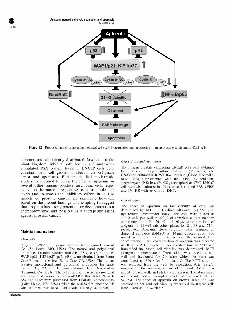
Members of the Bcl-2 family of proteins are criticalregulators of the apoptotic pathway (Yin et al., 1994).Bcl-2 is an upstream e�ector molecule in the apoptoticpathway and is identi®ed as a potent suppressor ofapoptosis (Hockenbery et al., 1993). Bcl-2 is found atinappropriately high levels in more than half of allhuman tumors (Reed, 1995). Bcl-2 has been shown toform a heterodimer with the pro-apoptotic memberBax and might thereby neutralize its pro-apoptotice�ects. Therefore, alterations in the levels of Bax andBcl-2, i.e. the ratio of Bax/Bcl-2 is a decisive factor andplays an important role in determining whether cellswill undergo apoptosis under experimental conditionsthat promote cell death. In our study, a decrease inBcl-2 protein expression was observed in androgen-responsive LNCaP cells. Importantly, the proteinexpression of Bax, however, was up-regulated inLNCaP cells after treatment up to 72 h, hence theratio of Bax to Bcl-2 was altered in favor of apoptosis.Our results suggest that up-regulation of Bax anddown-modulation of Bcl-2 may be another molecularmechanism through which apigenin induces apoptosis.Based on the outcome of this study and the availableliterature knowledge, as shown in the compositescheme in Figure 12, we suggest multiple pathwaysby which apigenin results in apoptotic cell death. Thismay be mediated via cell cycle arrest, modulation inBax/Bcl-2 ratio or NF-kB pathway. It is important tomention here, that based on the daily dietaryconsumption of ¯avonoids, the concentration used inour present study is physiologically achievable inhumans (Hollman and Katan, 1999a). This may, inpart, be supported by the epidemiological observationsthat regular consumption of fruits and vegetables,particularly cruciferous vegetables rich in ¯avonoids,may reduce the risk of prostate cancer (Kolonel et al.,2000; Cohen et al., 2000). Studies also indicate thatthese associations remain inconclusive because thechemopreventive e�ects achieved by ¯avonoids haveoften not been observed in in vivo situations (Hollmanand Katan, 1999b). Therefore, more detailed case ±control design studies are required to validate these®ndings.
Our study provides an in-depth experimentalevidence for the ®rst time that apigenin, the most
Oncogene
Apigenin induced cell-cycle regulation and apoptosisS Gupta et al
3735
common and abundantly distributed ¯avonoid in theplant kingdom, inhibits both serum- and androgen-stimulated PSA protein levels in LNCaP cells con-comitant with cell growth inhibition via G1-phasearrest and apoptosis. Further, detailed mechanisticstudies are required to de®ne the e�ect of apigenin onseveral other human prostate carcinoma cells, espe-cially on hormone-unresponsive cells at molecularlevels and to assess the inhibitory e�ects in in vivomodels of prostate cancer. In summary, however,based on the present ®ndings it is tempting to suggestthat apigenin has strong potential for development as achemopreventive and possibly as a therapeutic agentagainst prostate cancer.
Materials and methods
Materials
Apigenin (595% purity) was obtained from Sigma ChemicalCo. (St. Louis, MO, USA). The mono- and poly-clonalantibodies (human reactive anti-AR, PSA, cdk2, 4 and 6,WAF1/p21, KIP1/p27, p53, pRb) were obtained from SantaCruz Biotechnology Inc. (Santa Cruz, CA, USA). The humanreactive monoclonal and polyclonal antibodies for anti-cyclins D1, D2 and E were obtained from Neomarker(Fremont, CA, USA). The other human reactive monoclonaland polyclonal antibodies for anti-PARP, Bax, Bcl-2, NF-kB/p54 and IkBa were purchased from Upstate Biotechnology(Lake Placid, NY, USA) while the anti-Ser780-phospho-Rbwas obtained from MBL, Ltd. (Naka-ku Nagoya, Japan).
Cell culture and treatments
The human prostate carcinoma LNCaP cells were obtainedfrom American Type Culture Collection (Manassas, VA,USA) and cultured in RPMI 1640 medium (Gibco, Rockville,MD, USA), supplemented with 10% FBS, 1% penicillin/streptomycin (P-S) in a 5% CO2 atmosphere at 378C. LNCaPcells were also cultured in 10% charcoal-stripped FBS (cFBS)and 1% P-S with or without DHT.
Cell viability
The e�ect of apigenin on the viability of cells wasdetermined by MTT (3-[4,5-dimethylthiazol-2-yl]-2,5-diphe-nyl tetrazoliumbromide) assay. The cells were plated at16104 cells per well in 200 ml of complete culture mediumcontaining 1, 5, 10, 20, 40 and 80 mM concentrations ofapigenin in 96-well microtiter plates for 24, 48 and 72 h,respectively. Apigenin stock solutions were prepared indimethyl sulfoxide (DMSO) at 10 mM concentration, andmixed with fresh medium to achieve the desired ®nalconcentration. Each concentration of apigenin was repeatedin 10 wells. After incubation for speci®ed time at 378C in ahumidi®ed incubator, cell viability was determined. MTT(5 mg/ml in phosphate bu�ered saline) was added to eachwell and incubated for 2 h after which the plate wascentrifuged at 1800 g for 5 min at 48C. The MTT solutionwas removed from the wells by aspiration. After carefulremoval of the medium, 0.1 ml of bu�ered DMSO wasadded to each well, and plates were shaken. The absorbancewas recorded on a microplate reader at the wavelength of540 nM. The e�ect of apigenin on growth inhibition wasassessed as per cent cell viability where vehicle-treated cellswere taken as 100% viable.
Figure 12 Proposed model for apigenin-mediated cell cycle dysregulation and apoptosis of human prostate carcinoma LNCaP cells
Apigenin induced cell-cycle regulation and apoptosisS Gupta et al
3736
Oncogene
DNA cell cycle analysis
The cells (70% con¯uent) were starved for 36 h to arrestthem in G0 phase of the cell cycle, after which they weretreated with apigenin (1, 5, 10 and 20 mM doses) in RPMI-1640 complete media for 24 h. The cells were trypsinizedthereafter, washed twice with cold PBS, and centrifuged. Thepellet was resuspended in 50 ml cold PBS and 450 ml coldmethanol for 1 h at 48C. The cells were centrifuged at 110 gfor 5 min, pellet washed twice with cold PBS, suspended in500 ml PBS, and incubated with 5 ml RNAse (20 mg/ml ®nalconcentration) at 378C for 30 min. The cells were chilled overice for 10 min and stained with propidium iodide (50 mg/ml®nal concentration) for 1 h and analysed by ¯ow cytometry.
Protein extraction and Western blotting
The cells (70% con¯uent) were treated with apigenin (1, 5, 10and 20 mM doses) in RPMI-1640 complete media for 24 h.For time-dependent assay, the cells (50 ± 60% con¯uent) weretreated for 24, 48 and 72 h with 10 mM of apigenin. Afterwhich the media was aspirated, the cells were washed withcold PBS (10 mM, pH 7.4) and ice-cold lysis bu�er (50 mM
Tris-HCl, 150 mM NaCl, 1 mM EGTA, 1 mM EDTA, 20 mM
NaF, 100 mM Na3VO4, 0.5% NP-40, 1% Triton X-100,1 mM PMSF (pH 7.4)) with freshly added protease inhibitorcocktail (Protease Inhibitor Cocktail Set III, Calbiochem, LaJolla, CA, USA) over ice for 30 min. The cells were scraped,the lysate was collected in a microfuge tube and passedthrough a 21� G needle to break up the cell aggregates. Thelysate was cleared by centrifugation at 14 000 g for 15 min at48C and the supernatant (total cell lysate) was used orimmediately stored at 7808C. For NF-kB/p65 assay the cellswere processed for cytosolic and nuclear fractions asdescribed previously (Ahmad et al., 2000). The proteinconcentration was determined by DC Bio-Rad assay usingthe manufacturer's protocol (Bio Rad Laboratories, Hercules,CA, USA).For Western blotting, 25 ± 50 mg protein was resolved over
8 ± 12% polyacrylamide gels and transferred to a nitrocellu-lose membrane. The blot was blocked in blocking bu�er (5%nonfat dry milk/1% Tween 20; in 20 mM TBS, pH 7.6) for1 h at room temperature, incubated with appropriatemonoclonal or polyclonal primary antibody in blockingbu�er for 1 h to overnight at 48C, followed by incubationwith anti-mouse or anti-rabbit secondary antibody horse-radish peroxidase conjugate obtained from Amersham LifeScience Inc. (Arlington Height, IL, USA) and detected bychemiluminescence and autoradiography using XAR-5 ®lmobtained from Eastman Kodak Co. (Rochester, NY, USA).Densitometric measurements of the bands in Western blotanalysis were performed using digitalized scienti®c softwareprogram UN-SCAN-IT purchased from Silk Scienti®cCorporation (Orem, UT, USA).
DNA fragmentation assay
The cells were grown to about 70% con¯uence and treatedwith apigenin (1, 5, 10 and 20 mM concentration) for 48 h.Following this treatment, the cells were washed twice withphosphate-bu�ered saline (10 mM Tris, pH 7.5, 150 mM
NaCl, 5 mM MgCl2, and 0.5% Triton X-100), left on icefor 15 min, and pelleted by centrifugation (14 000 g) at 48C.The pellet was incubated with DNA lysis bu�er (10 mM Tris,pH 7.5, 400 mM NaCl, 1 mM EDTA, and 1% Triton X-100)for 30 min on ice and then centrifuged at 14 000 g at 48C.The supernatant obtained was incubated overnight with
RNAse (0.2 mg/ml) at room temperature and then withProteinase K (0.1 mg/ml) for 2 h at 378C. DNA wasextracted using phenol : chloroform (1 : 1) and precipitatedwith 95% ethanol for 2 h at 7808C. The DNA precipitatewas centrifuged at 14 000 g at 48C for 15 min and the pelletwas air-dried and dissolved in 20 ml of Tris-EDTA bu�er(10 mM Tris-HCl, pH 8.0, and 1 mM EDTA). Total amountof DNA was resolved over 1.5% agarose gel, containing0.3 mg/ml ethidium bromide in 16TBE bu�er (pH 8.3,89 mM Tris, 89 mM Boric acid and 2 mM EDTA) (BioWittaker, Inc., Walkersville, MD, USA). The bands werevisualized under UV transilluminator (Model # TM-36, UVPInc., San Gabriel, CA, USA) followed by polaroid photo-graphy (MP-4 Photographic System, Fotodyne Inc., Hart-land, WI, USA).
Apoptosis detection by fluorescence microscopy
The ApopNexin apoptosis detection kit (Oncor, Gaithers-burg, MD, USA) was used for the detection of apoptoticcells. This kit uses a dual-staining protocol in which theapoptotic cells are stained with annexin V (green ¯uores-cence), and the necrotic cells are stained with propidiumiodide (red ¯uorescence). Brie¯y, the cells were grown toabout 50% con¯uence in 6-well plate and then treated withapigenin (1, 5, 10 and 20 mM concentration) for 24 h.Apoptosis was detected by the use of the kit according tothe vendor's protocol using a Zeiss Axiovert 100 microscope.Brie¯y, the samples were excited at 330 ± 380 nm, and theimage was observed and photographed under a combinationof a 400-nm dichoric mirror and then 420-nm high-pass ®lter.Cells with green, condensed chromatin pattern were scored asapoptotic, whereas those with red nuclei without nuclearcondensation were considered necrotic.
Quantification of apoptosis
For quanti®cation of apoptosis, the cells were grown at adensity of 16106 cells in 100-mm culture dishes and weretreated with apigenin (1, 5, 10, and 20 mM concentration) for24 h. The cells were trypsinized, washed with PBS, andprocessed for labeling with ¯uorescein-tagged deoxyuridinetriphosphate nucleotide and propidium iodide by use of anAPO-DIRECT apoptosis kit obtained from Phoenix FlowSystems (San Diego, CA, USA) as per manufacturer'sprotocol. The labeled cells were then analysed by ¯owcytometry.
ELISA assay for PSA and NF-kB activity
Enzyme linked immunoabsorbent assay (ELISA) wasperformed for PSA and NF-kB/p65 activity. The humanPSA ELISA kit was obtained from Anogen (Ontario,Canada) for the quantitative determination of PSA in celllysate and culture medium according to the vendor'sprotocol. Brie¯y, the kit applies a technique of quantitativesandwich immunoassay for determination of PSA with anestimated sensitivity of 1 ng/ml PSA antigen.The commercially available Trans-AM kit was obtained
from Active Motif North America (Carlsbad, CA, USA) forassay of NF-kB/p65 activity according to the vendor'sprotocol. Brie¯y, the assay uses an oligonucleotide containingNF-kB consensus site (5'-GGGACTTTCC-3') that binds tothe cell extract and can detect NF-kB, which can recognizean epitope on p65 activated and bound to its target DNA.This assay is speci®c for NF-kB activation and is highlysensitive when compared with the gel retardation technique.
Oncogene
Apigenin induced cell-cycle regulation and apoptosisS Gupta et al
3737

AbbreviationsPSA, Prostate-speci®c antigen; AR, androgen receptor;NF-kB, nuclear factor-kappa B; pRb, hypo-phosphorylatedretinoblastoma; ppRb, hyper-phosphorylated retinoblasto-ma; DMSO, dimethyl sulphoxide; PBS, phosphate bu�eredsaline; TBS, tris bu�ered saline; DHT, 5a-dihydrotestoster-one; cdk, cyclin-dependent kinase; cki, cyclin kinaseinhibitor.
AcknowledgmentsSupported by grants from the United States Public HealthService (RO1CA 78809), American Institute for Cancerresearch (00A030), and Department of Defense (DAMD17-00-1-0527).
References
Adams PD. (2001). Biochim. Biophys. Acta, 1471, 123 ± 133.Agarwal ML, Taylor WR, Chernov MV, Chernova OB and
Stark GR. (1998). J. Biol. Chem., 273, 1 ± 4.Ahmad N, Gupta S and Mukhtar H. (1999). Oncogene, 18,
1891 ± 1896.Ahmad N, Gupta S and Mukhtar H. (2000). Arch. Biochem.
Biophys., 376, 338 ± 346.Birt DF, Mitchell D, Gold B, Pour P and Pinch HC. (1997).
Anticancer Res., 17, 85 ± 91.Birt DF, Shull JS and Yaktine AL. (1998). Chemoprevention
of cancer Shils ME, Olson JA, Shike M and Ross AC (eds).Baltimore: Williams & Wilkins. pp. 1263 ± 1295.
Bours V, Bentires-Al M, Hellin AC, Viatour P, Robe P,Delhalle S, Benoit V and Merville MP. (2000). Biochem.Pharmacol., 60, 1085 ± 1089.
Caltagirone S, Rossi C, Poggi A, Ranelletti FO, Natali PG,Brunetti M, Aiello FB and Piantelli M. (2000). Int. J.Cancer, 87, 595 ± 600.
Chaumontet C, Bex V, Gaillard-Sanchez I, Seillan-HeberdenC, Suschetet M and Martel P. (1994). Carcinogenesis, 15,2325 ± 2330.
Cohen JH, Kristal AR and Stanford JL. (2000). J. Natl.Cancer Inst., 92, 61 ± 68.
Duthie G and Crozier A. (2000). Curr. Opin. Clin. Nutr.Metab. Care., 3, 447 ± 451.
Fotsis T, Pepper MS, Montesano R, Aktas E, Breit S,Schweigerer L, Rasku S, Wahala K and Adlercreutz H.(1998). Baillieres Clin. Endocrinol. Metab., 12, 649 ± 666.
Gupta S, Afaq F and Mukhtar H. (2001). Biochem. Biophys.Res. Commun., 287, 914 ± 920.
Henttu P, Lukkarinen O and Vihko P. (1990). Int. J. Cancer,45, 654 ± 660.
Hockenbery DM, Oltvai ZN, Yin XM, Milliman CL andKorsmeyer SJ. (1993). Cell, 75, 241 ± 251.
Hollman PC and Katan MB. (1999a). Food Chem. Toxicol.,37, 937 ± 942.
Hollman PC and Katan MB. (1999b). Free Radic. Res., 31,75 ± 80.
Kim HP, Mani I, Iversen L and Ziboh VA. (1998).Prostaglandins Leukot. Essent. Fatty Acids., 58, 17 ± 24.
Kolonel LN, Hankin JH, Whittemore AS, Wu AH,Gallagher RP, Wilkens LR, John EM, Howe GR, DreonDM, West DW and Pa�enbarger Jr RS. (2000). CancerEpidemiol. Biomarkers Prev., 9, 795 ± 804.
Kuo ML, Lee KC and Lin JK. (1992). Mutat. Res., 270, 87 ±95.
Kuo ML, Lin JK, Huang TS and Yang NC. (1994). CancerLett., 87, 91 ± 97.
Lamb DJ, Weigel NL and Marcelli M. (2001). Vitam. Horm.,62, 199 ± 230.
Lee C, Sutkowski DM, Sensibar JA, Zelner D, Kim I, AmselI, Shaw N, Prins GS and Kozlowski JM. (1995).Endocrinology, 136, 796 ± 803.
Lee JI and Burckart GJ. (1998). J. Clin. Pharmacol., 38,981 ± 993.
Lepley DM and Pelling JC. (1997). Mol. Carcinog., 19, 74 ±82.
Lepley DM, Li B, Birt DF and Pelling JC. (1996).Carcinogenesis, 17, 2367 ± 2375.
Liang YC, Huang YT, Tsai SH, Lin-Shiau SY, Chen CF andLin JK. (1999). Carcinogenesis, 20, 1945 ± 1952.
Masciullo V, Khalili K and Giordano A. (2000). Int. J.Oncol., 17, 897 ± 902.
McVean M, Xiao H, Isobe K and Pelling JC. (2000).Carcinogenesis, 21, 633 ± 639.
Meyers FJ, Gumerlock PH, Chi SG, Borchers H, Deitch ADand deVere White RW. (1998). Cancer, 83, 2534 ± 2539.
Middleton E. (1984). Trends Pharmacol. Sci., 5, 335 ± 338.Motokura T and Arnold A. (1993). Curr. Opin. Genet. Dev.,
3, 5 ± 10.Mueller A, Odze R, Jenkins TD, Shahsesfaei A, Nakagawa
H, Inomoto T and Rustgi AK. (1997). Cancer Res., 57,5542 ± 5549.
Navone NM, Troncoso P, Pisters LL, Goodrow TL, PalmerJL, Nichols WW, von Eschenbach AC and Conti CJ.(1993). J. Natl. Cancer Inst., 85, 1657 ± 1669.
Naz RK and Herness EA. (2001). Front. Biosci., 6, 1083 ±1088.
Panes J, Gerritsen ME, Anderson DC, Miyasaka M andGranger DN. (1996). Microcirculation, 3, 279 ± 286.
Pestell RG, Albanese C, Reutens AT, Segall JE, Lee RJ andArnold A. (1999). Endocr. Rev., 20, 501 ± 534.
Pucci B, Kasten M and Giordano A. (2000). Neoplasia, 2,291 ± 299.
Reed JC. (1995). Curr. Opin. Oncol., 7, 541 ± 546.Sandhu C and Slingerland J. (2000). Cancer Detect. Prev., 24,
107 ± 118.Stamey TA, Yang N, Hay AR, McNeal JE, Freiha FS and
Redwine E. (1987). N. Engl. J. Med., 317, 909 ± 916.Takahashi T, Kobori M, Shinmoto H and Tsushida T.
(1998). Biosci. Biotechnol. Biochem., 62, 2199 ± 2204.Vidal A and Ko� A. (2000). Gene, 247, 1 ± 15.Vogelstein B, Lane D and Levine AJ. (2000). Nature, 408,
307 ± 310.Wang LG, Liu XM, Kreis W and Budman DR. (1997).
Cancer Res., 57, 714 ± 719.Wang W, Heideman L, Chung CS, Pelling JC, Koehler KJ
and Birt DF. (2000). Mol. Carcinog., 28, 102 ± 110.Wei H, Tye L, Bresnick E and Birt DF. (1990). Cancer Res.,
50, 499 ± 502.Yin F, Giuliano AE and Van Herle AJ. (1999). Thyroid, 9,
369 ± 376.Yin F, Giuliano AE, Law RE and Van Herle AJ. (2001).
Anticancer Res., 21, 413 ± 420.Yin XM, Oltvai ZN, Veis-Novack D, Linette GP and
Korsmeyer SJ. (1994). Cold Spring Harb. Symp. Quant.Biol., 59, 387 ± 393.
Zornig M, Hueber A, BaumW and Evan G. (2001). Biochim.Biophys. Acta, 1551, F1 ± F37.
Apigenin induced cell-cycle regulation and apoptosisS Gupta et al
3738
Oncogene


![DNA damage-induced nuclear factor-kappa B …...DNA damage-induced nuclear factor-kappa B activation and its roles in cancer progression Wei Wang 1, 2, ... human diseases and aging.[2]](https://img.pdfslide.us/doc/110x75/5f01c43b7e708231d400ef61/dna-damage-induced-nuclear-factor-kappa-b-dna-damage-induced-nuclear-factor-kappa.jpg)