Embed Size (px)
Citation preview
Kent Academic RepositoryFull text document (pdf)
Copyright & reuse
Content in the Kent Academic Repository is made available for research purposes. Unless otherwise stated all
content is protected by copyright and in the absence of an open licence (eg Creative Commons), permissions
for further reuse of content should be sought from the publisher, author or other copyright holder.
Versions of research
The version in the Kent Academic Repository may differ from the final published version.
Users are advised to check http://kar.kent.ac.uk for the status of the paper. Users should always cite the
published version of record.
Enquiries
For any further enquiries regarding the licence status of this document, please contact:
If you believe this document infringes copyright then please contact the KAR admin team with the take-down
information provided at http://kar.kent.ac.uk/contact.html
Citation for published version
Sumbayev, Vadim V. and Yasinska, Inna M. and Oniku, Abraham E. and Streatfield, Claire L.and Gibbs, Bernhard F. (2012) Involvement of Hypoxia-Inducible Factor-1 in the InflammatoryResponses of Human LAD2 Mast Cells and Basophils. PLoS ONE, 7 (3). e34259. ISSN 1932-6203.
DOI
https://doi.org/10.1371/journal.pone.0034259
Link to record in KAR
http://kar.kent.ac.uk/29575/
Document Version
Publisher pdf
Involvement of Hypoxia-Inducible Factor-1 in theInflammatory Responses of Human LAD2 Mast Cells andBasophils
Vadim V. Sumbayev.*, Inna Yasinska., Abraham E. Oniku, Claire L. Streatfield, Bernhard F. Gibbs*
Medway School of Pharmacy, University of Kent, Chatham Maritime, United Kingdom
Abstract
We recently showed that hypoxia-inducible factor 1 (HIF-1) plays a crucial role in the pro-allergic functions of humanbasophils by transcriptional control of energy metabolism via glycolysis as well as directly triggering expression of theangiogenic cytokine vascular endothelium growth factor (VEGF). Here, we investigated HIF-1 involvement in controlling thesynthesis of angiogenic and inflammatory cytokines from various human effector cells stimulated by IgE-dependent orinnate immune triggers. Purified primary human basophils, LAD2 human mast cells and THP-1 human myeloid cells wereused for investigations of FceRI and Toll-like receptor (TLR) ligand-induced responses. In contrast to basophils, LAD2 mastcells expressed background levels of HIF-1a, which was largely independent of the effects of stem cell factor (SCF). Bothmast cells and basophils expressed TLR2 and 4, albeit weakly compared to THP-1 cells. Cytokine production in mast cellsfollowing TLR ligand stimulation was markedly reduced by HIF-1a knockdown in LAD2 mast cells. In contrast, although HIF-1 is involved in IgE-mediated IL-4 secretion from basophils, it is not clearly induced by peptidoglycan (PGN). HIF-1aaccumulation is critical for sustaining human allergic effector cell survival and function. This transcription complex facilitatesgeneration of both pro-angiogenic and inflammatory cytokines in mast cells but has a differential role in basophilstimulation comparing IgE-dependent triggering with innate immune stimuli.
Citation: Sumbayev VV, Yasinska I, Oniku AE, Streatfield CL, Gibbs BF (2012) Involvement of Hypoxia-Inducible Factor-1 in the Inflammatory Responses of HumanLAD2 Mast Cells and Basophils. PLoS ONE 7(3): e34259. doi:10.1371/journal.pone.0034259
Editor: Michael B. Fessler, National Institute of Environmental Health Sciences, United States of America
Received January 13, 2012; Accepted February 24, 2012; Published March 28, 2012
Copyright: � 2012 Sumbayev et al. This is an open-access article distributed under the terms of the Creative Commons Attribution License, which permitsunrestricted use, distribution, and reproduction in any medium, provided the original author and source are credited.
Funding: Work was supported by the Asthma UK Grant (grant number 10/065 - to BFG and VS). The funders had no role in study design, data collection andanalysis, decision to publish, or preparation of the manuscript.
Competing Interests: The authors have declared that no competing interests exist.
* E-mail: [email protected] (BFG); [email protected] (VVS)
. These authors contributed equally to this work.
Introduction
Human allergic and inflammatory reactions are associated with
the activation of effector cells in a ligand-receptor dependent
manner [1,2]. Mast cells and basophils are key effectors of allergic
inflammation whereas other myeloid cells, such as monocytes/
macrophages and neutrophils, mediate pathogen-induced host
innate immune reactions [3]. Allergic inflammation is governed by
high-affinity IgE receptor (FceRI)-IgE-allergen complexes leading
to histamine release and production of pro-inflammatory cyto-
kines/eicosanoids [1,2]. Conversely, pathogen-induced inflamma-
tory responses are triggered via pathogen-associated molecular
patterns detected by pro-inflammatory Toll-like receptors (TLRs)
that induce the expression of inflammatory cytokines in effector
cells.
All the above inflammatory responses require effector cells to
adapt to stress induced by pro-inflammatory stimulation. The
adaptation process is controlled by a number of mechanisms,
where the crucial one is activation of the hypoxia-inducible factor
1 (HIF-1) transcription complex [4–6]. This complex plays a
pivotal role in cellular adaptation to low oxygen availability and to
inflammatory stress [4]. HIF-1 is a heterodimeric transcription
complex containing the constitutive beta and an inducible alpha
subunit, accumulation of which determines HIF-1 transcriptional
activity [4]. HIF-1 has a number of target genes (over 40) that
control angiogenesis, cell adhesion and glycolysis [4,5].
We recently reported that the pro-allergic (IgE-mediated)
responses of primary human basophils, as well as their capacity
to generate the angiogenic cytokine VEGF, involve HIF-1
activation [6]. Different kinds of inflammatory stress, such as
pathogen-associated molecular pattern-induced TLR-mediated
triggering, were also found to activate HIF-1 in THP-1 monocytes
[7–8]. In all cases HIF-1 directly controlled VEGF expression on
the transcriptional level and facilitated pro-inflammatory cytokine
expressions by upregulating glycolysis (thus controlling intracellu-
lar ATP levels and preventing their depletion). However, HIF-1
accumulation in THP-1 cells and basophils was governed by
differential intracellular signalling mechanisms [6–8].
Mast cells and basophils not only express FceRI receptors but
also several TLRs (in particular, TLR 2 and TLR 4 that recognise
molecular patterns shared mostly by bacterial pathogens [9–13]).
Thus, in case of an infection in an asthmatic airway mast cells and
basophils could be stimulated by pathogen-associated molecular
patterns through TLRs and by IgE-dependent mechanisms [9–
13]. Monocytes/macrophages, on the other hand, which are
usually regarded as key effectors of host innate immune defences,
also express FceRII (CD23) and thereby can participate in allergic
responses too [14]. Therefore, these cell types seem capable of
PLoS ONE | www.plosone.org 1 March 2012 | Volume 7 | Issue 3 | e34259
contributing to both innate immune and allergic responses.
However, it is unclear whether these inflammatory receptors
could simultaneously react to both IgE-dependent stimuli and
TLR ligands leading to dual inflammatory stress, thus affecting -
for example by potentiation- respective types of inflammatory
reactions. It is also unclear whether HIF-1 accumulation and
modulation of pro-inflammatory cytokine generation differs
between human basophils and mast cells. Given the above, in
the present study we addressed the role of HIF-1 in human mast
cell responses to IgE- and TLR-mediated triggering in comparison
to human basophils and THP-1 monocytes.
Materials and Methods
MaterialsAnti-human IgE, RPMI-1640 medium, foetal calf serum and
supplements, DOTAP transfection reagent, enhanced avian HS
RT-PCR kit, GenEluteTM mammalian total RNA miniprep kit,
LPS, PGN, Pam3Cys and HIF-1a-specific siRNA were purchased
from Sigma (Suffolk, UK). MaxisorpTM microtitre plates were
obtained from Nunc (Roskilde, Denmark). ELISA-based assay kits
for detection of VEGF, TNF-a and IL-6, as well as a caspase 3
assay kit, were bought from R&D Systems (Abingdon, UK). IL-4
ELISA kits were purchased from Biolegend (Cambridge BioSci-
ence Ltd, UK). Mouse monoclonal antibody to HIF-1a, mouse
monoclonal antibody to b-actin as well as rabbit polyclonal HRP-
labelled antibody to mouse IgG were from Abcam (Cambridge,
UK). Human polyclonal IgE was purchased from Amsbio
(Abingdon, UK). Stem-Pro-34 serum-free media and stem cell
factor (SCF) were obtained from Invitrogen (Paisley, UK).
Quantitative real-time PCR kit and real-time PCR plates were
bought from Roche (Burgess Hill, UK). All other chemicals were
of the highest grade of purity and commercially available (obtained
from Sigma (Suffolk, UK)).
LAD2 mast cellsLAD2 mast cells were kindly provided by A. Kirshenbaum and
D. Metcalfe (NIH, USA) [15]. Cells were cultured in the Stem-
Pro-34 serum-free media in the presence of 100 ng/ml SCF.
Primary human basophilsHuman basophils were obtained from buffy coats acquired from
healthy blood donors undergoing routine blood donation following
ethical approval. This research was approved by the NHS-REC
(ref. number: 07/Q1206/3), Blood samples were purchased from
the UK National Health Service (NHS) Blood and Transplant
(NHSBT) service and were collected in accordance with their
internal regulations. Basophils were purified by Ficoll density
centrifugation followed by negative selection using magnetic cell
sorting [16]. Basophils (9462% pure, as determined by alcian blue
staining with viability of .95%) were incubated for 15 min at
37uC in HEPES-buffered Tyrode’s solution and treated as
indicated. Following stimulation, histamine release analysis and
cell lysis was performed as previously described [6].
THP-1 human myeloid cells and HEK293 cellsTHP-1 human leukaemia monocytic macrophages were
purchased from the European collection of Cell Cultures (Salis-
bury, UK). Cells were grown in RPMI 1640 media supplemented
with 10% foetal calf serum, penicillin (50 IU/ml) and streptomy-
cin sulphate (50 mg/ml). HEK293 cells were kept at the same
conditions.
Transfer of HIF-1a siRNA to LAD2 mast cellsHIF-1a-specific siRNA, together with its mutated form (which
was used as an inactive control), was obtained from Sigma.
Transfection into LAD2 cells was performed using DOTAP
reagent [8,17] according to the manufacturer’s protocol. As we
previously reported [18], mutated siRNA did not impact the
investigated processes (data not shown), confirming that the effects
observed were caused by knocking down specific HIF-1a when
respective specific siRNAs were used. siRNA was transfected into
LAD2 cells using DOTAP transfection reagent according to the
manufacturer’s protocol. The efficiency of HIF-1a knockdown was
4767%.
Western blot analysisHIF-1a, TLR2 and TLR4 were determined by Western blot
analysis and compared to b-actin expressions to assess equal
loading, as described in our previous publication [8]. Li-Cor
(Lincoln, Nebraska USA) goat secondary antibodies conjugated
with fluorescent dyes for 60 min were employed according to the
manufacturer’s protocol and proteins visualized using a Li-Cor
Odyssey imaging system. The Western blot data were subjected to
quantitative analysis using Odyssey software and values were
normalised against respective actin bands.
Measurement of HIF-1a and VEGF by quantitative real-time reverse transcription PCR (qRT-PCR)Total RNA was isolated using GenEluteTM mammalian total
RNA miniprep kit, followed by HIF-1a mRNA reverse transcrip-
tase–polymerase chain reaction (RT-PCR) performed in accor-
dance with the manufacturer’s protocol. Then quantitative real-
time PCR was performed. Primer selection was as follows: HIF-
1a, 59-CTCAAAGTCGGACAGCCTCA-39, 59-CCCTGCAG-
TAGGTTTCTGCT-39 VEGF, 59-GTATAAGTCCTGGAGC-
GT-39, 59-CTCGGAGGGAGTCCCAAA-39; actin, 59-TGACG-
GGGTCACCCACACTGTGCCCATCTA-39, 59-CTAGAAG-
CATT-TGCGGTCGACGATGGAGGG-39 [8]. Reactions were
performed using a LightCyclerH 480 real-time PCR system and
respective SYBR Green I Master kit (Roche, Burgess Hill, UK).
Analysis was performed according to the manufacturer’s protocol.
Values representing VEGF and HIF-1a mRNA levels were
normalised against those of actin.
Measurement of TNF-a, VEGF, IL-4 and IL-6 productionProduction of TNF-a, VEGF, IL-4 and IL-6 by the cells was
analysed by ELISA according to the manufacturer’s protocols.
Histamine releaseMast cell and basophil histamine releases were assessed using
spectrofluorometic autoanalysis as previously described [6,19].
Percentage histamine releases were calculated from the total
histamine content in the sum of lysed cell pellet and supernatant
vials [6,19].
Detection of intracellular ATPThis was analysed using a luminometric kit (Sigma) according to
the manufacturer’s protocol.
Measurement of Caspase 3 activityThe activity of caspase 3 was assayed by a colorimetric method
based on the hydrolysis of the peptide substrate acetyl-Asp-Glu-
Val-Asp-p-nitroanilide (Ac-DEVD-pNA) and carried out accord-
ing to the manufacturer’s protocol.
HIF-1 in Mast Cells and Basophils
PLoS ONE | www.plosone.org 2 March 2012 | Volume 7 | Issue 3 | e34259
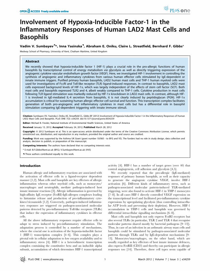
Figure 1. LAD2 human mast cells and primary human basophils express detectable amounts of TLRs 2 and 4. LAD2 cells, primaryhuman basophils (PHB) and THP-1 cells (positive control) were subjected to in-cell TLR2 (A) and TLR4 (B) assays (upper panel). TLRs 2 (A) and 4 (B)were also detected in the cell lysates (lower panel). Quantitative data are mean values+S.D. of at least three individual experiments. * indicatesp,0.01 vs. control. All Western blot data shown are from one representative experiment out of three that gave similar results.doi:10.1371/journal.pone.0034259.g001
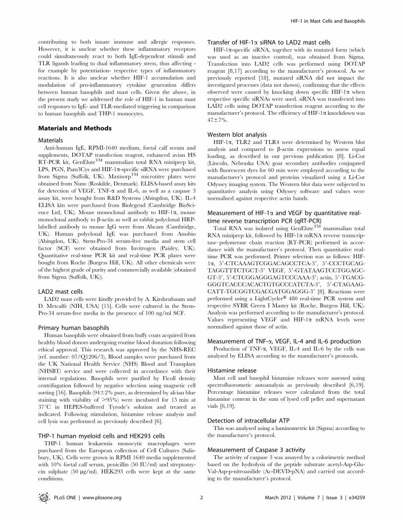
Figure 2. PGN and LPS induce HIF-1a accumulation in THP-1 but not in LAD2 cells. THP-1, LAD2 and HEK293 (negative control – lackingTLR2/4 expression) cells were exposed for 4 h to 1 mg/ml PGN or LPS. HIF-1a accumulation was then analysed. Quantitative data are meanvalues+S.D. of at least three individual experiments. * indicates p,0.01 vs. control. All Western blot data shown are from one representativeexperiment out of three that gave similar results.doi:10.1371/journal.pone.0034259.g002
HIF-1 in Mast Cells and Basophils
PLoS ONE | www.plosone.org 3 March 2012 | Volume 7 | Issue 3 | e34259
MTS cell viability assayCell viability was analysed using the Promega (Southampton,
UK) 3-(4,5-dimethylthiazol-2-yl)-5-(3-carboxymethoxyphenyl)-2-
(4-sulfophenyl)-2H-tetrazolium (MTS) cell viability assay kit
according to the manufacturer’s protocol.
In-cell TLR2 and TLR4 assaysThis was performed as recently described [20,21]. Briefly, cells
were centrifuged for 5 min at 1200 rpm, washed with respective
media and exposed to 2 mg/ml anti-TLR2 or anti-TLR4 antibody
for 2 h. This was followed by centrifugation for 5 min at
1200 rpm and washing with media. Cells were then incubated
for 2 h with fluorescent dye-labelled Li-Cor (Cambridge, UK)
secondary antibodies. Following centrifugation/washing, the cells
were transferred into wells of a 96-well plate which was scanned by
the Li-Cor Odyssey imaging system. Results were quantified using
Odyssey software.
Statistical analysisEach experiment was performed at least three times and
statistical analysis was conducted using two-tailed Student’s t
test. Statistical probabilities (p) were expressed as *, where
p,0.01.
Results
LAD2 mast cells and primary human basophils expressTLRs 2 and 4Human allergic effector cells (mast cells and basophils) are
known to produce detectable levels of TLRs 2 and 4 mRNA
[9,10]. Translation of these mRNAs into functional plasma
membrane-associated receptors is, however, still questionable.
We therefore studied whether LAD2 human mast cells and
primary human basophils express TLR2/4 proteins. To analyse
this we used Western blot analysis and in-cell TLR2/4 assays.
THP-1 human myeloid cells were used as a positive control as they
are known to express functional forms of both TLRs [7,22]. We
found that both mast cells and basophils expressed detectable
amounts of TLRs 2 and 4, although these expression levels were
significantly lower compared to those observed in THP-1 cells
(Figure 1).
LAD2 mast cells accumulate HIF-1a protein which is notupregulated in a TLR2/4-dependent mannerRecent evidence has demonstrated involvement of the HIF-1
transcription complex in TLR-mediated inflammatory reactions
[5–8]. We therefore asked whether this case is applicable to LAD2
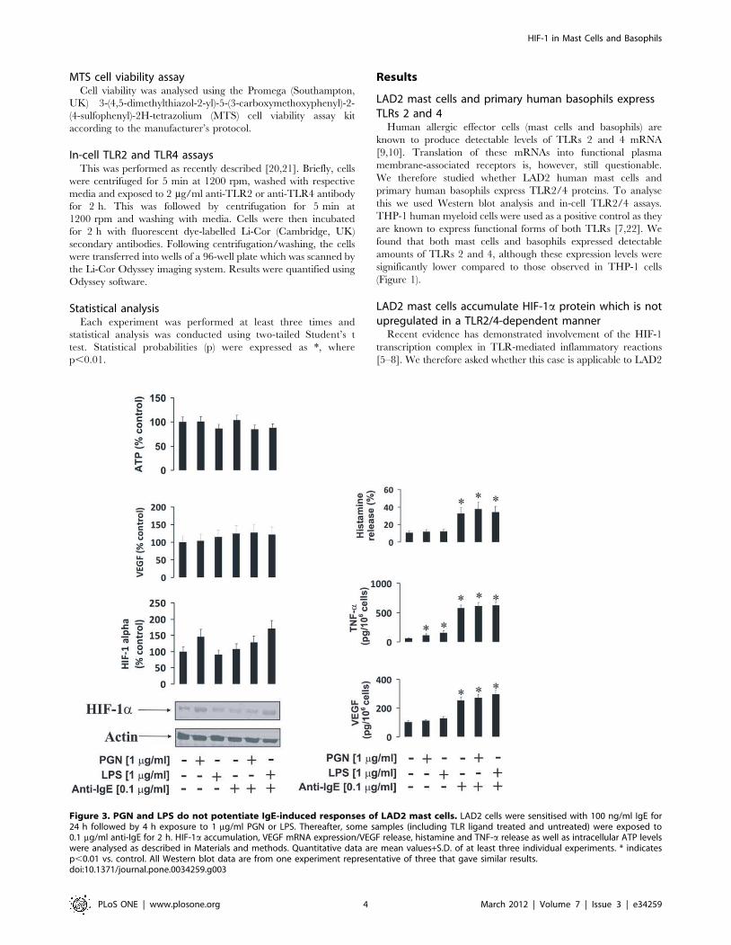
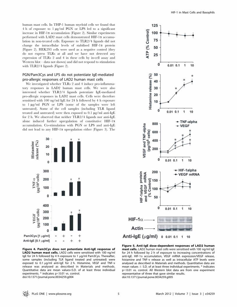
Figure 3. PGN and LPS do not potentiate IgE-induced responses of LAD2 mast cells. LAD2 cells were sensitised with 100 ng/ml IgE for24 h followed by 4 h exposure to 1 mg/ml PGN or LPS. Thereafter, some samples (including TLR ligand treated and untreated) were exposed to0.1 mg/ml anti-IgE for 2 h. HIF-1a accumulation, VEGF mRNA expression/VEGF release, histamine and TNF-a release as well as intracellular ATP levelswere analysed as described in Materials and methods. Quantitative data are mean values+S.D. of at least three individual experiments. * indicatesp,0.01 vs. control. All Western blot data are from one experiment representative of three that gave similar results.doi:10.1371/journal.pone.0034259.g003
HIF-1 in Mast Cells and Basophils
PLoS ONE | www.plosone.org 4 March 2012 | Volume 7 | Issue 3 | e34259
human mast cells. In THP-1 human myeloid cells we found that
4 h of exposure to 1 mg/ml PGN or LPS led to a significant
increase in HIF-1a accumulation (Figure 2). Similar experiments
performed with LAD2 mast cells demonstrated HIF-1a accumu-
lation in non-treated cells. Exposure to TLR2/4 ligands did not
change the intracellular levels of stabilised HIF-1a protein
(Figure 2). HEK293 cells were used as a negative control (they
do not express TLRs at all and we have not detected any
expression of TLRs 2 and 4 in these cells by in-cell assay and
Western blot – data not shown) and did not respond to stimulation
with TLR2/4 ligands (Figure 2).
PGN/Pam3Cys and LPS do not potentiate IgE-mediatedpro-allergic responses of LAD2 human mast cellsWe investigated whether TLRs 2 and 4 induce pro-inflamma-
tory responses in LAD2 human mast cells. We were also
interested whether TLR2/4 ligands potentiate IgE-mediated
pro-allergic responses in LAD2 mast cells. Cells were therefore
sensitised with 100 ng/ml IgE for 24 h followed by 4 h exposure
to 1 mg/ml PGN or LPS (some of the samples were left
untreated). Some of the cell samples (including TLR ligand
treated and untreated) were then exposed to 0.1 mg/ml anti-IgE
for 2 h. We observed that neither TLR2/4 ligands nor anti-IgE
alone induced further upregulation of constitutive HIF-1a
accumulation. Co-stimulation with PGN or LPS and anti-IgE
did not lead to any HIF-1a upregulation either (Figure 3). The
Figure 4. Pam3Cys does not potentiate Anti-IgE response ofLAD2 human mast cells. LAD2 cells were sensitised with 100 ng/mlIgE for 24 h followed by 4 h exposure to 1 mg/ml Pam3Cys. Thereafter,some samples (including TLR ligand treated and untreated) wereexposed to 0.1 mg/ml anti-IgE for 2 h. Histamine, VEGF and TNF-arelease was analysed as described in Materials and methods.Quantitative data are mean values+S.D. of at least three individualexperiments. * indicates p,0.01 vs. control.doi:10.1371/journal.pone.0034259.g004
Figure 5. Anti-IgE dose-dependent responses of LAD2 humanmast cells. LAD2 human mast cells were sensitised with 100 ng/ml IgEfor 24 h followed by 2 h of exposure to increasing concentrations ofanti-IgE. HIF-1a accumulation, VEGF mRNA expression/VEGF release,histamine and TNF-a release as well as intracellular ATP levels wereanalysed as described in Materials and methods. Quantitative data aremean values 6 S.D. of at least three individual experiments. * indicatesp,0.01 vs. control. All Western blot data are from one experimentrepresentative of three that gave similar results.doi:10.1371/journal.pone.0034259.g005
HIF-1 in Mast Cells and Basophils
PLoS ONE | www.plosone.org 5 March 2012 | Volume 7 | Issue 3 | e34259
same effect was observed in the case of the amounts of the VEGF
mRNA (Figure 3). Intracellular ATP levels were not significantly
affected, possibly because of basic HIF-1a accumulation in mast
cells. TLR2/4 ligands on their own did not induce histamine
release and were unable to potentiate IgE-mediated release of this
amine. The same effect was observed in the case of VEGF release
(Figure 3). Interestingly, both PGN and LPS were able to induce
TNF-a production but much lower quantities were released
compared to the cases when LAD2 cells were stimulated with
anti-IgE in the absence of PGN/LPS. Neither PGN nor LPS
were able to significantly potentiate anti-IgE-induced TNF-a
production in LAD2 cells (Figure 3). No detectable amounts of
IL-4 were released in any case by LAD2 mast cells (data not
shown). Since it was reported that PGN could display reduced
affinity of TLR2 [23] we controlled our observation by
performing exactly the same treatments of LAD2 cells with the
alternative TLR2 ligand – Pam3Cys and analysed TNF-a/VEGF
as well as histamine release. The results were, however, essentially
similar to those observed for PGN (Figure 4).
Increasing concentrations of anti-IgE were able to upregulate
TNF-a/VEGF and histamine release in LAD2 cells after 2 h
stimulation. But they were unable to significantly upregulate HIF-
1a accumulation and the levels of VEGF mRNA suggesting that
the existing basic expression of these two factors may be sufficient
to cover the respective functional requirements of the LAD2 cells
(Figure 5). No detectable amounts of IL-4 were released in any
case by LAD2 mast cells (data not shown).
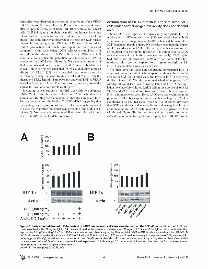
Accumulation of HIF-1a protein in non-stimulated LAD2cells under normal oxygen availability does not dependon SCFSince SCF was reported to significantly upregulate HIF-1a
stabilisation in different cell types [24], we asked whether basic
accumulation of this protein in LAD2 cells could be a result of
SCF-dependent priming effect. We therefore examined the impact
of SCF withdrawal on LAD2 cells that were either non-sensitised
or sensitised with 100 ng/ml IgE for 24 h in comparison to LAD2
cells that were cultured in the presence (as normally) of 100 ng/ml
SCF and either IgE-sensitized for 24 h or not. Some of the IgE-
sensitised cells were then exposed to 0.1 mg/ml anti-IgE for 2 h.
HIF-1a accumulation was then analysed.
We discovered that SCF non-significantly upregulated HIF-1a
accumulation in the LAD2 cells compared to those cultured in the
absence of SCF. In all other cases the levels of HIF-1a were very
similar (Figure 6A). We also examined whether long-term SCF
withdrawal could lead to a downregulation of HIF-1a accumu-
lation. We therefore cultured LAD2 cells in the absence of SCF for
24, 48 and 72 h. In addition, two positive controls of recognised
HIF-1a inducers were used. Here, LAD2 cells were cultured in the
presence of SCF but exposed for 4 h either to hypoxic (3% O2)
conditions or to 100 mM cobalt chloride. We observed, however,
that SCF withdrawal did not significantly downregulate HIF-1a
accumulation in LAD2 cells, regardless of the period of SCF
withdrawal (Figure 6B). Furthermore, neither hypoxia nor cobalt
chloride were able to significantly upregulate HIF-1a protein
Figure 6. Basic accumulation of HIF-1a protein in LAD2 humanmast cells does not depend on the SCF. A) Non-sensitised LAD2 cells andthose sensitised with 100 ng/ml IgE for 24 h were cultured in the presence or absence of 100 ng/ml SCF. Some of the IgE-sensitised cells were thenexposed to 0.1 mg/ml anti-IgE for 2 h. HIF-1a accumulation was then analysed by Western blot. VEGF mRNA levels were analysed by qRT-PCR. B)LAD2 cells were cultured in the absence of SCF for 24, 48 and 72 h. In addition, LAD2 cells, cultured as normally in the presence of SCF, were kept 4 hunder hypoxic (3% O2) conditions or exposed for 4 h to 100 mM cobalt chloride. HIF-1a accumulation was analysed by Western blot. Quantitativedata are mean values+S.D. of at least three individual experiments. * indicates p,0.01 vs. control. All Western blot data are from one experimentrepresentative of three that gave similar results.doi:10.1371/journal.pone.0034259.g006
HIF-1 in Mast Cells and Basophils
PLoS ONE | www.plosone.org 6 March 2012 | Volume 7 | Issue 3 | e34259

accumulation in LAD2 cells (Figure 6B). SCF, therefore, does not
appear to be a major contributor in determining the accumulation
of HIF-1a protein in non-stimulated LAD2 mast cells. Importantly,
the absence of marked increases in HIF-1a protein accumulation
following hypoxia/cobalt chloride treatment suggests that these cells
require baseline levels of this protein at near-maximum threshold
(those for mRNA levels were reported before [25]).
HIF-1a protein plays a crucial role in the activity of LAD2mast cellsTo characterise the role of the HIF-1 transcription complex in
LAD2 cell responses we used normal and HIF-1a knockdown LAD2
cells. The knockdown was achieved by transfection of the cells with
HIF-1a siRNA. HIF-1a knockdown was assayed by quantitative
real-time PCR (qPCR) and Western blot analysis. Both normal and
HIF-1a knockdown LAD2 cells were sensitised with 100 ng/ml IgE
and exposed to PGN, LPS or anti-IgE. We found that in both
stimulated and non-stimulated HIF-1a knockdown cells, the
expression of VEGF was significantly lower and ATP levels were
significantly downregulated (Figure 7). TNF-a and VEGF produc-
tion by LAD2mast cells were affected by HIF-1a knockdown, which
also resulted in increased caspase 3 activity (Figure 7). Only
histamine release was not affected in HIF-1a knockdown cells
(Figure 7).MTS assay demonstrated a slight, but significant, decrease
in cell viability after 2 h of exposure to 0.1 mg/ml anti-IgE (Figure 8)
following HIF-1a knockdown in IgE-sensitised LAD2 cells. This
observation is consistent with the results demonstrating an increase
in caspase 3 activity and ATP depletion (Figure 7).
Cross-interaction of IgE-mediated and PGN/LPS-mediated responses in primary human basophils andTHP-1 myeloid cellsWe investigated whether TLRs 2 and 4 are able to induce
inflammatory reactions in primary human basophils. We were also
Figure 7. HIF-1a protein plays a pivotal role in the inflammatory responses of LAD2 mast cells. (A) Normal and HIF-1a knockdown LAD2cells were sensitised with 100 ng/ml IgE for 24 h followed by 4 h exposure to 1 mg/ml PGN, LPS or 0.1 mg/ml anti-IgE. HIF-1a accumulation/mRNAlevels, VEGF mRNA expression/VEGF release, histamine and TNF-a release, intracellular ATP levels as well as caspase 3 activity were analysed asdescribed in Materials and methods. Quantitative data are mean values+S.D. of at least three individual experiments. * indicates p,0.01 vs. control.All Western blot data are from one experiment representative of three that gave similar results.doi:10.1371/journal.pone.0034259.g007
HIF-1 in Mast Cells and Basophils
PLoS ONE | www.plosone.org 7 March 2012 | Volume 7 | Issue 3 | e34259
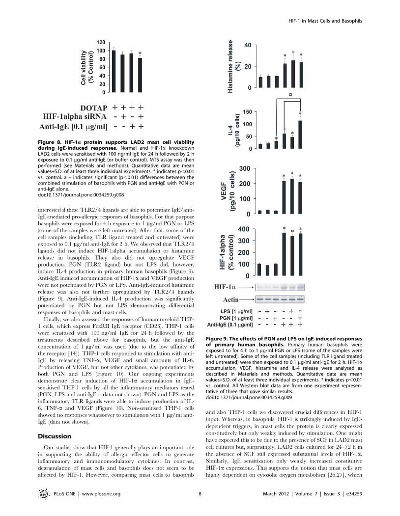
interested if these TLR2/4 ligands are able to potentiate IgE/anti-
IgE-mediated pro-allergic responses of basophils. For that purpose
basophils were exposed for 4 h exposure to 1 mg/ml PGN or LPS
(some of the samples were left untreated). After that, some of the
cell samples (including TLR ligand treated and untreated) were
exposed to 0.1 mg/ml anti-IgE for 2 h. We obesrved that TLR2/4
ligands did not induce HIF-1alpha accumulation or histamine
release in basophils. They also did not upregulate VEGF
production. PGN (TLR2 ligand) but not LPS did, however,
induce IL-4 production in primary human basophils (Figure 9).
Anti-IgE induced accumulation of HIF-1a and VEGF production
were not potentiated by PGN or LPS. Anti-IgE-induced histamine
release was also not further upregulated by TLR2/4 ligands
(Figure 9). Anti-IgE-induced IL-4 production was significantly
potentiated by PGN but not LPS demonstrating differential
responses of basophils and mast cells.
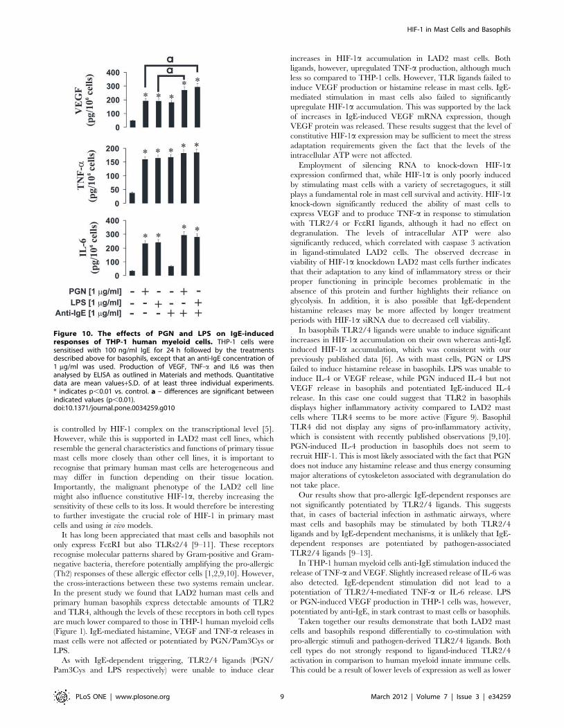
Finally, we also assessed the responses of human myeloid THP-
1 cells, which express FceRII IgE receptor (CD23). THP-1 cells
were sensitised with 100 ng/ml IgE for 24 h followed by the
treatments described above for basophils, but the anti-IgE
concentration of 1 mg/ml was used (due to the low affinity of
the receptor [14]). THP-1 cells responded to stimulation with anti-
IgE by releasing TNF-a, VEGF and small amounts of IL-6.
Production of VEGF, but not other cytokines, was potentiated by
both PGN and LPS (Figure 10). Our ongoing experiments
demonstrate clear induction of HIF-1a accumulation in IgE-
sensitised THP-1 cells by all the inflammatory mediators tested
(PGN, LPS and anti-IgE – data not shown). PGN and LPS as the
inflammatory TLR ligands were able to induce production of IL-
6, TNF-a and VEGF (Figure 10). Non-sensitised THP-1 cells
showed no responses whatsoever to stimulation with 1 mg/ml anti-
IgE (data not shown).
Discussion
Our studies show that HIF-1 generally plays an important role
in supporting the ability of allergic effector cells to generate
inflammatory and immunomodulatory cytokines. In contrast,
degranulation of mast cells and basophils does not seem to be
affected by HIF-1. However, comparing mast cells to basophils
and also THP-1 cells we discovered crucial differences in HIF-1
input. Whereas, in basophils, HIF-1 is strikingly induced by IgE-
dependent triggers, in mast cells the protein is clearly expressed
constitutively but only weakly induced by stimulation. One might
have expected this to be due to the presence of SCF in LAD2 mast
cell cultures but, surprisingly, LAD2 cells cultured for 24–72 h in
the absence of SCF still expressed substantial levels of HIF-1a.
Similarly, IgE sensitization only weakly increased constitutive
HIF-1a expressions. This supports the notion that mast cells are
highly dependent on cytosolic oxygen metabolism [26,27], which
Figure 8. HIF-1a protein supports LAD2 mast cell viabilityduring IgE-induced responses. Normal and HIF-1a knockdownLAD2 cells were sensitised with 100 ng/ml IgE for 24 h followed by 2 hexposure to 0.1 mg/ml anti-IgE (or buffer control). MTS assay was thenperformed (see Materials and methods). Quantitative data are meanvalues+S.D. of at least three individual experiments. * indicates p,0.01vs. control. a – indicates significant (p,0.01) differences between thecombined stimulation of basophils with PGN and anti-IgE with PGN oranti-IgE alone.doi:10.1371/journal.pone.0034259.g008
Figure 9. The effects of PGN and LPS on IgE-induced responsesof primary human basophils. Primary human basophils wereexposed to for 4 h to 1 mg/ml PGN or LPS (some of the samples wereleft untreated). Some of the cell samples (including TLR ligand treatedand untreated) were then exposed to 0.1 mg/ml anti-IgE for 2 h. HIF-1aaccumulation, VEGF, histamine and IL-4 release were analysed asdescribed in Materials and methods. Quantitative data are meanvalues+S.D. of at least three individual experiments. * indicates p,0.01vs. control. All Western blot data are from one experiment represen-tative of three that gave similar results.doi:10.1371/journal.pone.0034259.g009
HIF-1 in Mast Cells and Basophils
PLoS ONE | www.plosone.org 8 March 2012 | Volume 7 | Issue 3 | e34259
is controlled by HIF-1 complex on the transcriptional level [5].
However, while this is supported in LAD2 mast cell lines, which
resemble the general characteristics and functions of primary tissue
mast cells more closely than other cell lines, it is important to
recognise that primary human mast cells are heterogeneous and
may differ in function depending on their tissue location.
Importantly, the malignant phenotype of the LAD2 cell line
might also influence constitutive HIF-1a, thereby increasing the
sensitivity of these cells to its loss. It would therefore be interesting
to further investigate the crucial role of HIF-1 in primary mast
cells and using in vivo models.
It has long been appreciated that mast cells and basophils not
only express FceRI but also TLRs2/4 [9–11]. These receptors
recognise molecular patterns shared by Gram-positive and Gram-
negative bacteria, therefore potentially amplifying the pro-allergic
(Th2) responses of these allergic effector cells [1,2,9,10]. However,
the cross-interactions between these two systems remain unclear.
In the present study we found that LAD2 human mast cells and
primary human basophils express detectable amounts of TLR2
and TLR4, although the levels of these receptors in both cell types
are much lower compared to those in THP-1 human myeloid cells
(Figure 1). IgE-mediated histamine, VEGF and TNF-a releases in
mast cells were not affected or potentiated by PGN/Pam3Cys or
LPS.
As with IgE-dependent triggering, TLR2/4 ligands (PGN/
Pam3Cys and LPS respectively) were unable to induce clear
increases in HIF-1a accumulation in LAD2 mast cells. Both
ligands, however, upregulated TNF-a production, although much
less so compared to THP-1 cells. However, TLR ligands failed to
induce VEGF production or histamine release in mast cells. IgE-
mediated stimulation in mast cells also failed to significantly
upregulate HIF-1a accumulation. This was supported by the lack
of increases in IgE-induced VEGF mRNA expression, though
VEGF protein was released. These results suggest that the level of
constitutive HIF-1a expression may be sufficient to meet the stress
adaptation requirements given the fact that the levels of the
intracellular ATP were not affected.
Employment of silencing RNA to knock-down HIF-1a
expression confirmed that, while HIF-1a is only poorly induced
by stimulating mast cells with a variety of secretagogues, it still
plays a fundamental role in mast cell survival and activity. HIF-1a
knock-down significantly reduced the ability of mast cells to
express VEGF and to produce TNF-a in response to stimulation
with TLR2/4 or FceRI ligands, although it had no effect on
degranulation. The levels of intracellular ATP were also
significantly reduced, which correlated with caspase 3 activation
in ligand-stimulated LAD2 cells. The observed decrease in
viability of HIF-1a knockdown LAD2 mast cells further indicates
that their adaptation to any kind of inflammatory stress or their
proper functioning in principle becomes problematic in the
absence of this protein and further highlights their reliance on
glycolysis. In addition, it is also possible that IgE-dependent
histamine releases may be more affected by longer treatment
periods with HIF-1a siRNA due to decreased cell viability.
In basophils TLR2/4 ligands were unable to induce significant
increases in HIF-1a accumulation on their own whereas anti-IgE
induced HIF-1a accumulation, which was consistent with our
previously published data [6]. As with mast cells, PGN or LPS
failed to induce histamine release in basophils. LPS was unable to
induce IL-4 or VEGF release, while PGN induced IL-4 but not
VEGF release in basophils and potentiated IgE-induced IL-4
release. In this case one could suggest that TLR2 in basophils
displays higher inflammatory activity compared to LAD2 mast
cells where TLR4 seems to be more active (Figure 9). Basophil
TLR4 did not display any signs of pro-inflammatory activity,
which is consistent with recently published observations [9,10].
PGN-induced IL-4 production in basophils does not seem to
recruit HIF-1. This is most likely associated with the fact that PGN
does not induce any histamine release and thus energy consuming
major alterations of cytoskeleton associated with degranulation do
not take place.
Our results show that pro-allergic IgE-dependent responses are
not significantly potentiated by TLR2/4 ligands. This suggests
that, in cases of bacterial infection in asthmatic airways, where
mast cells and basophils may be stimulated by both TLR2/4
ligands and by IgE-dependent mechanisms, it is unlikely that IgE-
dependent responses are potentiated by pathogen-associated
TLR2/4 ligands [9–13].
In THP-1 human myeloid cells anti-IgE stimulation induced the
release of TNF-a and VEGF. Slightly increased release of IL-6 was
also detected. IgE-dependent stimulation did not lead to a
potentiation of TLR2/4-mediated TNF-a or IL-6 release. LPS
or PGN-induced VEGF production in THP-1 cells was, however,
potentiated by anti-IgE, in stark contrast to mast cells or basophils.
Taken together our results demonstrate that both LAD2 mast
cells and basophils respond differentially to co-stimulation with
pro-allergic stimuli and pathogen-derived TLR2/4 ligands. Both
cell types do not strongly respond to ligand-induced TLR2/4
activation in comparison to human myeloid innate immune cells.
This could be a result of lower levels of expression as well as lower
Figure 10. The effects of PGN and LPS on IgE-inducedresponses of THP-1 human myeloid cells. THP-1 cells weresensitised with 100 ng/ml IgE for 24 h followed by the treatmentsdescribed above for basophils, except that an anti-IgE concentration of1 mg/ml was used. Production of VEGF, TNF-a and IL6 was thenanalysed by ELISA as outlined in Materials and methods. Quantitativedata are mean values+S.D. of at least three individual experiments.* indicates p,0.01 vs. control. a – differences are significant betweenindicated values (p,0.01).doi:10.1371/journal.pone.0034259.g010
HIF-1 in Mast Cells and Basophils
PLoS ONE | www.plosone.org 9 March 2012 | Volume 7 | Issue 3 | e34259
activity of these receptors in mast cells and basophils compared to
myeloid cells. The presence of constitutive HIF-1 in LAD2 mast
cells was revealed to be essential for them in order to respond to
both IgE-dependent and TLR-mediated stimulation. However, in
contrast to basophils, increases in HIF-1 expressions to these
modes of activation were not pronounced. In basophils the protein
also plays an essential role in IgE-dependent activation but less so
for TLR2-mediated triggering. The results discussed above are
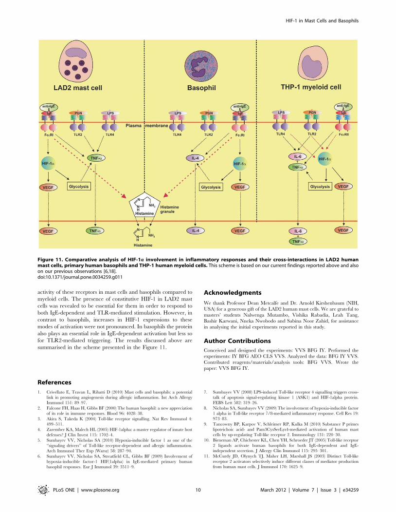
summarised in the scheme presented in the Figure 11.
Acknowledgments
We thank Professor Dean Metcalfe and Dr. Arnold Kirshenbaum (NIH,
USA) for a generous gift of the LAD2 human mast cells. We are grateful to
masters’ students Nalwenga Mutambo, Vishika Rabadia, Leah Tang,
Bashir Karwani, Nneka Nwobodo and Sabina Noor Zahid, for assistance
in analysing the initial experiments reported in this study.
Author Contributions
Conceived and designed the experiments: VVS BFG IY. Performed the
experiments: IY BFG AEO CLS VVS. Analyzed the data: BFG IY VVS.
Contributed reagents/materials/analysis tools: BFG VVS. Wrote the
paper: VVS BFG IY.
References
1. Crivellato E, Travan L, Ribatti D (2010) Mast cells and basophils: a potential
link in promoting angiogenesis during allergic inflammation. Int Arch Allergy
Immunol 151: 89–97.
2. Falcone FH, Haas H, Gibbs BF (2000) The human basophil: a new appreciation
of its role in immune responses. Blood 96: 4028–38.
3. Akira S, Takeda K (2004) Toll-like receptor signalling. Nat Rev Immunol 4:
499–511.
4. Zarember KA, Malech HL (2005) HIF-1alpha: a master regulator of innate host
defenses? J Clin Invest 115: 1702–4.
5. Sumbayev VV, Nicholas SA (2010) Hypoxia-inducible factor 1 as one of the
‘‘signaling drivers’’ of Toll-like receptor-dependent and allergic inflammation.
Arch Immunol Ther Exp (Warsz) 58: 287–94.
6. Sumbayev VV, Nicholas SA, Streatfield CL, Gibbs BF (2009) Involvement of
hypoxia-inducible factor-1 HIF(1alpha) in IgE-mediated primary human
basophil responses. Eur J Immunol 39: 3511–9.
7. Sumbayev VV (2008) LPS-induced Toll-like receptor 4 signalling triggers cross-
talk of apoptosis signal-regulating kinase 1 (ASK1) and HIF-1alpha protein.
FEBS Lett 582: 319–26.
8. Nicholas SA, Sumbayev VV (2009) The involvement of hypoxia-inducible factor
1 alpha in Toll-like receptor 7/8-mediated inflammatory response. Cell Res 19:
973–83.
9. Tancowny BP, Karpov V, Schleimer RP, Kulka M (2010) Substance P primes
lipoteichoic acid- and Pam3CysSerLys4-mediated activation of human mast
cells by up-regulating Toll-like receptor 2. Immunology 131: 220–30.
10. Bieneman AP, Chichester KL, Chen YH, Schroeder JT (2005) Toll-like receptor
2 ligands activate human basophils for both IgE-dependent and IgE-
independent secretion. J Allergy Clin Immunol 115: 295–301.
11. McCurdy JD, Olynych TJ, Maher LH, Marshall JS (2003) Distinct Toll-like
receptor 2 activators selectively induce different classes of mediator production
from human mast cells. J Immunol 170: 1625–9.
Figure 11. Comparative analysis of HIF-1a involvement in inflammatory responses and their cross-interactions in LAD2 humanmast cells, primary human basophils and THP-1 human myeloid cells. This scheme is based on our current findings reported above and alsoon our previous observations [6,18].doi:10.1371/journal.pone.0034259.g011
HIF-1 in Mast Cells and Basophils
PLoS ONE | www.plosone.org 10 March 2012 | Volume 7 | Issue 3 | e34259
12. Varadaradjalou S, Feger F, Thieblemont N, Hamouda NB, Pleau JM, et al.(2003) Toll-like receptor 2 (TLR2) and TLR4 differentially activate human mastcells. Eur J Immunol 33: 899–906.
13. Oldford SA, Haidl ID, Howatt MA, Leiva CA, Johnston B, et al. (2010) Acritical role for mast cells and mast cell-derived IL-6 in TLR2-mediatedinhibition of tumor growth. J Immunol 185: 7067–76.
14. Tu Y, Salim S, Bourgeois J, Di Leo V, Irvine EJ, et al. (2005) Perdue MH:CD23-mediated IgE transport across human intestinal epithelium: inhibition byblocking sites of translation or binding. Gastroenterology 2005, 129: 928–40.
15. Kirshenbaum AS, Akin C, Wu Y, Rottem M, Goff JP, et al. (2003)Characterization of novel stem cell factor responsive human mast cell linesLAD 1 and 2 established from a patient with mast cell sarcoma/leukemia;activation following aggregation of FcepsilonRI or FcgammaRI. Leuk Res 27:677–82.
16. Gibbs BF, Papenfuss K, Falcone FH (2008) A rapid two-step procedure for thepurification of human peripheral blood basophils to near homogeneity. Clin ExpAllergy 38: 480–5.
17. Nicholas SA, Bubnov VV, Yasinska IM, Sumbayev VV (2011) Involvement ofxanthine oxidase and hypoxia-inducible factor 1 in Toll-like receptor 7/8-mediated activation of caspase 1 and interleukin-1beta. Cell Mol Life Sci 68:151–8.
18. Lall H, Coughlan K, Sumbayev VV (2008) HIF-1alpha protein is an essentialfactor for protection of myeloid cells against LPS-induced depletion of ATP andapoptosis that supports Toll-like receptor 4-mediated production of IL-6. MolImmunol 45: 3045–9.
19. Gibbs BF, Rathling A, Zillikens D, Huber M, Haas H (2006) Initial Fc epsilonRI-mediated signal strength plays a key role in regulating basophil signaling anddeactivation. J Allergy Clin Immunol 118: 1060–7.
20. Pchejetski D, Nunes J, Coughlan K, Lall H, Pitson SM, et al. (2011) Theinvolvement of sphingosine kinase 1 in LPS-induced Toll-like receptor 4-mediated accumulation of HIF-1alpha protein, activation of ASK1 andproduction of the pro-inflammatory cytokine IL-6. Immunol Cell Biol 89:268–74.
21. Nicholas SA, Coughlan K, Yasinska I, Lall GS, Gibbs BF, et al. (2011)Dysfunctional mitochondria contain endogenous high-affinity human Toll-likereceptor 4 (TLR4) ligands and induce TLR4-mediated inflammatory reactions.Int J Biochem Cell Biol 43: 674–81.
22. Lee SA, Kim SM, Son YH, Lee CW, Chung SW, et al. (2011) Peptidoglycanenhances secretion of monocyte chemoattractants via multiple signallingpathways. Biochem Biophys Res Commun 408: 132–138.
23. Travassos LH, Girardin SE, Philpott DJ, Blanot D, Nahori MA, et al. (2004)Toll-like receptor 2-dependent bacterial sensing does not occur via peptidogly-can recognition. EMBO Rep 5: 1000–6.
24. Pedersen M, Lofstedt T, Sun J, Holmquist-Mengelbier L, Pahlman S, et al.(2008) Stem cell factor induces HIF-1alpha at normoxia in hematopoietic cells.Biochem Biophys Res Commun 377: 98–103.
25. Walczak-Drzewiecka A, Ratajewski M, Wagner W, Dastych J (2008) HIF-1a isup-regulated in activated mast cells by a process that involves calcineurin andNFAT. J Immunol 181: 1665–72.
26. Pendleton A, Pope B, Weeds A, Koffer A (2003) Latrunculin B or ATP depletioninduces cofilin-dependent translocation of actin into nuclei of mast cells. J BiolChem 278: 14394–400.
27. Kitahata Y, Nunomura S, Terui T, Ra C (2010) Prolonged culture of mast cellswith high-glucose medium enhances the Fc epsilon RI-mediated degranulationresponse and leukotriene C4 production. Int Arch Allergy Immunol 2010; 152Suppl 1: 22–31.
HIF-1 in Mast Cells and Basophils
PLoS ONE | www.plosone.org 11 March 2012 | Volume 7 | Issue 3 | e34259