Embed Size (px)
DESCRIPTION
Capsaicin; Chili
Citation preview

Capsaicin Displays Anti-Proliferative Activity againstHuman Small Cell Lung Cancer in Cell Culture and NudeMice Models via the E2F PathwayKathleen C. Brown1., Ted R. Witte2., W. Elaine Hardman2, Haitao Luo3, Yi C. Chen3, A. Betts Carpenter4,
Jamie K. Lau1, Piyali Dasgupta1*
1 Department of Pharmacology, Physiology and Toxicology, Joan C. Edwards School of Medicine, Marshall University, Huntington, West Virginia, United States of America,
2 Department of Biochemistry and Microbiology, Joan C. Edwards School of Medicine, Marshall University, Huntington, West Virginia, United States of America,
3 Department of Biology, Alderson-Broaddus College, Phillipi, West Virginia, United States of America, 4 Department of Anatomy and Pathology, Joan C. Edwards School
of Medicine, Marshall University, Huntington, West Virginia, United States of America
Abstract
Background: Small cell lung cancer (SCLC) is characterized by rapid progression and low survival rates. Therefore, noveltherapeutic agents are urgently needed for this disease. Capsaicin, the active ingredient of chilli peppers, displays anti-proliferative activity in prostate and epidermoid cancer in vitro. However, the anti-proliferative activity of capsaicin has notbeen studied in human SCLCs. The present manuscript fills this void of knowledge and explores the anti-proliferative effectof capsaicin in SCLC in vitro and in vivo.
Methodology/Principal Findings: BrdU assays and PCNA ELISAs showed that capsaicin displays robust anti-proliferativeactivity in four human SCLC cell lines. Furthermore, capsaicin potently suppressed the growth of H69 human SCLC tumors invivo as ascertained by CAM assays and nude mice models. The second part of our study attempted to provide insight intomolecular mechanisms underlying the anti-proliferative activity of capsaicin. We found that the anti-proliferative activity ofcapsaicin is correlated with a decrease in the expression of E2F-responsive proliferative genes like cyclin E, thymidylatesynthase, cdc25A and cdc6, both at mRNA and protein levels. The transcription factor E2F4 mediated the anti-proliferativeactivity of capsaicin. Ablation of E2F4 levels by siRNA methodology suppressed capsaicin-induced G1 arrest. ChIP assaysdemonstrated that capsaicin caused the recruitment of E2F4 and p130 on E2F-responsive proliferative promoters, therebyinhibiting cell proliferation.
Conclusions/Significance: Our findings suggest that the anti-proliferative effects of capsaicin could be useful in the therapyof human SCLCs.
Citation: Brown KC, Witte TR, Hardman WE, Luo H, Chen YC, et al. (2010) Capsaicin Displays Anti-Proliferative Activity against Human Small Cell Lung Cancer inCell Culture and Nude Mice Models via the E2F Pathway. PLoS ONE 5(4): e10243. doi:10.1371/journal.pone.0010243
Editor: Dong-Yan Jin, University of Hong Kong, Hong Kong
Received January 8, 2010; Accepted March 24, 2010; Published April 20, 2010
Copyright: � 2010 Brown et al. This is an open-access article distributed under the terms of the Creative Commons Attribution License, which permitsunrestricted use, distribution, and reproduction in any medium, provided the original author and source are credited.
Funding: This work was supported by a Center of Biochemical Research Excellence Pilot Research Grant from the National Institutes of Health (grant number5P20RR020180); the Pharmaceutical Manufacturers Association Foundation Research Starter Grant; the Marshall University ADVANCE fellowship; a Grant-in-Aidundergraduate research grant from National Aeronautics and Space Administration; and a Young Clinical Scientist Award from the Flight Attendant MedicalResearch Institute (grant number 82115). The funders had no role in study design, data collection and analysis, decision to publish, or preparation of themanuscript.
Competing Interests: The authors have declared that no competing interests exist.
* E-mail: [email protected]
. These authors contributed equally to this work.
Introduction
Small cell lung cancer (SCLC) is an aggressive malignancy
representing 13% of all lung cancer cases, with an overall 5-year
survival rate of less than 5% [1,2]. Such statistics emphasize the
need for novel treatment strategies for this disease. Recent
advances in the basic understanding of molecular events involved
in SCLC progression have led to the identification of potential
agents for therapeutic interventions [2,3,4,5]. These strategies
include growth factor/receptor-specific inhibitors, protein kinase
inhibitors and nutritional agents. The identification of nutritional
agents that display anti-proliferative activity may represent a novel
therapeutic avenue in human SCLC.
Capsaicin, the major active ingredient of chili peppers, is used
topically to treat pain and inflammation associated with a variety
of diseases [6,7]. Chemoprevention studies demonstrate that
capsaicin can suppress carcinogenesis of the skin, colon, lung,
tongue and prostate [8,9,10,11,12]. Although these studies have
addressed the chemopreventative potential of capsaicin, only a few
have addressed its potential as an anti-cancer agent. For example,
capsaicin has been shown to induce apoptosis in non-small cell
lung cancer (NSCLC), T-cell leukemia, esophageal carcinoma,
astroglioma, prostate, colon and gastric cancer cells in cell culture
models [13,14,15,16,17]. Additionally, the administration of
capsaicin has been shown to suppress prostate cancer tumor
growth in nude mice models [10,18].
PLoS ONE | www.plosone.org 1 April 2010 | Volume 5 | Issue 4 | e10243

Apart from causing apoptosis, capsaicin has been found to
induce cell cycle arrest in human cancer cells. Several convergent
studies have shown that capsaicin-induced G1 arrest in CE 81T/
VGH human epidermoid carcinoma cells and prostate cancer cells
occur via induction of p53 and the cyclin-dependent kinase (cdk)
inhibitor p21 [10,17,19,20,21]. The treatment of HL-60 human
leukemic cells with capsaicin caused G1 arrest via inhibition of
cdk2 activity. The anti-angiogenic activity of capsaicin is attributed
to its ability to cause G1 arrest in endothelial cells. Capsaicin-
induced G1 arrest is correlated with the suppression of cyclin D1
levels, inhibition of cdk4 activity and Rb phosphorylation in
endothelial and breast cancer cells [21,22]. These data raise the
possibility that the anti-proliferative activity of capsaicin is
mediated by its effects on the E2F-Rb pathway.
The E2F family of transcription factors, consisting of eight
member genes (E2F1-E2F8), plays a pivotal role in regulating cell
cycle progression and cell proliferation [23,24,25,26]. These E2Fs
have been further subclassified into two groups based on their
transcriptional regulatory properties on gene promoters. E2F1,
E2F2 and E2F3 are often referred to as ‘‘activator’’ E2Fs because
they transcriptionally activate E2F target proliferative genes such
as cyclin E, cdc25A and cdc6 [25,27,28,29]. These target genes
then induce the entry of cells into S-phase, thereby promoting cell
cycle progression. The second subclass, E2F4 and E2F5, are
referred to as the ‘‘repressor’’ E2Fs because they repress the
transcription of E2F target proliferative genes. E2F family
members E2F6, E2F7 and E2F8 are also repressors. E2F7 and
E2F8 are the most recently identified members of this family, and
much less is known about their function and regulation [25].
Despite the fact that all transcriptionally active E2Fs bind to the
same DNA recognition site on target promoters, they have
different functional roles in the cell [26,29].
Studies in recent years have shown that the activity of E2Fs is
stringently regulated by the pocket protein family, namely Rb,
p130 and p107. E2Fs 1–3 can bind to the Rb protein, but E2Fs 4
and 5 preferentially bind to p107 and p130 proteins
[24,25,26,29,30]. The Rb family proteins bind to a moiety within
the transcriptional activation region of E2Fs, effectively repressing
their activity. Thus, quiescent cells contain high levels of E2F
proteins bound to Rb, p130 and p107. The onset of mitogenic
stimuli causes phosphorylation of Rb, p130 and p107 by cyclin D
and E and their associated kinases. The phosphorylation of Rb,
p107 and p130 results in their inactivation and dissociation from
E2F proteins [25]. These free E2Fs subsequently bind to target
proliferative promoters like cyclin E, thymidylate synthase (TS),
cdc25A and cdc6, which regulate cell cycle progression [31,32]. Of
these, E2F1–3 stimulate the transcription of the aforementioned
genes, whereas E2F4 and E2F5 repress transcription [25]. A
majority of human SCLC are deficient in the Rb protein [3].
Therefore, it is probable that the other two pocket proteins, p107
and p130, play a vital role in E2F regulation and cell proliferation
in human SCLCs.
The anti-proliferative activity of capsaicin has not been studied
in human SCLCs. The present manuscript fills this gap of
knowledge and shows for the first time that capsaicin can potently
inhibit the proliferation of human SCLCs using multiple cell
culture and in vivo models. Previous studies have shown that a
majority of human SCLCs have mutations in Rb and p53, as well
as a dysregulation in the E2F-Rb pathway [3,4,5]. Data obtained
from microarrays, tissue samples from lung cancer patients and
human lung cancer cell lines indicate that cell cycle regulatory
molecules, like E2F1–5, p130, Skp2, cyclin D1 and p16, contribute
to the growth and progression of lung tumors [33]. Therefore, we
hypothesized that capsaicin displays anti-proliferative activity in
human SCLCs by regulating the activity of the E2F family of
transcription factors.
The present manuscript is an initial study aimed at investigating
the anti-proliferative activity of capsaicin in human SCLC in both
cell culture and animal models. Here, we demonstrate for the first
time that capsaicin displays potent anti-proliferative activity in four
human SCLC cell lines in vitro. Furthermore, our results show that
capsaicin suppresses the growth of human SCLC tumors in CAM
and nude mice models. We have also explored the contribution of
the E2F family of transcription factors in the growth-inhibitory
effects of capsaicin in SCLC cells. We observed that capsaicin
inhibits the proliferation of human SCLCs via a specific member
of the E2F family, namely E2F4. Ablation of E2F4 levels by two
independent siRNA was found to reverse the anti-proliferative
effect of capsaicin. Furthermore, capsaicin-induced growth arrest
was associated with a decrease in the expression of the E2F target
genes cyclin E, TS, cdc25A and cdc6. Finally, chromatin IP (ChIP)
analysis of human SCLC cells demonstrated that treatment of
capsaicin decreased the recruitment of activator E2Fs, namely
E2F2 and E2F3, to proliferative promoters like cyclin E, TS,
cdc25A and cdc6. On the other hand, the recruitment of repressor
E2F4 was enhanced by capsaicin treatment. Taken together, our
data suggest that capsaicin displays potent anti-proliferative
activity in human SCLC cells by differentially regulating the
E2F family of transcription factors. Furthermore, our results raise
the possibility that nutritional agents like capsaicin may be useful
for the therapy of human SCLC.
Materials and Methods
Ethics StatementNude mice were obtained from Charles River Laboratories and
acclimatized for one week. They were housed in autoclaved cages
with ad libitum access to food and water in HEPA-filtered racks and
closely monitored by animal facility staff. All procedures involving
nude mice were conducted according to the Animal Care and Use
guidelines in a facility accredited by the Association for Assessment
and Accreditation of Laboratory Animal Care (AAALAC)
International and were approved by the Institutional Animal
Care and Use Committee (IACUC) of Joan C. Edwards School of
Medicine, Marshall University (protocol#371).
Cell Culture and TransfectionThe human SCLC cell lines NCI-H69, NCI-H82 (hereafter
referred to as H69 and H82, respectively), DMS53 and DMS114
were obtained from American Type Culture Collection, Rockville,
MD. These cell lines were chosen because they have been
extensively studied, and their physical and molecular character-
istics closely resemble SCLC in patients. Gazdar et al., (1985)
originally isolated and characterized the H69 and H82 cell lines
[34,35]. They found that the morphology and growth character-
istics of H69 and H82 cells were typical of SCLC tumor cells
found in patients. Furthermore, the biochemical profile of these
cells (presence of L-dopa decarboxylase, neuroendocrine markers,
bombesin-like immunoreactivity, neuron-specific enolase and high
concentrations of brain isoenzyme of creatine kinase) was found to
be identical to human SCLC tumors observed in patients.
Similarly, DMS53 and DMS114 cells were first isolated from
human SCLC biopsies and characterized by Pettengill et al.,
(1980). They found that both DMS53 and DMS114 retained the
physical, morphological and biochemical profile of human SCLC
tumors observed in the clinic [36].
H69 and H82 were maintained in RPMI-1640 supplemented
with 2 mM glutamine, 100 units/ml penicillin, 50 mg/ml strepto-
Capsaicin and SCLC Therapy
PLoS ONE | www.plosone.org 2 April 2010 | Volume 5 | Issue 4 | e10243
mycin and 10% fetal bovine serum (FBS). DMS53 human SCLC
cells were cultured in Waymouth’s MB752/1 media containing
2 mM glutamine, 100 units/ml penicillin, 50 mg/ml streptomycin
and 10% FBS. The culture media for DMS114 human SCLC cells
was identical to DMS53, except that the media contained an
additional 2% sodium bicarbonate.
Primary normal human bronchial epithelial cells (NHBE) and
small airway epithelial cells (SAEC) were obtained from Lonza
Technologies, Switzerland. NHBEs were maintained in BEBM
media containing growth factors. Similarly SAECs were main-
tained in SABM media supplemented with growth factors. Both
BEMB and SABM media were prepared according to the
manufacturer’s instructions. All experiments using NHBEs and
SAECs were performed between passages 3–8 [31].
MTT AssayMTT assays were performed as described by Heo et al., (1990).
H69 and H82 cells were plated in 96-well plates at a density of
50,000 cells/well. DMS53 and DMS114 cells were plated in 96-
well plates at a density of 5,000 cells/well. The plates were
incubated for 24 hours to allow complete reattachment of the cells.
Subsequently, cells were treated with 50 mM capsaicin for
24 hours, 48 hours or 72 hours. After the indicated time points,
50 ml of MTT solution (5 mg/ml) was added to each well, and the
plates were incubated for 4 hours at 37uC [37]. Then, the media
was aspirated, and 150 ml of DMSO was added to each well to
solubilize the formazan crystals. The absorbance of the plates was
measured on an ELISA reader (Benchmark, BioRad) at a
wavelength of 540 nm. Each sample was performed in triplicate,
and the entire experiment was repeated twice.
BrdU and PCNA Proliferation AssaysBromodeoxyuridine (BrdU) labeling enzyme linked immuno-
sorbent assay (ELISA) kits were obtained from Roche Biochem-
icals and were used to examine the effects of capsaicin on the
proliferation of four human SCLC cell lines, H69, H82, DMS53
and DMS114. BrdU is a thymidine nucleotide analog that is
incorporated during S-phase (instead of thymidine) only in the
DNA of proliferating cells [31,32,38].
H69 and H82 cells were plated in 96-well plates at a density of
50,000 cells/well. DMS53, DMS114, SAEC and NHBE cells were
plated in 96-well plates at a density of 10,000 cells/well. DMS53
and DMS114 were incubated with serum-free media for 36 hours
to remove the effect of endogenous growth factors. After 36 hours,
these cells were then re-stimulated with 10% FBS in the presence
or absence of indicated concentrations of capsaicin for 18 hours,
which is the time required for S-phase entry [31,32,38]. The
NHBEs and SAECs were rendered quiescent in basal media
containing J the amount (v/v) of growth factors for 24 hours (cit).
Subsequently, the cells were stimulated with complete media
containing the full volume of growth factors for 18 hours as
described previously. The rate of BrdU incorporation was
measured by ELISA technique, and the percentage of cells in S-
phase quantitated by colorimetric evaluation (l= 405 nm). The
absorbance of cells treated with 10% FBS or complete media was
assumed to be 100%, and capsaicin-induced decreases in S-phase
were calculated as a percentage of the control FBS treated cells.
Each sample was tested in triplicate, and the assay was repeated
twice.
Cell proliferation was also assessed by measuring the levels of
proliferating cell nuclear antigen (PCNA) using a PCNA ELISA kit
from Calbiochem. H69, H82, DMS53 and DMS114 were plated
in 96-well plates and treated in an identical manner as the BrdU
assay described above. Subsequently, the media was removed, and
re-suspension buffer (50 mM Tris, pH 8, 5 mM EDTA, 0.2 mM
PMSF, 1 mg/ml pepstatin, 0.5 mg/ml leupeptin) was added to the
cells. The level of PCNA in the cells was quantitated by measuring
the absorbance at 405 nm, according to the manufacturer’s
protocol. Each sample was tested in duplicate, and the assay was
repeated twice for each cell type.
Cell Cycle AnalysisH69 SCLC cells were used for cell cycle analysis, using a
modification of the propidium iodide technique [15,39]. Briefly,
56105 cells were used per sample. Each sample was incubated
with serum-free media for 36 hours to remove the effect of
endogenous growth factors. After 36 hours, the cells were then re-
stimulated with 10% FBS in the presence or absence of 50 mM
capsaicin for 18 hours, which is the time required for S-phase
entry [30]. Cells were harvested, washed twice in buffer (1 mM
EDTA in DPBS without calcium and magnesium, supplemented
with 1% ultra low IgG FBS, Invitrogen Corporation), fixed in ice
cold 70% ethanol and re-suspended in propidium iodine staining
solution (50 mg/ml propidium iodide, 25 mg/ml RNase A in
DPBS/EDTA buffer) for 30 minutes at 37uC. The samples were
analyzed by a BD FACS Aria II flow cytometer (BD BioSciences).
Lysates and Western BlottingLysates for each cell line were made using the NP-40-based lysis
protocol [31,38,40]. DMS114 cells were grown in 100 cm dishes
to approximately 70% confluence. The cells were rendered
quiescent by incubating in serum-free media for 36 hours.
Subsequently, the cells were re-stimulated with 10% FBS in the
presence or absence of indicated doses of capsaicin for 18 hours.
Cells were harvested and washed three times with ice cold PBS.
Cells were then lysed with M2 lysis buffer (20 mM Tris, pH 7.6,
0.5% NP-40, 250 mM NaCl, 3 mM EGTA, 3 mM EDTA, 4 mM
DTT, 5 mM PMSF, 1 mM sodium fluoride, 1 mM sodium
orthovanadate, 25 mg/ml leupeptin, 5 mg/ml pepstatin, 5 mg/ml
aprotinin, 25 mg/ml trypsin-chymotrypsin inhibitor). Seventy
microliters of lysis buffer was added for every 20 ml of packed
cell volume. The lysate was rotated at 4uC for 30 minutes and
subsequently spun at 15000 g for 15 minutes at 4uC. The
supernatant was collected for further analysis. The protein
concentration of the lysate was measured using a Bradford
Reagent (Bio-Rad Labs). One hundred fifty microgram aliquot of
the protein was run on a 10% SDS-PAGE gel and transferred onto
nitrocellulose membranes (BioRad Labs).
The relative expression of the indicated proteins was analyzed
by western blotting. E2F1 monoclonal and polyclonal E2F2–6
antibodies were obtained from Santa Cruz Biotechnology.
Monoclonal antibodies to TS, cdc25A and cdc6 were also
obtained from Santa Cruz Biotechnology. Monoclonal antibody
to cyclin E was obtained from BD Biosciences. Monoclonal b-actin
antibody was obtained from Sigma Chemical Company, USA.
Polyclonal GAPDH antibody was obtained from Trevigen, Inc.
The secondary antibodies were obtained from Pierce Biotechnol-
ogies. The signal obtained in the western blot experiments was
detected by the SuperSignal West Dura Extended Duration
Substrate (Pierce Biotechnologies). The results of the western
blotting assays were quantitated by densitometric analysis (BioRad
Gel Documentation System) using the analysis software Quantity
4.5.2.
Chicken Embryo Chorioallantoic Membrane (CAM) AssaySpecific pathogen-free (SPF) fertile chicken eggs (Charles River
Laboratories, North Franklin, CT) were incubated at 37.5uC with
75% relative humidity, and continuously rotated slowly by an
Capsaicin and SCLC Therapy
PLoS ONE | www.plosone.org 3 April 2010 | Volume 5 | Issue 4 | e10243

automatic egg turner (G.Q.F. Manufacturing Company, Savan-
nah, GA). At Day 9, eggs were candled and windows opened on
the shell to expose the CAM [41]. H69 cells (36106) were
suspended in 100 ml cold serum-free medium, mixed with 100 ml
cold BD Matrigel Matrix (BD Biosciences, San Jose, CA) and
50 mM capsaicin. These cells were applied to the CAM of each
chicken embryo. Eggs were incubated at 37uC for 4 days before
tumor implants were removed, photographed and weighed. A
total of 12 eggs were assayed for each group [41].
Antitumor Studies in Nude MiceEight 4-week-old male nude mice were obtained from Charles
River Laboratories and acclimatized for one week. They were
housed in autoclaved cages with ad libitum access to food and water
in HEPA-filtered racks and closely monitored by animal facility
staff. All procedures were conducted according to the Animal Care
and Use guidelines in a facility accredited by the Association for
Assessment and Accreditation of Laboratory Animal Care
(AAALAC) International and were approved by the Institutional
Animal Care and Use Committee (IACUC) of Joan C. Edwards
School of Medicine, Marshall University (protocol#371).
H69 cells were harvested and re-suspended in a 1:1 (v/v)
solution of serum-free media and Matrigel matrix (BD
Biosciences). Two million cells in 100 mL were injected
subcutaneously between the scapulae of each mouse [42]. After
the tumors reached 100 mm3, the mice were switched to control
AIN-76A based diet containing 10% corn oil until the tumors
reached 800 mm3. Subsequently, the mice were divided into
two groups. The treatment group (N = 4) was changed to a diet
containing 50 mg capsaicin/kg food (which is about 10 mg
capsaicin/kg body weight of mouse per day). The control group
(N = 4) was continued on the control diet. Mice were weighed
once per week. Their food consumption was monitored by
weighing the leftover food once per week. The administration of
capsaicin caused no discomfort or weight loss in mice.
Additionally, food intake was similar between control and
capsaicin-treated mice.
The drug treatment was continued until tumors of the control
group reached 2000 mm3. Tumor lengths (l), widths (w) and
height (h) were measured daily (for 6 days out of a week) for each
mouse. Tumor volumes were calculated as (l x w x h)/2 [43,44].
After euthanizing the mice, the tumors were excised. Half of the
tumor was snap frozen in liquid nitrogen and used to make lysates.
Tumor lysates were prepared using T-Per lysis buffer (Pierce
Biotechnology), according to manufacturer’s protocol [38]. The
other half of the tumor was fixed in formalin and used for
immunohistochemistry.
Caspase Cleavage AssayCaspase cleavage assay was performed with tumor lysates
prepared from control mice and capsaicin-treated mice using the
caspase-3 cleavage kit (Chemicon, Temecula). Tumor lysates were
prepared using T-Per lysis buffer as described above. An aliquot of
60 ml lysate was used for each reaction. Additionally, 60 ml of
cisplatin-treated H69 lysate (made out of treating H69 cells with
30 mM of cisplatin for 72 hours) was used as the positive control,
according to manufacturer’s protocol. Each sample was tested in
duplicate, and the assay was repeated twice for each cell type.
ImmunohistochemistryImmunostaining was performed using the M.O.M. staining kit
(Vector Laboratories, Burlingame, CA, USA). Paraffin-embedded
H69 xenograft mouse tissue sections (4 mm) were dewaxed in
xylene and subsequently rehydrated in ethanol. Sections were
subjected to antigen retrieval treatment using the Antigen Retrival
kit (BioGenex Inc.), according to the manufacturer’s protocol. The
sections were then treated with Proteinase K treatment (20 mg/ml)
for 15 minutes and quenched of endogenous peroxidases in 0.3%
H2O2 solution in methanol for 30 minutes. The sections were
blocked using the Avidin-Biotin blocking kit (Vector Laboratories,
Burlingame, CA, USA). The sections were incubated with anti-
PCNA (BioGenex Inc.) monoclonal primary antibody (1:100
dilution) for one hour at room temperature. The sections were
washed in PBS to remove excess antibody and developed using the
M.O.M. and peroxidase DAB kit, obtained from Vector
Laboratories (Burlingame, CA). Sections were counterstained with
hematoxylin, dehydrated, mounted in Permount Mounting
Medium (Fisher Biotech) and photographed under Olympus
BX41 bright field microscope. Hemotoxylin and eosin (H and E)
pictures were photographed at 40X magnification. PCNA staining
was photographed at 1000X magnification using oil. PCNA
positive cells, which are the proliferating cells, were quantitated by
counting 5 fields of 100 cells. Data is presented as the percentage
of PCNA positive cells.
siRNA Transfection and AssaysChemically synthesized, double-stranded siRNA for E2F1,
E2F2, E2F3, E2F4, E2F5 and E2F6 was purchased from Santa
Cruz Biotechnology. The transfection experiments were per-
formed in H69 and DMS114 cells [31,45]. Asynchronous cells
were harvested and re-plated in 96-well plates at about 40%
confluence in growth media containing 10% FBS in the absence of
antibiotics. The transfection of the above mentioned siRNA was
performed by using Oligofectamine reagent (Invitrogen Corpora-
tion), according to the manufacturer’s protocol. Eighteen hours
post transfection, the cells were rendered quiescent for 36 hours by
incubation in serum-free media. Subsequently, the cells were
treated with 10% FBS in the presence of 50 mM capsaicin for
18 hours. The capsaicin was added 30 minutes prior to addition of
the media containing 10% FBS. After 18 hours, the percentage of
cells in S-phase was measured by the BrdU ELISA kit (Roche
Laboratories). A non-targeting siRNA sequence (Santa Cruz
Biotechnology) was used as a negative control for the transfection
experiments. Each transfection was performed in duplicate, and
the whole assay was repeated twice.
The results of the E2F4 transfection experiments were verified
using a second set of independent siRNA obtained from Ambion
Biotechnologies [31,45]. The protocol of the transfection was
same as previously described. A non-targeting control-siRNA was
used as the negative control in all experiments. Each transfection
was performed in duplicate, and the whole assay was repeated
twice.
Western blotting experiments were performed to assess the
expression of proteins after siRNA transfection [31,45] in both
H69 and DMS114 cells. Each transfection in the H69 cells was
done using 56105 cells seeded in T-10 flasks (Midwest Scientific)
in RPMI containing 10% FBS without antibiotics. In the case of
the DMS114 cells, the transfection was performed in 6–well plates,
using 56105 cells/well. In both the H69 cells and DMS114 cells,
each transfection was performed in duplicate, and the whole
experiment was repeated twice. The transfection of the indicated
siRNA was performed using Oligofectamine reagent (Invitrogen
Corporation), according to the manufacturer’s protocol. A non-
targeting control-siRNA was used as the negative control. After
36 hours of transfection, lysates were made as described above and
the expression of E2F family of proteins was determined by
western blotting analysis.
Capsaicin and SCLC Therapy
PLoS ONE | www.plosone.org 4 April 2010 | Volume 5 | Issue 4 | e10243

Figure 1. Capsaicin inhibited the proliferation of human SCLC cells in a time- and concentration-dependent manner. (A) MTT assaysshow at the treatment of H69 and H82 cells with 50 mM capsaicin causes a decrease in cell viability starting from 24 hours to 72 hours. (B) The MTTassay was repeated in DMS53 and DMS114 cells. Similar results were obtained. (C) Capsaicin displays potent dose-dependent anti-proliferativeactivity in H69 human SCLC cells. H69 cells were serum-starved for 36 hours and then re-stimulated with 10% FBS for 18 hours in the presence or
Capsaicin and SCLC Therapy
PLoS ONE | www.plosone.org 5 April 2010 | Volume 5 | Issue 4 | e10243

Real-time PCRH69 cells were subjected to serum starvation for 36 hours and
subsequently stimulated with 10% FBS for 8 hours [46]. Total
RNA was isolated using RNeasy miniprep kit from QIAGEN.
One microgram of RNA was DNase treated using RQ1 DNase
(Promega), followed by first-strand cDNA synthesis using the
iScript cDNA synthesis kit (Bio-Rad). A fraction (1/20) of the final
cDNA reaction volume was used in each PCR [46]. Primer
sequences [46,47] are as follows:
Cyclin E (forward primer): 59TTCTTGAGCAACACCCT-
CTTCTGCAGCC39.
Cyclin E (reverse primer): 59TCGCCATATACCGGTCAAA-
GAAATCTTGTGCC39.
TS (forward primer): 59CTGCCAGCTGTACCAGAGAT39.
TS (reverse primer): 59ATGTGCATCTCCCAAAGTGT39.
Cdc6 (forward primer): 59CCCCATGATTGTGTTGGTAT39.
Cdc6 (reverse primer): 59TTCAACAGCTGTGGCTTACA39.
18S (forward primer): 59CTCAACACGGGAAACCTCAC39.
18S (reverse primer): 59AAATCGCTCCACCAACTAAGAA39.
Real-time PCR was performed on a Bio-Rad iCycler.
Chromatin Immunoprecipitation (ChIP) AssayH69 were serum-starved for 36 hours by incubating them in
serum-free RPMI-1640 [31,32,40,48]. Subsequently, these quies-
cent H69 cells were re-stimulated with 10% FBS in the presence or
absence of 50 mM capsaicin for 8 hours. Twenty-five million cells
were used per IP reaction. Cells were treated with 1%
formaldehyde for 10 minutes at room temperature to cross-link
the proteins and the DNA. Cross-linking was terminated by the
addition of 0.125 M glycine. Rabbit anti-mouse secondary
antibody was used as the control for all reactions. PCR reactions
were performed using 5 mL of DNA from the immunoprecipita-
tion reactions or 1 mL of DNA from the input reaction as template.
PCR cycling conditions for TS and cdc6 were as follows: 94uC for
2 min; then 35 cycles of 94uC for 30 s, 56uC for 30 s and 65uC for
30 s; followed by 65uC for 2 min. PCR cycling conditions for
cyclin E were as follows: 94uC for 2 min; then 35 cycles of 94uCfor 30 s, 66uC for 30 s and 72uC for 30 s; followed by 72uC for
2 min. The primer for cyclin E encompassed two of three E2F
binding sites on the promoter as described in Nevins et al., (1994).
These binding sites comprise the high affinity E2F binding sites on
the human cyclin E promoter [49]. PCR for the c-Fos promoter
(which is not regulated by E2F) was used as a negative control for
all experiments. The sequences of the PCR primers used in the
PCRs were as follows:
Cyclin E (forward primer): 59CCCCGTCCCTGCGCCTC-
GCTG39.
Cyclin E promoter (reverse primer): 59CGGCGGCGG-
CGACGGCAGTGG39.
Cdc6 promoter (forward primer): 59GGCCTCACAGC-
GACTCTAAGA39.
Cdc6 promoter (reverse primer): 59CTCGGACTCACCA-
CAAGC39.
TS promoter (forward primer): 59TGGCGCACGCTCTCTA-
GAGC39.
TS promoter (reverse primer): 59GACGGAGGCAGGC-
CAAGTG39.
The cdc25A, c-fos primers and PCR conditions are described in
[29].
Statistical AnalysisAll data was expressed as the mean 6 SEM and represented
using Graph Pad Prism 5. Differences between control and
treated samples were analyzed by analysis of variance (ANOVA),
followed by a Dunnett’s multiple comparison test. The tumor
growth rates in athymic mice were analyzed by ANOVA followed
by Neuman-Keuls test. In the immunohistochemistry experi-
ments, all PCNA positive nuclei were counted in five independent
fields by two independent observers in a randomized double blind
fashion. Chi-square analysis was done to determine differences in
the percentage of PCNA positive nuclei. BrdU assays were
performed in triplicate for each data point and the whole assay
was repeated twice. PCNA ELISAs and caspase cleavage assays
were performed in duplicate and the whole assay was repeated
twice. All analyses were completed using a 95% confidence
interval. Data was considered significant when the P value was
less than 05.
Results
Capsaicin Displays Anti-proliferative Activity in HumanSCLC Cells in vitro
MTT assays were performed to determine whether capsaicin
can suppress the growth of human SCLC cells in vitro [37].
Treatment of H69 and H82 human SCLC cells with 50 mM
capsaicin can potently suppress the growth of these cells in a time
dependent manner, with the maximal growth-inhibitory effect of
capsaicin being displayed at 72 hours post treatment (Fig. 1, A).
We repeated the experiment in DMS53 and DMS114 human
SCLC cells and obtained similar results (Fig. 1, B).
The next question we asked was whether capsaicin could cause
inhibition of cell proliferation in human SCLC cells. BrdU assays
were used to examine the anti-proliferative effects of capsaicin in
four human SCLC cell lines, namely H69, H82, DMS53 and
DMS114 [31,48] and two normal lung epithelial cell lines (NHBE
and SAEC). Serum-starved H69 cells were re-stimulated with 10%
FBS in the presence or absence of capsaicin. BrdU is a thymidine
nucleotide analog which is incorporated (instead of thymidine)
only into the replicating DNA of proliferating S-phase cells. The
amount of incorporation is measured by ELISA technique. The
absorbance of cells treated with 10% FBS was assumed to be
100%, and capsaicin-induced decrease in S-phase was calculated
as a percentage of the 10% FBS treated cells.
absence of the indicated doses of capsaicin. BrdU incorporation assays were performed to assess the anti-proliferative effects of capsaicin. (D) Theexperiment was repeated in H82, DMS114 and DMS53 human SCLC cell lines and similar results were obtained. Most interestingly, capsaicindisplayed relatively little anti-proliferative activity in SAEC and NHBE normal lung epithelial cells, indicating that the anti-proliferative activity ofcapsaicin was specific for lung cancer cells. (E) The results obtained from the BrdU assays were verified by the measurement of PCNA levels in humanSCLC cells. Capsaicin decreased the number of PCNA positive cells in all the four SCLC cell lines. (F) FACS analysis confirms that capsaicin causes G1/Sarrest in H69 human SCLC cells. H69 cells were serum-starved for 36 hours and subsequently re-stimulated with 10% FBS for 18 hours (to induce S-phase entry) in the presence or absence of 50 mM capsaicin. Cells were then stained with propidium iodide and analyzed by FACS to quantitate therelative percentage of cells in G1 and S phase. The percentage of cells in G1 and S-phase of untreated cells were taken to be 100%, and the relativepercentages of capsaicin-treated cells were calculated relative to the control. Capsaicin increased the percentage of cells in G1 phase andconcomitantly decreased the percentage of cells in S-phase, indicating the presence of G1/S arrest in capsaicin-treated H69 cells. Values indicated byan ‘‘*’’ or a different letter are statistically significant.doi:10.1371/journal.pone.0010243.g001
Capsaicin and SCLC Therapy
PLoS ONE | www.plosone.org 6 April 2010 | Volume 5 | Issue 4 | e10243

Figure 2. The treatment with capsaicin correlated with decreased expression of E2F-responsive proliferative S-phase genes. (A) Real-time PCR analysis indicated that capsaicin decreased the mRNA levels of cyclin E, TS, cdc25A and cdc6. (B) Western blotting analysis showed thatcapsaicin caused a concentration-dependent decrease in the levels of cyclin E, TS, cdc25A and cdc6 in H69 human SCLC cells. b-actin was used as theloading control for the western blotting experiments and the results were quantitated by densitometric analysis. (C) The western blotting experimentwas repeated in H82, DMS53 and DMS114 human SCLC cells treated with 50 mM capsaicin and similar results were observed. Values indicated by an‘‘*’’ are statistically significant.doi:10.1371/journal.pone.0010243.g002
Capsaicin and SCLC Therapy
PLoS ONE | www.plosone.org 7 April 2010 | Volume 5 | Issue 4 | e10243

Capsaicin and SCLC Therapy
PLoS ONE | www.plosone.org 8 April 2010 | Volume 5 | Issue 4 | e10243
Capsaicin inhibited the proliferation of H69 human SCLC cells
in a concentration dependent manner (Fig. 1, C). The anti-
proliferative activity of capsaicin was maximal at 50 mM and
remained constant thereafter until 100 mM. Therefore, the
concentration of 50 mM capsaicin was used for all subsequent
experiments. BrdU assays showed that 50 mM capsaicin displayed
relatively little anti-proliferative activity in both NHBE and
SAECs, whereas it displayed robust growth-inhibitory activity in
the human SCLC cell lines at the same concentrations (Fig. 1, D).
We believe these results constitute an important finding; capsaicin
suppresses cell growth in lung cancer cells and minimally affects
normal lung cells.
The results of the BrdU assays were further verified by
measurement of the PCNA levels in human SCLC cells treated
with capsaicin. Quiescent human SCLC cells were stimulated with
serum for 18 hours (to induce S-phase entry) in the presence or
absence of 50 mM capsaicin. After 18 hours, PCNA levels were
measured by ELISA. PCNA ELISA assays showed that treatment
of H69, H82, DMS53 and DMS114 human SCLC cells with
50 mM capsaicin resulted in about a 50% inhibition of S-phase
entry of cells (Fig. 1, E).
Finally, we performed cell cycle analysis by flow cytometry to
confirm the results obtained from the BrdU and PCNA
experiments [15,39]. Serum-starved H69 cells were re-stimulated
with 10% FBS in the presence or absence of 50 mM capsaicin.
Subsequently, the cells were fixed with ethanol, stained with
propidium iodide and analyzed by flow cytometry. The percentage
of cells in the G1-phase and S-phase of untreated cells were taken
to be 100%, and the relative distribution of cells in G1- and S-
phase of capsaicin-treated cells were calculated as a percentage of
the control untreated cells (Fig. 1, F). Fifty micromole capsaicin
caused an increase in the fraction of G1 cells and a concomitant
decrease in the percentage of H69 cells in S-phase (Fig. 1, F).
Taken together, our results show for the first time that capsaicin
displays potent anti-proliferative activity in human SCLC cells in
vitro.
Anti-proliferative Effect of Capsaicin Was Correlated withDecrease of E2F S-phase Genes
Next, we wanted to examine whether capsaicin-induced growth
arrest was correlated with any changes in the expression of E2F-
target proliferative genes. We selected cyclin E, TS, cdc25A and
cdc6. Since these are E2F target genes, we examined whether
capsaicin could decrease the mRNA level by quantitative real-
time-PCR using RNA extracted from serum-stimulated cells and
cells treated with serum and 50 mM capsaicin. Our results showed
that serum-stimulated expression of cyclin E, TS, cdc25A, and
cdc6 was significantly down-regulated by 50 mM capsaicin (P,01;
Fig. 2, A). These results were confirmed by western blotting
analysis. Capsaicin suppressed the protein levels of all the above
genes in a concentration dependent manner in H69 human SCLC
cells (Fig. 2, B). We repeated the western blotting experiments in
H82, DMS53 and DMS114 cells treated with 50 mM capsaicin
and obtained similar results (Fig. 2, C). Taken together, capsaicin
suppressed the expression of cyclin E, TS, cdc25A and cdc6 both
at mRNA and protein levels in human SCLCs.
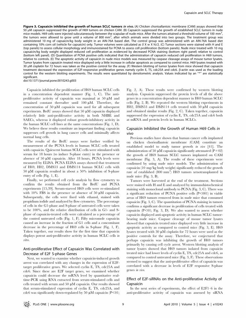
Capsaicin Inhibited the Growth of Human H69 Cells invivo
Previous studies have shown that human cancer cells implanted
on chicken chorioallantoic membrane (CAM) constitute an
established model to study tumor growth in vivo [41]. The
administration of 50 mM capsaicin significantly attenuated (P = 02)
the growth of H69 human SCLC tumors implanted on CAM
membrane (Fig. 3, A). The results of these experiments were
confirmed by using nude mice models. The administration of
capsaicin (10 mg/kg body weight) significantly reduced the growth
rate of established (800 mm3) H69 tumors xenotransplanted in
nude mice (Fig. 3, B).
Tumors were harvested at the end of the treatment. Sections
were stained with H and E and analyzed by immunohistochemical
staining with monoclonal antibody to PCNA (Fig. 3, C). There was
a significant reduction of PCNA positive cells (P,001) in viable
areas of H69 tumors removed from in nude mice that consumed
capsaicin (Fig. 3, C). The quantitation of PCNA staining in tumors
confirms a significant decrease in proliferation of cells treated with
capsaicin (P,01; Fig. 3, D). We also wanted to assess whether
capsaicin displayed anti-apoptotic activity in human SCLC tumor-
bearing nude mice. Caspase cleavage of mouse tumor lysates
showed that capsaicin treated mice displayed very little increase in
apoptotic activity as compared to control mice (Fig. 3, E). H69
lysates treated with 30 mM cisplatin for 72 hours were used as the
positive controls for the assay. Therefore, we conjectured that
perhaps capsaicin was inhibiting the growth of H69 tumors
primarily by causing cell cycle arrest. Western blotting analysis of
tumor lysates showed that H69 tumors isolated from capsaicin
treated mice had lower levels of cyclin E, TS, cdc25A and cdc6, as
compared to control untreated mice (Fig. 3, F). These observations
seemed to suggest that the anti-proliferative effect of capsaicin was
correlated with a decrease in levels of E2F responsive S-phase
genes in vivo.
Effect of E2F-siRNAs on the Anti-proliferative Activity ofCapsaicin
In the next series of experiments, the effect of E2F1–6 in the
anti-proliferative activity of capsaicin was assessed by siRNA
Figure 3. Capsaicin inhibited the growth of human SCLC tumors in vivo. (A) Chicken chorioallantoic membrane (CAM) assays showed that50 mM capsaicin suppressed the growth of H69 tumors on chicken CAM. (B) Capsaicin suppressed the growth of established SCLC tumors in nudemice models. H69 cells were injected subcutaneously between the scapulae of nude mice. After the tumors attained a threshold volume of 100 mm3,the tumors were allowed to grow until a volume of 800 mm3, after which animals were divided into two groups. The treatment group wasadministered 10 mg of capsaicin/kg body weight in an AIN-76A based diet. The control group was administered with an AIN-76A based dietcontaining 10% corn oil (vehicle for capsaicin) only. Tumor volumes were calculated as (l X w X h)/2. (C) Tumor sections were stained with H and E(top panels) to assess cellular morphology and immunostained for PCNA to assess cell proliferation (bottom panels). Nude mice treated with 10 mgcapsaicin/kg body weight displayed reduced cell proliferation as evidenced by decreased PCNA staining (bottom right panel) relative to control(bottom left panel). (D) Quantitation of PCNA positive cells indicated that the administration of capsaicin reduced cell proliferation in H69 tumors,relative to controls. (E) The apoptotic activity of capsaicin in nude mice models was measured by caspase cleavage assays of mouse tumor lysates.Tumor lysates from capsaicin treated mice displayed only a little increase in cellular apoptosis as compared to control mice. H69 lysates treated with30 mM cisplatin for 72 hours was taken as the positive control for the assay. (F) Western blotting of tumor lysates from mice showed that capsaicintreatment decreased the expression of E2F-responsive proliferative genes namely cyclin E, TS, cdc25A and cdc6. b-actin was used as the loadingcontrol for the western blotting experiments. The results were quantitated by densitometric analysis. Values indicated by an ‘‘*’’ are statisticallysignificant.doi:10.1371/journal.pone.0010243.g003
Capsaicin and SCLC Therapy
PLoS ONE | www.plosone.org 9 April 2010 | Volume 5 | Issue 4 | e10243

Figure 4. BrdU assays demonstrated that depletion of E2F4 ablates the anti-proliferative activity of capsaicin. (A) H69 and DMS114cells were transfected with the indicated E2F-siRNA as detailed in ‘‘Materials and Methods.’’ Eighteen hours post transfection, the cells were serum-starved for 36 hours and re-stimulated with media containing 10% FBS in the presence of 50 mM capsaicin for 18 hours. Subsequently, BrdU assayswere performed to measure cell proliferation. The anti-proliferative activity of capsaicin was ablated by E2F4-siRNA but not affected by a non-targeting control-siRNA. (B) Western blotting analysis confirmed the suppression of E2F1–6 expression upon siRNA transfection. GAPDH was used asthe loading control for the western blotting experiments, and the results were quantitated by densitometric analysis. (C) Western blotting
Capsaicin and SCLC Therapy
PLoS ONE | www.plosone.org 10 April 2010 | Volume 5 | Issue 4 | e10243
methodology. E2F1, E2F2, E2F3, E2F4, E2F5 and E2F6 siRNAs
were individually transfected in H69 cells. Eighteen hours after
transfection, H69 cells were serum-starved for 36 hours (by
incubating them in serum-free RPMI) and subsequently re-
stimulated with 10% FBS in the presence of 50 mM capsaicin
for 18 hours. BrdU assays were performed to measure the
percentage of cells undergoing S-phase entry [31].
We observed that E2F4-siRNA reversed the anti-proliferative
effect of capsaicin in H69 and DMS114 cells, whereas E2F1–3-,
E2F5- and E2F6-siRNA did not have any effect on the growth-
inhibitory effects of capsaicin (Fig. 4, A). The control non-targeting
siRNAs did not have any effect on the anti-proliferative effect of
capsaicin (Fig. 4, A). Western blotting analysis showed that the
transfection of the above mentioned siRNAs suppressed E2F1–6
protein levels in both H69 and DMS114 cells (Fig. 4, B and C,
respectively).
Since the E2F-family of transcription factors have been shown
to play a pivotal role in cell proliferation, we wanted to examine
whether E2F-siRNAs would affect proliferation of human SCLC
cells untreated with capsaicin. E2F1–6-siRNAs were individually
transfected in H69 cells using Oligofectamine reagent. Eighteen
hours post-transfection, H69 cells were serum-starved for 36 hours
(by incubating them in serum-free RPMI) and subsequently re-
stimulated with 10% FBS for 18 hours. BrdU assays were
performed to measure the percentage of cells undergoing S-phase
entry [31]. We found that none of the E2F-siRNAs individually
affected the proliferation of SCLC cells stimulated with 10% FBS
(Fig. 4, D). The experiment was repeated in a second SCLC cell
line, DMS114, and similar results were observed (Fig. 4, D). Taken
together, our results show that E2F4-siRNA specifically abrogates
the anti-proliferative effect of capsaicin and does not affect serum-
induced proliferation of SCLC cells.
The results of the siRNA experiments were verified by using two
independent sets of E2F-siRNAs. BrdU assays showed that both
independent E2F4-siRNA ablated the effects of 50 mM capsaicin
in H69 cells (Fig. 5, A), whereas a non-targeting control-siRNA did
not have any effect. The data obtained from the BrdU assays was
further verified by PCNA-ELISA assays, and similar results were
obtained (Fig. 5, B). Western blotting analysis showed that the
transfection of either sets of E2F4-siRNAs produced efficient
suppression of E2F4 protein levels in H69 cells (Fig. 5, C).
Capsaicin Promotes the Recruitment of E2F4 toE2F-responsive Proliferative Promoters
Our data seemed to indicate that E2F4 plays a vital role in the
anti-proliferative effects of capsaicin. Therefore, ChIP assays were
performed to examine the differential occupancy of E2F family of
proteins on E2F-responsive proliferative promoters (cyclin E, TS,
cdc25A and cdc6) upon capsaicin-treatment of H69 human SCLC
cells [31,32,40,45].
High amounts of the proliferative E2Fs, namely E2F1, E2F2
and E2F3, were bound to all E2F-responsive promoters in the
control serum-stimulated H69 cells (Fig. 6, left panel, lanes 3,4,5
from the left). In contrast, no E2F4 or very low levels of E2F4 were
bound to these promoters in proliferating H69 cells (Fig. 6, left
panel, lane 6 from left). However, the treatment of these cells with
50 mM capsaicin for 8 hours resulted in the recruitment of robust
amounts of E2F4 on cyclin E, TS, cdc25A and cdc6 promoters
(Fig. 6, right panel, lane 6 from left), accompanied with
concomitant dissociation of E2F1, E2F2 and E2F3 (Fig. 6, right
panel, lane 3,4,5 from left). Furthermore, the enhanced binding of
E2F4 was accompanied by recruitment of the E2F4-binding
partner p130 on the cyclin E, TS, cdc25A and cdc6 promoters in
capsaicin-treated cells. In contrast, very low levels of p130 were
bound to these promoters in proliferating cells (Fig. 6, left panel,
last lane from left). Therefore, our data seem to suggest that
capsaicin selectively recruits E2F4 and p130 to proliferative
promoters, thereby repressing their transcription and inhibiting S-
phase entry in human SCLC cells. PCR for the c-Fos promoter
(which is not regulated by E2F) was used as a negative control for
the ChIP experiments. We observed that there were no E2F family
proteins associated with this promoter (Fig. 6, bottom lanes).
Discussion
The dysfunction of the E2F/Rb pathway is a hallmark of
greater than 90% of lung cancers [3,4,5]. Several convergent
studies have indicated that the majority of human SCLC contains
mutations/deletions in vital tumor suppressor genes like Rb, p130,
p16, cyclin D1 and p53, which facilitate its growth and distant
metastasis [3]. Therefore, it is probable that the anti-proliferative
activity of nutritional agents like capsaicin is mediated by its effects
on the cell cycle machinery in human SCLC cells.
Emerging evidence shows that capsaicin is a promising anti-
cancer agent that causes potent apoptosis in prostate cancer, non
small cell lung cancer, gliomas and gastric cancers
[10,13,14,15,16,21,50]. However, the growth-inhibitory activity
of capsaicin in human SCLC is unknown. To our knowledge, our
pilot study showed for the first time that capsaicin displays potent
anti-proliferative activity in human SCLCs both in cell culture
models and in two in vivo models. Our results showed that
capsaicin suppresses the growth of established H69 human SCLC
tumors in both CAM and nude mice models. We observed that the
administration of capsaicin did not cause any gross discomfort,
toxicity or weight loss in these animals. Taken together, our data
suggest that capsaicin has the potential for therapy and
management of human SCLCs.
The majority of studies exploring the anti-cancer activity of
capsaicin have focused on the mechanisms underlying capsaicin-
induced apoptosis [10,13,14,15,16,21,50]. Only a few studies have
investigated the signaling pathways underlying capsaicin-induced
cell cycle arrest [10,17,19,21]. Our results showed for the first time
that capsaicin induces G1 arrest in four human SCLC cell lines in
a concentration dependent manner. Our data are in agreement
with results of previous studies, which have shown that capsaicin
induces G1 arrest in prostate cancer, breast cancer, epidermoid
cancer and human leukemic cells [10,17,19,21]. Min et al., (2004)
have found that capsaicin can inhibit the proliferation of
endothelial cells and displays anti-angiogenic activity in both cell
culture and mouse models [22]. Similarly, capsaicin analogs like
capsiate and dihydrocapsiate have been found to inhibit VEGF-
induced angiogenesis [51]. It is probable that the anti-angiogenic
activity of capsaicin is, at least in part, responsible for its observed
anti-tumor activity in mice models and the CAM model. We
quantified the extent of angiogenesis observed in our CAM
experiments and found that the capsaicin-treated H69 tumor-
bearing CAM contained fewer blood vessels (3.460.6) than those
of the untreated controls (8.560.9). Other mechanisms of the anti-
experiments demonstrated that levels of E2F1–6 were decreased upon siRNA transfection in DMS114 cells. (D) BrdU assay showed that transfection ofE2F1–6 siRNA without capsaicin did not affect the proliferation of cells in response to 10% FBS. This indicates that the effects of E2F-siRNAs observedin (A) are specifically mediated by capsaicin treatment. Values indicated by an ‘‘*’’ are statistically significant.doi:10.1371/journal.pone.0010243.g004
Capsaicin and SCLC Therapy
PLoS ONE | www.plosone.org 11 April 2010 | Volume 5 | Issue 4 | e10243

proliferative activity of capsaicin included regulation of cyclin/
cdks, p21 and p53 in the aforementioned cells. However, no
studies have explored the role of downstream E2F family of
proteins in the anti-proliferative effects of capsaicin.
One of the interesting findings of our study was that E2F1-,
E2F2- and E2F3-siRNA by themselves had no effect on the serum-
induced proliferation of H69 cells. This observation can be
explained by the fact that there is considerable redundancy in the
proliferative functions of the E2F family of proteins
[25,27,28,29,30]. E2F1–3 are proliferative E2Fs. Therefore, if
one of them, for example E2F1, is knocked down by E2F1-siRNA,
it is probable that its mitogenic functions can be compensated by
other E2F2 and E2F3 [25,27,28,29,30]. Thus, no effect of these
siRNAs on serum-induced proliferation of H69 cells is observed.
Figure 5. Two independent E2F4-siRNA reversed the anti-proliferative effect of capsaicin in H69 cells. (A) The transfection of siRNA andsubsequent BrdU assay was performed as described in ‘‘Materials and Methods.’’ (B) The results obtained by BrdU assay are confirmed with PCNAELISA assay. Two independent E2F4-siRNA were transfected as described in ‘‘Materials and Methods.’’ PCNA assays were then performed to assess thelevels of E2F4-siRNA on the anti-proliferative activity of capsaicin. (C) Western blotting analysis indicated that E2F4 levels are suppressed upon siRNAtransfection. GAPDH was used as the loading control for the western blotting experiments, and the results were quantitated by densitometricanalysis. Values indicated by an ‘‘*’’ are statistically significant.doi:10.1371/journal.pone.0010243.g005
Capsaicin and SCLC Therapy
PLoS ONE | www.plosone.org 12 April 2010 | Volume 5 | Issue 4 | e10243

E2F4, E2F5 and E2F6 are repressive E2F proteins; therefore, their
siRNAs would not be expected to have an impact on cell
proliferation.
Our study demonstrated for the first time that E2F4 primarily
mediates the anti-proliferative activity of capsaicin. Our data is
consistent with previous studies that have shown that E2F4 is a
repressor of transcription and inhibits cell proliferation. In
addition, the E2F4/p130 pathway has been implicated in the
growth and progression of lung cancer. E2F42/2 mice were
found to have defects in small airway epithelial cells, suggesting
a role for this protein in lung development [52]. Studies from
E2F4 (2/2) Rb (2/2) chimeric mice have suggested that E2F4
may play a role in early stages of small cell lung cancer [53].
Bankovic et al., (2009) studied genomic instability in NSCLC
patients by DNA fingerprinting and discovered that E2F4 was
among the group of genes responsible for growth and metastasis
of NSCLCs [33]. Ren et al., (2002) performed a ChIP analysis
combined with microarray experiments to determine transcrip-
tional targets of E2F4. Their results indicated that E2F4 target
genes include those regulating DNA damage checkpoint, DNA
repair, mitotic spindle checkpoint and chromatin assembly/
condensation [54]. Many of these processes are involved in
neoplastic transformation. Similarly, the E2F4 binding pocket
protein, p130, has been suggested as a novel molecular target
for diagnosis and therapy of lung cancers [55,56]. Gene therapy
studies have shown that over expression of p130 in advanced
stage lung tumors could attenuate their growth [55,57].
Furthermore, p130 is involved in tumor angiogenesis and plays
a vital role in the differentiation and mobilization of bone
marrow-derived endothelial cell precursors and endothelial
sprouting from neighboring vessels [58]. We believe that
nutritional agents like capsaicin recruit the E2F4/p130 pathway
to exert anti-proliferative effects in human SCLCs.
ChIP assays were performed to examine the relative distribution
of E2Fs on E2F-responsive promoters in human SCLC cells. Our
studies revealed that the treatment of human SCLC cells with
capsaicin leads to differential recruitment of E2Fs on E2F-
responsive promoters like cyclin E, TS, cdc25A and cdc6. Control
H69 SCLC cells contained E2F1, E2F2 and E2F3 bound to
proliferative promoters. Our results are consistent with previous
studies in that proliferative responses are primarily mediated by
E2F1, E2F2 and E2F3, whereas the recruitment of E2F4 on the
promoters suppresses cell proliferation [31,32,38]. The treatment
of H69 human SCLC cells with capsaicin leads to a switch in E2F
subtypes on cyclin E, TS, cdc25A and cdc6 promoters; E2F1–3
are dissociated from the promoter, and E2F4 and p130 are
recruited. We did not detect any E2F5 on any of the promoters
(data not shown).
In summary, the data presented in this paper show that
capsaicin displays potent anti-proliferative activity against
human SCLC, and this effect is mediated by the E2F4
pathway. Aberrancies in the E2F pathway are one of the
hallmarks of human SCLC [3,4,30,57]; therefore, nutritional
agents like capsaicin, which target the E2F pathway, may
represent new avenues for the treatment of lethal malignancies
like small cell lung cancer. We believe that the data obtained
from this pilot study establishes the ‘‘proof–of-principle’’ for
these concepts. Future research will focus on the identification
of second generation capsaicin-mimetics and the exploration of
their anti-proliferative activity and signaling pathways in
human SCLCs.
Acknowledgments
We thank Dr. Srikumar Chellappan and his laboratory for their continuous
support. We would also like to thank Jennifer M. Napper for help with the
flow cytometry experiments and the Histopathology Facility (Cabell
Figure 6. ChIP assays showed that capsaicin induces the recruitment of E2F4 and p130 on proliferative promoters. Serum-stimulatedH69 cells contained robust amounts of proliferative E2Fs, namely E2F1, E2F2 and E2F3, associated with E2F-responsive proliferative promoters likecyclin E, TS, cdc25A and cdc6 (left panel). The treatment of H69 cells with 50 mM capsaicin causes dissociation of proliferative E2Fs and recruitment ofthe repressive E2F4 and p130 on these promoters (right panel). PCR for the c-Fos promoter (which is not regulated by E2F) was taken as the controlfor the experiment.doi:10.1371/journal.pone.0010243.g006
Capsaicin and SCLC Therapy
PLoS ONE | www.plosone.org 13 April 2010 | Volume 5 | Issue 4 | e10243

Huntington Hospital, Huntington, WV). The advice and suggestions of Dr.
Nalini Santanam and Carla Cook are gratefully acknowledged.Author Contributions
Conceived and designed the experiments: WEH PD. Performed the
experiments: KCB TRW HL YCC JKL PD. Analyzed the data: KCB HL
YCC ABC JKL. Contributed reagents/materials/analysis tools: WEH
ABC. Wrote the paper: JKL PD.
References
1. Krug LM, Kris MG, Rosenweig K, Travis WD (2008) Small cell and otherneuroendocrine tumors of the lung; DeVita VT, Lawrence TS, Rosenburg SA,
eds. Philadelphia: Lippincott Williams and Wilkins.
2. Metro G, Cappuzzo F (2009) Emerging drugs for small-cell lung cancer. ExpertOpin Emerg Drugs 14: 591–606.
3. Salgia R, Skarin AT (1998) Molecular abnormalities in lung cancer. J Clin
Oncol 16: 1207–1217.
4. Minna JD (1993) The molecular biology of lung cancer pathogenesis. Chest 103:449S–456S.
5. Minna JD, Kurie JM, Jacks T (2003) A big step in the study of small cell lung
cancer. Cancer Cell 4: 163–166.
6. Biro T, Acs G, Acs P, Modarres S, Blumberg PM (1997) Recent advances inunderstanding of vanilloid receptors: a therapeutic target for treatment of pain
and inflammation in skin. J Investig Dermatol Symp Proc 2: 56–60.
7. Caterina MJ, Schumacher MA, Tominaga M, Rosen TA, Levine JD, et al.(1997) The capsaicin receptor: a heat-activated ion channel in the pain pathway.
Nature 389: 816–824.
8. Jang JJ, Kim SH, Yun TK (1989) Inhibitory effect of capsaicin on mouse lungtumor development. In Vivo 3: 49–53.
9. Anandakumar P, Kamaraj S, Jagan S, Ramakrishnan G, Vinodhkumar R, et al.
(2007) Stabilization of pulmonary mitochondrial enzyme system by capsaicinduring benzo(a)pyrene induced experimental lung cancer. Biomed Pharmac-
other.
10. Mori A, Lehmann S, O’Kelly J, Kumagai T, Desmond JC, et al. (2006)Capsaicin, a component of red peppers, inhibits the growth of androgen-
independent, p53 mutant prostate cancer cells. Cancer Res 66: 3222–3229.
11. Park KK, Surh YJ (1997) Effects of capsaicin on chemically-induced two-stagemouse skin carcinogenesis. Cancer Lett 114: 183–184.
12. Tanaka T, Kohno H, Sakata K, Yamada Y, Hirose Y, et al. (2002) Modifying
effects of dietary capsaicin and rotenone on 4-nitroquinoline 1-oxide-induced rat
tongue carcinogenesis. Carcinogenesis 23: 1361–1367.
13. Amantini C, Mosca M, Nabissi M, Lucciarini R, Caprodossi S, et al. (2007)
Capsaicin-induced apoptosis of glioma cells is mediated by TRPV1 vanilloid
receptor and requires p38 MAPK activation. J Neurochem 102: 977–990.
14. Athanasiou A, Smith PA, Vakilpour S, Kumaran NM, Turner AE, et al. (2007)Vanilloid receptor agonists and antagonists are mitochondrial inhibitors: how
vanilloids cause non-vanilloid receptor mediated cell death. Biochem BiophysRes Commun 354: 50–55.
15. Bhutani M, Pathak AK, Nair AS, Kunnumakkara AB, Guha S, et al. (2007)
Capsaicin is a novel blocker of constitutive and interleukin-6-inducible STAT3activation. Clin Cancer Res 13: 3024–3032.
16. Gil YG, Kang MK (2008) Capsaicin induces apoptosis and terminal
differentiation in human glioma A172 cells. Life Sci 82: 997–1003.
17. Tsou MF, Lu HF, Chen SC, Wu LT, Chen YS, et al. (2006) Involvement of Bax,Bcl-2, Ca2+ and caspase-3 in capsaicin-induced apoptosis of human leukemia
HL-60 cells. Anticancer Res 26: 1965–1971.
18. Sanchez AM, Sanchez MG, Malagarie-Cazenave S, Olea N, Diaz-Laviada I(2006) Induction of apoptosis in prostate tumor PC-3 cells and inhibition of
xenograft prostate tumor growth by the vanilloid capsaicin. Apoptosis 11: 89–99.
19. Wu CC, Lin JP, Yang JS, Chou ST, Chen SC, et al. (2006) Capsaicin inducedcell cycle arrest and apoptosis in human esophagus epidermoid carcinoma CE
81T/VGH cells through the elevation of intracellular reactive oxygen species
and Ca2+ productions and caspase-3 activation. Mutat Res 601: 71–82.
20. Lehen’kyi V, Flourakis M, Skryma R, Prevarskaya N (2007) TRPV6 channel
controls prostate cancer cell proliferation via Ca(2+)/NFAT-dependent
pathways. Oncogene 26: 7380–7385.
21. Thoennissen NH, O’Kelly J, Lu D, Iwanski GB, La DT, et al. (2009) Capsaicincauses cell-cycle arrest and apoptosis in ER-positive and -negative breast cancer
cells by modulating the EGFR/HER-2 pathway. Oncogene Epub ahead ofprint.
22. Min JK, Han KY, Kim EC, Kim YM, Lee SW, et al. (2004) Capsaicin inhibits
in vitro and in vivo angiogenesis. Cancer Res 64: 644–651.
23. Nevins JR, Chellappan SP, Mudryj M, Hiebert S, Devoto S, et al. (1991) E2Ftranscription factor is a target for the RB protein and the cyclin A protein. Cold
Spring Harb Symp Quant Biol 56: 157–162.
24. Chellappan SP, Hiebert S, Mudryj M, Horowitz JM, Nevins JR (1991) The E2Ftranscription factor is a cellular target for the RB protein. Cell 65: 1053–1061.
25. Chen HZ, Tsai SY, Leone G (2009) Emerging roles of E2Fs in cancer: an exit
from cell cycle control. Nat Rev Cancer 9: 785–797.
26. Swiss VA, Casaccia P (2009) Cell-context specific role of the E2F/Rb pathway indevelopment and disease. Glia.
27. DeGregori J, Johnson DG (2006) Distinct and Overlapping Roles for E2F
Family Members in Transcription, Proliferation and Apoptosis. Curr Mol Med6: 739–748.
28. Johnson DG, Degregori J (2006) Putting the Oncogenic and Tumor SuppressiveActivities of E2F into Context. Curr Mol Med 6: 731–738.
29. Wang S, Fusaro G, Padmanabhan J, Chellappan SP (2002) Prohibitin co-
localizes with Rb in the nucleus and recruits N-CoR and HDAC1 for
transcriptional repression. Oncogene 21: 8388–8396.
30. Nevins JR (2001) The Rb/E2F pathway and cancer. Hum Mol Genet 10:699–703.
31. Dasgupta P, Rastogi S, Pillai S, Ordonez-Ercan D, Morris M, et al. (2006)
Nicotine induces cell proliferation by beta-arrestin-mediated activation of Srcand Rb-Raf-1 pathways. J Clin Invest 116: 2208–2217.
32. Dasgupta P, Sun J, Wang S, Fusaro G, Betts V, et al. (2004) Disruption of theRb—Raf-1 interaction inhibits tumor growth and angiogenesis. Mol Cell Biol
24: 9527–9541.
33. Bankovic J, Stojsic J, Jovanovic D, Andjelkovic T, Milinkovic V, et al. (2009)Identification of genes associated with non-small-cell lung cancer promotion and
progression. Lung Cancer.
34. Gazdar AF, Carney DN, Nau MM, Minna JD (1985) Characterization of
variant subclasses of cell lines derived from small cell lung cancer havingdistinctive biochemical, morphological, and growth properties. Cancer Res 45:
2924–2930.
35. Gazdar AF, Carney DN, Russell EK, Sims HL, Baylin SB, et al. (1980)Establishment of continuous, clonable cultures of small-cell carcinoma of lung
which have amine precursor uptake and decarboxylation cell properties. Cancer
Res 40: 3502–3507.
36. Pettengill OS, Sorenson GD, Wurster-Hill DH, Curphey TJ, Noll WW, et al.(1980) Isolation and growth characteristics of continuous cell lines from small-
cell carcinoma of the lung. Cancer 45: 906–918.
37. Heo DS, Park JG, Hata K, Day R, Herberman RB, et al. (1990) Evaluation of
tetrazolium-based semiautomatic colorimetric assay for measurement of humanantitumor cytotoxicity. Cancer Res 50: 3681–3690.
38. Kinkade R, Dasgupta P, Carie A, Pernazza D, Carless M, et al. (2008) A small
molecule disruptor of Rb/Raf-1 interaction inhibits cell proliferation, angio-genesis, and growth of human tumor xenografts in nude mice. Cancer Res 68:
3810–3818.
39. Knudsen ES, Angus SP (2003) Functional analysis of the antimitogenic activity
of tumor suppressors. Methods Mol Biol 218: 3–15.
40. Dasgupta P, Betts V, Rastogi S, Joshi B, Morris M, et al. (2004) Direct binding ofapoptosis signal-regulating kinase 1 to retinoblastoma protein: novel links
between apoptotic signaling and cell cycle machinery. J Biol Chem 279:
38762–38769.
41. Gu JW, Bailey AP, Sartin A, Makey I, Brady AL (2005) Ethanol stimulatestumor progression and expression of vascular endothelial growth factor in chick
embryos. Cancer 103: 422–431.
42. Song P, Sekhon HS, Fu XW, Maier M, Jia Y, et al. (2008) Activated cholinergicsignaling provides a target in squamous cell lung carcinoma. Cancer Res 68:
4693–4700.
43. Tomayko MM, Reynolds CP (1989) Determination of subcutaneous tumor size
in athymic (nude) mice. Cancer Chemother Pharmacol 24: 148–154.
44. Shabbir M, Thompson CS, Mikhailidis D, Morgan RJ, Burnstock G (2003)Extracellular ATP attenuates the growth of hormone refractory prostate cancer
in vivo. Eur Urology Supplements 2: 24–27.
45. Dasgupta P, Kinkade R, Joshi B, Decook C, Haura E, et al. (2006) Nicotine
inhibits apoptosis induced by chemotherapeutic drugs by up-regulating XIAPand survivin. Proc Natl Acad Sci U S A 103: 6332–6337.
46. Rastogi S, Joshi B, Dasgupta P, Morris M, Wright K, et al. (2006) Prohibitin
facilitates cellular senescence by recruiting specific corepressors to inhibit E2Ftarget genes. Mol Cell Biol 26: 4161–4171.
47. Potemski P, Pluciennik E, Bednarek AK, Kusinska R, Jesionek-Kupnicka D,et al. (2006) Cyclin E expression in operable breast cancer quantified using real-
time RT-PCR: a comparative study with immunostaining. Jpn J Clin Oncol 36:142–149.
48. Dasgupta P, Chellappan SP (2006) Nicotine-mediated cell proliferation and
angiogenesis: new twists to an old story. Cell Cycle 5: 2324–2328.
49. Ohtani K, DeGregori J, Nevins JR (1995) Regulation of the cyclin E gene by
transcription factor E2F1. Proc Natl Acad Sci U S A 92: 12146–12150.
50. Chow J, Norng M, Zhang J, Chai J (2007) TRPV6 mediates capsaicin-inducedapoptosis in gastric cancer cells—Mechanisms behind a possible new ‘‘hot’’
cancer treatment. Biochim Biophys Acta 1773: 565–576.
51. Pyun BJ, Choi S, Lee Y, Kim TW, Min JK, et al. (2008) Capsiate, a nonpungent
capsaicin-like compound, inhibits angiogenesis and vascular permeability via adirect inhibition of Src kinase activity. Cancer Res 68: 227–235.
52. Danielian PS, Bender Kim CF, Caron AM, Vasile E, Bronson RT, et al. (2007)
E2f4 is required for normal development of the airway epithelium. Dev Biol 305:564–576.
Capsaicin and SCLC Therapy
PLoS ONE | www.plosone.org 14 April 2010 | Volume 5 | Issue 4 | e10243

53. Parisi T, Bronson RT, Lees JA (2009) Inhibition of pituitary tumors in Rb
mutant chimeras through E2f4 loss reveals a key suppressive role for the pRB/E2F pathway in urothelium and ganglionic carcinogenesis. Oncogene 28:
500–508.
54. Ren B, Cam H, Takahashi Y, Volkert T, Terragni J, et al. (2002) E2F integratescell cycle progression with DNA repair, replication, and G(2)/M checkpoints.
Genes Dev 16: 245–256.55. Ho VM, Schaffer BE, Karnezis AN, Park KS, Sage J (2009) The retinoblastoma
gene Rb and its family member p130 suppress lung adenocarcinoma induced by
oncogenic K-Ras. Oncogene 28: 1393–1399.
56. Simpson DS, Mason-Richie NA, Gettler CA, Wikenheiser-Brokamp KA (2009)
Retinoblastoma family proteins have distinct functions in pulmonary epithelial
cells in vivo critical for suppressing cell growth and tumorigenesis. Cancer Res
69: 8733–8741.
57. Claudio PP, Caputi M, Giordano A (2000) The RB2/p130 gene: the latest
weapon in the war against lung cancer? Clin Cancer Res 6: 754–764.
58. Gabellini C, Del Bufalo D, Zupi G (2006) Involvement of RB gene family in
tumor angiogenesis. Oncogene 25: 5326–5332.
Capsaicin and SCLC Therapy
PLoS ONE | www.plosone.org 15 April 2010 | Volume 5 | Issue 4 | e10243


![Capsaicin. Scoville heat unitsExamples 15,000,000– 16,000,000 Pure capsaicin [9]capsaicin [9] 8,600,000–9,100,000 Various capsaicinoids (e.g., homocapsaicin,](https://img.pdfslide.us/doc/110x75/56649ec95503460f94bd6e61/capsaicin-scoville-heat-unitsexamples-15000000-16000000-pure-capsaicin.jpg)