Embed Size (px)
Citation preview
I N V I T E D R E V I E W
Disorders of memory
Michael D. Kopelman
Neuropsychiatry and Memory Disorders Clinic, University
Department of Psychiatry and Psychology, Guy's, King's
and St Thomas' Medical School (KCL), London, UK
Correspondence to: Michael D. Kopelman,
Neuropsychiatry and Memory Disorders Clinic, University
Department of Psychiatry and Psychology, Guy's, King's
and St Thomas' Medical School (KCL), St Thomas'
Campus, London, SE1 7EH, UK
E-mail: [email protected]
SummaryThis paper reviews disorders of memory. After abrief survey of the clinical varieties of the amnesicsyndrome, transient and persistent, selected theor-etical issues will be considered by posing a series ofquestions. (i) What is impaired and what is sparedin anterograde amnesia? (ii) Do temporal lobe, dien-cephalic and frontal lobe amnesias differ? (iii) Howindependently semantic is semantic memory? (iv)What determines the pattern and extent of retro-
grade memory loss? (v) Can retrograde amnesia ever
be `isolated'? (vi) Does psychogenic amnesia involve
the same mechanisms as organic amnesia? (vii) How
and when do false memories arise? Commonalities as
well as differences across separate literatures will be
emphasized, and the case for a more `dynamic'
(interactionist) approach to the investigation of amne-
sia will be advocated.
Keywords: amnesia; anterograde; confabulation; psychogenic; retrograde
Abbreviations: AA = anterograde amnesia; FDG PET = [18H]¯uoro-deoxy glucose PET; GABA = gamma-amino butyric
acid; K = `know'; PTA = post-traumatic amnesia; PTSD = post-traumatic stress disorder; R = `remember'; RA =
retrograde amnesia; SPECT = single photon emission tomography; TEA = transient epileptic amnesia; TGA = transient
global amnesia; TPP = thiamine pyrophosphate; WAIS-R = Wechsler Adult Intelligence ScaleÐRevised; WMS-R =
Wechsler Memory ScaleÐRevised
Introduction`So we beat on, boats against the current, borne back
ceaselessly into the past.'
(F. Scott Fitzgerald, The Great Gatsby, 1925.)
`The places that we have known belong nowonly to the little world of space on which wemap them for our own convenience. None ofthem was ever more than a thin slice, heldbetween the contiguous impressions that com-posed our life at that time; remembrance of aparticular form is but regret for a particularmoment; and houses, roads, avenues are as fugi-tive, alas, as the years.'
(M. Proust, Remembrance of Things Past, 1913.)
The literature on memory and its disorders has proliferated,
particularly since the advent of recent neuroimaging tech-
niques. However, controversy remains concerning certain
broad issues, selected examples of which will be considered
in turn in this review, following a brief survey of the clinical
disorders that give rise to an amnesic syndrome. The focus of
the review will be on neuropsychological studies of patients
with disorders of episodic or semantic memory, and there will
be only brief reference to functional neuroimaging studies of
healthy individuals.
Varieties of amnesic syndromeThe amnesic syndrome can be de®ned as: `An abnormal
mental state in which memory and learning are affected out of
ã Guarantors of Brain 2002
Brain (2002), 125, 2152±2190
by guest on October 6, 2016
http://brain.oxfordjournals.org/D
ownloaded from
all proportion to other cognitive functions in an otherwise
alert and responsive patient' (Victor et al., 1971).
The Korsakoff syndrome can be de®ned as the same but
with the following phrase added: `... resulting from nutritional
depletion, notably thiamine de®ciency.'
In fact, Victor et al. (1971) used the ®rst description as a
de®nition of the Korsakoff syndrome, but the present author
feels that it is important to distinguish between amnesic
syndromes in general (for which the Victor et al. de®nition
suf®ces) and the particular clinical condition described by
Korsakoff (1889), whose cases (with hindsight) can all be
viewed as having suffered nutritional depletion, whether of
alcoholic or non-alcoholic causation. Various disorders can
give rise to an amnesic syndrome, including herpes
encephalitis, severe hypoxia, certain vascular lesions, head
injury, deep midline tumours, basal forebrain lesions and
occasionally early dementia.
The Korsakoff syndromeAs just mentioned, the Korsakoff syndrome is the result of
nutritional depletion, i.e. thiamine de®ciency. Korsakoff
(1889) described this condition as resulting from alcohol
abuse or from a number of other causes, but by far the most
common nowadays is alcohol abuse.
There are frequent misunderstandings about the nature of
this disorder. `Short term memory' is intact in the Jamesian
sense of recall over a matter of seconds (Zangwill, 1946), but
learning over more prolonged periods is severely impaired,
and there is usually a retrograde memory loss which
characteristically extends back many years or decades
(Kopelman, 1989; Parkin et al., 1990b). Korsakoff (1889)
himself noted that his patients `reason about everything
perfectly well, draw correct deductions from given premises,
make witty remarks, play chess or a game of cards, in a word
comport themselves as mentally sound persons.' However, he
also noted repetitive questionings, the extensive nature of the
retrograde memory loss, and a particular problem in remem-
bering the temporal sequence of events, associated with
severe disorientation in time. As will be discussed below, he
gave examples of confabulation that re¯ected the problem
with temporal sequence memory, such that real memories
were jumbled up and retrieved inappropriately, out of
temporal context.
Many cases of the Korsakoff syndrome are diagnosed
following an acute Wernicke encephalopathy, involving
confusion, ataxia, nystagmus and ophthalmoplegia. Not all
these features are always present, and the ophthalmoplegia in
particular responds rapidly to treatment with high-dose
vitamins. These features are often associated with a periph-
eral neuropathy. However, the disorder can also have an
insidious onset (Cutting, 1978a), and such cases are more
likely to come to the attention of psychiatrists: in these cases,
there may be either no known history or only a transient
history of Wernicke features. There are also reports that the
characteristic Wernicke±Korsakoff neuropathology (see
below) is diagnosed much more commonly at autopsy in
alcoholics than in life (Torvik et al., 1982; Harper, 1983),
implying that many cases are being missed.
The Korsakoff syndrome is unusual among memory
disorders in that there is a distinct neurochemical pathology
with important implications for treatment. Following animal
research in the 1930s and 1940s, and the important observa-
tions of de Wardener and Lennox (1947) and others in
malnourished prisoners-of-war, thiamine depletion was
established as the mechanism giving rise to the acute
Wernicke episode, and the subsequent (Korsakoff) memory
impairment. However, the genetic factor, predisposing cer-
tain heavy drinkers to develop this syndrome before hepatic
or gastro-intestinal complications, remains unclear. It was
thought that a transketolase gene might account for this,
because transketolase is the enzyme that requires thiamine
pyrophosphate (TPP) as a co-factor. Such a gene has been
identi®ed (McCool et al., 1993), but it does not account for all
the properties of transketolase and only very weakly for
predisposition to the Korsakoff syndrome. Moreover, it
remains unclear how thiamine depletion produces the
particular neuropathology found in Wernicke±Korsakoff
patients, although Witt (1985) pointed out that six neuro-
transmitter systems (including acetylcholine, GABA, gluta-
mate) are affected by thiamine depletion, either by reduction
of TPP-dependent enzyme activity or by direct structural
damage (see also Butterworth, 1989). Whatever the precise
mechanism, treatment as soon as possible with high doses of
parenterally administered multi-vitamins is essential. The
Wernicke features respond well to high doses of vitamins, and
such treatment can prevent the occurrence of a chronic
Korsakoff state (Victor et al., 1971; Lishman, 1998). The
small risk of anaphylaxis is completely outweighed by the
large risk of severe brain damage if such treatment is not
administered.
The characteristic neuropathology in what is often known
as the `Wernicke±Korsakoff syndrome' consists of neuronal
loss, micro-haemorrhages and gliosis in the paraventricular
and peri-aqueductal grey matter (Victor et al., 1971).
However, there has been debate as to which particular lesions
are critical for the chronic memory disorder to arise. Victor
et al. (1971) pointed out that all 24 of their cases, in whom the
medial-dorsal nucleus of the thalamus was affected, had a
clinical history of persistent memory impairment (Korsakoff
syndrome), whereas ®ve cases, in whom this nucleus was
unaffected, had a history of Wernicke features without any
recorded clinical history of subsequent memory disorder. By
contrast, the mamillary bodies were implicated in all the
Wernicke cases, whether or not there was subsequent
memory impairment. However, Mair et al. (1979) provided
a careful pathological and neuropsychological description of
two Korsakoff patients, whose autopsies showed lesions in
the mamillary bodies and the anterior and midline thalamus,
including the paratenial but not the medial-dorsal nuclei.
Mair et al. suggested that the lesions they described might
`disconnect' a critical memory circuit running between the
Disorders of memory 2153
by guest on October 6, 2016
http://brain.oxfordjournals.org/D
ownloaded from
temporal lobes and the frontal cortex. Mayes et al. (1988)
obtained very similar ®ndings in two further Korsakoff
patients, who were also very carefully described both
neuropsychologically and at autopsy. More recently,
Harding et al. (2000) found neuronal loss and atrophy in
the mamillary bodies and medial-dorsal thalamic nucleus of
13 Wernicke patients, whether or not they developed
(Korsakoff) amnesia. Comparison of eight Korsakoff with
®ve `Wernicke only' patients showed differences con®ned to
the anterior principal thalamic nucleus, suggesting that
atrophy in this structure is critical for the development of
amnesia.
There is also neuropathological evidence of general
cortical atrophy in Korsakoff patients particularly involving
the frontal lobes (Torvik et al., 1982; Harper et al., 1987), and
this is associated with neuropsychological evidence of
`frontal' or `executive' test dysfunction in these patients
(Leng and Parkin, 1988; Jacobson et al., 1990; Shoqeirat
et al., 1990; Kopelman, 1991).
There have been a number of neuroimaging studies in the
Korsakoff syndrome. CT scan studies indicated a degree of
general cortical atrophy, particularly involving the frontal
lobes (Shimamura et al., 1988; Jacobson and Lishman, 1990).
MRI studies have indicated more speci®c atrophy in
diencephalic and frontal structures (Jernigan et al., 1991;
Colchester et al., 2001). The ®ndings in single photon
emission tomography (SPECT) and PET studies have been
more variable, some studies showing widespread hypoperfu-
sion and hypometabolism (Hunter et al., 1989; Paller et al.,
1997), other studies showing very little change relative to
healthy controls (Martin et al., 1992). White matter and
diencephalic changes have also been implicated (Reed et al.,
2002).
Victor et al. (1971) reported that 25% of Korsakoff patients
`recovered', 50% showed improvement through time and
25% remained unchanged. Whilst it is unlikely that any
established Korsakoff patient shows complete recovery, the
present author's experience is that substantial improvement
does occur over a matter of years, if patients refrain from
alcohol: it is probably correct to say that 75% of Korsakoff
patients show a variable degree of improvement, whilst 25%
show no change.
Herpes encephalitisThis can give rise to a particularly severe form of amnesic
syndrome (Wilson and Wearing, 1995). The majority of cases
are said to be primary infections, and a history of a preceding
`cold sore' on the lip is uncommon. There is characteristically
a fairly abrupt onset of acute fever, headache and nausea.
Seizures can occur, and there may be behavioural changes.
The fully developed clinical picture with neck rigidity,
vomiting, and motor and sensory de®cits may take several
days to emerge (Peto and Juel-Jensen, 1996). Diagnosis is by
a raised titre of antibodies to the virus in the CSF, but often
this is missed and a presumptive diagnosis is made on the
basis of the clinical picture as well as severe signal alteration
in the temporal lobes on MRI brain imaging, associated with
loss of tissue volume, haemorrhage and oedema.
Neuropathological and neuroimaging studies show that
there is usually extensive bilateral temporal lobe damage
(Hierons et al., 1978; N. Kapur et al., 1994; Yoneda et al.,
1994; Colchester et al., 2001) although, occasionally, the
changes are surprisingly unilateral (Stanhope and Kopelman,
2000). There are often frontal changes, most commonly in the
orbito-frontal regions, and there is a variable degree of
general cortical atrophy. The medial temporal lobe structures
are particularly severely affected, including the hippocampi,
amygdalae, and the entorhinal, perirhinal and parahippocam-
pal cortices. Evidence from studies of bilateral temporal
lobectomy (Scoville and Milner, 1957; Milner, 1966), as well
as animal lesion studies (Zola-Morgan et al., 1989; Murray
et al., 1993; Zola et al., 2000), has indicated that these
structures are particularly critical in memory formation.
The chronic memory disorder in herpes encephalitis shows
many resemblances to that in the Korsakoff syndrome,
consistent with the fact that there are many neural connec-
tions between the thalami, mamillary bodies and the
hippocampi (Aggleton and Saunders, 1997). Encephalitis,
like head injury, can also implicate basal forebrain structures,
which give cholinergic outputs to the hippocampi; since these
are thought to modulate hippocampal function, this may
further exacerbate the damage (Damasio et al., 1985; Phillips
et al., 1987; von Cramon and Schuri, 1992).
Contrary to what was postulated in the 1980s, there appears
to be no difference between Korsakoff patients and herpes
encephalitis patients in terms of forgetting rates (Kopelman
and Stanhope, 1997), but herpes patients show better `insight'
into the nature of their disorder (Kopelman et al., 1998a) and
a `¯atter' temporal gradient to their retrograde memory loss
(i.e. less sparing of early memories). They may have a
particularly severe de®cit in spatial memory, especially when
the right hippocampus is implicated (Kopelman et al., 1997).
Semantic memory is more commonly affected in herpes
patients, and this results from the involvement of the left
infero-lateral temporal lobe, producing impairments in nam-
ing, reading (`surface dyslexia') and other aspects of lexical-
semantic memory. Right temporal lobe damage may lead to a
particularly severe impairment in face recognition memory or
knowledge of people (e.g. Eslinger et al., 1996).
Severe hypoxiaSevere hypoxia can give rise to an amnesic syndrome
following carbon monoxide poisoning, cardiac and respira-
tory arrests, or suicide attempts by hanging or poisoning with
the exhaust pipe from a car. Drug overdoses may precipitate
prolonged unconsciousness and cerebral hypoxia, and this
quite commonly occurs in heroin abusers. A recent review has
provided a timely reminder that the neuropathological and
cognitive consequences of hypoxia are variable and can be
widespread (Caine and Watson, 2000), a point widely
2154 M. D. Kopelman
by guest on October 6, 2016
http://brain.oxfordjournals.org/D
ownloaded from
accepted in clinical practice but sometimes neglected in the
neuropsychological literature.
Zola-Morgan et al. (1986) described a patient who,
following repeated episodes of hypoxia and cardio-respira-
tory arrest, developed moderately severe anterograde amne-
sia. At autopsy 6 years later, this patient was shown to have a
severe loss of pyramidal cells in the CA1 region of the
hippocampi bilaterally, whereas the rest of the brain appeared
relatively normal. Press et al. (1989) reported hippocampal
atrophy on MRI in three amnesic patients, and Kopelman
et al. (2001) found medial temporal lobe atrophy in hypoxic
patients although these patients also showed reduced glucose
metabolism in the thalami on [18H]¯uoro-deoxy glucose PET
(FDG PET) (Reed et al., 1999; compare Markowitsch et al.,
1997b). In brief, the memory disorder is likely to result from a
combination of hippocampal and thalamic changes, related to
the many common neural pathways between these two
structures (Fazio et al., 1992; Aggleton and Saunders, 1997).
A recent ®nding of fornix atrophy and memory de®cits
following carbon monoxide poisoning (Kesler et al., 2001) is
consistent with this view.
Vascular disordersVascular disorders can particularly affect memory, as
opposed to general cognitive functioning, in (i) thalamic,
medial temporal or retrosplenial infarction, and (ii) sub-
arachnoid haemorrhage.
In an elegant CT scan study, von Cramon et al. (1985)
showed that it was damage to the anterior thalamus that was
critical in producing an amnesic syndrome (compare with
Harding et al., 2000). When the pathology was con®ned to the
more posterior regions of the thalamus, memory function was
relatively unaffected. The anterior region of the thalamus is
variably supplied by the polar or paramedian arteries in
different individuals, both of which are, ultimately, branches
of the posterior cerebral circulation. More recently, Van der
Werf et al. (2000), in a review of the literature, argued that it
is damage to the mamillo-thalamic tract, which projects into
the anterior nuclei of the thalamus, that is critical in
producing an amnesic syndrome. A relatively pure lesion of
the anterior thalamus produces anterograde amnesia (AA)
with minimal retrograde amnesia (RA) (Graff-Radford et al.,
1990; Parkin and Hunkin, 1993; Kapur et al., 1996a). Cases
of more extensive RA (Hodges and McCarthy, 1993) or
dementia have been described, probably re¯ecting the extent
to which fronto-cortical projections are implicated in the
infarction.
The hippocampi are supplied by the anterior and posterior
choroidal arteries, branches of the internal carotid and
posterior cerebral arteries, respectively (Walsh, 1987).
Unilateral infarction tends to give rise to material-speci®c
memory loss and bilateral damage to global amnesia
(O'Connor and Verfaellie, 2002). The retrosplenium is also
supplied from the posterior cerebral artery, and infarction or
haemorrhage can produce amnesia by disrupting connections
to anterior thalamus, entorhinal and parahippocampal cortices
(Valenstein et al., 1987; Maguire, 2001).
Subarachnoid haemorrhage following rupture of an
aneurysm can result in memory impairment, whether the
anterior cerebral or posterior cerebral circulation in the Circle
of Willis is involved (Richardson, 1989, 1991). Ruptured
aneurysms in the anterior communicating artery can impli-
cate the basal forebrain and ventro-medial frontal structures.
Gade and others have argued that it is whether or not the
septal nucleus of the basal forebrain is damaged that
determines whether a persistent amnesic syndrome occurs
in such patients (Gade, 1982; Gade and Mortensen, 1990).
Others have attributed the ¯orid confabulation that sometimes
occurs to associated ventro-medial frontal damage (e.g. De
Luca and Cicerone, 1991; Moscovitch and Melo, 1997;
Gilboa and Moscovitch, 2002).
Head injuryHead injury can give rise to either transient or persisting
amnesia. It is important to distinguish between RA, which is
usually (but not necessarily) relatively brief, post-traumatic
amnesia (PTA), and `islands' of preserved memory within the
amnesic gap (Russell and Nathan, 1946; Lishman, 1968,
1973, 1998). Occasionally, PTA may exist without any RA,
although this is more common in cases of penetrating lesions.
Sometimes there is a particularly vivid memory for images or
sounds occurring immediately before the injury, on regaining
consciousness, or during a lucid interval between the injury
and the onset of PTA.
PTA is generally assumed to re¯ect the degree of
underlying diffuse brain pathology, in particular rotational
forces giving rise to diffuse axonal injury. King (1997) has
reported that PTA can be assessed with reasonable reliability;
the length of PTA is predictive of eventual cognitive (Brooks,
1984), psychiatric (Lishman, 1968), and social outcome
(Russell and Smith, 1961; Brooks, 1991). However, the
duration of PTA is often not well documented in medical
records, and these relationships are often weaker than is
generally assumed. PTA needs to be distinguished from
persisting anterograde memory impairment, which may be
detected on clinical assessment or cognitive testing long after
the period of PTA has ended. Levin et al. (1988a) found that,
during PTA, head injury patients showed accelerated forget-
ting of learned information, whereas after PTA forgetting
rates were normal (compare with Baddeley et al., 1987).
Damage to the frontal and anterior temporal lobes by direct
trauma results in contusion, haematoma and haemorrhage.
Contre-coup damage, intracranial haemorrhage and hypoxia
can all result (Teasdale and Mendelow, 1984). Rotational and
acceleration±deceleration forces may produce shearing or
tensile stretching of axons with subsequent gliosis (Strich
et al., 1956; Oppenheimer, 1968). The resulting intra-axonal
changes lead to a failure in axoplasmic transport, axonal
swelling, and, ultimately, to disconnection, de-afferentation
Disorders of memory 2155
by guest on October 6, 2016
http://brain.oxfordjournals.org/D
ownloaded from
and loss of synaptic boutons over a matter of hours or days
(Blumbergs et al., 1995; Povlishock and Christman, 1995).
Memory is commonly the last cognitive function to show
improvement following an acute trauma (Conkey, 1938), and
patients can show the characteristic features of an amnesic
syndrome (Baddeley et al., 1987; Levin et al., 1988b).
Disproportionate RA has been described (see below),
particularly in association with damage to the frontal or
anterior temporal regions. Forgetfulness is also a common
complaint within the context of mild concussion (Lishman,
1988; Fleminger, 2000). Whilst research studies show that
recovery from mild head injury is typically expected 1±3
months post-injury, some patients demonstrate symptoms far
beyond this time (Barth et al., 1996): long periods of
disability and enduring post-concussion symptoms are
related, in part, to individual vulnerabilities. In such cases,
complaints characteristically persist long after the settlement
of any compensation issues (Merskey and Woodforde, 1972;
Miller, 1979; Tarsh and Royston, 1985).
Recent controversy has concerned whether severe head
injury and amnesia exclude the possibility of post-traumatic
stress disorder (PTSD) symptoms. O'Brien and Nutt (1998)
argued that a head injury causing coma and severe amnesia
prevents the `re-experiencing' symptoms of PTSD. However,
McMillan (1996) described 10 patients with head injury of
varying severity, in whom PTSD arose. These 10 patients
reported `windows' of experience, in which emotional
disturbance was suf®cient to cause PTSD, even though
PTA was of relatively long duration (three patients had PTAs
of more than 2 weeks). These `windows' involved recall of
events close to impact when RA was brief (e.g. of a lorry
bearing down), or of distressing events soon after the accident
(when PTA was short) or of `islands' of memory (e.g. hearing
the screaming of others). Others reported distressing recol-
lections that appeared to be self-generated or that had been
reported to the patient by others. McNeil and Greenwood
(1996) reported PTSD symptoms in a young traf®c accident
victim, who had an RA of 2 days and a PTA of 4 weeks.
Despite these cases, the topic remains controversial, and
some authors have even disputed whether victims of mild
head injury can have PTSD (for review, see Harvey et al.,
2002).
Transient global amnesiaTransient global amnesia (TGA) most commonly occurs in
the middle-aged or elderly, more frequently in men, and
results in a period of amnesia lasting several hours. As is well
known, it is characterized by repetitive questioning, and there
may be some confusion, but patients do not report any loss of
personal identity. It is sometimes preceded by headache or
nausea, a stressful life event, a medical procedure, intense
emotion or vigorous exercise. Hodges and Ward (1989) found
that the mean duration of amnesia was 4 h and the maximum
12 h. In 25% of their sample, there was a past history of
migraine, which was considered to have a possible aetiolo-
gical role. In a further 7%, the patients subsequently
developed unequivocal features of epilepsy in the absence
of any previous history of seizures. There was no association
with either a past history of or risk factors for vascular
disease, nor with clinical signs indicating a vascular path-
ology. In particular, there was no association with transient
ischaemic attacks. In 60±70% of the sample, the underlying
aetiology was unclear.
Somewhat similarly, Miller et al. (1987), in a sample of
150 men and 127 women, found that the mean duration of the
episode was 6.2 h, ranging from 2 to 12 h. A seizure disorder
was noted in eight patients and, as in the Hodges and Ward
(1989) report, the incidence of cerebro-vascular events
(stroke) was no higher than would be expected in this age
group.
Where neuropsychological tests have been administered to
patients during their acute episode of transient amnesia
(Kritchevsky et al., 1988; Hodges and Ward, 1989), the
patients have shown a profound AA on tests of both verbal
and non-verbal memory, but RA was variable, usually being
relatively brief but occasionally prolonged, and worse for
recall of episodes than facts (Evans et al., 1993; Guillery
et al., 2000). Following the attack, Kritchevsky and Squire
(1989) reported that there was complete recovery after
several weeks. Miller et al. (1987) stated that `a selective
verbal memory de®cit' was found `in a few patients. ..
[although] persisting memory complaints or frank dementia
following TGA were seldom noted.' However, Hodges and
Oxbury (1990), in a follow-up of 41 cases at 6 months, found
statistically signi®cant impairments on tests measuring the
recall of stories, famous faces, and autobiographical memor-
ies.
The general consensus is that the amnesic disorder results
from transient dysfunction in limbic-hippocampal circuits,
crucial to memory formation. For example, Stillhard et al.
(1990) reported severe bitemporal hypoperfusion during an
episode of TGA using SPECT and, after recovery from the
episode, cerebral perfusion returned to normal. Evans et al.
(1993) obtained similar ®ndings, also in a SPECT study, with
recovery to normal 7 weeks later. Fujii et al. (1989) used FDG
PET in TGA patients 3 months after the episode, obtaining
normal ®ndings in all but one of the patients. Simons and
Hodges (2000) and Goldenberg (2002) have cited evidence
that changes on diffusion-weighted MRI might indicate
ischaemic disfunction in these brain regions.
Transient epileptic amnesiaThis refers to the minority of TGA cases in whom epilepsy
appears to be the underlying cause (Heath®eld et al., 1973;
Fisher, 1982; Miller et al., 1987; Hodges and Ward, 1989).
The main predictive factors for an epileptic aetiology are
brief episodes of memory loss (1 h or less) and multiple
attacks (Miller et al., 1987; Hodges and Warlow, 1990;
Kapur, 1990). It is important to note that standard EEG and
CT scan ®ndings are often normal. However, an epileptic
2156 M. D. Kopelman
by guest on October 6, 2016
http://brain.oxfordjournals.org/D
ownloaded from
basis to the disorder may be revealed on sleep EEG
(Kopelman et al., 1994a). Kapur (1990) coined the phrase
`transient epileptic amnesia' (TEA) to describe such attacks:
this is a useful term, although it does not distinguish between
episodes that are ictal (Palmini et al., 1992; Vuilleumier et al.,
1996) and those that are post-ictal in nature (Tassinari et al.,
1991).
Patients with TEA may show residual de®cits in
between their attacks, associated with their underlying
neuropathology. These may involve anterograde memory
(Kopelman et al., 1994a) or aspects of remote memory
(Kapur et al., 1986, 1989; Zeman et al., 1998). Most
commonly, the patients complain of `gaps' in their past
memory. It seems plausible that the patients may have
had brief runs of seizure activity in the past: these would
have been undetected clinically but resulted in faulty
(anterograde) encoding of very speci®c items in autobio-
graphical memory (Kopelman, 2000a). An alternative
interpretation is that the current epilepsy somehow
prevents the retrieval of old, autobiographical memories
(Kapur, 2000; Manes et al., 2001).
Epilepsy may, of course, give rise to automatisms or
post-ictal confusional states (Logsdail and Toone, 1988;
Fenwick, 1990; Lishman 1998). Where there is an
automatism, there is always bilateral involvement of the
limbic structures involved in memory formation, including
the hippocampal and parahippocampal structures bilat-
erally as well as the mesial diencephalon (Fenton, 1972).
Consequently, amnesia for the period of automatic
behaviour is always present and is usually complete
(Knox 1968; Fenwick, 1990, 1993).
SummaryThe above examples do not, of course, comprise a compre-
hensive list of the disorders which give rise to transient or
persistent amnesia. What they have in common is that
temporary or permanent dysfunction or damage in medial
temporal/diencephalic circuitry (or in the basal forebrain
cholinergic inputs to that circuitry) produces the amnesia. The
remainder of this article concerns theoretical issues that arise
in our understanding of the functioning of this circuitry and
its interaction with frontal lobe and temporal neocortical
mechanisms.
What is impaired and what is spared inanterograde amnesia?As is well known, a distinction is usually drawn between so-
called `working memory', which holds information for brief
periods of time (a matter of seconds) and allocates resources,
and secondary memory in which information is stored on a
permanent or semi-permanent basis (James, 1890; Hebb,
1949; Baddeley and Warrington, 1970; Shallice and
Warrington, 1970). Secondary memory can, in turn, be
subdivided into an episodic (or `explicit') component,
semantic memory and implicit memory. Episodic memory
refers to incidents or events from a person's past (allowing
that person `to travel back mentally in time') and it is
characteristically affected in the amnesic syndrome, whereas
semantic memory refers to knowledge of facts, concepts and
language (Tulving, 1972). Implicit memory includes classical
conditioning, the procedural learning of perceptuo-motor
skills and the facilitation of responses in the absence of
explicit memory known as `priming'. Much neuropsycholo-
gical research has examined which of these components are
affected/spared in amnesia, and the underlying mechanisms
of de®cit.
Encoding, storage and retrievalOver the years, there has been extensive debate concerning
whether the primary de®cit in the amnesic syndrome lies in
either (i) the initial encoding of episodic memories, or (ii)
some kind of physiological `consolidation' into secondary
memory, or (iii) faulty encoding and storage of contextual
information, or (iv) accelerated forgetting of information, or
(v) in retrieval processes (Warrington and Weiskrantz, 1970;
Butters and Cermak, 1980; Meudell and Mayes, 1982).
(i) Faulty encodingSome theories propose that there is a de®cit in the psycho-
logical processes involved in the initial `registration' or
representation of information. In particular, it has been
argued that, whilst Korsakoff patients are able to `encode' the
direct, sensory properties of information, they have dif®cul-
ties in `processing' its more meaningful (semantic) qualities
(e.g. Butters and Cermak, 1980). For example, they perform
particularly badly at learning word-pairs (e.g. hungry±thin),
the recall of which is normally facilitated by thinking of
semantic links between the words (Cutting, 1978b; Butters
and Cermak, 1980). On the other hand, giving instructions or
orienting tasks that encourage the extraction of meaning from
a stimulus produces, at most, a relatively small enhancement
in the amnesic patients' subsequent recall of that stimulus, an
effect closely similar to that seen in healthy controls (Meudell
et al., 1979; McDowall, 1981; Meudell and Mayes, 1982).
Consequently, it seems unlikely that a failure to encode
semantic information is the fundamental memory de®cit in
amnesia.
(ii) Faulty consolidationA second type of hypothesis proposes that there is impairment
in the physiological processes that are assumed to occur
shortly after an initial representation is laid down in order to
establish (`consolidate') information into some relatively
permanent form (e.g. Meudell et al., 1979; Moscovitch,
1982). These processes were traditionally thought to involve
the `transfer' of information from primary to secondary
Disorders of memory 2157
by guest on October 6, 2016
http://brain.oxfordjournals.org/D
ownloaded from
memory, operating during a time-period of something less
than a minute. The hypothesis was suggested by the ®nding
that span test scores and other measures of primary memory
(requiring the recall of information within a few seconds)
were relatively intact in Korsakoff, medial temporal lesion
and head injury patients (e.g. Milner, 1966; Brooks, 1984). In
one particularly intriguing study, Lynch and Yarnell (1973)
interviewed six concussed American footballers within 30 s
of their injury and at intervals thereafter. Although they were
initially able to give a lucid account of events occurring just
before the blow, they subsequently developed a `relatively
complete' RA, suggesting that their immediate memories had
not been effectively consolidated. A problem with this theory
is that it cannot by itself explain why RA of more than a few
seconds or minutes should occur unless one postulates a
distinct `slow' consolidation process lasting years or decades.
This issue will be discussed below.
(iii) Faulty encoding or storage of contextualinformationHuppert and Piercy proposed a very speci®c de®cit in
amnesic patients' acquisition of contextual (e.g. temporal/
spatial) information (Huppert and Piercy, 1976, 1978), and
this idea was adopted by several other groups (e.g. Mayes
et al., 1985). There is certainly substantial evidence of
disproportionate contextual memory de®cits in amnesic
patients, but dif®culties with this theory include (a) the fact
that the pattern of contextual memory de®cits may vary
substantially across patient groups (e.g. Parkin et al., 1990a),
and that disproportionate contextual memory de®cits are not
always found (Squire, 1982; Cave and Squire 1991); and (b)
such de®cits have often been attributed (perhaps erroneously)
to concomitant frontal lobe pathology, rather than to the
medial temporal/diencephalic lesions critical to the develop-
ment of anterograde amnesia. Recently, this theory has
evolved into a more generalized notion of a de®cit in binding
complex associations (Mayes and Downes, 1997) or memory
for relations between items (Cohen et al., 1997). In turn, this
notion relates to various distinctions to be considered below:
between recall and recognition memory, remembering and
knowing, and between explicit and implicit memory.
(iv) Accelerated forgettingA fourth possibility focuses upon `storage' (retention) rather
than learning processes. On the basis of ®ndings in a single-
case (HM), Huppert and Piercy (1979) argued that patients
with hippocampal lesions show accelerated forgetting, even
after material has been adequately learned; however, this was
not the case in patients with diencephalic pathology. Some
support for this ®nding has been obtained in other patients
with hippocampal pathology (Parkin and Leng, 1988; Frisk
and Milner, 1990) and also in Alzheimer patients (Hart et al.,
1987). However, there were ceiling effects in the ®rst two of
these studies, and a failure to use equivalent measures at
different time-periods in the third. Moreover, Freed et al.
(1987) failed to replicate Huppert and Piercy's original
observation in HM (Huppert and Piercy, 1979) in HM when
he was studied repeatedly across the same time delays; and
Kopelman (1985) and McKee and Squire (1992) found no
difference in forgetting between diencephalic patients and
those with Alzheimer dementia or focal temporal lobe
lesions. Recent studies have demonstrated an apparent
difference between recognition memory, in which forgetting
rates in amnesic patients were normal once initial learning
had been accomplished, and recall measures in which
amnesic patients showed accelerated forgetting over delays
of a few minutes (Kopelman and Stanhope 1997; Christensen
et al., 1998; Isaac and Mayes 1999a, b; Green and Kopelman,
2002). However, there were no differences in these latter
studies between patients with primarily diencephalic or
temporal lobe pathology (Kopelman and Stanhope, 1997;
Green and Kopelman, 2002). One interpretation of such
®ndings is that the major de®cit in amnesia involves
acquisition or learning processes, since impairments in new
learning can be detected on standard measures of either
recognition or recall memory, but that there is an additional,
more subtle impairment in retention (or `consolidation') over
a period of minutes, detectable only on recall tests (Kopelman
and Stanhope, 1997, 2001; Green and Kopelman, 2002).
(v) Faulty retrievalTwo types of retrieval hypothesis have been postulated.
`Pure' retrieval hypotheses postulate a retrieval de®cit arising
independently of any failure of `acquisition' processes. For
example, Warrington and Weiskrantz postulated that amnesic
patients are unable to suppress inappropriate responses during
recall or recognition tasks (Warrington and Weiskrantz, 1968,
1970). They noted that amnesic patients sometimes respond
erroneously to memory tests with what had been the correct
replies to previous tasks and, secondly, that the provision of
retrieval cues can improve their performance. On the other
hand, it has been shown that healthy subjects also exhibit
these phenomena when given recall tests at relatively long
retention intervals (e.g. a week), suggesting that they may be
a consequence of poor memory rather than its cause (Woods
and Piercy, 1974; Mayes and Meudell, 1981). Moreover,
restricting the number of choices in a recognition or cued
recall test does not necessarily improve amnesic patients'
performance relative to controls (Huppert and Piercy, 1976;
Warrington and Weiskrantz, 1978). In recent years, the
Warrington and Weiskrantz ®nding of a bene®cial effect of
cues has been reinterpreted as demonstrating preserved
priming in the presence of impaired explicit memory (e.g.
Shimamura, 1986), and there is also evidence that intact
priming can precipitate interference effects when explicit
memory is impaired (Mayes et al., 1987). A modi®ed
retrieval hypothesis stresses evidence that retrieval processes
are heavily dependent upon the nature of initial encoding, and
2158 M. D. Kopelman
by guest on October 6, 2016
http://brain.oxfordjournals.org/D
ownloaded from
that retrieval de®cits arise as a consequence of an initial
encoding impairment (Tulving and Thompson, 1973).
Moreover, a retrieval de®cit may be important where there
is an extensive RA (Weiskrantz, 1985; Kopelman, 1989; see
below).
In summary, several studies point to a problem in the initial
acquisition or `consolidation' of information with a more
subtle impairment in retention (forgetting) detectable only on
recall testing. There may also be a secondary de®cit in
retrieval processes. However, the precise nature of this
acquisition de®cit remains ill de®ned. One possibility is that
there is a de®cit in `binding' different types of material
including contextual information, and that this relates to
various distinctions drawn within episodic memory, which
will now be considered.
Recall and recognition memoryThere may be a differentiation between recall and recognition
memory. On the basis of a meta-analysis of single case and
small group studies of amnesic patients, Aggleton and Shaw
(1996) claimed that patients whose pathology was con®ned to
circuitry involving the hippocampi, fornices, mamillary
bodies, mamillo-thalamic tract and anterior thalami, showed
impairments on recall but not recognition testing. They
argued that in such patients, memory based on familiarity
judgements (recognition) was intact, whereas recall memory,
involving recollection of such contextual features as time and
spatial location, was impaired (see also Aggleton and Brown,
1999). They postulated that damage to other structures, such
as the perirhinal cortex, was required to produce an impair-
ment in recognition memory. A potential problem with this
single-dissociation is that it might simply re¯ect the severity
of amnesia, milder amnesic patients (with less extensive
pathology) showing relatively spared performance at recog-
nition memory. Aggleton and Shaw (1996) tried to argue
against this by comparing the recognition memory scores of
(i) patients with `limbic' lesions and (ii) Korsakoff/`other'
amnesic patients, where the revised Wechsler Memory Scale
(WMS-R) scores of the two groups did not differ signi®-
cantly. However, there were trend differences in recall scores,
several of which were close to `¯oor', making any interpret-
ation equivocal.
Consistent with the Aggleton hypothesis, Vargha-Khadem
et al. (1997) described three patients with a developmental
amnesia for everyday events, resulting from brain injury in
infancy or early childhood. These patients showed a
pronounced loss of hippocampal volume bilaterally, and
their neuropsychological test performance showed impair-
ments on recall but not recognition memory, the latter being
tested with material that included lists of words, non-words,
familiar faces and unfamiliar faces. These ®ndings suggested
that, whilst recall of episodic memories (involving a temporal
and spatial context) was impaired as a result of these patients'
hippocampal pathology, recognition memory and semantic
memory were spared, because they do not necessarily involve
recollection of temporal/spatial context: this allowed these
individuals to cope within mainstream education (see also
Baddeley et al., 2001).
In contrast, Squire and colleagues have shown impaired
performance in a series of studies on measures of recognition
memory by patients whose pathology was apparently limited
to the hippocampal formation: their studies included 29
measures of verbal and non-verbal recognition memory (yes/
no, forced-choice; Reed and Squire, 1997) and the Doors and
People test (Manns and Squire, 1999). Moreover, Zola et al.
(2000) showed that monkeys with lesions limited to the
hippocampal region were impaired on two tasks of recogni-
tion memory (but see Baxter and Murray, 2001a, b; Zola and
Squire, 2001, on animal ®ndings). In human amnesic patients,
Kopelman et al. (2001) found no obvious differences between
recall and recognition memory tests in the size of their
correlations with an MRI measure of hippocampal atrophy.
The major difference between the Vargha-Khadem and
Squire ®ndings may be in the severity of the memory disorder
in their respective patients. This is somewhat dif®cult to
determine because different tests were employed, but the
Vargha-Khadem et al. (1997) subjects had a mean Wechsler
Adult Intelligence ScaleÐRevised (WAIS-R) verbal IQ
minus Wechsler (Wechsler, 1945) memory quotient discrep-
ancy of 11.67 points (SD 6 11.15), whereas the Reed and
Squire (1997) patients with hippocampal lesions had a mean
WAIS-R IQ minus WMS-R general memory index difference
of 33.67 points (SD 6 17.92). In this connection, patient YR,
reported by Mayes et al. is relevant (Mayes et al., 2001,
2002). YR had bilateral hippocampal atrophy and a 34-point
WAIS-R IQ minus WMS-R general memory discrepancy.
Across 43 recognition memory tests, YR did show signi®cant
impairment relative to controls, but the impairment was very
minor (mean z = ±0.5) and clinically signi®cant (>2 SD) in
only 10% of tests. In contrast, her impairment on recall tests
was disproportionately severe (mean z = ±3.6) and clinically
signi®cant in 95% of tests.
In summary, it has been hypothesized that hippocampal
damage produces a de®cit only in recall memory, whereas
perirhinal pathology implicates recognition memory as well.
The possibility that completely spared recognition memory in
the hippocampal cases simply re¯ects a relatively mild
memory impairment cannot be excluded. However, there is
now at least one case-report suggesting relative sparing of
recognition memory in a patient with a fairly severe
impairment of recall memory.
Remembering and knowing; recollection andfamiliarityGardiner and Richardson-Klavehn (2000) have distinguished
between the subjective states of `remembering' and `know-
ing'. They de®ned remembering as `intensely personal
experiences of the past, those in which we seem to recreate
Disorders of memory 2159
by guest on October 6, 2016
http://brain.oxfordjournals.org/D
ownloaded from
previous events and experiences with the awareness of
reliving these events and experiences mentally.' Knowing
referred to `experiences of the past, in which we are aware of
knowledge that we possess but in a more impersonal
way . . . no awareness of reliving . . . [a] general sense of
familiarity . . . [of] facts, without reliving them mentally.'
Various experimental studies have identi®ed a number of
factors differentiating `remember' (R) and `know' (K)
memories (Gardiner and Java, 1990; Gardiner and Parkin,
1990; Rajaram, 1993). One issue that arises is whether R
memories are (i) a special form of, and have the potential to
become K memories (`redundancy'), (ii) the outcome of two
overlapping (`independent') processes (one of which is
`knowing'), or (iii) the manifestation of an entirely separate
process (`exclusivity'). A second issue is how these subject-
ive states (R, K) relate to more objective measures of
`recollection' and `familiarity'.
In amnesic patients, Knowlton and Squire (1995) found
reduced R and K memories (responses) on a recognition
memory test, relative to healthy controls. The controls'
performance 1 week later resembled that of the amnesic
patients 10 min after stimuli presentation. Moreover, many of
the controls' R responses at 10 min became K responses at 1
week, implying that R and K memories were either redundant
or independent, but not mutually exclusive. The authors
concluded that amnesic patients show impairments in both
the R and K components of explicit memory, and that K
responses cannot be identi®ed as re¯ecting `implicit mem-
ory'.
Others have argued that recognition memory judgements
can be made either on the basis of feelings of familiarity,
which may be relatively preserved in amnesia (see above), or
on the basis of contextual memories (e.g. when or where
something happened) i.e. conscious recollection (e.g.
Huppert and Piercy, 1978; Yonelinas, 2001). There have
been various attempts to quantify these memory components:
for example, Jacoby and colleagues' process dissociation
procedure relies on subjects' ability/inability to include or
exclude items whose source or context is recollected (Jacoby
et al., 1993). Yonelinas (2001) has argued that recollection
and familiarity are independent processes with very different
response characteristics. In recognition memory, recollection
produces all-or-none `high con®dence' responses when the
contextual information retrieved exceeds a certain threshold.
Familiarity produces a much more variable pattern of
responding according to trace strength (d¢) and response
bias (B) (signal detection theory), and the pattern of these
responses (hits : false alarms) at different levels of con®dence
(response bias) can be plotted on a so-called receiver
operating characteristics (ROC) curve.
Yonelinas et al. (1998) emphasized the importance of
controlling for response bias in both R/K and process
dissociation experiments. They used a signal-detection pro-
cedure to recalculate ®ndings from such studies in amnesia
(e.g. Verfaellie and Treadwell, 1993; Schacter et al., 1996a),
and they also reported their own ®ndings plotting ROC curves
in a small group of amnesic patients. Across all three
techniques, amnesia produced a pronounced reduction in
recollection memory and a smaller but consistent reduction in
familiarity responses: this suggested that R and K are
subjective states measuring (or re¯ecting) recollection and
familiarity and, conversely, that objective measures of the
latter can be used to predict the occurrence of these conscious
states (a point contested by Gardiner, 2001).
In summary, the evidence suggests that memories based on
`recollection' (and its subjective counterpart, `remembering')
are severely impaired in amnesia, and that those based on
familiarity judgements (or `knowing') are also signi®cantly
impaired, although less severely so.
Explicit and implicit memoryDespite their severe impairment of explicit or episodic
memory, many aspects of implicit memory are preserved in
amnesic patients. For example, they show intact acquisition
and retention of the classically conditioned eye-blink
response (Weiskrantz and Warrington, 1979; Daum et al.,
1989), provided no interval is interposed between the offset of
the conditioned stimulus and the onset of the unconditioned
stimulus (Clark and Squire, 1998). Amnesic patients can also
show preserved acquisition and retention of perceptuo-motor
skills (procedural memory) (Corkin, 1965; Moscovitch, 1982;
Cermak and O'Connor, 1983; Wilson and Wearing, 1995). In
contrast, procedural memory is impaired in certain subcor-
tical dementias such as Parkinson's disease and Huntington's
disease on, for example, pursuit rotor and the `Tower of
Hanoi' tasks (Heindel et al., 1988; Saint-Cyr et al., 1988).
More recently, Reber and Squire (1999) have demonstrated
that skill learning is not a single entity: Parkinson patients
were impaired at `habit learning', implicating the neostria-
tum, but showed intact learning of arti®cial grammars and dot
pattern prototypes, which were postulated to re¯ect brain
regions outside both the neostriatum and the medial temporal
lobes.
Priming refers to the facilitation of a response in the
absence of conscious awareness (explicit memory) that the
stimulus has occurred before. This facilitation can occur
across perceptually or conceptually similar/identical stimuli.
Many studies have demonstrated intact priming in amnesic
patients on tasks such as stem-completion (e.g. Graf et al.,
1984; Shimamura and Squire, 1984; Graf and Schacter,
1985), and Hamann and Squire (1997) reported fully intact
priming in a profoundly amnesic patient (from herpes
encephalitis), despite his performing at chance on recognition
memory tests. Such ®ndings have been interpreted as
demonstrating that priming is not dependent on diencepha-
lic/medial temporal brain structures, but that cortical regions
must be critical (e.g. Shimamura, 1986; Schacter, 1987;
Alvarez and Squire, 1995; Reber and Squire, 1999).
Keane et al. (1995) contrasted the performance of two
patients on measures of explicit and implicit memory. The
®rst was LH, who had suffered a severe closed head injury 23
2160 M. D. Kopelman
by guest on October 6, 2016
http://brain.oxfordjournals.org/D
ownloaded from
years earlier: he had undergone a right temporal lobectomy
and insertion of a shunt for hydrocephalus. In addition, he had
damage to the right parietal and occipital lobes and a left
hemisphere white matter lesion, which extended from the
inferior temporal gyrus to just below the occipital horn. The
medial temporal structures were spared gross structural
damage. The second patient was HM (Scoville and Milner,
1957; Corkin et al., 1997), who became profoundly amnesic
following a bilateral medial temporal lobectomy. His MRI
showed bilateral pathology including the medial temporal
polar cortex, the amygdalae and the entorhinal cortex. The
resection involved approximately the anterior 2 cm of the
hippocampal formation, and there was atrophy in the
posterior 2 cm of the hippocampal ®elds. The posterior
perirhinal and parahippocampal cortices were only slightly
damaged. Keane et al. (1995) showed that LH was intact at
measures of (explicit) recognition memory, but impaired at
perceptual priming (e.g. perceptual identi®cation, word-
completion priming). In contrast, HM showed severely
impaired recognition memory, but intact perceptual priming.
On the basis of these ®ndings, Keane et al. (1995) argued that
visuo-perceptual priming depends on the integrity of occipital
circuits that were compromised in LH but were preserved in
HM. In contrast, explicit recognition memory is critically
dependent on intact medial temporal lobes.
An alternative view has been postulated by Ostergaard,
who argued that, when a task is easy and subjects achieve
high levels of baseline performance (producing the `correct'
words to cues in the absence of any primes), the possible
magnitude of priming effects is constrained and may not
re¯ect the amount of information available from the study
(`priming') episode (Ostergaard, 1994, 1998, 1999). When a
task was made more dif®cult, a signi®cant correlation
between priming and explicit memory (recognition) scores
emerged in healthy subjects (Ostergaard, 1998) and amnesic
patients showed a priming impairment not seen on an easier
task (Ostergaard, 1999). Moreover, two studies employing
quantitative structural MRI (Jernigan and Ostergaard, 1993;
Jernigan et al., 2001) found correlations between impaired
priming and medial temporal/hippocampal volume loss in
mixed groups of amnesic, dementing, and healthy control
subjects. Ostergaard and colleagues have concluded that
amnesic patients show impaired priming, related to medial
temporal damage, after controlling for baseline performance
(Ostergaard, 1994, 1999). More recently, Kinder and Shanks
(2001) have postulated that a single memory system may
underlie apparent `dissociations' in performance on recogni-
tion memory and priming tasks, arguing that differential rates
of learning between patients and controls and differing task
demands across procedures can explain the pattern of results
obtained.
Gooding et al. (2000) have put forward a third position.
They conducted a meta-analysis of studies of implicit
memory for familiar or novel information in amnesic patients
and healthy controls. In 36 studies involving 59 separate
measures, the patients and controls showed equivalent
priming on tests using familiar information (the control
group doing better 11 times out of 23 investigations).
However, the controls performed signi®cantly better than
the amnesic patients on priming tasks involving novel items
(18 out of 23 studies) or novel associative information (e.g.
unrelated word pairs such as mountain±stamp) (nine out of 13
studies). Somewhat similarly, Chun and Phelps (1999) found
that amnesic patients showed normal perceptuo-motor (pro-
cedural) skill learning on a visual search task, but unlike
healthy subjects, they failed to show additional bene®ts from
contextual cueing: the bene®ts of this contextual cueing were
`implicit' in the healthy subjects because they could not
identify the context on subsequent recognition testing. From
such ®ndings, Gooding et al. (2000) concluded that the
hippocampi are essential for encoding and storing (or
`binding') novel information with its associates including
context, or in making new associations between established
items, whether implicitly or explicitly.
In summary, the conventional wisdom is that priming is
spared in amnesia, and that damage to sites of pathology
beyond the medial temporal lobes is required to produce
impaired priming (e.g. involving occipital circuitry).
However, amnesic patients do show impaired priming in
certain experimental conditions, e.g. in `dif®cult' tasks where
baseline responding has been controlled or in associative
learning. This implicates a contribution of medial temporal/
diencephalic structures in priming in these circumstances.
Do temporal lobe, diencephalic and frontallobe amnesias differ?Lesion studiesThere has been considerable interest in whether or not the
pattern of memory de®cits differs across patients with either
medial temporal, diencephalic, or frontal pathology. As
already mentioned, Huppert and Piercy (1979) argued that
temporal lobe pathology gives rise to accelerated forgetting,
whereas diencephalic amnesia does not, but this observation
has generally not been supported. An alternative hypothesis is
that the diencephalon and medial temporal lobes differ in
their contribution to context memory. Parkin and others
argued that diencephalic lesions produce larger de®cits in
temporal order memory than does medial temporal lobe
pathology, whereas spatial context de®cits are larger for the
latter group (Parkin et al., 1990a; Hunkin et al., 1994). Other
studies have provided some support for these ®ndings, but the
®ndings are generally much less clear-cut (Chalfonte et al.,
1996; Kopelman et al., 1997).
Context memory has also been invoked as a particular role
of the frontal lobes. Milner (1982) and Butters et al. (1994)
found impairments in temporal context memory in patients
with large frontal lesions, although Milner et al. (1991) and
Kopelman et al. (1997) found that this de®cit was con®ned to
patients with lesions penetrating the dorso-lateral frontal
cortical margins. Owen et al. (1990) described spatial
Disorders of memory 2161
by guest on October 6, 2016
http://brain.oxfordjournals.org/D
ownloaded from
working memory impairments following frontal lobe lesions,
and Schacter et al. (1984) reported that de®cits in memory for
the source of information were correlated with impaired
performance on tests of frontal/executive function. More
generally, pathology to the frontal lobes has been thought to
be damaging to executive functions such as planning, the
organization of material, monitoring of responses, the
inhibition of inappropriate responses, and memory for context
(e.g. Shimamura et al., 1991; Shimamura, 1994; Baldo and
Shimamura, 2002). Studies have drawn attention, for
example, to impairment on subject-ordered tasks (Petrides
and Milner, 1982), release from proactive interference
(Moscovitch, 1982), or aspects of the `executive' component
of working memory (Baddeley, 1986; Petrides, 2000). This
combination of planning, organizational and context memory
de®cits was considered to explain why, at least in some
studies, patients with frontal lobe lesions showed dispropor-
tionate impairment on measures of recall memory relative to
recognition (Janowsky et al., 1989; Shimamura et al., 1990).
Furthermore, the abnormality in temporal context memory
has been postulated as the basis for spontaneous confabula-
tion sometimes found in patients with frontal lobe lesions (see
below).
There are various problems with a simple notion that the
frontal lobes play only a specialized `executive' role in
memory. First, it has become clear that the limbic-
diencephalic memory system also contributes to various
aspects of context memory, as already indicated. Studies in
amnesic patients have shown that de®cits in memory for
temporal context (Kopelman, 1989; Parkin et al., 1990a),
spatial context (Shoqeirat and Mayes, 1991) or the modality
of information (Pickering et al., 1989) are often attributable to
damage in the diencephalon or medial temporal lobes, rather
than the frontal cortex. Secondly, some animal studies have
indicated a critical role for the frontal lobes in `target'
memory (Bachevalier and Mishkin, 1986; Parker and Gaffan,
1998). Thirdly, Wheeler et al. (1995) re-examined the effects
of frontal lesions on measures of human recall and recogni-
tion memory. In a meta-analysis of the literature, they found
that, in virtually all studies, frontal lesion patients were
impaired on tests of recall memory, relative to age-matched
healthy controls, even if the decrement did not actually reach
statistical signi®cance in all individual studies. Moreover,
there was a (lesser) degree of impairment in frontal lesion
patients on tests of recognition memory in the vast majority of
studies. Fourthly, individual patients have been described, in
whom frontal lesions have produced a disproportionate
impairment in recognition memory, resulting from a high
rate of false-positive errors, rather than in recall memory,
which is normally more demanding of `executive' resources
(Delbecq-Derouesne et al., 1990; Parkin et al., 1996; Schacter
et al., 1996b).
Following such observations, Kopelman and Stanhope
conducted a series of investigations comparing groups of
memory-disordered patients, whose primary pathology was
either in the frontal lobes, the diencephalon or the temporal
lobes. Although de®nite differences were found between the
frontal and other groups, these were relatively subtle, and
there was considerable overlap in the groups' patterns of
memory impairment. Memory-disordered patients with
frontal lobe lesions performed similarly to other amnesic
patients in making frequency judgements (Stanhope et al.,
1998) and in their overall performance at recall and
recognition memory measures (Kopelman and Stanhope,
1998), although their scores were enhanced by the provision
of `blocked' category cues in a way that was not seen in the
other amnesic patients (Kopelman and Stanhope, 1998).
Secondly, patients whose frontal lesions penetrated the dorso-
lateral cortex demonstrated severe impairment on a measure
of temporal context memory, but not on a measure of spatial
context memory, whereas other patients with large frontal
lesions showed intact performance on both measures
(Kopelman et al., 1997). Thirdly, diencephalic and temporal
lobe patients showed accelerated forgetting, relative to
controls, on measures of recall memory, whereas frontal
lesion patients showed only a non-signi®cant trend in the
same direction (Kopelman and Stanhope, 1997; but also see
Jetter et al., 1986). Fourthly, the frontal patients showed
impairment on two measures of remote memory (recall of
autobiographical incidents and of famous news events),
comparable in severity to other amnesic patients, but (unlike
the latter group) they were not impaired in the retrieval of
well-rehearsed personal facts. In the latter case, accurate
retrieval presumably demands less active or `effortful'
reconstruction processes.
Neuroimaging investigationsFunctional neuroimaging studies have been reviewed else-
where (e.g. Rugg, 2002), and it is beyond the scope of this
paper to provide a detailed account. One of the earliest
investigations (Grasby et al., 1993) pointed to the strength of
frontal activations in memory encoding and retrieval. The
HERA (hemispheric encoding and retrieval asymmetry)
hypothesis (Tulving et al., 1994a) was based on ®ndings
that the left frontal region was particularly involved in the
encoding of episodic memories, whereas the right frontal
region, together with the precuneus, was of particular
importance in episodic memory retrieval (S. Kapur et al.,
1994; Shallice et al., 1994; Tulving et al., 1994b; Fletcher
et al., 1995). In contrast, a more recent functional MRI
(fMRI) study has produced evidence of traditional left±right
material-speci®c asymmetries during encoding in both the
medial temporal lobes and the pre-frontal cortex (Golby et al.,
2001). As in lesion studies, it has become important to
differentiate the role of frontal and medial temporal structures
in episodic memory encoding and retrieval.
Fletcher and Henson (2001) have reviewed evidence for a
subclassi®cation of frontal functioning. They argued that
ventro-lateral frontal activation is associated with the updat-
ing/maintenance of information, dorso-lateral activation with
the selection/manipulation/monitoring of that information,
2162 M. D. Kopelman
by guest on October 6, 2016
http://brain.oxfordjournals.org/D
ownloaded from
and anterior activation with the selection of processes/
subgoals. On the other hand, Rugg and Wilding (2000)
postulated four different classes of retrieval process but
argued that, to date, there is little evidence to fractionate their
neural correlates.
Other studies have examined medial temporal lobe
activation. For example, Schacter et al. (1996c) found that
hippocampal activation was associated with the successful
retrieval of episodic memories, whereas left prefrontal
activation was associated with less successful but `effortful'
retrieval. Brewer et al. (1998) found that parahippocampal
activation and a region of right dorsolateral frontal activation
were predictive of subsequent remembering of pictures.
Gabrieli et al. (1997) found that encoding of novel informa-
tion activated posterior medial temporal regions, whereas
successful retrieval was associated with more anterior medial
temporal activations. However, on the basis of meta-analyses,
Lepage et al. (1998) argued that anterior medial temporal
activations were associated with memory encoding and
posterior activations with retrieval, whereas Schacter and
Wagner (1999) found that encoding activations were associ-
ated with both anterior and posterior activations. Wagner et al.
(1998) have argued that the parahippocampal gyri engage
general encoding mechanisms, and that the left prefrontal
regions may contribute to the organization of event attributes
in working memory, serving as input to the medial temporal
memory system. Kopelman et al. (1998b) found that verbal
encoding of new stimuli was associated with both a left
hippocampal and a left prefrontal activation, but that, as
incremental learning to repeated stimuli occurred, the left
medial temporal activation remained but the left frontal
activation disappeared. Repeated presentation of stimuli was
associated with activation in a right prefrontal and precuneus
`retrieval' circuit, suggesting that `consolidation' of learning
engages both the right hemisphere circuit (to retrieve
previously learned material) and the left medial temporal
region (during incremental learning). Other studies have
indicated that medial temporal activation may be particularly
related to associative encoding. This has been found using
fMRI and verbal stimuli (Mayes et al., 1998), SPECT and
pictures of complex scenes (Montaldi et al., 1998), and the
encoding of inter-item associations between pictures or words
using PET (Henke et al., 1997).
SummaryRecent lesion and neuroimaging studies indicate a closer
interaction and overlap in function than was previously
assumed between the frontal lobe and medial temporal/
diencephalic structures engaged in memory formation. Whilst
a broad distinction between executive processes and encod-
ing/retrieval mechanisms remains valid, there is considerable
overlap in the effect of focal lesions involving these brain
regions, and in the brain activations associated with speci®c
memory tasks. However, lesion studies indicate that dien-
cephalic/medial temporal patients show faster forgetting on
recall tasks than frontal patients, and are less likely to respond
to the provision of speci®c category cues, suggesting a more
fundamental amnesic de®cit. Similarly, some functional
imaging studies suggest that medial temporal activations
are more closely related to successful or incremental
remembering than are frontal activations. Lesion studies
indicate that there may be differences between speci®c sites
of frontal pathology on temporal context memory and in
effects upon recall and recognition memory; in functional
imaging, dissociations in frontal activation patterns are
currently being explored.
How independently semantic is semanticmemory?The distinction between semantic and episodic memory was
made by Tulving (1972), although it had historical precedents
(e.g. Gillespie, 1937). Semantic memory is the system which
processes, stores, and retrieves information about the mean-
ing of words, concepts and facts. It is affected in semantic
dementia, a variant of fronto-temporal dementia, and also in
herpes encephalitis, Alzheimer dementia, and occasionally in
head injury. It seems to be rarely affected in cerebro-vascular
disorders (`stroke') because of dual blood supply from the
middle and posterior cerebral arteries to the critical regions in
the left infero-lateral temporal lobe (Patterson and Hodges,
1995, 2000).
Category-speci®c de®citsCategory-speci®c impairments are frequently reported in
semantic memory disorders. Warrington and Shallice (1984)
demonstrated that, in herpes encephalitis patients, identi®ca-
tion of living things and foods was severely impaired, relative
to the patients' ability to identify inanimate objects, and that
this was independent of the modality of presentation. Other
dissociations have also been postulated: for example,
between physical objects and their word forms, abstract and
concrete words, high and low frequency words, nouns and
verbs, and content and function words (Warrington and
Shallice, 1984; Caramazza, 1998).
Several problems have arisen. On the one hand, not all
studies have controlled adequately for properties such as
word frequency and familiarity, as pointed out by Funnell and
Sheridan (1992) and Stewart et al. (1992), although the more
recent studies have done so (e.g. Lambon Ralph et al., 1998;
Moss et al., 1998). Secondly, unusual dissociations (e.g.
patients who are severely impaired in processing animals but
not other living things such as fruits and vegetables), highly
speci®c impairments (e.g. con®ned to fruits and vegetables),
or reversed dissociations (animals intact, inanimate objects
impaired) have all been reported (Warrington and McCarthy,
1987; Caramazza, 1998). Most particularly, there is little
agreement about the interpretation of these category-speci®c
impairments. It has been suggested that living things may be
Disorders of memory 2163
by guest on October 6, 2016
http://brain.oxfordjournals.org/D
ownloaded from
primarily processed in terms of their sensory properties,
whereas inanimate objects are processed mainly in terms of
their function (e.g. Warrington and Shallice, 1984; Basso
et al., 1988; Borgo and Shallice, 2001). However, Moss et al.
(1998) reported a patient in whom the visual and functional
attributes of living things were implicated equally in a
naming task, whereas both types of attribute were relatively
spared with respect to objects. Many alternative theories have
been proposed. Not only is there disagreement about the
impaired processes underlying category-speci®c de®cits but,
more generally, about whether they provide unambiguous
proof of underlying brain mechanisms dedicated to those
categories (Caramazza, 1998). An alternative is that category-
speci®c de®cits are simply an `emergent property' of
differences in the structure and content of concepts them-
selves (Tyler and Moss, 2001).
Some functional imaging studies have provided some
support for a neurobiological basis to category speci®city.
Martin and colleagues found differing patterns of activation
for generating colour words, action words and form words
(Martin et al., 1995, 1996; Chao et al., 1999). Different
classes of item were associated with activation of differing
networks in discrete cortical regions: identifying and/or
naming animals was associated with activations in the lateral
fusiform gyrus, medial occipital cortex and superior temporal
sulcus, whereas identifying and/or naming tools was associ-
ated with activations in the medial fusiform gyrus, left middle
temporal gyrus and left premotor cortex. Some of these brain
regions overlapped with those that are atrophied in patients
with category-speci®c de®cits (Moss et al., 1998; Moss and
Tyler, 2000). On the other hand, Devlin et al. (2002)
identi®ed a neural system common to living things and
objects, but failed to ®nd robust evidence of functional
segregation by domain or category.
Semantic dementiaWarrington (1975) described three cases of a degenerative
disorder, selectively affecting semantic memory. Snowden
et al. (1989) coined the term `semantic dementia' to describe
some similar cases, and this term was subsequently adopted
by Hodges et al. (1992) and others. Semantic dementia is
really a `temporal lobe' variant of fronto-temporal dementia
or degeneration (Snowden et al., 1996b; Hodges et al., 1998).
It is characterized by a progressive disorder of semantic
knowledge: this involves a profound loss of meaning,
encompassing verbal and non-verbal material and resulting
in severe impairments in naming and word comprehension.
Speech becomes increasingly empty and lacking in substan-
tives, but output is ¯uent, effortless, grammatically correct
and free from phonemic paraphasias. Perceptual and reason-
ing abilities are intact, and episodic memory is relatively
preserved (Snowden et al., 1989, 1996a; Hodges et al., 1992;
Murre et al., 2001). Primary progressive non-¯uent aphasia is
another disorder resulting from focal temporal lobe atrophy
but in this, unlike semantic dementia, speech output is non-
¯uent, comprehension is relatively preserved (at least
initially), and there are frequent phonemic errors (Snowden
et al., 1996b).
On MRI, semantic dementia patients characteristically
show severe atrophy of the inferior and lateral temporal gyri
with relative preservation of medial temporal structures
(Snowden et al., 1989; Hodges et al., 1992). A recent study
using voxel-based morphometry found pronounced atrophy
of the left temporal pole and left antero-lateral temporal lobe
region (Mummery et al., 1999). A PET activation study,
examining a semantic decision task relative to a visual
decision task, showed reduced activity in the left posterior
inferior temporal gyrus: this was interpreted as re¯ecting
disrupted temporal lobe connections (Mummery et al., 1999).
The right temporal lobe is usually affected to a lesser (but
variable) degree, and it is predominantly affected in cases in
whom the most prominent problems are in visual semantics
(Evans et al., 1995). Although the primary site(s) of cortical
pathology are consistent across studies, there is variability in
the histopathological ®ndings, which may involve spongi-
form changes and neuronal loss (Snowden et al., 1996b), Pick
cells and bodies (Hodges et al., 1998), or ubiquitin-positive
tau-negative inclusion bodies identical to motor neurone
disease but without involvement of brainstem or spinal cord
motor neurones (Rossor et al., 2000).
In semantic dementia, Warrington (1975) found that
knowledge of subordinate categories (e.g. the name of a
speci®c animal) was more impaired than knowledge of
superordinate categories (e.g. animals or birds). Similar
®ndings were obtained by Snowden et al. (1989) and
Hodges et al. (1992). Hodges et al. (1995) monitored the
deterioration of a single patient over the course of 2
years, ®nding that the pattern of relative preservation of
superordinate knowledge was demonstrable both during a
particular test session and across test sessions longitudin-
ally. However, over the course of time, there was a
progression from subordinate to superordinate errors.
Warrington (1975) had interpreted this pattern of deteri-
oration as re¯ecting the hierarchical manner in which
knowledge is organized (compare Collins and Quillian,
1969), but Hodges et al. (1995) argued that superordinate
words have multiple connections within the semantic
network, and consequently are less vulnerable to degrad-
ation, a view that does not necessarily imply an
hierarchical structure to the organization of knowledge.
A third view, advocated by Funnell (1995), is that both
superordinate and subordinate knowledge are affected,
after controlling for word frequency and familiarity
effects, suggesting that the apparent sparing of super-
ordinate knowledge is an artefact of factors such as word
frequency and familiarity. Other classes of knowledge,
e.g. number and the ability to perform mental calcula-
tions, may be surprisingly well preserved despite the
severe disruption of verbal and visual semantics in this
disorder (Butterworth et al., 2001; Cappelletti et al.,
2001).
2164 M. D. Kopelman
by guest on October 6, 2016
http://brain.oxfordjournals.org/D
ownloaded from
Interaction of semantic and episodic memory insemantic dementiaIn a series of publications, Snowden and colleagues have
pointed to a putative interaction between autobiographical
experience and semantic knowledge (Snowden et al.,
1994, 1995, 1996a). They found that patients with
semantic dementia recognized the names of personal
acquaintances better than those of celebrities, personally
familiar place names better than high frequency, imper-
sonal place names, and personal objects better than
alternative examples of the same object (Snowden et al.,
1994). Comprehension of words in conversation tended to
be limited to those aspects of meaning relevant to the
patient's own life, and de®nitions given by a semantic
dementia patient had a markedly autobiographical quality
(Snowden et al., 1995). Furthermore, Snowden et al.
(1996a) found that such patients' knowledge was better
for contemporary than for past or historical celebrities, for
the contemporary decimal monetary system compared
with the old (imperial) British monetary system, and for
recent personal facts and incidents compared with earlier
ones (see below). Snowden and colleagues concluded that
current autobiographical experience interacts with and
facilitates the maintenance and/or retrieval of residual
semantic knowledge in semantic dementia patients
(Snowden et al., 1994, 1995, 1996a; Snowden, 2002).
Descriptions of two more patients have provided further
evidence of autobiographical cues facilitating semantic
knowledge in semantic dementia patients (Funnell, 2001;
Westmacott et al., 2001).
Graham and colleagues have contested this view
(Graham et al., 1997, 1999, 2000). They examined
knowledge of golf and bowls in two patients with
semantic dementia, who were regular participants in
these sports. Graham et al. (1997) failed to ®nd a direct
effect of autobiographical experience on previously
learned knowledge of these sports, although one of their
patients showed some preserved knowledge of people she
saw frequently. Graham and colleagues argued that this
knowledge was mediated by the hippocampi, and they
characterized it as `semantic-like' and `highly autobio-
graphically constrained' (Graham et al., 1997, 1999). In
reply, Snowden et al. (1999) argued that this preserved
knowledge is indeed `semantic', albeit impoverished, and
that what determines whether previously established
knowledge is available is not the time at which it was
acquired (as Graham et al., 1997, had suggested) but its
relevance to contemporary autobiographical experience.
Dissociation of semantic and episodic memory?Consistent with the argument of Graham et al. (1997),
Kitchener et al. (1998) provided further evidence for the
apparent separation of the neurobiological systems under-
lying semantic and episodic memory (compare Vargha-
Khadem et al., 1997; Verfaellie et al., 2000). They described
a severely amnesic 49-year-old man, whose MRI showed
complete destruction of the left hippocampus, parahippo-
campal gyrus, entorhinal and perirhinal cortex. There was
ischaemia in the left thalamus, and infarction involving the
left medial frontal lobe and basal forebrain. There was also a
circumscribed area of infarction in the right posterior
hippocampus and partial atrophy in the remainder of the
right hippocampus. There was relative preservation of infero-
lateral temporal neocortex. The patient showed severe
impairment across a range of standard episodic memory
tests. However, the patient did show some residual knowledge
on tests involving recognition memory for personal events or
personally known individuals, and on more semantic tasks
involving famous faces, events, names, or premorbidly
acquired vocabulary. From their ®ndings, the authors argued
that new semantic information can be acquired in the absence
of `any' signi®cant anterograde episodic memory. They
postulated that this occurs by means of a `slow-learning'
system in the (non-medial) temporal neocortex as a result of
repeated presentation of the relevant information. Although
some of the authors' ®ndings were striking (for example, the
patient did not know that his daughter had grown up in the last
13 years, despite knowing who Ronald Reagan was), their
interpretation has to be taken with caution. The authors
compared a very severely impaired episodic memory with a
better but still very poor semantic memory, and there was
some preservation of the right medial temporal structures.
Some of the tests in which the patient performed relatively
well (famous faces, famous news events) are those in which
other studies have shown an important right temporal lobe
contribution (Kitchener et al., 1999; Kopelman et al., 1999).
SummarySemantic memory is severely affected in semantic dementia,
and is also affected in disorders such as herpes encephalitis
and Alzheimer dementia. Category-speci®c effects and rela-
tive preservation of superordinate knowledge have commonly
been reported in these disorders. However, the underlying
nature of these phenomena remains controversial, as do the
degree of semantic±episodic independence in focal lesions
and semantic±episodic interaction in semantic dementia.
What determines the pattern and extent ofretrograde memory loss?Temporal gradientsRibot (1882) argued that: `The progressive destruction of
memory follows a logical order ± a law . . . it begins at the
most recent recollections which, being . . . rarely repeated
and . . . having no permanent associations, represent organ-
ization in its feeblest form.' Since 1971, Ribot's `law' has
been tested across a number of different tasks, measuring
knowledge of famous people/faces, news events and other
Disorders of memory 2165
by guest on October 6, 2016
http://brain.oxfordjournals.org/D
ownloaded from
public information, facts about oneself (personal semantic
memory), and `autobiographical' incidents (events) from a
person's past.
Sanders and Warrington (1971) administered tests of
famous faces and public news events to ®ve amnesic patients,
including three alcoholic Korsakoff patients. On recall and
recognition versions of their famous faces test, the amnesic
group was severely impaired compared with healthy subjects,
but on neither test was there any clear evidence of relative
sparing of early memories, known as a `temporal gradient'.
However, subsequent studies have found such a gradient.
Marslen-Wilson and Teuber (1975) obtained only mild
impairment in Korsakoff patients on a famous faces test
covering the 1920s to the 1950s, but severe impairment on the
faces from the 1960s. Albert and colleagues (Albert et al.,
1979, 1981) obtained evidence of a temporal gradient
extending back 30 years (from the 1970s to the 1940s) and
Cohen and Squire (1981) for the most recent two decades. On
a measure of the recall of autobiographical incidents,
requiring subjects to retrieve memories related to a cue-
word and to date them (the `Crovitz test'; Crovitz and
Schiffman, 1974), Zola-Morgan et al. (1983) found that
Korsakoff patients had to delve much further back into their
remote past in order to retrieve autobiographical memories
(mean = 30.4 years) than did comparison groups of non-
Korsakoff alcoholics (mean = 20.1 years) or healthy controls
(mean = 12.7 years).
In general, the earlier studies in Korsakoff patients did not
document the duration from the Wernicke episode until the
time of testing, so that it was dif®cult to separate out the
`retrograde' and `anterograde' contributions to `recent'
memory impairment. However, in the Kopelman (1989)
study, the mean duration of illness was documented as 5.75
years, and there was a retrograde component to the Korsakoff
patients' amnesia of 15±25 years. In addition, studies in
Korsakoff patients have shown the following: (i) the severity
of RA is not well correlated with the severity of AA
(Shimamura et al., 1986; Kopelman, 1989; Parkin, 1991;
Mayes et al., 1994); (ii) the impairment in recalling recent
memories cannot be attributed to a speci®c de®cit in encoding
or retrieving contextual information (Parkin et al., 1990b);
and (iii) there is also RA with a temporal gradient for more
purely semantic information, e.g. knowledge of words which
entered the language at different times (Verfaellie et al.,
1995b).
Other groups of amnesic patients show either a temporal
gradient comparable with that seen in Korsakoff patients
(Squire et al., 1989) or a somewhat ¯atter or `gentler'
gradient (Kopelman, 1989; Kopelman et al., 1999). Even in
Alzheimer patients, there is generally a `gentle' temporal
gradient across a number of different tasks with relative
sparing of early memories (Beatty et al., 1988; Sagar et al.,
1988; Kopelman, 1989; Greene et al., 1995; Greene and
Hodges, 1996). More controversial is whether there is a
temporal gradient in patients with Huntington's dementia or
frontal lobe pathology. Albert et al. (1981) and Beatty et al.
(1988) failed to obtain a temporal gradient in Huntington
patients, as did Mangels et al. (1996) on recall tests in patients
with frontal lobe lesions. On the other hand, Kopelman et al.
(1999) found a statistically signi®cant temporal gradient on a
news events test in frontal patients, as did D'Esposito et al.
(1996) on a famous faces test, and Gade and Mortensen
(1990) obtained near-signi®cance (P = 0.053) on a public
events test.
In summary, many studies have produced evidence of a
temporal gradient, consistent with Ribot's `law'. However,
the steepness of that gradient appears to differ across patient
groups, generally being gentler in patients with dementia or
frontal lobe atrophy than in patients with purer amnesic
syndromes from, for example, hypoxia or the Korsakoff
syndrome. Moreover, differing task demands may produce
somewhat varying patterns of performance: retrieving auto-
biographical memories in an `unconstrained' manner to a
cue-word in the Crovitz test (Sagar et al., 1988), providing a
`free narrative' of important events from a person's life
(Fromholt and Larsen, 1991, 1992), or retrieving autobio-
graphical memories when constrained to particular time-
periods (Kopelman, 1989; MacKinnon and Squire, 1989) may
each produce somewhat different patterns of results.
Why do temporal gradients occur?One view of the temporal gradient in Korsakoff patients was
that it re¯ected both (i) the consequences of damage at the
time of the Wernicke episode and (ii) a progressive deteri-
oration in the anterograde acquisition of memories during the
years of heavy drinking (Albert et al., 1979, 1981). There are
three pieces of evidence against the latter idea: (a) Butters and
Cermak (1986) obtained a temporal gradient in an eminent
scientist, PZ, who developed the alcoholic Korsakoff syn-
drome shortly after writing his autobiography. He was tested
on information derived from this book that he had apparently
been aware of at the time of writing. Nevertheless, he showed
a striking temporal gradient in recalling this information after
his Wernicke episode. (b) There is no signi®cant correlation
between the severity/extensiveness of the RA loss in
Korsakoff patients and the estimated duration of their heavy
drinking (Kopelman et al., 1999). (c) Temporal gradients
have also been described in non-Korsakoff amnesic patients
of acute onset (Kopelman et al., 1989; Squire et al., 1989). To
the extent that Korsakoff patients have a steeper gradient than
other amnesic patients (Kopelman et al., 1999), this may
result from an additional, progressive anterograde impair-
ment arising from their period of heavy drinking; however,
this is likely to be a relatively minor factor, adding to a
temporal gradient which appears acutely for other reasons
(Kopelman, 1989; Parkin et al., 1990b).
A second view is that the medial temporal and diencephalic
structures are critical not only in initial memory formation,
but also in the physiological `consolidation' of memories
over 2 or 3 years or longer, after which memories are stored in
neocortex and are no longer dependent upon the medial
2166 M. D. Kopelman
by guest on October 6, 2016
http://brain.oxfordjournals.org/D
ownloaded from

temporal/diencephalic system (`structural reallocation'; e.g.
Squire et al., 1984; Squire and Alvarez, 1995; Teng and
Squire, 1999). This is consistent with the ®nding that patients
with medial temporal lobe or diencephalic lesions often show
only a brief or even absent RA (Zola-Morgan et al., 1986;
Dusoir et al., 1990; Graff-Radford et al., 1990; Reed and
Squire, 1998; Kopelman et al., 1999). In contrast, patients
with an RA extending back many years tend to have either
extensive pathology beyond the medial temporal lobes/
diencephalon or an alcoholic history. Where that pathology
is particularly widespread, as in Alzheimer dementia or
herpes encephalitis, the RA tends to extend a long way back
with a relatively `¯at' temporal gradient. Connectionist
networks have been used to model the temporal gradients
associated with prolonged consolidation (Alvarez and Squire,
1995; Squire and Alvarez, 1995; McClelland et al., 1995;
Murre, 1997). A fundamental problem for this theory is that
an extensive temporal gradient, going back 20 to 30 years,
would imply that physiological consolidation must continue
for a very long time indeed (Nadel and Moscovitch, 1997).
A third view is that, as episodic memories are rehearsed,
they adopt a more semantic form, losing their contextual
immediacy or vividness, but protecting them from the effects
of brain damage (Cermak, 1984). Early memories are
particularly salient and well rehearsed (Cermak, 1984;
Weiskrantz, 1985; Sagar et al., 1988; Kopelman, 1989), and
therefore protected from any retrieval de®cit. There are
reasons for supposing that the RA results, at least in part, from
a de®cit in retrieval processes, rather than from a destruction
of memory storage per se. (a) Amnesic and dementing
patients can manifest a disproportionate bene®t from recog-
nition or contextual cues (compared with healthy controls)
despite very poor recall performance (Kopelman, 1989;
Parkin et al., 1990a; Verfaellie et al., 1995; Kopelman et al.,
1999). (b) A de®cit in retrieving remote memories superim-
posed upon an anterograde learning de®cit is consistent with
the observation that there are generally low and non-
signi®cant correlations between the severity of AA and RA
(Shimamura and Squire, 1986; Kopelman, 1989; Parkin,
1991). (c) Signi®cant correlations between remote memory
scores and frontal/executive performance (Kopelman, 1991;
Verfaellie et al., 1995; D'Esposito et al., 1996) suggest the
putative role of frontal/neocortical mechanisms in organizing
the retrieval of remote memories. To the extent that early
memories are `semanticized' or highly rehearsed, they are
protected from such a retrieval de®cit, and a temporal
gradient results (Cermak, 1984). However, a major problem
for this theory is the ®nding that memories which are
semantic virtually from the outset, such as knowledge of the
meaning of new words, can also show a temporal gradient
(Verfaellie et al., 1995a, b).
A fourth view postulated by Nadel and Moscovitch is that
the hippocampi are continuously involved in the storage and
retrieval (reactivation) of memory traces (Nadel and
Moscovitch, 1997; Moscovitch and Nadel, 1998, 1999). In
their model, a hippocampal±neocortical ensemble constitutes
the memory trace for an episode, within which a distributed
group of hippocampal neurones acts as an index or pointer to
neocortical (or other) neurones that represent the information,
binding the activations of these neurones into a coherent
memory trace (compare Morton et al., 1985; Murre, 1997).
Because the hippocampi are always involved in encoding
processes when attention is actively engaged, Nadel and
Moscovitch (1997) proposed that the reactivation of a
memory trace results in a newly encoded trace: repeated
reactivations will produce `multiple traces' and the `extrac-
tion' of factual information from an episode and its integra-
tion into pre-existing (neocortical) semantic memory stores.
Retrieval becomes easier as the number of traces proliferates
with rehearsal, resulting in a temporal gradient. The authors
predicted that the extent of RA and slope of the temporal
gradient for episodic memories would depend upon the size
of a hippocampal lesion, and that complete hippocampal
transection would result in a ¯at gradient. However, factual
information (e.g. for public events and personalities) is
eventually stored in neocortex independently of the (hippo-
campal-mediated) episode in which it was acquired: this
aspect of the theory resembles `structural reallocation'
approaches and predicts a steeper gradient for `semantic'
than for autobiographical remote memories. (In the most
recent accounts of this theory, the role previously attributed to
the hippocampi is now accorded to the medial temporal lobes;
see e.g. Fujii et al., 2000.)
Nadel and Moscovitch (1997) cited a large number of
studies, in which medial temporal lobe pathology was
accompanied by a temporally extensive RA (see also Fujii
et al., 2000), and Viskontas et al. (2000) found a ¯at
gradient in memory for autobiographical incidents in
temporal lobe epilepsy patients, whereas there was a
steeper gradient in the recall of personal semantic facts.
Similarly, Cipolotti et al. (2001) reported an extensive
RA in a patient with severe hippocampal atrophy
(although there was some evidence of a gentle gradient
in their data). On the other hand, virtually all of the
patients cited by Nadel and Moscovitch (1997) had
extensive pathology elsewhere. Other reports suggest
only a brief RA in patients with medial temporal (or
diencephalic) lesions (Zola-Morgan et al., 1986; Reed and
Squire, 1998); and Kopelman and colleagues did not ®nd
any correlation between MRI measures of medial tem-
poral volume and the extent or gradient of RA in herpes
and hypoxic patients (M. D. Kopelman, D. Lasserson, D.
Kingsley, F. Bello, C. Rush, N. Stanhope et al., manu-
script submitted), contrary to Nadel and Moscovitch's
prediction.
Reversed temporal gradients in semanticdementiaAs mentioned brie¯y above, Snowden et al. (1996) found
recency effects (reversed temporal gradients) in semantic
Disorders of memory 2167
by guest on October 6, 2016
http://brain.oxfordjournals.org/D
ownloaded from
dementia patients' memory for personal facts and incidents.
They contrasted this gradient with that obtained more
typically in Alzheimer or amnesic patients. Graham and
Hodges (1997) replicated this ®nding on autobiographical
memory tests and Hodges and Graham (1998) on a famous
names test, although the latter ®nding was somewhat
equivocal because of `ceiling' and `¯oor' effects and the
failure to report group by time-period interactions. One
interpretation of these ®ndings is in terms of structural
reallocation: in semantic dementia, the temporal neocortex is
severely damaged and early memories are lost, whereas more
recent memories are dependent upon medial temporal
structures, which are relatively preserved (Graham and
Hodges, 1997). An alternative explanation is that the
`autobiographical' impairment in these patients mainly
re¯ects their linguistic and semantic de®cit, associated with
left temporal damage, and that current everyday experience
provides cues for the retrieval of recent autobiographical
memories stored elsewhere (Moscovitch and Nadel, 1999;
Kopelman, 2000), just as it helps them to make conversa-
tional use of words they cannot de®ne (Snowden et al., 1996,
1999). Consistent with this, Moss and colleagues (Moss et al.,
2002) have both, found a pronounced bene®cial effect of
cueing in autobiographical memory retrieval in semantic
dementia patients, and this cueing effect was only very
weakly related to time period.
Dissociable forms of retrograde amnesiaOn the basis of an extensive review of the literature, Kapur
(1999) proposed a `provisional' framework, delineating
major dissociable forms of neurological retrograde amnesia.
He advocated a distinction between episodic RA and
semantic RA, and subdivisions within these major groupings.
In episodic RA, he distinguished between `pre-ictal' and
`extended' RA, and in semantic RA between knowledge of
people and events. Within `people', Kapur argued that
knowledge of names and faces can be differentially affected
and, within `faces', he differentiated knowledge of famous
versus personally familiar faces.
A problem with such dissociations, as will be discussed
below, is that they are largely based upon single cases and,
when larger groups of patients are investigated, considerable
overlap in performance can be found, and correlates of
performance may emerge which might account for apparent
dissociations (Kopelman, 2000a). Factors such as levels of
education, media exposure and intelligence can strongly
affect performance on semantic tests, and executive dysfunc-
tion may adversely affect performance on episodic tests:
single case studies do not always examine the in¯uence of
such factors. Moreover, cognitive±brain relationships are
sometimes postulated on the basis of patients with either very
subtle markers of brain dysfunction (e.g. on EEG or SPECT)
or patients with multiple sites of pathology, which means that
the attribution of causality to a particular site of `pathology'
may be equivocal even in the presence of an apparent `double
dissociation' (Kopelman, 2000a). The same issue arises as in
Caramazza's discussion of category-speci®c de®cits
(Caramazza, 1998): does a differential pattern of performance
on cognitive tasks, even if supported by a double dissociation,
necessarily imply that speci®c brain mechanisms should be
postulated as underlying the categories affected or spared?
Despite these quali®cations, Kapur's (Kapur, 1999)
categorization does provide a useful framework for viewing
the pattern of performance of different patients on particular
types of test. Moreover, some general observations about
localized effects can be made. Quantitative MRI evidence
suggests that widespread networks within the temporal lobe,
frontal neocortex and thalamus are involved in the storage
and retrieval of remote memories (M. D. Kopelman, D.
Lasserson, D. Kingsley, F. Bello, C. Rush, N. Stanhope et al.,
submitted), but there are almost certainly focal or localized
contributions within these regions. It seems likely that the
frontal lobes contribute particularly to the planning, moni-
toring and organization of retrieval processes in remote
memory, particularly where more `effortful' or active recon-
struction is required (Mangels et al., 1996; Kopelman et al.,
1999). The right temporal lobe appears to be particularly
important for the reconstructive processes engaging visual or
multi-modal imagery necessary for retrieving autobiograph-
ical or `event' memories (O'Connor et al., 1992; Ogden,
1993; Rubin and Greenberg, 1998; Kopelman et al., 1999; but
contrast Kitchener et al., 1999). On the other hand, the left
temporal lobe may be particularly crucial for the storage and
access of remote semantic and lexical-semantic information
(De Renzi et al., 1987; Kopelman et al., 1999).
Neuroimaging of remote memoryTo date, there have been relatively few activation studies of
remote memory retrieval. Fink et al. (1996) showed wide-
spread activation of right frontal and temporal lobe (and left
medial temporal) regions when healthy subjects imagined
incidents from their remote past, relative to their brain state
when imagining events unrelated to their past. Maguire and
Mummery (1999) required subjects to listen to statements
that differed according to whether or not they constituted
autobiographical or general knowledge, and whether or not
they had a speci®c focus in time. There was enhanced activity
for time-speci®c autobiographical events in the left hippo-
campus, medial prefrontal cortex and left temporal pole.
Bilateral temporo-parietal regions were activated preferen-
tially for personal memories, regardless of time-speci®city.
Conway et al. (1999) obtained left frontal, inferior temporal
and occipito-parietal activations in verbal cueing of autobio-
graphical memories and, like Ryan et al. (2001) and Maguire
et al. (2001), did not obtain any medial temporal differences
in retrieving remote or recent memories. In contrast, Haist
et al. (2001) reported temporally graded medial temporal
activations in retrieving the names of famous faces from
different decades.
2168 M. D. Kopelman
by guest on October 6, 2016
http://brain.oxfordjournals.org/D
ownloaded from
SummaryTemporal gradients of varying steepness are widely reported
in RA, but their underlying basis is disputed. A progressive
deterioration in acquiring new memories cannot account for
temporal gradients in patients with an acute onset of amnesia.
Consolidation theory requires physiological changes lasting
years or decades. The semantic protection hypothesis is
undermined by temporal gradients in `purely' semantic
memories. The multiple trace hypothesis has problems with
cases of brief RA in patients with isolated medial temporal or
diencephalic pathology. The precise role of speci®c brain
structures in storing/retrieving remote memories is currently
an active topic of research; and the neurobiological status of
the many dissociations postulated in RA remains unclear.
Can retrograde amnesia ever be `isolated'?Case reports in the literatureIn the early 1980s, a series of patients with apparently isolated
RA were described (Roman-Campos et al., 1980; Goldberg
et al., 1981, 1982; Andrews et al., 1982; Rousseaux et al.,
1984). Of these, the most convincing study was by Goldberg
et al. (1981), who described a 36-year-old patient with an
open skull fracture associated with extensive right temporal
lobe changes on CT scan and a lesser degree of left temporal
abnormality. The patient had an initially `profound' AA, but
improved to a state in which a severe impairment in remote
memory was accompanied by only minimal anterograde
de®cits.
Kapur et al. (1992) described a 26-year-old woman who
had fallen from a horse. She had sustained left and right
frontal contusions, evident on CT scan with associated
regions of low attenuation. An MRI scan 18 months later
showed lesions in the left and right temporal poles, with the
hippocampi apparently intact. On cognitive testing, the
patient had an estimated pre-morbid IQ (NART) of 105
with a current (WAIS-R) IQ of 98. Frontal/executive test
performance was intact, but the patient was impaired across
all the remote memory tests employed. Most anterograde
memory tasks were performed normally, but it should be
noted that the patient performed poorly at immediate logical
memory (13th percentile) and on the faces component of the
recognition memory test (4th percentile; Warrington, 1984).
Somewhat similarly, Levine et al. (1998) described a
patient, ML, who had suffered a road traf®c accident resulting
in a right frontal lesion, involving the uncinate fasciculus, as
well as left prefrontal haemorrhages. This patient had an
initially severe AA, consistent with a reported post-traumatic
amnesia of at least 34 days (and coma for 6 days), but this
resolved leaving a verbal memory index (WMS-R) of 128 and
a severe RA. However, ®ndings on a task requiring the
subject to make remember/know judgements in anterograde
memory over varying delays, as well as in a PET activation
study, indicated that there were, in fact, subtle impairments of
anterograde memory.
A further study, commonly cited in this literature, is that by
O'Connor et al. (1992). These authors described a patient
who, following extensive right temporal lobe damage from
herpes encephalitis, manifested a severe loss of autobio-
graphical memories, relative to knowledge of public infor-
mation. The patient also suffered a visual agnosia, and the
authors suggested that the loss of autobiographical memories
might be related to a problem in conjuring up visual imagery.
The authors wrote: `whilst she has only a mild to moderate
verbal [my emphasis] learning de®cits, she continues to
demonstrate very severe retrograde amnesia.' Indeed, this
patient had a severe de®cit in visuo-spatial anterograde
memory as well as her RA.
There are many other cases cited in this literature, which
has been reviewed extensively elsewhere (Kopelman, 2000a).
Some of these patients had de®nite structural brain damage
(e.g. Maravita et al., 1995; Evans et al., 1996; Mattioli et al.,
1996; Carlesimo et al., 1998). In other cases, the evidence for
brain pathology was equivocal (Roman-Campos et al., 1980;
Stuss and Guzman, 1988; Starkstein et al., 1997; Venneri and
Caffarra, 1998), or negligible (e.g. Stracciari et al., 1994; De
Renzi et al., 1995; Lucchelli et al., 1995; Papagno, 1998). De
Renzi et al. (1997) argued that such cases should be called
`functional amnesia' on the grounds that organic change may
eventually be discovered `at the cellular level' (see also
Lucchelli et al., 1995, 1998; Kapur, 1996; Spinnler et al.,
1996).
Critique of the literatureIn a recent review of this topic (Kopelman, 2000a; see also
Kapur, 2000; Kopelman, 2000b), the present author con-
cluded such cases can be classi®ed under a number of
different categories, as follows.
(i) Some cases had severe autobiographical amnesia and
have been widely cited in this literature, despite the fact that
there was evidence of very poor anterograde memory,
sometimes particularly for visuo-spatial material (e.g.
O'Connor et al., 1992; Markowitsch et al., 1993; Ogden,
1993; Brown and Chobor, 1995). Such cases cannot be
described as instances of `isolated', `focal' or `disproportion-
ate' RA, despite the fact that the relationship between
memories for visuo-spatial material and autobiographical
amnesia poses interesting theoretical questions.
(ii) There are other cases in the literature who showed poor
anterograde memory in moderate or more subtle form across
a number of anterograde tests, particularly logical memory,
face recognition memory and tests of delayed recall (Kapur
et al., 1986, 1989, 1992, 1996b; Stuss and Guzman, 1988;
Hunkin et al., 1995; Maravita et al., 1995; Calabrese et al.,
1996; Mattioli et al., 1996; Carlesimo et al., 1998; Gainotti
et al., 1998; Lucchelli and Spinnler, 1998; Zeman et al.,
1998). In these instances, the phrase `disproportionate', rather
than `focal' or `isolated', RA seems more appropriate.
Moreover, these cases pose the questions of why there are
such characteristic patterns, and whether `like' has really
Disorders of memory 2169
by guest on October 6, 2016
http://brain.oxfordjournals.org/D
ownloaded from
been compared with `like' across retrograde and anterograde
memory? Possibly the RA tasks require greater use of visual
or multi-modal imagery, which may be critical for autobio-
graphical incident recall (O'Connor et al., 1992; Ogden,
1993; Brown and Chobor, 1995; Van der Linden et al., 1996;
Gainotti et al., 1998; Rubin and Greenberg, 1998).
Alternatively, remote memory tasks may be more `effortful',
requiring more spontaneous strategies and `executive' plan-
ning and organization in retrieval (Baddeley and Wilson,
1986; Della Sala et al., 1993) than do anterograde tasks. A
third possibility is that the remote memory de®cit re¯ects
impaired consolidation, manifest on recall tests at prolonged
delays (De Renzi and Lucchelli, 1993; Maravita et al., 1995;
Evans et al., 1996; Kapur et al., 1996b; Mattioli et al., 1996).
The latter is an attractive hypothesis, although insuf®ciently
tested as yet, but it implies that the problem is not speci®c to
retrograde memory. Only one recent study (Manning, 2002)
has seriously addressed the issue of comparing `like' with
`like', but the patient performed normally or near normally at
standardized remote memory tasks (AMI, Dead-or-Alive).
(iii) Some of the cases in the literature showed a speci®c
impairment in autobiographical memory (Evans et al., 1996),
others showed poor recall for public or semantic knowledge
(e.g. Kapur et al., 1986, 1989; Kapur, 1994), whilst yet others
showed RA across a wide range of tasks (e.g. Kapur et al.,
1992). All these cases tend to get cited as instances of `focal
RA', but it is important to emphasize that they are not the
same. If dissociations are to be postulated, it ought to be
shown that the patient fails on more than one task re¯ecting a
purported de®cit, otherwise the ®ndings may simply re¯ect
chance variation in test performance. Secondly, some patterns
of ®ndings can be explained in other terms: for example, poor
recall for public or semantic knowledge may re¯ect low
intelligence or poor education and media exposure
(Kopelman, 1989; Kapur et al., 1999), whereas profoundly
impaired autobiographical memory retrieval in the presence
of surprisingly good semantic knowledge might re¯ect severe
frontal/executive impairment (Kopelman, 2000a).
(iv) The most convincing cases in this literature have been
those in whom there was an initially severe anterograde
amnesia, as well as an extensive RA, particularly following
head injury. The RA remained profound, whereas the AA
became only moderate, mild or minimal (Rousseaux et al.,
1984; Goldberg et al., 1981; Hunkin et al., 1995; Calabrese
et al., 1996; Evans et al., 1996; Kapur et al., 1996b; Mattioli
et al., 1996; Carlesimo et al., 1998; Levine et al., 1998;
Markowitsch et al., 1999). In such cases, the issue is not so
much whether RA can occur without AA, but what
determines differential patterns of recovery. This is a
somewhat different question, but one on which we have
surprisingly little information. It is likely that both physio-
logical (such as neural reorganization) and psychological
factors in¯uence recovery processes, particularly following
head injury (Lishman, 1973, 1988; Newcombe, 1982). The
fact that recovery from AA and RA can occur at differential
rates does not necessarily mean that they are completely
dissociated.
(v) A second group of patients were those with TEA, who
commonly report `gaps' in their autobiographical memory
and have often been cited in this literature. Some of these
cases had either moderately severe (Kopelman et al., 1994a)
or more subtle (Kapur et al., 1989) anterograde memory
impairment, and they often performed well on standardized
tests of autobiographical and remote memory (Kapur et al.,
1986; Kapur, 1993; Kopelman et al., 1994a; Zeman et al.,
1998; Manes et al., 2001). In such cases, the most parsimo-
nious explanation is that the patients had brief runs of seizure
activity in the past, which were not detected clinically, and
that these resulted in faulty (anterograde) encoding of very
speci®c items in autobiographical memory (Kopelman,
2000a; but see Kapur, 2000).
(vi) A ®nal group of cases were those in whom
psychogenic factors might well have been predominant.
Brain disease and psychiatric problems commonly co-occur
(Kopelman and Crawford, 1996; Markowitsch, 1996), and
psychological factors may exacerbate cognitive de®cits and/
or in¯uence patterns of recovery. This may have been
particularly true of patients in whom mild concussion was
accompanied by disproportionate impairments in retrograde
memory. In some case descriptions of focal RA, evidence of
possible psychiatric factors was only brie¯y or scantily
discussed (Roman-Campos et al., 1980; Andrews et al., 1982;
Stuss and Guzman, 1988; Stracciari et al., 1994; De Renzi
et al., 1995; Mattioli et al., 1996), although a fuller discussion
has been included in more recent studies (Barbarotto et al.,
1996; Lucchelli et al., 1998; Papagno, 1998; Mackenzie Ross,
2000). Cases in whom RA has been reversed during an
interview under sodium amytal (Stuss and Guzman, 1988;
Kopelman, 2000a) add weight to the possibility that psycho-
logical factors can be crucial, even in the presence of some
evidence of organic brain damage.
SummaryDisproportionate RA does occur, although `focal' or `isol-
ated' RA is much more equivocal. It is an example of a
postulated dissociation whose neurobiological status remains
unclear. The evidence that it is related to a speci®c, localized
brain pathology is thin, when account is taken of the above
factors. Future research needs to concentrate not so much on
the existence of `dissociation', but on the determinants of
differential recovery processes in head trauma, the nature of
memory `gaps' in TEA, and on the interaction of psychogenic
factors and brain pathology in producing RA.
Does psychogenic amnesia involve the samemechanisms as organic amnesia?Psychological forms of memory loss can either be `global' or
`situation speci®c'. The former refers to a profound loss of
2170 M. D. Kopelman
by guest on October 6, 2016
http://brain.oxfordjournals.org/D
ownloaded from

past memories, often encompassing the sense of personal
identity, and the latter to memory loss for a particular incident
or part of an incident.
Global amnesiaThis is exempli®ed by the so-called `fugue state' (Hunter,
1964), sometimes known as `functional retrograde amnesia'
(Schacter et al., 1982). The fugue state refers, in essence, to a
syndrome consisting of a sudden loss of all autobiographical
memories and knowledge of self or personal identity, usually
associated with a period of wandering, for which there is a
subsequent amnesic gap upon recovery. Fugue states usually
last a few hours or days only: if prolonged, the suspicion of
simulation must always arise.
There appear to be three main predisposing factors for
fugue episodes. First, fugue states are always preceded by a
severe, precipitating stress, such as marital or emotional
discord (Kanzer, 1939), bereavement (Schacter et al., 1982),
®nancial problems (Kanzer, 1939), a charge of offending
(Wilson et al., 1950) or stress during wartime (Sargant and
Slater, 1941; Par®tt and Gall, 1944; Hunter, 1964). Secondly,
depressed mood is an extremely common antecedent for
psychogenic fugue states. Berrington et al. (1956) wrote: `In
nearly all fugues, there appears to be one common factor,
namely a depressive mood. Whether the individual in the
fugue is psychotic, neurotic, or psychopathic, depression
seems to start off the fugue.' For example, Abeles and
Schilder (1935) described a woman who deserted her husband
for another man: after a week, she determined to return to her
family but, as she descended into the underground railway
station, she was contemplating suicide. The authors tersely
reported that `instead amnesia developed'. The third factor is
a history of a transient, organic amnesia from such causes as
epilepsy (Stengel, 1941), head injury (Berrington et al., 1956)
or alcoholic blackouts (Goodwin et al., 1969). For example,
Berrington et al. (1956) reported that 19 of their 37 fugue
cases had previously experienced a head injury of some
degree of severity. It appears that patients who have
previously experienced a transient organic amnesia, and
then become depressed and/or suicidal, are particularly likely
to go into a fugue in the face of a severe precipitating stress.
The clinical and neuropsychological phenomena in such
cases bear interesting resemblances to organic amnesia. For
example, there may be islets or fragments of preserved
memory within the amnesic gap. A woman who was due to
meet her husband to discuss divorce recalled that she was
`supposed to meet someone' (Kanzer, 1939). A young man,
who slipped into a fugue following his grandfather's funeral,
recalled a cluster of details from the year which he described
(after recovery) as having been the happiest of his life
(Schacter et al., 1982). The subject may adopt a detached
attitude to these memory fragments, describing them as
`strange and unfamiliar' (Coriat, 1907; Markowitsch, 1996).
In many cases, semantic knowledge remains unimpaired (e.g.
Kanzer, 1939; Schacter et al., 1982), whereas in others it is
also implicated (Coriat, 1907; Abeles and Schilder, 1935;
Kanzer, 1939). Similarly, performance of verbal learning
tests can be unaffected (Abeles and Schilder, 1935;
Kopelman et al., 1994b), mildly impaired (Schacter et al.,
1982) or more severely impaired (Gudjonsson and Taylor,
1985). Memory for procedural skills is often preserved (e.g.
Coriat, 1907; Bradford and Smith, 1979) and this may also be
true of other aspects of implicit memory (Campodonico and
Rediess, 1996; Kihlstrom and Schacter, 2000). Deliberate
cueing of memories is seldom successful (Coriat, 1907;
Kanzer, 1939; Kopelman et al., 1994b), but memory retrieval
is often facilitated by chance cues in the environment (e.g.
Abeles and Schilder, 1935; Schacter et al., 1982). Kopelman
(1995b) gave the example of a patient who, on seeing an
author's name on the spine of a book, recalled that he had a
friend of that name who was dying of cancer. On transfer to a
psychiatric ward, he recollected the details of another
psychiatric hospital admission from years earlier.
Situation-speci®c amnesiaSituation-speci®c amnesia can arise in a variety of circum-
stances, including committing an offence, being the victim of
an offence or of child sexual abuse, and in a variety of other
circumstances resulting in post-traumatic stress disorder
(PTSD).
OffencesOffenders as well as victims of crimes commonly claim
amnesia, particularly in violent offences, and psychiatrists or
neurologists are often called upon to comment upon this.
Amnesia is claimed by 25±45% of offenders in cases of
homicide, approximately 8% of perpetrators of other violent
crimes and a small percentage of non-violent offenders
(Kopelman, 1987b, 1995b). Such ®ndings have been made in
different settings, including a remand prison (Taylor and
Kopelman, 1984), a forensic psychiatric outpatient clinic
(Bradford and Smith, 1979) and a Home Of®ce Register of
Life Offenders (Pyzsora et al., 2002).
It is necessary to exclude underlying neurological factors
such as an epileptic automatism, post-ictal confusional state,
head injury, hypoglycaemia, sleepwalking (Howard and
d'Orban, 1987; Fenwick, 1990, 1993), or REM (rapid eye
movement) sleep disorder (Schenck et al., 1986; Clarke et al.,
2000). Such pathology can be grounds for a so-called `insane'
automatism in English law (if the result of a brain disease) or
a `sane' automatism (if the consequence of an external agent).
For example, the present author saw a young diabetic patient,
normally obsessive in maintaining his control, who took his
insulin before dinner, delayed preparing his meal when he
was intrigued by a television programme and who subse-
quently attacked his ¯atmate with a bread knife. On the
arrival of the police, the man showed characteristic features
of hypoglycaemia. He was acquitted on the grounds of a
Disorders of memory 2171
by guest on October 6, 2016
http://brain.oxfordjournals.org/D
ownloaded from
`sane' automatism. In other circumstances, amnesia per se
does not constitute grounds for alleviation of responsibility.
Amnesia for an offence is most commonly associated with:
(i) states of extreme emotional arousal, in which the offence is
unpremeditated, and the victim usually a lover, wife, or
family member, most commonly in homicide (`crimes of
passion'). In such cases, there is often a preceding history of
depression and, on `coming round', the person realizes what
has been done and may well call the police himself/herself
(Gudjonsson and MacKeith, 1988; Kopelman, 1995b). (ii)
Alcoholic intoxication (sometimes in association with other
substances), usually involving very high peak levels as well
as a long history of alcohol abuse. Alcohol can produce
amnesia from a so-called `blackout' or as a state-dependent
phenomenon (Goodwin et al., 1969). The offence may vary
from criminal damage, through assault, to homicide, and the
victim may be unknown to the offender. (iii) Florid psychotic
states: occasionally, offenders describe a delusional account
of what has happened (Taylor and Kopelman, 1984), quite at
odds with what was observed by others, sometimes resulting
in confessions to `crimes' that the person could not actually
have committed (a `paramnesia' or `delusional memory').
PTSDPTSD can occur in association with head injury, road traf®c
accidents, being the victim of violent crime, or in major
disasters (e.g. the sinking of the Herald of Free Enterprise at
Zeebrugge, the King's Cross ®re). As is well known, it is
characterized by intrusive thoughts and memories (`¯ash-
backs') about the traumatic experience. Brewin and col-
leagues have postulated a dual system of memory
representation to try to account for the spontaneity and
invasiveness of these ¯ashbacks (Brewin et al., 1996; Brewin,
2001). However, there may be instances of partial memory
loss (`fragmentary' memories), distortions, or even frank
confabulations. For example, the author saw a victim of the
Herald of Free Enterprise at Zeebrugge, who described trying
to rescue a close friend still on board the ship, when other
witnesses reported that this close friend had not been seen
from the moment the ship turned over. Although factors such
as head injury or hypothermia may confound the interpret-
ation of such cases, it is of interest that PTSD symptoms have
been reported to occur even when the subject appears to have
been completely amnesic for the episode (McNeil and
Greenwood, 1996; Harvey et al., 2002). Moreover, PTSD
victims may show de®cits in anterograde memory on formal
tasks many years after the original trauma (Bremner et al.,
1993), and there are claims that they show a loss of
hippocampal volume on MRI brain scan (Bremner et al.,
1995), which has been attributed to effects of glucocorticoid
metabolism (Markowitsch, 1996). These MRI ®ndings have
to be interpreted with caution in view of the rather crude
measurements employed: in the Bremner et al. study, even
the control values were a long way out of line with those in
most other investigations (see Colchester et al., 2001).
A recent review documented evidence of amnesia in
the victims of lightning ¯ashes, ¯ood disasters, pipeline
explosions, earthquakes, concentration camp and holocaust
survivors, refugees and traumatized soldiers from the two
world wars and Vietnam (Brown et al., 1999). Other
reviews have cited instances of kidnap and torture (van
der Kolk and Fisler, 1995). The psychological impact of a
road traf®c accident may also give rise to amnesia
(Harvey, 2000; Kopelman, 2000c), although it may be
dif®cult to separate this from the effects of concussion.
Brown et al. (1999) also found evidence of forgetting in
all 68 studies that they reviewed on the fraught issue of
memory for child sexual abuse. Whilst there are indeed
problems in evaluating self-reports of amnesia for child
abuse, some smaller scale studies have examined the
corroborative evidence for the trauma and the subsequent
forgetting in some detail (Schooler et al., 1997), and
others have proposed mechanisms whereby such forget-
ting and subsequent (possibly erroneous) re-retrieval
might occur (Schacter, 1996; Shimamura, 1997), particu-
larly in relation to certain cues or triggers (Andrews et al.,
2000).
Mechanisms involved in psychogenic andorganic amnesiaIt has been claimed that situation-speci®c amnesia involves a
narrowing of consciousness with attention focused on central
perceptual details, sometimes evolving into amnesia
(Christianson, 1984), and/or that emotional or traumatic
events are processed differently from `ordinary memories'
(van der Kolk and Fisler, 1995). In particular, emotional
memories, especially those involving fear, may implicate
amygdaloid circuits, distinct from those involved in `normal'
learning (Adolphs et al., 1994; Cahill et al., 1995; Young
et al., 1995; Fine and Blair, 2000). However, it may also be
the case that, when something extraordinary happens, we ask
ourselves to recall far more detail than we would normally
expect, and experience the shortfall as `gaps' in memory
(Kopelman, 2000c).
Markowitsch (1996) pointed to various commonalities
between psychogenic and organic amnesia, and Kopelman
(2000a) proposed a model to try to take account of the
interplay of psychosocial factors and brain systems in
producing psychogenic amnesia. In Fig. 1, social and
psychological factors are indicated in the ovals, whilst the
relevant brain systems are in the boxes. Whilst stress
sometimes has a direct effect upon the medial temporal/
diencephalic system, thereby producing an impairment in
new learning, the model postulates that, in other cases,
stress predominantly affects frontal control/executive sys-
tems, such that the retrieval of autobiographical memories
is inhibited. This inhibition will be exacerbated, or made
more likely, when a subject is extremely aroused, very
depressed, or when there has been a `learning experience'
2172 M. D. Kopelman
by guest on October 6, 2016
http://brain.oxfordjournals.org/D
ownloaded from
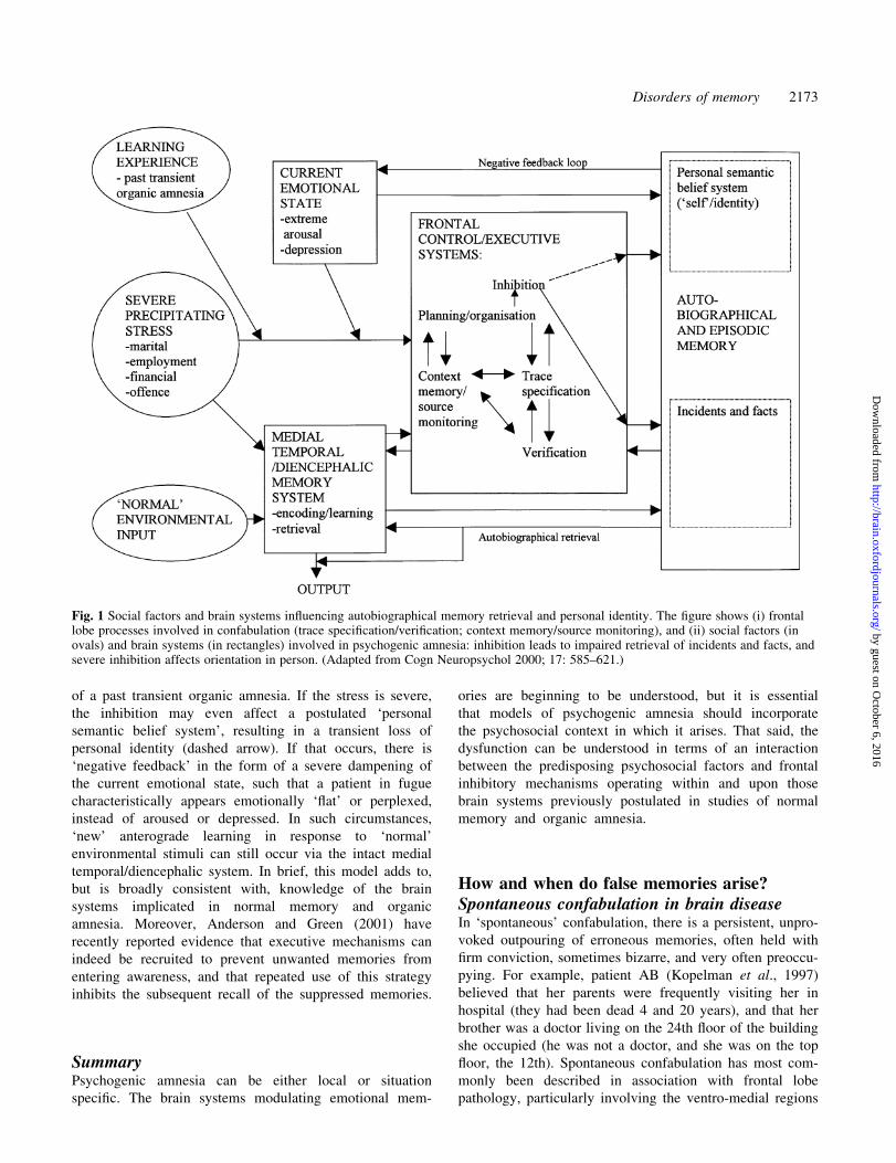
of a past transient organic amnesia. If the stress is severe,
the inhibition may even affect a postulated `personal
semantic belief system', resulting in a transient loss of
personal identity (dashed arrow). If that occurs, there is
`negative feedback' in the form of a severe dampening of
the current emotional state, such that a patient in fugue
characteristically appears emotionally `¯at' or perplexed,
instead of aroused or depressed. In such circumstances,
`new' anterograde learning in response to `normal'
environmental stimuli can still occur via the intact medial
temporal/diencephalic system. In brief, this model adds to,
but is broadly consistent with, knowledge of the brain
systems implicated in normal memory and organic
amnesia. Moreover, Anderson and Green (2001) have
recently reported evidence that executive mechanisms can
indeed be recruited to prevent unwanted memories from
entering awareness, and that repeated use of this strategy
inhibits the subsequent recall of the suppressed memories.
SummaryPsychogenic amnesia can be either local or situation
speci®c. The brain systems modulating emotional mem-
ories are beginning to be understood, but it is essential
that models of psychogenic amnesia should incorporate
the psychosocial context in which it arises. That said, the
dysfunction can be understood in terms of an interaction
between the predisposing psychosocial factors and frontal
inhibitory mechanisms operating within and upon those
brain systems previously postulated in studies of normal
memory and organic amnesia.
How and when do false memories arise?Spontaneous confabulation in brain diseaseIn `spontaneous' confabulation, there is a persistent, unpro-
voked outpouring of erroneous memories, often held with
®rm conviction, sometimes bizarre, and very often preoccu-
pying. For example, patient AB (Kopelman et al., 1997)
believed that her parents were frequently visiting her in
hospital (they had been dead 4 and 20 years), and that her
brother was a doctor living on the 24th ¯oor of the building
she occupied (he was not a doctor, and she was on the top
¯oor, the 12th). Spontaneous confabulation has most com-
monly been described in association with frontal lobe
pathology, particularly involving the ventro-medial regions
Fig. 1 Social factors and brain systems in¯uencing autobiographical memory retrieval and personal identity. The ®gure shows (i) frontallobe processes involved in confabulation (trace speci®cation/veri®cation; context memory/source monitoring), and (ii) social factors (inovals) and brain systems (in rectangles) involved in psychogenic amnesia: inhibition leads to impaired retrieval of incidents and facts, andsevere inhibition affects orientation in person. (Adapted from Cogn Neuropsychol 2000; 17: 585±621.)
Disorders of memory 2173
by guest on October 6, 2016
http://brain.oxfordjournals.org/D
ownloaded from
(Luria, 1976; Stuss et al., 1978; Kapur and Coughlan, 1980;
Shapiro et al., 1981; Baddeley and Wilson, 1986; Moscovitch
and Melo, 1997). However, spontaneous confabulation can
also occur in other, more generalized disorders, such as
confusional states (DeLuca and Cicerone, 1991) or the
generalized (metabolic) effects of carcinoma (Kopelman
et al., 1997). On the other hand, frontal dysfunction seems to
be a necessary, but perhaps not a suf®cient, condition for
spontaneous confabulation (Kopelman et al., 1997; Dalla
Barba et al., 1999): it is a truism that many patients with
frontal pathology do not confabulate, and this does not seem
to be related purely to the precise site of pathology.
A number of explanatory theories for spontaneous
confabulation have been proposed. The ®rst group of theories
emphasizes faulty speci®cation and veri®cation in memory
retrieval. Burgess and Shallice (1996) postulated de®cits in a
descriptor process, an editor process, and a mediator process,
which all contribute differently to the clinical phenomena of
confabulation. The `descriptor' speci®es the type of trace that
would satisfy the demands of a retrieval task, and `noisy'
speci®cation increases the chance of an inappropriate repre-
sentation being produced as a candidate memory. The `editor'
checks that the output from long-term storage ®ts with
previously retrieved memory elements and with overall task
requirements. When this `editor' is impaired, confabulators
respond to a question without giving it adequate consider-
ation, checking, or self-correction. The `mediator' controls
strategic/problem-solving operations concerning the ade-
quacy or plausibility of retrieved memory elements; impair-
ment results in reasoning errors and in bizarre or `fantastic'
responses (see also Burgess and McNeil, 1999). Moscovitch
and Melo (1997) proposed a somewhat similar theory, also
identifying a number of putative de®cits in cue-retrieval,
strategic search or faulty monitoring, the last resulting in
erroneous memories not being edited out or suppressed.
Likewise, Schacter et al. (1998) argued that an insuf®ciently
`focused' retrieval description, or an impairment in post-
retrieval monitoring and veri®cation, may give rise to
confabulation. In addition, these latter authors argued that
encoding impairments may make subjects more liable to
confabulatory errors at retrieval.
The second group of theories emphasizes contextual
memory de®cits, particularly with reference to temporal
sequence and/or source monitoring de®cits. Korsakoff (1889)
himself gave several examples of confusions in temporal
sequence, resulting in confabulation, such as a woman whose
account of past vacations confused journeys to Finland and
the Crimea so that wholly inappropriate descriptions were
given. Other authorities have also emphasized temporal
context confusions as the basis of confabulation (e.g. Moll,
1915; Van der Horst, 1932; Talland, 1965; Victor et al.,
1971). Most recently, Schnider et al. (1996) found that a
small group of spontaneous confabulators were differentiated
from other amnesic patients and healthy controls on the basis
of errors at an `implicit' temporal context memory task, but
not on other memory or executive tasks (see also Schnider
and Peak, 1999). In a variant of this hypothesis, Dalla Barba
and colleagues have proposed that `temporal consciousness'
is intact but malfunctioning in confabulating patients (Dalla
Barba, 1993a, b, 1999; Dalla Barba et al., 1997, 1999). Such
patients are aware of a past, present and future (unlike
severely amnesic patients) but, in making temporal judge-
ments, they employ only the most stable elements from their
long-term memory stores. Asked what they did yesterday or
will do tomorrow, the patients reply with well-established
routines or the habits of a lifetime, however irrelevant to their
present situation. More generally, Johnson and colleagues
have argued that confabulation may result from a wider range
of context, source, or reality monitoring de®cits (Johnson,
1991; Johnson et al., 1993, 2000). Source memory or
monitoring refers to the ability to identify the `source' of
information (Schacter et al., 1984), and reality monitoring to
the ability to discriminate `real' from imagined experience.
A third group of theories emphasizes that multiple de®cits
may contribute to confabulation. Shapiro et al. (1981)
emphasized (i) impaired self-monitoring, (ii) a resulting
failure to inhibit memory errors and (iii) frequent persevera-
tions as the basis of confabulation: each of these de®cits could
be related to a different aspect of frontal/executive function.
In a comparison of a confabulating patient and other patients
with frontal lesions, Johnson et al. (1997) concluded that
confabulation may re¯ect an interaction between (i) a vivid
imagination, (ii) an inability to retrieve autobiographical
memories systematically and (iii) source monitoring de®cits.
Kopelman et al. (1997) found that (i) many confabulations
might plausibly result from the confounding and inappropri-
ate retrieval of `real' memory fragments out of temporal
sequence, but that (ii) other confabulations result from
perseverations, particularly in semantic memory, and (iii)
yet others appear to be instantaneous, ill-considered and
unchecked responses to immediate environmental and social
cues.
In summary, it seems likely that temporal context and
source monitoring de®cits contribute to, but are not the sole
basis of, the impairments of memory speci®cation (descrip-
tion; focused retrieval) and veri®cation (editing; monitoring)
postulated in other theories. It seems plausible that these
various de®cits re¯ect dysfunction in differing components of
frontal control mechanisms and that they interact together
(see Fig. 1 and Kopelman, 1999; Gilboa and Moscovitch,
2002).
Momentary (`provoked') confabulationMuch more commonly, amnesic patients show `momentary'
or `provoked' confabulations. These are ¯eeting intrusion
errors or distortions made in response to a challenge to
memory, such as a memory test (Berlyne, 1972; Kopelman,
1987). In amnesic patients, these intrusion errors are second-
ary to the memory de®cit itself, rather than necessarily
re¯ecting frontal/executive dysfunction, just as, in healthy
subjects, they arise when memory is `weak' for any reason.
2174 M. D. Kopelman
by guest on October 6, 2016
http://brain.oxfordjournals.org/D
ownloaded from
Such errors re¯ect the essentially `reconstructive' nature of
memory retrieval: where the trace is weak (e.g. at long
delays), this reconstruction becomes distorted or frankly
erroneous, but this does not necessarily imply a conscious/
deliberate attempt to `®ll in' memory gaps. The errors
themselves, although ¯eeting, can be quite striking and
surprising. Momentary confabulation in healthy subjects has
been demonstrated in many experimental investigations
(Bartlett, 1932; Hammersley and Read, 1986; Kopelman,
1987; Lindsay and Read, 1994; Read and Lindsay, 1997;
Schacter et al., 1998).
In a neuroimaging study, Schacter et al. (1996d) found that
the left medial temporal (parahippocampal) memory system
was activated during both accurate (`veridicial') and faulty
(`illusory') recognition of word stimuli. Accurate recognition
was associated with signi®cantly increased blood ¯ow in the
left temporo-parietal cortex, whereas faulty recognition was
associated with activation in the prefrontal and orbito-frontal
cortices and the cerebellum, which were postulated as
involved in error detection. The overlap between the brain
systems activated by true and faulty memories may help to
explain the particular salience of false memories in many
circumstances.
False memories in other contextsFalse memories can also arise in many other circumstances
(Kopelman, 1999). Delusional memories are a relatively rare
phenomenon, displaying many of the features of spontaneous
confabulation, except that they tend to revolve around a
single theme and are not necessarily related to frontal/
executive dysfunction (David and Howard, 1994; Kopelman
et al., 1995; Baddeley et al., 1996). Delusional misidenti®ca-
tion syndromes are often associated with the concurrence of
bilateral frontal lobe pathology and right posterior lesions
(Alexander et al., 1979; Ellis and Young, 1990; Young et al.,
1995; Feinberg and Roane, 1997; Box et al., 1999; Feinberg
et al., 1999). Confabulations have also been described in
schizophrenic patients during story recall (Nathaniel-James
and Frith, 1996; Nathaniel-James et al., 1996), although it is
possible that these are simply `momentary' confabulations,
coloured by the preoccupying delusional beliefs of these
patients (Kopelman, 1999). There are also rare psychiatric
syndromes in which patients adopt a false identity or
identities, associated with `memories' in which they have
come to believe (Kopelman, 1999). These include `pseudo-
logia fantastica', sometimes known as `pathological lying'
(Fish, 1967), rare instances of people who adopt a new role
and identity following a typical fugue episode (Kopelman
et al., 1994b; Markowitsch et al., 1997a) and so-called
multiple personality disorder (dissociative identity disorder),
in which the widely varying geographical prevalence almost
certainly re¯ects differences in the reinforcing behaviour of
doctors, psychologists, and the outside world (Merskey, 1992,
1995).
Most controversial are cases of allegedly false memory for
child sexual abuse. The polarized positions of of®cial reports
(Brandon et al., 1998; Morton et al., 1995) illustrate the
unresolved and political aspects of this debate. However,
certain common themes have emerged in the scienti®c
literature on this topic. Even the most forthright protagonists
of false memory have found that 19% of victims of abuse
reported forgetting at some time in the past (Loftus et al.,
1994). Other researchers (Schacter, 1996; Schacter et al.,
1997; Shimamura, 1997) have suggested that memories for
abuse are never actually completely forgotten, but they are
retained in a vague unelaborated form, poorly located in
temporal and spatial context. Such memories would be
vulnerable to the normal processes of decay and interference,
as well as to conscious avoidance and suppression.
Appropriate cueing might result in the re-retrieval of these
memories, but the `weak trace' would make them very
vulnerable to distortion and augmentation. It also seems very
plausible that false memories for sexual abuse are most likely
to arise in particular social contexts (Kopelman, 1999).
A related phenomenon is false confession to an offence, in
which the victim is oneself rather than another. Gudjonsson
et al. (1999) described a man whose conviction of homicide
was quashed after he had served 25 years in prison. At 17
years of age, he had persuaded himself of his `guilt' during
48 h of police interviews, in which he was in an extremely
distraught and agitated state without a doctor or lawyer in
attendance. Shortly before the trial, and just after it had
begun, he was given three interviews under sodium
amylobarbitone, which were subsequently part of the grounds
for appeal. Recently, somewhat similar cases from 1952 and
1983 have had their convictions overturned (R. versus Hay
Gordon, 2000; R. versus Fell, 2001). Gudjonsson and
colleagues have described the circumstances in which people
make such `confessions', which they often come to believe or
`internalize' (Gudjonsson and MacKeith, 1982, 1988, 1990;
Gudjonsson, 1992). In many of these cases, source memory
errors (about where particular information had been learned
about the offence or victim) seem to have arisen in the context
of personal self-doubt (low self-esteem and/or depression)
and/or external coercion by the interrogators. This resulted in
a faulty attribution, held subsequently with varying degrees of
conviction.
SummaryIt seems that various de®cits in frontal control mechanisms,
involving trace speci®cation or veri®cation processes and
context or source memory, may contribute to neurological
confabulation. But false memories can also arise in the
absence of brain disease, innocuously in the case of
momentary confabulation, or more sinisterly in the case of
false memory for sexual abuse or offences (false confes-
sions). In these latter instances also, impairments in context-
ual or source memory have been postulated.
Disorders of memory 2175
by guest on October 6, 2016
http://brain.oxfordjournals.org/D
ownloaded from
ConclusionsThis review has attempted an overview of clinical memory
disorders and selected theoretical controversies that arise
from them. Many of the previously postulated distinctions
between temporal lobe and diencephalic amnesia (e.g. in
forgetting rates) have broken down. There is even substantial
overlap between these forms of amnesia and that arising from
large frontal lobe lesions, although important distinctions
remain. Recent research in organic amnesia has emphasized
impairments in `binding' different types of material in
anterograde memory, including contextual information and
associations, and various distinctions have been drawn to
emphasize the types of memory characteristically affected or
relatively spared in AA: recall/recognition memory, remem-
bering/knowing, recollection/familiarity and explicit/implicit
memory. How these various distinctions map precisely on to
one another remains controversial. One view of RA is that
medial temporal/diencephalic structures are critical to mem-
ory formation, but that damage to these structures gives rise to
a relatively brief (3 years or less) RA. According to this view,
a superimposed retrieval de®cit, related to widespread frontal
and/or temporal neocortical pathology, is required to produce
a temporally extensive RA (greater than 3 years) (e.g.
Kopelman, 1989, 1991). Psychogenic factors could precipi-
tate or exacerbate a retrieval de®cit. However, this time-
limited view of medial temporal lobe function is now
contested by those who postulate a more uniform effect of
medial temporal lesions upon RA and AA (Nadel and
Moscovitch 1997; Fujii et al., 2000; Cipolotti et al., 2001).
In this review, I have tried to emphasize commonalities (as
well as differences) between largely separate literatures
where they exist: for example, between the mechanisms
underlying the neurological and psychogenic forms of
amnesia and of false memory (confabulation). An important
point is that neuropsychological approaches have been
heavily in¯uenced by the success of modular models in
explaining other types of cognitive disorder, such as acquired
dyslexia and prosopagnosia. But this approach has sometimes
led to a somewhat limiting and static perspective. There are
probably only a ®nite number of functional dissociations to be
discovered; they may have a number of alternative explan-
ations; and their neurobiological status often remains unclear
(Caramazza, 1998; Kopelman, 2000a; Tyler and Moss,
2001). Greater emphasis needs to be placed on a more
dynamic approach which examines: (i) the interaction
between different cognitive systems (and the neural networks
or regions which underlie them), as in Snowden's investiga-
tions of the interplay between autobiographical and semantic
memory in semantic dementia (Snowden, 2002); (ii) the
determinants of differential patterns of cognitive change
through time such as the relative rates of recovery of RA and
AA following head injury; and (iii) the interaction of
psychogenic and motivational factors with speci®c brain
systems or pathology, e.g. following head injury, in
psychogenic amnesia, or in the neurological and psycho-
logical forms of false memory (confabulation). In this topic,
there is still great scope for clinicians and researchers to
inform and learn from one another.
AcknowledgementsThe author is grateful for helpful comments from Dr Eli
Jaldow and an anonymous referee; his empirical work has
been supported by a number of grants from the Wellcome
Trust.
References
Abeles M, Schilder P. Psychogenic loss of personal identity. Arch
Neurol Psychiat 1935; 34: 587±604.
Adolphs R, Tranel D, Damasio H, Damasio A. Impaired recognition
of emotion in facial expressions following bilateral damage to the
human amygdala. Nature 1994; 372: 669±72.
Aggleton JP, Brown MW. Episodic memory, amnesia, and the
hippocampal anterior thalamic axis. [Review]. Behav Brain Sci
1999; 22: 425±44.
Aggleton JP, Saunders RC. The relationships between temporal
lobe and diencephalic structures implicated in anterograde amnesia.
[Review]. Memory 1997; 5: 49±71.
Aggleton JP, Shaw C. Amnesia and recognition memory: a re-
analysis of psychometric data. Neuropsychologia 1996; 34: 51±62.
Albert MS, Butters N, Levin J. Temporal gradients in the retrograde
amnesia of patients with alcoholic Korsakoff's disease. Arch Neurol
1979; 36: 211±6.
Albert MS, Butters N, Brandt J. Patterns of remote memory in
amnesic and demented patients. Arch Neurol 1981; 38: 495±500.
Alexander MP, Stuss DT, Benson DF. Capgras syndrome: a
reduplicative phenomenon. Neurology 1979; 29: 334±9.
Alvarez P, Squire LR. Memory consolidation and the medial
temporal lobe: a simple network model. [Review]. Proc Natl Acad
Sci USA 1994; 91: 7041±5.
Anderson MC, Green C. Suppressing unwanted memories by
executive control. Nature 2001; 410: 366±9.
Andrews B, Brewin CR, Ochera J, Morton J, Bekerian DA, Davies
GM, et al. The timing, triggers and qualities of recovered memories
in therapy. Br J Clin Psychol 2000; 39: 11±26.
Andrews E, Poser CM, Kessler M. Retrograde amnesia for forty
years. Cortex 1982; 18: 441±58.
Bachevalier J, Mishkin M. Visual recognition impairment follows
ventromedial but not dorsolateral prefrontal lesions in monkeys.
Behav Brain Res 1986; 20: 249±61.
Baddeley AD. Working memory. Oxford: Clarendon Press; 1986.
Baddeley A. Exploring the central executive. Q J Exp Psychol A
1996; 49: 5±28.
Baddeley AD, Warrington EK. Amnesia and the distinction
between long- and short-term memory. J Verb Learn Verb Behav
1970; 9: 176±89.
2176 M. D. Kopelman
by guest on October 6, 2016
http://brain.oxfordjournals.org/D
ownloaded from
Baddeley A, Wilson B. Amnesia, autobiographical memory, and
confabulation. In: Rubin DC, editor. Autobiographical memory.
Cambridge: Cambridge University Press; 1986. p. 225±52.
Baddeley AD, Harris JE, Sunderland A, Watts K, Wilson B. Closed
head injury and memory. In: Levin H, editor. Neurobehavioural
recovery from head injury. Oxford, Oxford University Press; 1987.
p. 295±317.
Baddeley A, Thornton A, Chua SE, McKenna P. Schizophrenic
delusions and the construction of autobiographical memory. In:
Rubin DC, editor. Remembering our past: Studies in
autobiographical memory. Cambridge: Cambridge University
Press, 1996. p. 384±428.
Baddeley A, Vargha-Khadem F, Mishkin M. Preserved recognition
in a case of developmental amnesia: implications for the acquisition
of semantic memory? J Cogn Neurosci 2001; 13: 357±69.
Baldo JV, Shimamura AP. Frontal lobes and memory. In:
Baddeley AD, Kopelman MD, Wilson BA, editors. Handbook of
memory disorders. 2nd ed. Chichester (UK): John Wiley; In
press 2002 p. 363±80.
Barbarotto R, Laiacona M, Cocchini G. A case of simulated,
psychogenic or focal pure retrograde amnesia: did an entire life
become unconscious? Neuropsychologia 1996; 34: 575±85.
Bartlett FC. Remembering: a study in experimental and social
psychology. Cambridge: Cambridge University Press; 1932.
Barth JT, Diamond A, Errico A. Mild head injury and post
concussion syndrome: does anyone really suffer? Clin
Electroencephalogr 1996; 27: 183±6.
Basso A, Capitani E, Laiacona M. Progressive language impairment
without dementia: a case with isolated category speci®c semantic
defect. J Neurol Neurosurg Psychiatry 1988; 51: 1201±7.
Baxter MG, Murray EA. Opposite relationship of hippocampal and
rhinal cortex damage to delayed nonmatching-to-sample de®cits in
monkeys. Hippocampus 2001a; 11: 61±71.
Baxter MG, Murray EA. Effects of hippocampal lesions on delayed
nonmatching-to-sample in monkeys: a reply to Zola and Squire
(2001). Hippocampus 2001b; 11: 201±3.
Beatty WW, Salmon DP, Butters N, Heindel WC, Granholm EL.
Retrograde amnesia in patients with Alzheimer's disease or
Huntington's disease. Neurobiol Aging 1988; 9: 181±6.
Berlyne N. Confabulation. Br J Psychiatry 1972; 120: 31±9.
Berrington WP, Liddell DW, Foulds GA. A re-evaluation of the
fugue. J Ment Sci 1956; 102: 280±6.
Blumbergs PC, Scott G, Manavis J, Wainwright H, Simpson DA,
McLean AJ. Topography of axonal injury as de®ned by amyloid
precursor protein and sector scoring method in mild and severe
closed head injury. J Neurotrauma 1995; 12: 565±72.
Borgo F, Shallice T. When living things and other `sensory quality'
categories behave in the same fashion: a novel category speci®city
effect. Neurocase 2001; 7: 201±20.
Box O, Laing H, Kopelman MD. The evolution of spontaneous
confabulation, delusional misidenti®cation, and a related delusion
in a case of severe head injury. Neurocase 1999; 5: 251±62.
Bradford JM, Smith SM. Amnesia and homicide: the Padola case
and a study of thirty cases. Bull Am Acad Psychiatry Law 1979; 7:
219±31.
Brandon S, Boakes J, Glaser D, Green R. Recovered memories of
childhood sexual abuse. Implications for clinical practice. [Review.]
Br J Psychiatry 1998; 172: 296±307.
Bremner JD, Scott TM, Delaney RC, Southwick SM, Mason JW,
Johnson DR, et al. De®cits in short-term memory in post-traumatic
stress disorder. Am J Psychiatry 1993; 150: 1015±9.
Bremner JD, Randall P, Scott TM, Bronen RA, Seibyl JP,
Southwick SM, et al. MRI-based measurement of hippocampal
volume in patients with combat-related posttraumatic stress
disorder. Am J Psychiatry 1995; 152: 973±81.
Brewer JB, Zhao Z, Desmond JE, Glover GH, Gabrieli JD. Making
memories: brain activity that predicts how well visual experience
will be remembered. Science 1998; 281: 1185±7.
Brewin CR. A cognitive neuroscience account of posttraumatic
stress disorder and its treatment. [Review]. Behav Res Ther 2001;
39: 373±93.
Brewin CR, Dalgleish T, Joseph S. A dual representation theory of
posttraumatic stress disorder. [Review]. Psychol Rev 1996; 103:
670±86.
Brooks DN. Cognitive de®cits after head injury. In: Brooks N,
editor. Closed head injury: psychological, social and family
consequences. Oxford: Oxford University Press; 1984. p. 44±73.
Brooks DN. The head-injured family. J Clin Exp Neuropsychol
1991; 13: 155±88.
Brown JW, Chobor KL. Severe retrograde amnesia. Aphasiology
1995; 9: 163±70.
Brown D, Sche¯in AW, Whit®eld CL. Recovered memories: the
current weight of the evidence in science and in the courts. J
Psychiatry Law 1999; 27: 5±156.
Burgess PW, McNeill JE. Content-speci®c confabulation. Cortex
1999; 35: 163±82.
Burgess PW, Shallice T. Confabulation and the control of
recollection. [Review]. Memory 1996; 4: 359±411.
Butters N, Cermak LS. Alcoholic Korsakoff's syndrome: an
information-processing approach to amnesia. New York:
Academic Press; 1980.
Butters N, Cermak LS. A case study of the forgetting of
autobiographical knowledge: implications for the study of
retrograde amnesia. In: Rubin DC, editor. Autobiographical
memory. Cambridge: Cambridge University Press; 1986. p. 253±72.
Butters MA, Kaszniak AW, Glisky EL, Eslinger PJ, Schacter DL.
Recency discrimination de®cits in frontal lobe patients.
Neuropsychology 1994; 8: 343±53.
Butterworth RF. Effects of thiamine de®ciency on brain
metabolism: implications for the pathogenesis of the Wernicke-
Korsakoff syndrome. [Review]. Alcohol Alcohol 1989; 24: 271±9.
Butterworth B, Cappelletti M, Kopelman M. Category speci®city in
reading and writing: the case of number words. Nat Neurosci 2001;
4: 784±6.
Disorders of memory 2177
by guest on October 6, 2016
http://brain.oxfordjournals.org/D
ownloaded from
Cahill L, Babinsky R, Markowitsch HJ, McGaugh JL. The
amygdala and emotional memory. Nature 1995; 377: 295±6.
Caine D, Watson JD. Neuropsychological and neuropathological
sequelae of cerebral anoxia: a critical review. J Int Neuropsychol
Soc 2000; 6: 86±99.
Calabrese P, Markowitsch HJ, Durwen HF, Widlitzek H, Haupts M,
Holinka B, et al. Right temporofrontal cortex as critical locus for the
ecphory of old episodic memories. J Neurol Neurosurg Psychiatry
1996; 61: 304±10.
Campodonico JR, Rediess S. Dissociation of implicit and explicit
knowledge in a case of psychogenic retrograde amnesia. J Int
Neuropsychol Soc 1996; 2: 146±58.
Cappelletti M. Butterworth B, Kopelman MD. Spared numerical
abilities in a case of semantic dementia. Neuropsychologia 2001;
39: 1224±39.
Caramazza A. The interpretation of semantic category-speci®c
de®cits: what do they reveal about the organization of conceptual
knowledge in the brain? Neurocase 1998; 4: 265±72.
Carlesimo GA, Sabbadini M, Loasses A, Caltagirone C. Analysis of
the memory impairment in a post-encephalitic patient with focal
retrograde amnesia. Cortex 1998; 34: 449±60.
Cave CB, Squire LR. Equivalent impairment of spatial and
nonspatial memory following damage to the human hippocampus.
Hippocampus 1991; 1: 329±40.
Cermak LS. The episodic-semantic distinction in amnesia. In:
Squire LR, Butters N, editors. The neuropsychology of memory.
New York: Guilford Press; 1984. p. 55±62.
Cermak LS, O'Connor M. The anterograde and retrograde retrieval
ability of a patient with amnesia due to encephalitis.
Neuropsychologia 1983; 21: 213±34.
Chao LL, Haxby JV, Martin A. Attribute-based neural substrates in
temporal cortex for perceiving and knowing about objects. Nat
Neurosci 1999; 2: 913±9.
Chalfonte BL, Verfaellie M, Johnson MK, Reiss L. Spatial location
memory in amnesia: binding item and location information under
incidental and intentional encoding conditions. Memory 1996; 4:
591±614.
Christensen H, Kopelman MD, Stanhope N, Lorentz L, Owen P.
Rates of forgetting in Alzheimer dementia. Neuropsychologia 1998;
36: 547±57.
Christianson S-A. The relationship between induced emotional
arousal and amnesia. Scand J Psychol 1984; 25: 147±60.
Chun MM, Phelps DA. Memory de®cits for implicit contextual
information in amnesic subjects with hippocampal damage. Nat
Neurosci 1999; 2: 844±7.
Cipolotti L, Shallice T, Chun D, Fox N, Scahill R, Harrison G, et al.
Long-term retrograde amnesia . . . the crucial role of the
hippocampus. Neuropsychologia 2001; 39: 151±72.
Clark RE, Squire LR. Classical conditioning and brain systems: the
role of awareness. Science 1998; 280: 77±81.
Clarke NA, Williams AJ, Kopelman MD. Rapid eye movement
sleep behaviour disorder, depression and cognitive impairment:
case study. Br J Psychiatry 2000; 176: 189±92.
Cohen NJ, Squire LR. Retrograde amnesia and remote memory
impairment. Neuropsychologia 1981; 19: 337±56.
Cohen NJ, Poldrack RA, Eichenbaum H. Memory for items and
memory for relations in the procedural/declarative memory
framework. Memory 1997; 5: 131±78.
Colchester A, Kingsley D, Lasserson D, Kendall B, Bello F,
Rush C, et al. Structural MRI volumetric analysis in patients
with organic amnesia. I: methods and comparative ®ndings
across diagnostic groups. J Neurol Neurosurg Psychiatry 2001;
71: 13±22.
Collins AM, Quillian MR. Retrieval time from semantic memory. J
Verb Learn Verb Behav 1969; 8: 240±7.
Conkey RC. Psychological changes associated with head injuries.
Arch Psychol 1938; 232: 1±62.
Conway MA, Turk DJ, Miller SL, Logan J, Nebes RD, Meltzer CC,
et al. A positron emission tomography (PET) study of
autobiographical memory retrieval. Memory 1999; 7: 679±702.
Coriat IH. The Lowell case of amnesia. J Abnorm Psychol 1907; 2:
93±111.
Corkin S. Tactually-guided maze learning in man: effects of
unilateral cortical excisions and bilateral hippocampal lesions.
Neuropsychologia 1965; 3: 339±51.
Corkin S, Amaral DG, Gonzalez RG, Johnson KA, Hyman BT.
H.M.'s medial temporal lobe lesion: ®ndings from magnetic
resonance imaging. J Neurosci 1997; 17: 3964±79.
Costello A, Fletcher PC, Dolan RJ, Frith CD, Shallice T. The
origins of forgetting in a case of isolated retrograde amnesia
following a haemorrhage: evidence from functional imaging.
Neurocase 1998; 4: 437±46.
Crovitz HF, Schiffman H. Frequency of episodic memories as a
function of their age. Bull Psychonom Soc 1974; 4: 517±8.
Cutting J. The relationship between Korsakoff's syndrome and
`alcoholic dementia'. Br J Psychiatry 1978a; 132: 240±51.
Cutting J. A cognitive approach to Korsakoff's syndrome. Cortex
1978b; 14: 485±95.
D'Esposito M, Alexander MP, Fischer R, McGlinchey-Berroth R,
O'Connor M. Recovery of memory and executive function
following anterior communicating artery aneurysm rupture. J Int
Neuropsychol Soc 1996; 2: 565±70.
Dalla Barba G. Confabulation: knowledge and recollective
experience. Cogn Neuropsychol 1993a; 10: 1±20.
Dalla Barba G. Different patterns of confabulation. Cortex 1993b;
29: 567±81.
Dalla Barba G, Cappelletti JY, Signorini M, Denes G.
Confabulation: remembering `another' past, planning `another'
future. Neurocase 1997; 3: 425±36.
Dalla Barba G, Nedjam Z, Dubois B. Confabulation, executive
functions, and source memory in Alzheimer's disease. Cogn
Neuropsychol 1999; 16: 385±98.
2178 M. D. Kopelman
by guest on October 6, 2016
http://brain.oxfordjournals.org/D
ownloaded from
Damasio AR, Graff-Radford NR, Eslinger PJ, Damasio H, Kassell
N. Amnesia following basal forebrain lesions. Arch Neurol 1985;
42: 263±71.
Daum I, Channon S, Canavan AG. Classical conditioning in
patients with severe memory problems. J Neurol Neurosurg
Psychiatry 1989; 52: 47±51.
David AS, Howard R. An experimental phenomenological approach
to delusional memory in schizophrenia and late paraphrenia.
Psychol Med 1994; 24: 515±24.
De Luca J, Cicerone KD. Confabulation following aneurysm of the
anterior communicating artery. Cortex 1991; 27: 417±23.
DeRenzi E, Lucchelli F. Dense retrograde amnesia, intact learning
capability and abnormal forgetting rate: a consolidation de®cit?
Cortex 1993; 29: 449±66.
DeRenzi E, Liotti M, Nichelli P. Semantic amnesia with
preservation of autobiographic memory. Cortex 1987; 23: 575±97.
DeRenzi E, Lucchelli F, Muggia S, Spinnler H. Persistent
retrograde amnesia following a minor trauma. Cortex 1995; 31:
531±42.
DeRenzi E, Lucchelli F, Muggia S, Spinnler H. Is memory loss
without anatomical damage tantamount to a psychogenic de®cit?
The case of pure retrograde amnesia. Neuropsychologia 1997; 35:
781±94.
deWardener HE, Lennox B. Cerebral beriberi (Wernicke's
encephalopathy): review of 52 cases in a Singapore prisoner-of-
war hospital. Lancet 1947; 1: 11±7.
Delbecq-Derouesne J, Beauvois MF, Shallice T. Preserved recall
versus impaired recognition. Brain 1990; 113: 1045±74.
DellaSala S, Laiacona M, Spinnler H, Trivelli C. Autobiographical
recollection and frontal damage. Neuropsychologia 1993; 31: 823±39.
Devlin JT, Russell RP, Davis MH, Price CJ, Moss HE, Fadili MJ, et al.
Is there an anatomical basis for category-speci®city? Semantic
memory studies in PET and fMRI. Neuropsychologia 2002; 40: 54±75.
Dusoir H, Kapur N, Byrnes DP, McKinstry S, Hoare RD. The role
of diencephalic pathology in human memory disorder: evidence
from a penetrating paranasal injury. Brain 1990; 113: 1695±706.
Ellis HD, Young AW. Accounting for delusional misidenti®cations.
[Review]. Br J Psychiatry 1990; 157: 239±48.
Eslinger PJ, Easton A, Grattan LM, Van Hoesen GW. Distinctive
forms of partial retrograde amnesia after asymmetric temporal lobe
lesions. Cereb Cortex 1996; 6: 530±9.
Evans JJ, Wilson B, Wraight EP, Hodges JR. Neuropsychological
and SPECT scan ®ndings during and after transient global amnesia:
evidence for the differential impairment of remote episodic
memory. J Neurol Neurosurg Psychiatry 1993; 56: 1227±30.
Evans JJ, Heggs AJ, Antoun N, Hodges JR. Progressive
prosopagnosia associated with selective right temporal lobe
atrophy: a new syndrome? Brain 1995; 118: 1±13.
Evans JJ, Breen EK, Antoun N, Hodges JR. Focal retrograde
amnesia for autobiographical events following cerebral vasculitis.
Neurocase 1996; 2: 1±11.
Fazio F, Perani D, Gilardi MC, Colombo F, Cappa SF, Vallar
G, et al. Metabolic impairment in human amnesia: a PET study
of memory networks. J Cereb Blood Flow Metab 1992; 12:
353±8.
Feinberg TE, Roane DM. Anosognosia, completion and
confabulation: the neutral-personal dichotomy. Neurocase 1997;
3: 73±85.
Feinberg TE, Eaton LA, Roane DM, Giacino JT. Multiple Fregoli
delusions after traumatic brain injury. Cortex 1999; 35: 373±87.
Fenton GW. Epilepsy and automatism. Br J Hosp Med 1972; 7: 57±64.
Fenwick P. Automatism, medicine and the law. [Review]. Psychol
Med Monogr Suppl 1990; 17: 1±27.
Fenwick P. Brain, mind and behaviour: some medico-legal aspects.
[Review]. Br J Psychiatry 1993; 163: 565±73.
Fine C, Blair RJ. The cognitive and emotional effects of amygdala
damage. Neurocase 2000; 6: 435±50.
Fink GR, Markowitsch HJ, Reinkemeier M, Bruckbauer T, Kessler
J, Heiss W. Cerebral representation of one's own past: neural
networks involved in autobiographical memory. J Neurosci 1996;
16: 4275±82.
Fish FJ. Clinical psychopathology: signs and symptoms in
psychiatry. Bristol: John Wright; 1967.
Fisher CM. Transient global amnesia. Arch Neurol 1982; 39: 605±8.
Fleminger S. Dementia due to head injury. In: Gelder MG, Lopez-Ibor
JJ Jr, Andreason NC, editors. New Oxford textbook of psychiatry, Vol.
1. Oxford: Oxford University Press; 2000. p. 440±51.
Fletcher PC, Henson RN. Frontal lobes and human memory.
Insights from functional neuroimaging. [Review]. Brain 2001; 124:
849±81.
Fletcher PC, Frith CD, Grasby PM, Shallice T, Frackowiak RS, Dolan
RJ. Brain systems for encoding and retrieval of auditory-verbal
memory. An in vivo study in humans. Brain 1995; 118: 401±16.
Freed DM, Corkin S, Cohen NJ. Forgetting in H.M.: a second look.
Neuropsychologia 1987; 25: 461±71.
Freed DM, Corkin S, Growdon JH, Nissen MJ. Selective attention
in Alzheimer's disease: characterizing cognitive subgroups of
patients. Neuropsychologia 1989; 27: 325±39.
Frisk V, Milner B. The role of the left hippocampal region in the
acquisition and retention of story content. Neuropsychologia 1990;
28: 349±59.
Fromholt P, Larsen SF. Autobiographical memory in normal aging
and primary degenerative dementia (dementia of Alzheimer type). J
Gerontol 1991; 46: P85±91.
Fromholt P, Larsen SF. Autobiographical memory and life-history
narratives in aging and dementia (Alzheimer type). In: Conway
MA, Rubin DC, Spinnler H, Wagenaar WA, editors. Theoretical
perspectives on autobiographical memory. Dordrecht: Kluwer
Academic; 1992. p. 413±26.
Fujii K, Sadoshima S, Ishitsuka T, Kusuda K, Kuwabara Y,
Ichiya Y, et al. Regional cerebral blood ¯ow and metabolism in
patients with transient global amnesia: a positron emission
tomography study. J Neurol Neurosurg Psychiatry 1989; 52:
622±30.
Disorders of memory 2179
by guest on October 6, 2016
http://brain.oxfordjournals.org/D
ownloaded from
Fujii T, Moscovitch M, Nadel L. Memory consolidation, retrograde
amnesia, and the temporal lobe. In: Boller F, Grafman J, editors.
Handbook of neuropsychology. 2nd ed. Amsterdam: Elsevier; 2000.
p. 223±50.
Funnell E. Objects and properties: a study of the breakdown of
semantic memory. Memory 1995; 3: 497±518.
Funnell E. Evidence for scripts in semantic dementia: implications
for theories of semantic memory. Cogn Neuropschol 2001; 18: 323±
41.
Funnell E, Sheridan J. Categories of knowledge? Unfamiliar aspects
of living and nonliving things. Cogn Neuropsychol 1992; 9: 135±
53.
Gabrieli JD, Brewer JB, Desmond JE, Glover GH. Separate neural
bases of two fundamental memory processes in the human medial
temporal lobe. Science 1997; 276: 264±6.
Gade A. Amnesia after operations on aneurysms of the anterior
communicating artery. Surg Neurol 1982; 18: 46±9.
Gade A, Mortensen EL. Temporal gradient in the remote memory
impairment of amnesic patients with lesions in the basal forebrain.
Neuropsychologia 1990; 28: 985±1001.
Gainotti G, Almonti S, Di Betta AM, Silveri MC. Retrograde
amnesia in a patient with retrosplenial tumour. Neurocase 1998; 4:
519±26.
Gardiner JM. Episodic memory and autonoetic consciousness: a
®rst-person approach. [Review]. Philos Trans R Soc Lond B Biol
Sci 2001; 356: 1351±61.
Gardiner JM, Java RI. Recollective experience in word and non-
word recognition. Mem Cognit 1990; 18: 23±30.
Gardiner JM, Parkin AJ. Attention and recollective experience in
memory recognition. Mem Cognit 1990; 18: 579±83.
Gardiner JM, Richardson-Klavehn A. Remembering and knowing.
In: Tulving E, Craik FIM, editors. The Oxford handbook of
memory. Oxford: Oxford University Press; 2000. p. 229±44.
Gilboa A, Moscovitch M. The cognitive neuroscience of
confabulation; a review and a model. In: Baddeley AD,
Kopelman MD, Wilson BA, editors. Handbook of memory
disorders. 2nd ed. Chichester (UK): John Wiley. 2002; p. 315±42.
Gillespie RD. Amnesia. Arch Neurol Psychiat 1937; 37: 748±64.
Golby AJ, Poldrack RA, Brewer JB, Spencer D, Desmond JE, Aron
AP, et al. Material-speci®c lateralization in the medial temporal
lobe and prefrontal cortex during memory encoding. Brain 2001;
124: 1841±54.
Goldberg E, Antin SP, Bilder RM Jr, Gerstman LJ, Hughes JE,
Mattis S. Retrograde amnesia: possible role of mesencephalic
reticular activation in long-term memory. Science 1981; 213: 1392±
4.
Goldberg E, Hughes JE, Mattis S, Antin SP. Isolated retrograde
amnesia: different etiologies, same mechanisms? Cortex 1982; 18:
459±62.
Goldenberg G. Transient global amnesia. In: Baddeley AD,
Kopelman MD, Wilson BA, editors. Handbook of memory
disorders. 2nd ed. Chichester (UK): John Wiley. In press 2002.
Gooding PA, Mayes AR, van Eijk R. A meta-analysis of indirect
memory tests for novel material in organic amnesics.
Neuropsychologia 2000; 38: 666±76.
Goodwin DW, Crane JB, Guze SE. Phenomenological aspects of
the alcoholic `blackout'. Br J Psychiatry 1969; 115: 1033±8.
Graf P, Schacter DL. Implicit and explicit memory for new
associations in normal and amnesic subjects. J Exp Psychol Learn
Mem Cogn 1985; 11: 501±18.
Graf P, Squire LR, Mandler G. The information that amnesic
patients do not forget. J Exp Psychol Learn Mem Cogn 1984; 10:
164±78.
Graff-Radford NR, Tranel D, Van Hoesen GW, Brandt JP.
Diencephalic amnesia. Brain 1990; 113: 1±25.
Graham KS, Hodges JR. Differentiating the roles of the
hippocampal complex and the neocortex in long-term memory
storage: evidence from the study of semantic dementia and
Alzheimer's disease. Neuropsychology 1997; 11: 77±89.
Graham KS, Lambon Ralph MA, Hodges JR. Determining the
impact of autobiographical experience on `meaning': new insights
from investigating sports-related vocabulary and knowledge in two
cases with semantic dementia. Cogn Neuropsychol 1997; 14: 801±
37.
Graham KS, Lambon Ralph MA, Hodges JR. A questionable
semantics: the interaction between semantic knowledge and
autobiographical experience in semantic dementia. Cogn
Neuropsychol 1999; 16: 689±98.
Graham KS, Simons JS, Pratt KH, Patterson K, Hodges JR. Insights
from semantic dementia on the relationship between episodic and
semantic memory. Neuropsychologia 2000; 38: 313±24.
Grasby PM, Frith CD, Friston KJ, Bench C, Frackowiak RS, Dolan
RJ. Functional mapping of brain areas implicated in auditory±verbal
memory function. Brain 1993; 116: 1±20.
Green R, Kopelman MD. Contribution of recollection memory and
familiarity judgements to forgetting rates in organic amnesia.
Cortex 2002; 38: 161±78.
Greene JD, Hodges JR. Identi®cation of famous faces and famous
names in early Alzheimer's disease: relationship to anterograde
episodic and general semantic memory. Brain 1996; 119: 111±28.
Greene JD, Hodges JR, Baddeley AD. Autobiographical memory
and executive function in early dementia of Alzheimer type.
Neuropsychologia 1995; 33: 1647±70.
Gudjonsson GH. The psychology of interrogations, confessions and
testimony. Chichester (UK): John Wiley; 1992.
Gudjonsson GH, Mackeith JAC. False confessions. Psychological
effects of interrogation. A discussion paper. In: Trankell A, editor.
Reconstructing the past: the role of psychologists in criminal trials.
Deventer: Kluwer; 1982. p. 253±69.
Gudjonsson GH, MacKeith JA. Retracted confessions: legal,
psychological and psychiatric aspects. [Review]. Med Sci Law
1988; 28: 187±94.
Gudjonsson GH, MacKeith JA. A proven case of false confession:
psychological aspects of the coerced-compliant type. Med Sci Law
1990; 30: 329±35.
2180 M. D. Kopelman
by guest on October 6, 2016
http://brain.oxfordjournals.org/D
ownloaded from
Gudjonsson GH, Taylor PJ. Cognitive de®cit in a case of retrograde
amnesia. Br J Psychiatry 1985; 147: 715±8.
Gudjonsson GH, Kopelman MD, MacKeith JA. Unreliable
admissions to homicide: a case of misdiagnosis of amnesia and
misuse of abreaction technique. Br J Psychiatry 1999; 174: 455±9.
Guillery B, Desgranges B, Piolino P, Laville P, de la Sayette V,
Eustache F. Extensive temporally graded retrograde amnesia for
personal±episodic facts in transient global amnesia. Neurocase
2000; 6: 205±10.
Guinan EM, Lowy C, Stanhope N, Lewis PD, Kopelman MD.
Cognitive effects of pituitary tumours and their treatments: two case
studies and an investigation of 90 patients. J Neurol Neurosurg
Psychiatry 1998; 65: 870±6.
Haist F, Bowden Gore J, Mao H. Consolidation of human memory
over decades revealed by functional magnetic resonance imaging.
Nat Neurosci 2001; 4: 1139±45.
Hamann SB, Squire LR. Intact perceptual memory in the absence of
conscious memory. Behav Neurosci 1997; 111: 850±4.
Hammersley R, Read JD. What is integration? Remembering a
story and remembering false implications about the story. Br J
Psychol 1986; 77: 329±41.
Harding A, Halliday G, Caine D, Kril J. Degeneration of anterior
thalamic nuclei differentiates alcoholics with amnesia. Brain 2000;
123: 141±54.
Harper C. The incidence of Wernicke's encephalopathy in
Australia: a neuropathological study of 131 cases. J Neurol
Neurosurg Psychiatry 1983; 46: 593±8.
Harper C, Kril J, Daly J. Are we drinking our neurones away? Br
Med J 1987; 294: 534±6.
Hart RP, Kwentus JA, Taylor JR, Harkins SW. Rate of forgetting in
dementia and depression. J Consult Clin Psychol 1987; 55: 101±5.
Harvey AG, Brewin CR, Jones C, Kopelman MD. Co-existence of
posttraumatic stress disorder and traumatic brain injury: towards
solving the paradox. J. Int Neuropsychol Soc In press 2002
Harvey P. Fear can interrupt the continuum of memory. J Neurol
Neurosurg Pychiatry 2000; 69: 562.
Heath®eld KW, Croft PB, Swash M. The syndrome of transient
global amnesia. Brain 1973; 96: 729±36.
Hebb DO. The organization of behaviour. New York: Chapman and
Hall; 1949.
Heindel WC, Butters N, Salmon DP. Impaired learning of a motor
skill in patients with Huntington's disease. Behav Neurosci 1988;
102: 141±7.
Henke K, Buck A, Weber B, Wieser HG. Human hippocampus
establishes associations in memory. Hippocampus 1997; 7: 249±
56.
Hierons R, Janota I, Corsellis JA. The late effects of necrotizing
encephalitis of the temporal lobes and limbic areas: a clinico-
pathological study of 10 cases. Psychol Med 1978; 8: 21±42.
Hodges JR, Graham KS. A reversal of the temporal gradient for
famous person knowledge in semantic dementia: implications for
the neural organisation of long-term memory. Neuropsychologia
1998; 36: 803±25.
Hodges JR, McCarthy RA. Autobiographical amnesia resulting
from bilateral paramedian thalamic infarction: a case study in
cognitive neurobiology. Brain 1993; 116: 921±40.
Hodges JR, Oxbury SM. Persistent memory impairment following
transient global amnesia. J Clin Exp Neuropsychol 1990; 12: 904±
20.
Hodges JR, Ward CD. Observations during transient global
amnesia: a behavioural and neuropsychological study of ®ve
cases. Brain 1989; 112: 595±620.
Hodges JR, Warlow CP. The aetiology of transient global amnesia.
[Review]. Brain 1990; 113: 639±57.
Hodges JR, Patterson K, Oxbury S, Funnell E. Semantic dementia;
progressive ¯uent aphasia with temporal lobe atrophy. Brain 1992;
115: 1783±1806.
Hodges JR, Graham N, Patterson K. Charting the progression in
semantic dementia. Memory 1995; 3: 463±95.
Hodges JR, Garrard P, Patterson K. Semantic dementia. In: Kertesz
A, Munoz DG, editors. Pick's disease and Pick complex. New
York: Wiley-Liss; 1998. p. 83±104.
Howard C, D'OrbaÂn PT. Violence in sleep: medico-legal issues and
two case reports. [Review]. Psychol Med 1987; 17: 915±25.
Hunkin NM, Parkin AJ, Longmore BE. Aetiological variation in the
amnesic syndrome: comparisons using the list discrimination task.
Neuropsychologia 1994; 32: 819±25.
Hunkin NM, Parkin AJ, Bradley VA, Burrows EH, Aldrich FK,
Jansari A, et al. Focal retrograde amnesia following closed head
injury: a case study and theoretical account. Neuropsychologia
1995; 33: 509±23.
Hunter IML. Memory. Harmondsworth: Penguin; 1964.
Hunter R, McLuskie R, Wyper D, Patterson J, Christie JE,
Brooks DN, et al. The pattern of function-related regional
cerebral blood ¯ow investigated by single photon emission
tomography with 99mTc-HMPAO in patients with presenile
Alzheimer's disease and Korsakoff's psychosis. Psychol Med
1989; 19: 847±55.
Huppert FA, Piercy M. Recognition memory in amnesic patients:
effect of temporal context and familiarity of material. Cortex 1976;
12: 3±20.
Huppert FA, Piercy M. The role of trace strength in recency and
frequency judgements by amnesic and control subjects. Q J Exp
Psychol 1978; 30: 347±54.
Huppert FA, Piercy M. Normal and abnormal forgetting in organic
amnesia: effect of locus of lesion. Cortex 1979; 15: 385±90.
Isaac CL, Mayes AR. Rate of forgetting in amnesia: 1. Recall and
recognition of prose. [Review.] J Exp Psychol Learn Mem Cogn
1999a; 25: 942±62.
Isaac CL, Mayes AR. Rate of forgetting in amnesia: II. Recall and
recognition of word lists at different levels of organization. J Exp
Psychol Learn Mem Cogn 1999b; 25: 963±77.
Jacobson RR, Lishman WA. Cortical and diencephalic lesions in
Disorders of memory 2181
by guest on October 6, 2016
http://brain.oxfordjournals.org/D
ownloaded from
Korsakoff's syndrome: a clinical and CT scan study. Psychol Med
1990; 20: 63±75.
Jacobson RR, Acker C, Lishman WA. Patterns of
neuropsychological de®cit in alcoholic Korsakoff's syndrome.
Psychol Med 1990; 20: 321±34.
Jacoby LL, Toth JP, Yonelinas AP. Separating conscious and
unconscious in¯uences of memory: measuring recollection. J Exp
Psychol Gen 1993; 122: 139±54.
James W. The principles of psychology, Vol. 1. London:
Macmillan; 1890.
Janowsky JS, Shimamura AP, Kritchevsky M, Squire LR. Cognitive
impairment following frontal lobe damage and its relevance to
human amnesia. Behav Neurosci 1989; 103: 548±60.
Jernigan TL, Ostergaard AL. Word priming and recognition
memory are both affected by mesial temporal lobe damage.
Neuropsychology 1993; 7: 14±26.
Jernigan TL, Schafer K, Butters N, Cermak LS. Magnetic
resonance imaging of alcoholic Korsakoff patients.
Neuropsychopharmacology 1991; 4: 175±86.
Jernigan TL, Ostergaard AL, Fennema-Notestine C. Mesial
temporal, diencephalic, and striatal contributions to de®cits in
single word reading, word priming, and recognition memory. J Int
Neuropsychol Soc 2001; 7: 63±78.
Jetter W, Posner U, Freeman RB Jr, Markowitsch HJ. A verbal long
term memory de®cit in frontal lobe damaged patients. Cortex 1986;
22: 229±42.
Johnson MK. Reality monitoring: evidence from confabulation in
organic brain disease patients. In: Prigatano GP, Schacter DL,
editors. Awareness de®cit after brain injury: clinical and theoretical
issues. New York: Oxford University Press; 1991. p. 176±97.
Johnson MK, Hashtroudi S, Lindsay DS. Source monitoring.
[Review]. Psychol Bull 1993; 114: 3±28.
Johnson MK, O`Connor M, Cantor J. Confabulation, memory
de®cits, and frontal dysfunction. Brain Cogn 1997; 34: 189±206.
Johnson MK, Hayes SM, D'Esposito M, Raye CL. Confabulation.
In: Boller F, Grafman J, editors. Handbook of neuropsychology,
Vol. 2. 2nd ed. Amsterdam: Elsevier; 2000. p. 383±408.
Kanzer M. Amnesia: a statistical study. Am J Psychiatry 1939; 96:
711±6.
Kapur N. Transient epileptic amnesia: a clinically distinct form of
neurological memory disorder. In: Markowitsch HJ, editor.
Transient global amnesia and related disorders. Toronto: Hogrefe
and Huber; 1990. p. 140±51.
Kapur N. Focal retrograde amnesia in neurological disease: a
critical review. [Review]. Cortex 1993; 29: 217±34.
Kapur N. Remembering Norman Schwarzkopf: evidence for two
distinct long-term fact learning mechanisms. Cogn Neuropsychol
1994; 11: 661±70.
Kapur N. The `petites Madeleines' phenomenon in two amnesic
patients. Sudden recovery of forgotten memories [letter]. Brain
1996; 119: 1401±4.
Kapur N. Syndromes of retrograde amnesia: a conceptual and
empirical synthesis. [Review]. Psychol Bull 1999; 125: 800±25.
Kapur N. Focal retrograde amnesia and the attribution of causality:
an exceptionally benign commentary. Cogn Neuropsychol 2000;
17: 623±37.
Kapur N, Coughlan AK. Confabulation and frontal lobe
dysfunction. J Neurol Neurosurg Psychiatry 1980; 43: 461±3.
Kapur N, Heath P, Meudell P, Kennedy P. Amnesia can facilitate
memory performance: evidence from a patient with dissociated
retrograde amnesia. Neuropsychologia 1986; 24: 215±21.
Kapur N, Young A, Bateman D, Kennedy P. Focal retrograde
amnesia: a long term clinical and neuropsychological follow-up.
Cortex 1989; 25: 387±402.
Kapur N, Ellison D, Smith MP, McLellan DL, Burrows EH. Focal
retrograde amnesia following bilateral temporal lobe pathology: a
neuropsychological and magnetic resonance study. Brain 1992;
115: 73±85.
Kapur N, Barker S, Burrows EH, Ellison D, Brice J, Illis LS, et al.
Herpes simplex encephalitis: long-term magnetic resonance
imaging and neuropsychological pro®le. J Neurol Neurosurg
Psychiatry 1994; 57: 1334±42.
Kapur N, Thompson S, Cook P, Lang D, Brice J. Anterograde but
not retrograde memory loss following combined mammillary body
and medial thalamic lesions. Neuropsychologia 1996a; 34: 1±8.
Kapur N, Scholey K, Moore E, Barker S, Brice J, Thompson S, et al.
Long-term retention de®cits in two cases of disproportionate
retrograde amnesia. J Cogn Neurosci 1996b; 8: 416±34.
Kapur N, Thompson P, Kartsounis LD, Abbott P. Retrograde
amnesia: clinical and methodological caveats. Neuropsychologia
1999; 37: 27±30.
Kapur S, Craik FI, Tulving E, Wilson AA, Houle S, Brown GM.
Neuroanatomical correlates of encoding in episodic memory: levels
of processing effect. Proc Natl Acad Sci USA 1994; 91: 2008±11.
Keane MM, Gabrieli JD, Mapstone HC, Johnson KA, Corkin S.
Double dissociation of memory capacities after bilateral occipital-
lobe or medial temporal-lobe lesions. Brain 1995; 118: 1129±48.
Kesler SR, Hopkins RO, Blatter DD, Edge-Booth H, Bigler ED.
Verbal memory de®cits associated with fornix atrophy in carbon
monoxide poisoning. J Int Neuropsychol Soc 2001; 7: 640±6.
Kihlstrom JF, Schacter DL. Functional amnesia. In: Boller F,
Grafman J, editors. Handbook of neuropsychology, Vol. 2. 2nd ed.
Amsterdam: Elsevier; 2000. p. 409±27.
Kinder A, Shanks, DR. Amnesia and the declarative/nondeclarative
distinction: a recurrent network model of classi®cation, recognition,
and repetition priming. J Cogn Neurosci 2001; 13: 648±69.
King N. Mild head injury: neuropathology, sequelae, measurement
and recovery. [Review]. Br J Clin Psychol 1997; 36: 161±84.
Kitchener EG, Hodges JR. Impaired knowledge of famous people
and events with intact autobiographical memory in a case of
progressive right temporal lobe degeneration: implications for the
organisation of remote memory. Cogn Neuropsychol 1999; 16:
589±607.
2182 M. D. Kopelman
by guest on October 6, 2016
http://brain.oxfordjournals.org/D
ownloaded from
Kitchener EG, Hodges JR, McCarthy R. Acquisition of post-morbid
vocabulary and semantic facts in the absence of episodic memory.
Brain 1998; 121: 1313±27.
Knowlton BJ, Squire LR. Remembering and knowing: two different
expressions of declarative memory. J Exp Psychol Learn Mem
Cogn 1995; 21: 699±710.
Knox SJ. Epileptic automatism and violence. Med Sci Law 1968; 8:
96±104.
Kopelman MD. Rates of forgetting in Alzheimer-type dementia and
Korsakoff's syndrome. Neuropsychologia 1985; 23: 623±38.
Kopelman MD. Two types of confabulation. J Neurol Neurosurg
Psychiatry 1987a; 50: 1482±7.
Kopelman MD. Crime and amnesia: a review. Behav Sci Law
1987b; 5: 323±42.
Kopelman MD. Remote and autobiographical memory, temporal
context memory and frontal atrophy in Korsakoff and Alzheimer
patients. Neuropsychologia 1989; 27: 437±60.
Kopelman MD. Frontal dysfunction and memory de®cits in the
alcoholic Korsakoff syndrome and Alzheimer-type dementia. Brain
1991; 114: 117±37.
Kopelman MD. The Korsakoff syndrome. [Review]. Br J Psychiatry
1995a; 166: 154±73.
Kopelman MD. The assessment of psychogenic amnesia. In:
Baddeley AD, Wilson BA, Watts FN, editors. Handbook of
memory disorders. Chichester (UK): John Wiley; 1995b. p. 427±48.
Kopelman MD. Varieties of false memory. Cogn Neuropsychol
1999; 16: 197±214.
Kopelman MD. Focal retrograde amnesia and the attribution of
causality: an exceptionally critical review. Cogn Neuropsychol
2000a; 17: 585±621.
Kopelman MD. Comments on focal retrograde amnesia and the
attribution of causality: an exceptionally benign commentary by
Narinder Kapur. Cogn Neuropsychol 2000b; 17: 639±40.
Kopelman MD. Fear can interrupt the continuum of memory. J
Neurol Neurosurg Psychiatry 2000c; 69: 431±2.
Kopelman MD, Crawford S. Not all memory clinics are dementia
clinics. Neuropsychol Rehabil 1996; 6: 187±202.
Kopelman MD, Stanhope N. Rates of forgetting in organic amnesia
following temporal lobe, diencephalic, or frontal lobe lesions.
Neuropsychology 1997; 11: 343±56.
Kopelman MD, Stanhope N. Recall and recognition memory in
patients with focal frontal, temporal lobe and diencephalic lesions.
Neuropsychologia 1998; 36: 785±95.
Kopelman MD, Stanhope N. Anterograde and retrograde amnesia
following frontal, temporal lobe, or diencephalic lesions. In Squire
LR, Schacter DL, editors. The neuropsychology of memory. 3rd ed.
New York: Guilford Press; 2002. p. 47±60.
Kopelman MD, Wilson BA, Baddeley AD. The Autobiographical
Memory Interview: a new assessment of autobiographical and
personal semantic memory in amnesic patients. J Clin Exp
Neuropsychol 1989; 11: 724±44.
Kopelman MD, Panayiotopoulos CP, Lewis P. Transient epileptic
amnesia differentiated from psychogenic `fugue':
neuropsychological, EEG, and PET ®ndings. J Neurol Neurosurg
Psychiatry 1994a; 57: 1002±4.
Kopelman MD, Christensen H, Puffett A, Stanhope N. The great
escape: a neuropsychological study of psychogenic amnesia.
Neuropsychologia 1994b; 32: 675±91.
Kopelman MD, Guinan EM, Lewis PDR. Delusional memory,
confabulation, and frontal lobe dysfunction: a case study in de
CleÂrambault's syndrome. Neurocase 1995; 1: 71±7.
Kopelman MD, Stanhope N, Kingsley D. Temporal and spatial
context memory in patients with focal frontal, temporal lobe, and
diencephalic lesions. Neuropsychologia 1997; 35: 1533±45.
Kopelman MD, Stanhope N, Guinan E. Subjective memory
evaluations in patients with focal frontal, diencephalic, and
temporal lobe lesions. Cortex 1998a; 34: 191±207.
Kopelman MD, Stevens TG, Foli S, Grasby P. PET activation
of the medial temporal lobe in learning. Brain 1998b; 121: 875±
87.
Kopelman MD, Stanhope N, Kingsley D. Retrograde amnesia in
patients with diencephalic, temporal lobe or frontal lesions.
Neuropsychologia 1999; 37: 939±58.
Kopelman MD, Lasserson D, Kingsley D, Bello F, Rush C,
Stanhope N, et al. Structural MRI volumetric analysis in patients
with organic amnesia, 2: correlations with anterograde memory and
executive tests in 40 patients. J Neurol Neurosurg Psychiatry 2001;
71: 23±8.
Korsakoff SS. Psychic disorder in conjunction with peripheral
neuritis, 1889. Translated and republished by Victor M, Yakovlev
PI. Neurology 1955; 5: 394±406.
Kritchevsky M, Squire LR. Transient global amnesia: evidence for
extensive, temporally graded retrograde amnesia. Neurology 1989;
39: 213±8.
Kritchevsky M, Squire LR, Zouzounis JA. Transient global
amnesia: characterization of anterograde and retrograde amnesia.
Neurology 1988; 38: 213±9.
Lambon Ralph MA, Howard D, Nightingale G, Ellis AW. Are
living and non-living category-speci®c de®cits linked to impaired
perceptual or associative knowledge? Evidence from a category-
speci®c double dissociation. Neurocase 1998; 4: 311±38.
LeDoux JE, Muller J. Emotional memory and psychopathology.
[Review]. Philos Trans R Soc Lond B Biol Sci 1997; 352:
1719±26.
Leng NR, Parkin AJ. Double dissociation of frontal dysfunction in
organic amnesia. Br J Clin Psychol 1988; 27: 359±62.
Lepage M, Habib R, Tulving E. Hippocampal PET activations of
memory encoding and retrieval: the HIPER model. Hippocampus
1998; 8: 313±22.
Levin HS, High WM Jr, Eisenberg HM. Learning and forgetting
during post-traumatic amnesia in head injured patients. J Neurol
Neurosurg Psychiatry 1988a; 51: 14±20.
Levin HS, Goldstein FC, High WM Jr, Eisenberg HM.
Disproportionately severe memory de®cit in relation to normal
Disorders of memory 2183
by guest on October 6, 2016
http://brain.oxfordjournals.org/D
ownloaded from
intellectual functioning after closed head injury. J Neurol Neurosurg
Psychiatry 1988b; 51: 1294±301.
Levine B, Black SE, Cabeza R, Sinden M, Mcintosh AR, Toth JP,
et al. Episodic memory and the self in a case of isolated retrograde
amnesia. Brain 1998; 121: 1951±73.
Lewis P, Kopelman MD. Forgetting rates in neuropsychiatric
disorders. J Neurol Neurosurg Psychiatry 1998; 65: 890±8.
Lindsay DS, Read JD. Psychotherapy and memories of childhood
sexual abuse: a cognitive perspective. Appl Cogn Psychol 1994; 8:
281±338.
Lishman WA. Brain damage in relation to psychiatric disability
after head injury. Br J Psychiatry 1968; 114: 373±410.
Lishman WA. The psychiatric sequelae of head injury: a review.
[Review]. Psychol Med 1973; 3: 304±18.
Lishman WA. Physiogenesis and psychogenesis in the `post-
concussional syndrome'. [Review]. Br J Psychiatry 1988; 153:
460±9.
Lishman WA. Organic psychiatry: the psychological consequences
of cerebral disorder. 3rd ed. Oxford: Blackwell Science; 1998.
Loftus EF, Polonsky S, Fullilove MT. Memories of childhood
sexual abuse: remembering and repressing. Psychol Women Q
1994; 18: 67±84.
Logsdail SJ, Toone BK, Post-ictal psychoses: a clinical and
phenomenological description. Br J Psychiatry 1988; 152: 246±52.
Lucchelli F, Spinnler H. Ephemeral new traces and evaporated
remote engrams: a form of neocortical temporal lobe amnesia? A
preliminary case report. Neurocase 1998; 4: 447±59.
Lucchelli F, Muggia S, Spinnler H. The `Petites Madeleines'
phenomenon in two amnesic patients. Sudden recovery of forgotten
memories. Brain 1995; 118: 167±83.
Lucchelli F, Muggia S, Spinnler H. The syndrome of pure
retrograde amnesia. Cogn Neuropsychiatry 1998; 3: 91±117.
Luria AR. The neuropsychology of memory. New York: John
Wiley; 1976.
Lynch S, Yarnell PR. Retrograde amnesia: delayed forgetting after
concussion. Am J Psychol 1973; 86: 643±5.
Mackenzie Ross S. Profound retrograde amnesia following mild
head injury: organic or functional? Cortex 2000; 36: 521±37.
MacKinnon DF, Squire LR. Autobiographical memory and
amnesia. Psychobiology 1989; 17: 247±56.
Maguire EA. The retrosplenial contribution to human navigation: a
review of lesion and neuroimaging ®ndings. [Review]. Scand J
Psychol 2001; 42: 225±38.
Maguire EA, Mummery CJ. Differential modulation of a common
memory retrieval network revealed by positron emission
tomography. Hippocampus 1999; 9: 54±61.
Maguire EA, Henson RN, Mummery CJ, Frith CD. Activity in
prefrontal cortex, not hippocampus, varies parametrically with the
increasing remoteness of memories. Neuroreport 2001; 12: 441±4.
Mair WG, Warrington EK, Weiskrantz L. Memory disorder in
Korsakoff's psychosis: a neuropathological and neuropsychological
investigation of two cases. Brain 1979; 102: 749±83.
Manes F, Hodges JR, Graham KS, Zeman A. Focal
autobiographical amnesia in association with transient epileptic
amnesia. Brain 2001; 124: 499±509.
Mangels JA, Gershberg FB, Shimamura AP, Knight RT. Impaired
retrieval from remote memory in patients with frontal lobe damage.
Neuropsychology 1996; 10: 32±41.
Manning L. Focal retrograde amnesia documented with matching
anterograde and retrograde procedures. Neuropsychologia 2002; 40:
28±38.
Manns JR, Squire LR. Impaired recognition memory on the Doors
and People Test after damage limited to the hippocampal region.
Hippocampus 1999; 9: 495±9.
Maravita A, Spadoni A, Mazzucchi A, Parma M. A new case of
retrograde amnesia with abnormal forgetting rate. Cortex 1995; 31:
653±67.
Markowitsch HJ. Organic and psychogenic retrograde amnesia: two
sides of the same coin? Neurocase 1996; 2: 357±71.
Markowitsch HJ, Calabrese P, Haupts M, Durwen HF, Liess J,
Gehlen W. Searching for the anatomical basis of retrograde
amnesia. J Clin Exp Neuropsychol 1993; 15: 947±67.
Markowitsch HJ, Fink GR, ThoÈne A, Kessler J, Heiss W-D. A PET
study of persistent psychogenic amnesia covering the whole life
span. Cogn Neuropsychiatry 1997a; 2: 135±58.
Markowitsch HJ, Weber-Luxenburger G, Ewald K, Kessler J, Heiss
W-D. Patients with heart attacks are not valid models for medial
temporal lobe amnesia. A neuropsychological and FDG-PET study
with consequences for memory research. Eur J Neurol 1997b; 4:
178±84.
Markowitsch HJ, Calabrese P, Neufeld H, Gehlen W, Durwen
HF. Retrograde amnesia for world knowledge and preserved
memory for autobiographic events: a case report. Cortex 1999;
35: 243±52.
Marslen-Wilson WD, Teuber HL. Memory for remote events in
anterograde amnesia. Neuropsychologia 1975; 13: 353±64.
Martin A, Haxby JV, Lalonde FM, Wiggs CL, Ungerleider LG.
Discrete cortical regions associated with knowledge of color and
knowledge of action. Science 1995; 270: 102±5.
Martin A, Wiggs CL, Ungerleider LG, Haxby JV. Neural correlates
of category-speci®c knowledge. Nature 1996; 379: 649±52.
Martin PR, Rio D, Adinoff B, Johnson JL, Bisserbe JC, Rawlings
RR, et al. Regional cerebral glucose utilization in chronic organic
mental disorders associated with alcoholism. J Neuropsychiatry
Clin Neurosci 1992; 4: 159±67.
Mattioli F, Grassi F, Perani D, Cappa SF, Miozzo A, Fazio F.
Persistent post-traumatic retrograde amnesia: a neuropsychological
and (18) FDG PET study. Cortex 1996; 32: 121±9.
Mayes AR, Downes JJ. What do theories of the functional de®cit(s)
underlying amnesia have to explain? [Review]. Memory 1997; 5:
3±36.
Mayes AR, Meudell PR. How similar is immediate memory in
2184 M. D. Kopelman
by guest on October 6, 2016
http://brain.oxfordjournals.org/D
ownloaded from
amnesic patients to delayed memory in normal subjects? A
replication, extension and reassessment of the amnesic cueing
effect. Neuropsychologia 1981; 19: 647±54.
Mayes AR, Meudell PR, Pickering A. Is organic amnesia caused by
a selective de®cit in remembering contextual information?
[Review]. Cortex 1985; 21: 167±202.
Mayes AR, Pickering A, Fairbairn A. Amnesic sensitivity to
proactive interference: its relationship to priming and the causes of
amnesia. Neuropsychologia 1987; 25: 211±20.
Mayes AR, Meudell PR, Mann D, Pickering A. Location of lesions
in Korsakoff's syndrome: neuropsychological and neuropatho-
logical data on two patients. Cortex 1988; 24: 367±88.
Mayes AR, Downes JJ, McDonald C, Poole V, Rooke S, Sagar HJ,
et al. Two tests for assessing remote public knowledge: a tool for
assessing retrograde amnesia. Memory 1994; 2: 183±210.
Mayes AR, Gooding PA, Hunkin NM, Nunn JA, Gregory LJ,
Brammer MJ, et al. Storage of verbal associations is suf®cient to
activate the left medial temporal lobe. Behav Neurol 1998; 11: 163±
72.
Mayes AR, Holdstock JS, Isaac CL, Hunkin NM, Roberts N.
Relative sparing of item recognition memory in a patient with adult-
onset damage limited to the hippocampus. Hippocampus. In press
2002.
Mayes AR, Isaac CL, Holdstock JS, Hunkin NM, Montaldi D,
Downes JJ, et al. Memory for single items, word pairs, and temporal
order of different kinds in a patient with selective hippocampal
lesions. Cogn Neuropsychol 2001; 18: 97±123.
McClelland JL, McNaughton BL, O'Reilly RC. Why there are
complementary learning systems in the hippocampus and
neocortex: insights from the successes and failures of
connectionist models of learning and memory. [Review]. Psychol
Rev 1995; 102: 419±57.
McCool BA, Plonk SG, Martin PR, Singleton CK. Cloning of
human transketolase cDNAs and comparison of the nucleotide
sequence of the coding region in Wernicke-Korsakoff and non-
Wernicke-Korsakoff individuals. J Biol Chem 1993; 268: 1397±
404.
McDowall J. Effects of encoding instructions on recall and
recognition in Korsakoff patients. Neuropsychologia 1981; 19:
43±8.
McKee RD, Squire LR. Equivalent forgetting rates in long-term
memory for diencephalic and medial temporal lobe amnesia. J
Neurosci 1992; 12: 3765±72.
McMillan TM. Post-traumatic stress disorder following minor and
severe closed head injury: 10 single cases. Brain Inj 1996; 10: 749±
58.
McNeil JE, Greenwood R. Can PTSD occur with amnesia for the
precipitating event? Cogn Neuropsychiatry 1996; 1: 239±46.
Merskey H. The manufacture of personalities. The production of
multiple personality disorder. [Review]. Br J Psychiatry 1992; 160:
327±40.
Merskey H. Multiple personality disorder and false memory
syndrome. Br J Psychiatry 1995; 166: 281±83.
Merskey H, Woodforde JM. Psychiatric sequelae of minor head
injury. Brain 1972; 92: 521±8.
Meudell PR, Mayes AR. Normal and abnormal forgetting: some
comments on the human amnesic syndrome. In: Ellis AW, editor.
Normality and pathology in cognitive functions. London: Academic
Press; 1982. p. 203±38.
Meudell PA, Mayes AR, Neary D. Is amnesia caused by a
consolidation impairment? In: Oborne DJ, Gruneberg MM, Eiser
JR, editors. Research in psychology and medicine, Vol. 1. London:
Academic Press; 1979. p. 323±30.
Miller E. The long-term consequences of head injury: a discussion
of the evidence with special reference to the preparation of legal
reports. Br J Soc Clin Psychol 1979; 18: 87±98.
Miller JW, Petersen RC, Metter EJ, Millikan CH, Yanagihara T.
Transient global amnesia: clinical characteristics and prognosis.
Neurology 1987; 37: 733±7.
Milner B. Amnesia following operation on the temporal lobes. In:
Whitty CWM, Zangwill OL, editors. Amnesia. 1st ed. London:
Butterworths; 1966. p. 109±33.
Milner B. Some cognitive effects of frontal-lobe lesions in man.
Philos Trans R Soc Lond B Biol Sci 1982; 298: 211±26.
Milner B, Corsi P, Leonard G. Frontal-lobe contribution to recency
judgements. Neuropsychologia 1991; 29: 601±18.
Moll JM. The amnesic or Korsakoff's syndrome with alcoholic
aetiology: an analysis of 30 cases. J Ment Sci 1915; 61: 423±37.
Montaldi D, Mayes AR, Barnes A, Pirie H, Hadley DM, Patterson J,
et al. Associative encoding of pictures activates the medial temporal
lobes. Hum Brain Mapp 1998; 6: 85±104.
Morton J, Hammersley RH, Bekerian DA. Headed records: a model
for memory and its failures. Cognition 1985; 20: 1±23.
Morton J, Andrews B, Bekerian D, Brewin CR, Davies GM, Mollon
P. Recovered memories. Leicester: British Psychologial Society;
1995.
Moscovitch M. Multiple dissociations of function in amnesia. In:
Cermak LS, editor. Human memory and amnesia. Hillsdale (NJ):
Lawrence Erlbaum; 1982. p. 337±70.
Moscovitch M, Melo B. Strategic retrieval and the frontal lobes:
evidence from confabulation and amnesia. Neuropsychologia 1997;
35: 1017±34.
Moscovitch M, Nadel L. Consolidation and the hippocampal
complex revisited: in defense of the multiple-trace model.
[Review]. Curr Opin Neurobiol 1998; 8: 297±300.
Moscovitch M, Nadel L. Multiple-trace theory and semantic
dementia: response to K.S. Graham (1999). Trends Cogn Sci
1999; 3: 87±9.
Moss HE, Tyler LK. A progressive category-speci®c semantic
de®cit for non-living things. Neuropsychologia 2000; 38: 60±82.
Moss HE, Tyler LK, Durrant-Peat®eld M, Bunn EM. `Two eyes of a
see-through': impaired and intact semantic knowledge in a case of
selective de®cit for living things. Neurocase 1998; 4: 291±310.
Moss HE, Kopelman MD, Cappelletti M, de Mornay Davies P,
Disorders of memory 2185
by guest on October 6, 2016
http://brain.oxfordjournals.org/D
ownloaded from
Jadlow E. Lost for words or loss of memory? Autobiographical
memory in semantic dementia. Cogn Neuropsychol 2002. In press.
Mummery CJ, Patterson K, Wise RJ, Vandenbergh R, Price CJ,
Hodges JR. Disrupted temporal lobe connections in semantic
dementia. Brain 1999; 122: 61±73.
Murray EA, Gaffan D, Mishkin M. Neural substrates of visual
stimulus-stimulus association in rhesus monkeys. J Neurosci 1993;
13: 4549±61.
Murre JM. Implicit and explicit memory in amnesia: some
explanations and predictions by the TraceLink model. Memory
1997; 5: 213±32.
Murre JM, Graham KS, Hodges JR. Semantic dementia: relevance
to connectionist models of long-term memory. [Review]. Brain
2001; 124: 647±75.
Nadel L, Moscovitch M. Memory consolidation, retrograde amnesia
and the hippocampal complex. [Review]. Curr Opin Neurobiol
1997; 7: 217±27.
Nathaniel-James DA, Frith CD. Confabulation in schizophrenia:
evidence of a new form? Psychol Med 1996; 26: 391±9.
Nathaniel-James DA, Foong J, Frith CD. The mechanisms of
confabulation in schizophrenia. Neurocase 1996; 2: 475±83.
Neary D, Snowden JS, Bowen DM, Sims NR, Mann DM, Benton JS,
et al. Neuropsychological syndromes in presenile dementia due to
cerebral atrophy. J Neurol Neurosurg Psychiatry 1986; 49: 163±74.
Newcombe F. The psychological consequences of closed head
injury: assessment and rehabilitation. [Review]. Injury 1982; 14:
111±36.
O'Brien M, Nutt D. Loss of consciousness and post-traumatic stress
disorder. Br J Psychiatry 1998; 173: 102±4.
O'Connor M, Vefaillie M. The amnesic syndrome: overview and
subtypes. In Baddeley AD, Kopelman MD, Wilson BA, editors.
Handbook of Memory Borders, 2nd ed. Chichester, UK: John Wiley
& Co 2002. p. 145±166.
O'Connor M, Butters N, Miliotis P, Eslinger P, Cermak LS. The
dissociation of anterograde and retrograde amnesia in a patient with
herpes encephalitis. J Clin Exp Neuropsychol 1992; 14: 159±78.
O'Connor M, Sieggreen MA, Ahern G, Schomer D, Mesulam
M. Accelerated forgetting in association with temporal lobe
epilepsy and paraneoplastic encephalitis. Brain Cogn 1997; 35:
71±84.
Ogden JA. Visual object agnosia, prosopagnosia, achromatopsia,
loss of visual imagery, and autobiographical amnesia following
recovery from cortical blindness: case M.H. Neuropsychologia
1993; 31: 571±89.
Oppenheimer DR. Microscopic lesions in the brain following head
injury. J Neurol Neurosurg Psychiatry 1968; 31: 299±306.
Ostergaard AL. Dissociations between word priming effects in
normal subjects and patients with memory disorders: multiple
memory systems or retrieval? Q J Exp Psychol A 1994; 47: 331±64.
Ostergaard AL. The effects on priming of word frequency, number
of repetitions, and delay depend on the magnitude of priming. Mem
Cognit 1998; 26: 40±60.
Ostergaard AL. Priming de®cits in amnesia: now you see them, now
you don't. J Int Neuropsychol Soc 1999; 5: 175±90.
Owen AM, Downes JJ, Sahakian BJ, Polkey CE, Robbins TW.
Planning and spatial working memory following frontal lobe lesions
in man. Neuropsychologia 1990; 28: 1021±34.
Paller KA, Acharya A, Richardson BC, Plaisant O, Shimamura AP,
Reed BR, et al. Functional neuroimaging of cortical dysfunction in
alcoholic Korsakoff's syndrome. J Cogn Neurosci 1997; 9: 277±93.
Palmini AL, Gloor P, Jones-Gotman M. Pure amnestic seizures in
temporal lobe epilepsy. Brain 1992; 115: 749±69.
Papagno C. Transient retrograde amnesia associated with impaired
naming of living categories. Cortex 1998; 34: 111±21.
Par®tt DN, Gall CMC. Psychogenic amnesia: the refusal to
remember. J Ment Sci 1944; 90: 511±27.
Parker A, Gaffan D. Interaction of frontal and perirhinal cortices in
visual object recognition memory in monkeys. Eur J Neurosci 1998;
10: 3044±57.
Parkin AJ. Recent advances in the neuropsychology of memory. In:
Weinman J, Hunter J, editors. Memory: neurochemical and
abnormal perspectives. London: Harwood Academic; 1991. p.
141±62.
Parkin AJ, Hunkin NM. Impaired temporal context memory on
anterograde but not retrograde tests in the absence of frontal
pathology. Cortex 1993; 29: 267±80.
Parkin AJ, Leng NRC. Comparative studies of human amnesia:
syndrome or syndromes? In: Markowitsch HJ, editor. Information
processing by the brain. Toronto: Hans Huber; 1988. p. 107±23.
Parkin AJ, Leng NR, Hunkin NM. Differential sensitivity to context in
diencephalic and temporal lobe amnesia. Cortex 1990a; 26: 373±80.
Parkin AJ, Montaldi D, Leng NR, Hunkin NM. Contextual cueing
effects in the remote memory of alcoholic Korsakoff patients and
normal subjects. Q J Exp Psychol A 1990b; 42: 585±96.
Parkin AJ, Bindschaedler C, Harsent L, Metzler C. Pathological
false alarm rates following damage to the left frontal cortex. Brain
Cogn 1996; 32: 14±27.
Patterson K, Hodges JR. Disorders of semantic memory. In
Baddeley AD, Wilson BA, Watts FN, editors. Handbook of
memory disorders. Chichester (UK): John Wiley; 1995. p. 167±86.
Patterson K, Hodges JR. Semantic dementia: one window on the
structure and organisation of semantic memory. In: Boller F,
Grafman J, editors. Handbook of neuropsychology, Vol. 2. 2nd ed.
Amsterdam: Elsevier; 2000. p. 313±34.
Peto TEA, Jeul-Jensen BE. Herpes simplex virus infections. In:
Weatherall DJ, Ledingham JGG, Warrell DA, editors. Oxford
textbook of medicine, Vol. 1. 3rd ed. Oxford: Oxford University
Press; 1996. p. 341±6.
Petrides M. Frontal lobes and memory. In: Boller F, Grafman J,
editors. Handbook of neuropsychology, Vol. 2. 2nd ed. Amsterdam:
Elsevier; 2000. p. 67±84.
Petrides M, Milner B. De®cits on subject-ordered tasks after frontal-
and temporal-lobe lesions in man. Neuropsychologia 1982; 20:
249±62.
2186 M. D. Kopelman
by guest on October 6, 2016
http://brain.oxfordjournals.org/D
ownloaded from
Phillips S, Sangalang V, Sterns G. Basal forebrain infarction. A
clinicopathologic correlation. Arch Neurol 1987; 44: 1134±8.
Pickering AD, Mayes AR, Fairbairn AF. Amnesia and memory for
modality information. Neuropsychologia 1989; 27: 1249±59.
Povlishock JT, Christman CW. The pathobiology of traumatically
induced axonal injury in animals and humans: a review of current
thoughts. [Review]. J Neurotrauma 1995; 12: 555±64.
Press GA, Amaral DG, Squire LR. Hippocampal abnormalities in
amnesic patients revealed by high-resolution magnetic resonance
imaging. Nature 1989; 341: 54±7.
Pyzsora NM, Barker AF, Kopelman MD. Amnesia for criminal
offences: a study of life sentence prisoners. J Forensic Psychiatry. In
press 2002.
Rajaram S. Remembering and knowing: two means of access to the
personal past. Mem Cognit 1993; 21: 89±102.
Read JD, Lindsay DS, editors. Recollections of trauma: scienti®c
research and clinical practice. New York: Plenum Press; 1997.
Reber PJ, Squire LR. Intact learning of arti®cial grammars and
intact category learning by patients with Parkinson's disease. Behav
Neurosci 1999; 113: 235±42.
Reed JM, Squire LR. Impaired recognition memory in patients with
lesions limited to the hippocampal formation. Behav Neurosci
1997; 111: 667±75.
Reed JM, Squire LR. Retrograde amnesia for facts and events:
®ndings from four new cases. J Neurosci 1998; 18: 3943±54.
Reed LJ, Marsden P, Lasserson D, Sheldon N, Lewis P, Stanhope
N, et al. FDG-PET analysis and ®ndings in amnesia resulting from
hypoxia. Memory 1999; 7: 599±612.
Reed LJ, Lasserson D, Marsden P, Stanhope N, Stevens TG, Bello
F, et al. FDG-PET ®ndings in Wernicke-Korsakoff syndrome.
Cortex 2002. In press.
Ribot T. Diseases of memory. New York: Appleton; 1882.
Richardson JT. Performance in free recall following rupture and
repair of intracranial aneurysm. Brain Cogn 1989; 9: 210±26.
Richardson JT. Cognitive performance following rupture and repair
of intracranial aneurysm. Acta Neurol Scand 1991; 83: 110±22.
Roman-Campos G, Poser CM, Wood FB. Persistent retrograde
memory de®cit after transient global amnesia. Cortex 1980; 16:
509±18.
Rossor MN, Revesz T, Lantos PL, Warrington EK. Semantic
dementia with ubiquitin-positive tau-negative inclusion bodies.
Brain 2000; 123: 267±76.
Rousseaux M, Delafosse A, Cabaret M, Lesoin F, Jomin M.
Amnesic retrograde post-traumatique. Cortex 1984; 20: 575±83.
Rubin DC, Greenberg DL. Visual memory-de®cit amnesia: a
distinct amnesic presentation and etiology. Proc Natl Acad Sci USA
1998; 95: 5413±6.
Rugg MD. Functional neuroimaging of memory. In: Baddeley AD,
Kopelman MD, Wilson BA, editors. Handbook of memory
disorders. 2nd ed. Chichester (UK): John Wiley. 2002. p. 57±80.
Rugg MD, Wilding EL. Retrieval processing and episodic memory.
Trends Cogn Sci 2000; 4: 108±15.
Russell WR, Nathan PW. Traumatic amnesia. Brain 1946; 69: 280±
300.
Russell WR. Smith A. Post-traumatic amnesia in closed head injury.
Arch Neurol 1961; 5: 4±17.
Ryan L, Nadel L, Keil K, Putnam K, Schnyer D, Trouard T, et al.
Hippocampal complex and retrieval of recent and very remote
autobiographical memories: evidence from functional magnetic
resonance imaging in neurologically intact people. Hippocampus
2001; 11: 707±14.
Sagar HJ, Cohen NJ, Sullivan EV, Corkin S, Growdon JH. Remote
memory function in Alzheimer's disease and Parkinson's disease.
Brain 1988; 111: 185±206.
Saint-Cyr JA, Taylor AE, Lang AE. Procedural learning and
neostriatal dysfunction in man. Brain 1988; 111: 941±59.
Sargant W, Slater E. Amnesic syndromes in war. Proc Soc Med
1941; 34: 757±64.
Sanders HI, Warrington EK. Memory for remote events in amnesic
patients. Brain 1971; 94: 661±8.
Schacter DL. Implicit memory: history and current status. J Exp
Psychol Learn Mem Cogn 1987; 13: 501±18.
Schacter DL. Searching for memory: the brain, the mind, and the
past. New York: Basic Books; 1996.
Schacter DL, Wagner AD. Medial temporal lobe activations in
fMRI and PET studies of episodic encoding and retrieval. [Review].
Hippocampus 1999; 9: 7±24.
Schacter DL, Wang PL, Tulving E, Freeman M. Functional
retrograde amnesia: a quantitative case study. Neuropsychologia
1982; 20: 523±32.
Schacter DL, Harbluk JL, McLachlan DR. Retrieval without
recollection: an experimental analysis of source amnesia. J Verb
Learn Verb Behav 1984; 23: 593±611.
Schacter DL, Verfaellie M, Pradere D. The neuropsychology of
memory illusions: false recall and recognition in amnesic patients. J
Mem Lang 1996a; 35: 319±34.
Schacter DL, Curran T, Galluccio L, Milberg M, Bates J. False
recognition and the right frontal lobe: a case study.
Neuropsychologia 1996b; 34: 793±808.
Schacter DL, Alpert NM, Savage CR, Rauch SL, Albert MS.
Conscious recollection and the human hippocampal formation. Proc
Natl Acad Sci USA 1996c; 93: 321±5.
Schacter DL, Reiman E, Curran T, Yun LS, Bandy D, McDermott
KB, et al. Neuroanatomical correlates of veridical and illusory
recognition memory: evidence from positron emission tomography.
Neuron 1996d; 17: 267±74.
Schacter DL, Norman KA, Koutstaal W. The recovered memories
debate: a cognitive neuroscience perspective. In Conway MA,
editor. Recovered memories and false memories. Oxford: Oxford
University Press; 1997. p. 63±99.
Schacter DL, Norman KA, Koutstaal W. The cognitive
Disorders of memory 2187
by guest on October 6, 2016
http://brain.oxfordjournals.org/D
ownloaded from
neuroscience of constructive memory. [Review]. Annu Rev Psychol
1998; 49: 289±318.
Schacter DL, Badgaiyan RD, Alpert NM. Visual word stem
completion priming within and across modalities: a PET study.
Neuroreport 1999; 10: 2061±5.
Schenck CH, Bundlie SR, Ettinger MG, Mahowald MW. Chronic
behavioral disorders of human REM sleep: a new category of
parasomnia. Sleep 1986; 9: 293±308.
Schmidtke K, Vollmer H. Retrograde amnesia: a study of its
relation to anterograde amnesia and semantic memory de®cits.
Neuropsychologia 1997; 35: 505±18.
Schnider A, Ptak R. Spontaneous confabulators fail to suppress
currently irrelevant memory traces. Nat Neurosci 1999; 2: 677±81.
Schnider A, von DaÈniken C, Gutbrod K. The mechanisms of
spontaneous and provoked confabulations. Brain 1996; 119: 1365±
75.
Schooler JW, Bendiksen M, Ambadar Z. Taking the middle line:
can we accommodate both fabricated and recovered memories of
sexual abuse? In: Conway MA, editor. Recovered memories and
false memories. Oxford: Oxford University Press; 1997. p. 251±92.
Scoville WB, Milner B. Loss of recent memory after bilateral
hippocampal lesions. J Neurol Neurosurg Psychiatry 1957; 20: 11±
21.
Shallice T, Warrington EK. Independent functioning of verbal
memory stores: a neuropsychological study. Q J Exp Psychol 1970;
22: 261±73.
Shallice T, Fletcher P, Frith CD, Grasby P, Frackowiak RS, Dolan
RJ. Brain regions associated with acquisition and retrieval of verbal
episodic memory. Nature 1994; 368: 633±5.
Shapiro B, Alexander MP, Gardner H, Mercer B. Mechanisms of
confabulation. Neurology 1981; 31: 1070±6.
Shimamura AP. Priming effects in amnesia: evidence for a
dissociable memory function. [Review]. Q J Exp Psychol A 1986;
38: 619±44.
Shimamura AP. Memory and frontal lobe function. In: Gazzaniga
MS, editor. The cognitive neurosciences. Cambridge (MA): MIT
Press; 1994 p. 803±813.
Shimamura AP. Recollection: perspectives on reinstated memory
and child trauma. In: Read JD, Lindsay DS, editors. Recollections
of trauma: scienti®c research and clinical practice. New York:
Plenum Press; 1997. p. 253±66.
Shimamura AP, Squire LR. Paired-associate learning and priming
effects in amnesia: a neuropsychological study. J Exp Psychol: Gen
1984; 113: 556±70.
Shimamura AP, Jernigan TL, Squire LR. Korsakoff's syndrome:
radiological (CT) ®ndings and neuropsychological correlates.
J Neurosci 1988; 8: 4400±10.
Shimamura AP, Janowsky JS, Squire LR. Memory for the temporal
order of events in patients with frontal lobe lesions and amnesic
patients. Neuropsychologia 1990; 28: 803±13.
Shimamura AP, Janowsky JS, Squire LR. What is the role of frontal
lobe damage in memory disorders? In: Levin HS, Eisenberg HM,
Benton AL, editors. Frontal lobe function and dysfunction. New
York: Oxford University Press; 1991. p. 173±95.
Shoqeirat MA, Mayes AR. Disproportionate incidental spatial
memory and recall de®cits in amnesia. Neuropsychologia 1991; 29:
749±69.
Shoqeirat MA, Mayes A, MacDonald C, Meudell P, Pickering A.
Performance on tests sensitive to frontal lesions by patients with
organic amnesia: Leng and Parkin revisited. Br J Clin Psychol 1990;
29: 401±8.
Simons JS, Hodges JR. Transient global amnesia. Neurocase 2000;
6: 211±30.
Snowden JS. Disorders of semantic memory. In: Baddeley AD,
Kopelman MD, Wilson BA, editors. Handbook of memory
disorders. 2nd ed. Chichester (UK): John Wiley. 2002. p. 293±314
Snowden JS, Goulding PJ, Neary D. Semantic dementia: a form of
circumscribed cerebral atrophy. Behav Neurol 1989; 2: 167±82.
Snowden J, Grif®ths H, Neary D. Semantic dementia:
autobiographical contribution to preservation of meaning. Cogn
Neuropsychol 1994; 11: 265±88.
Snowden JS, Grif®ths HL, Neary D. Autobiographical experience
and word meaning. Memory 1995; 3: 225±46.
Snowden JS, Grif®ths HL, Neary D. Semantic±episodic memory
interactions in semantic dementia: implications for retrograde
memory function. Cogn Neuropsychol 1996a; 13: 1101±37.
Snowden JS, Neary D, Mann DMA. Fronto-temporal lobar
degeneration: fronto-temporal dementia, progressive aphasia,
semantic dementia. New York: Churchill Livingstone; 1996b.
Snowden JS, Grif®ths HL, Neary D. The impact of autobiographical
experience on meaning: reply to Graham, Lambon Ralph, and
Hodges. Cogn Neuropsychol 1999; 16: 673±87.
Spinnler H, Lucchelli F, Muggia S. The `Petites Madeleines'
phenomenon in two amnesic patients. Sudden recovery of forgotten
memories [letter]. Brain 1996; 119: 1403±4.
Squire LR. Two forms of human amnesia: an analysis of forgetting.
J Neurosci 1981; 1: 635±40.
Squire LR. Comparisons between forms of amnesia: some de®cits
are unique to Korsakoff's syndrome. J Exp Psychol Learn Mem
Cogn 1982; 8: 560±71.
Squire LR, Alvarez P. Retrograde amnesia and memory
consolidation: a neurobiological perspective. [Review]. Curr Opin
Neurobiol 1995; 5: 169±77.
Squire LR, Cohen NJ, Nadel L. The medial temporal region and
memory consolidation: a new hypothesis. In: Weingartner H, Parker
ES, editors. Memory consolidation. Hillsdale (NJ): Lawrence
Erlbaum; 1984. p. 185±210.
Squire LR, Haist F, Shimamura AP. The neurology of memory:
quantitative assessment of retrograde amnesia in two groups of
amnesic patients. J Neurosci 1989; 9: 828±39.
Stanhope N, Kopelman MD. Art and memory: a 7-year follow-up of
herpes encephalitis in a professional artist. Neurocase 2000; 6: 99±
110.
Stanhope N, Guinan E, Kopelman MD. Frequency judgements of
2188 M. D. Kopelman
by guest on October 6, 2016
http://brain.oxfordjournals.org/D
ownloaded from
abstract designs by patients with diencephalic, temporal lobe or
frontal lobe lesions. Neuropsychologia 1998; 36: 1387±96.
Starkstein SE, Sabe L, Dorrego MF. Severe retrograde amnesia
after a mild closed head injury. Neurocase 1997; 3: 105±9.
Stengel E. On the aetiology of the fugue state. J Ment Sci 1941; 87:
572±99.
Stewart F, Parkin AJ, Hunkin NM. Naming impairments following
recovery from herpes simplex encephalitis: category-speci®c? Q J
Exp Psychol A 1992; 44: 261±84.
Stillhard G, Landis T, Schiess, Regard M, Sialer G. Bitemporal
hypoperfusion in transient global amnesia: 99m-Tc-HM-PAO
SPECT and neuropsychological ®ndings during and after an
attack. J Neurol Neurosurg Psychiatry 1990; 53: 339±42.
Stracciari A, Ghidoni E, Guarino M, Poletti M, Pazzaglia P. Post-
traumatic retrograde amnesia with selective impairment of
autobiographical memory. Cortex 1994; 30: 459±68.
Strich SJ. Diffuse degeneration of the cerebral white matter in
severe dementia following head injury. J Neurol Neurosurg
Psychiatry 1956; 19: 163±85.
Stuss DT, Guzman DA. Severe remote memory loss with minimal
anterograde amnesia: a clinical note. Brain Cogn 1988; 8: 21±30.
Stuss DT, Alexander MP, Lieberman A, Levine H. An
extraordinary form of confabulation. Neurology 1978; 28: 1166±72.
Talland GA. Deranged memory. New York: Academic Press; 1965.
Tarsh MJ, Royston C. A follow-up study of accident neurosis. Br J
Psychiatry 1985; 146: 18±25.
Tassinari CA, Ciarmatori C, Alesi C, Cardinaletti L, Salvi F,
Rubboli G, et al. Transient global amnesia as a postictal state from
recurrent partial seizures. Epilepsia 1991; 32: 882±5.
Taylor PJ, Kopelman MD. Amnesia for criminal offences. Psychol
Med 1984; 14: 581±8.
Teasdale G, Mendelow D. Pathophysiology of head injuries. In:
Brooks DN, editor. Closed head injury: psychological, social,
and family consequences. Oxford: Oxford Unviersity Press;
1984. p. 4±36.
Teng E, Squire LR. Memory for places learned long ago is intact
after hippocampal damage. Nature 1999; 400: 675±7.
Torvik A, Lindboe CF, Rogde S. Brain lesions in alcoholics. J
Neurol Sci 1982; 56: 233±48.
Tulving E. Episodic and semantic memory. In: Tulving E,
Donaldson W, editors. Organization of memory. New York:
Academic Press; 1972. p. 381±403.
Tulving E, Thomson DM. Encoding speci®city and retrieval
processes in episodic memory. Psychol Rev 1973; 80: 352±73.
Tulving E, Kapur S, Craik FI, Moscovitch M, Houle S. Hemispheric
encoding/retrieval asymmetry in episodic memory: positron
emission tomography ®ndings. [Review]. Proc Natl Acad Sci
USA 1994a; 91: 2016±20.
Tulving E, Kapur S, Markowitsch HJ, Craik FI, Habib R, Houle S.
Neuroanatomical correlates of retrieval in episodic memory:
auditory sentence recognition. Proc Natl Acad Sci USA 1994b;
91: 2012±5.
Tyler LK, Moss HE. Towards a distributed account of conceptual
knowledge. Trends Cogn Sci 2001; 5: 244±52.
Valenstein E, Bowers D, Verfaellie M, Heilman KM, Day A,
Watson RT. Retrosplenial amnesia. Brain 1987; 110: 1631±46.
Van der Horst L. Ueber die Psychologie des Korsakowsyndroms.
Mschr Psychiat Neurol 1932; 83: 65±84.
van der Kolk BA, Fisler R. Dissociation and the fragmentary nature
of traumatic memories: overview and exploratory study. [Review]. J
Trauma Stress 1995; 8: 505±25.
Van der Linden M, BreÂdart S, Depoorter N, Coyette F. Semantic
memory and amnesia: a case study. Cogn Neuropsychol 1996; 13:
391±413.
Van der Werf Y, Witter MP, Uylings HB, Jolles J.
Neuropsychology of infarctions in the thalamus: a review.
[Review]. Neuropsychologia 2000; 38: 613±27.
Vargha-Khadem F, Gadian DG, Watkins KE, Connelly A, Van
Paesschen W, Mishkin M. Differential effects of early hippocampal
pathology on episodic and semantic memory. Science 1997; 277:
376±80.
Venneri A, Caffarra P. Transient autobiographical amnesia: EEG
and single-photon emission CT evidence of an organic aetiology.
Neurology 1998; 50: 186±191.
Verfaellie M, Treadwell JR. Status of recognition memory in
amnesia. Neuropsychology 1993; 7: 5±13.
Verfaellie M, Croce P, Milberg WP. The role of episodic memory in
semantic learning: an examination of vocabulary acquisition in a
patient with amnesia due to encephalitis. Neurocase 1995a; 1: 291±
304.
Verfaellie M, Reiss L, Roth HL. Knowledge of New English
vocabulary in amnesia: an examination of premorbidly acquired
semantic memory. J Int Neuropsychol Soc 1995b; 1: 443±53.
Verfaellie M, Koseff P, Alexander MP. Acquisition of novel
semantic information in amnesia: effects of lesion location.
Neuropsychologia 2000; 38: 484±92.
Victor M, Adams RD, Collins GH. The Wernicke-Korsakoff
syndrome. Philadelphia: F.A. Davis; 1971.
Viskontas IV, McAndrews MP, Moscovitch M. Remote episodic
memory de®cits in patients with unilateral temporal lobe epilepsy
and excisions. J Neurosci 2000; 20: 5853±7.
von Cramon DY, Schuri U. The septo-hippocampal pathways and
their relevance to human memory: a case report. Cortex 1992; 28:
411±22.
von Cramon DY, Hebel N, Schuri U. A contribution to the
anatomical basis of thalamic amnesia. Brain 1985; 108: 993±1008.
Vuilleumier P, Despland PA, Regli F. Failure to recall (but not to
remember): pure transient amnesia during nonconvulsive status
epilepticus. Neurology 1996; 46: 1036±9.
Wagner AD, Schacter DL, Rotte M, Koutstaal W, Maril A, Dale
AM, et al. Building memories: remembering and forgetting of
Disorders of memory 2189
by guest on October 6, 2016
http://brain.oxfordjournals.org/D
ownloaded from
verbal experiences as predicted by brain activity. Science 1998;
281: 1188±91.
Walsh KA. Neuropsychology: a clinical approach. 2nd edn.
Edinburgh: Churchill Livingstone; 1987.
Warrington EK. The selective impairment of semantic memory. Q J
Exp Psychol 1975; 27: 635±57.
Warrington EK. Recognition Memory Test. Windsor (UK); NFER-
Nelson; 1984.
Warrington EK, McCarthy RA. Categories of knowledge: further
fractionations and an attempted integration. Brain 1987; 110: 1273±96.
Warrington EK, Shallice T. Category speci®c semantic
impairments. Brain 1984; 107: 829±54.
Warrington EK, Weiskrantz L. New method of testing long-term
retention with special reference to amnesic patients. Nature 1968;
217: 972±4.
Warrington EK, Weiskrantz L. Amnesic syndrome: consolidation or
retrieval? Nature 1970; 228: 628±30.
Warrington EK, Weiskrantz L. Further analysis of the prior learning
effect in amnesic patients. Neuropsychologia 1978; 16: 169±77.
Wechsler D. A standardized memory scale for clinical use. J
Psychol 1945; 19: 87±95.
Weiskrantz L. On issues and theories of the human amnesic
syndrome. In: Weinberger NM, McGaugh JL, Lynch G, editors.
Memory systems of the brain. New York: Guilford Press; 1985. p.
380±415.
Weiskrantz L, Warrington EK. Conditioning in amnesic patients.
Neuropsychologia 1979; 17: 187±94.
Westmacott R, Leach L, Freedman M, Moscovitch M. Different
patterns of autobiographical memory loss in semantic dementia and
medial temporal lobe amnesia. Neurocase 2001; 7: 37±55.
Wheeler MA, Stuss DT, Tulving E. Frontal lobe damage produces
episodic memory impairment. J Int Neuropsychol Soc 1995; 1:
525±36.
Wilson BA, Wearing D. Prisoner of consciousness: a state of just
awakening following herpes simplex encephalitis. In: Campbell R,
Conway MA, editors. Broken memories. Oxford: Blackwell; 1995.
p. 14±30.
Wilson G, Rupp C, Wilson WW. Amnesia. Am J Psychiatry 1950;
106: 481±5.
Witt ED. Neuroanatomical consequences of thiamine de®ciency: a
comparative analysis. Alcohol Alcohol 1985; 20: 201±21.
Woods RT, Piercy M. Similarity between amnesic memory and
normal forgetting. Neuropsychologia 1974; 12: 437±45.
Yoneda Y, Mori E, Yamashita H, Yamadori A. MRI volumetry of
medial temporal lobe structures in amnesia following herpes
simplex encephalitis. Eur Neurol 1994; 34: 243±52.
Yonelinas AP. Components of episodic memory: the contribution of
recollection and familiarity. [Review]. Philos Trans R Soc Lond B
Biol Sci 2001; 356: 1363±74.
Yonelinas AP, Kroll NE, Dobbins I, Lazzara M, Knight RT.
Recollection and familiarity de®cits in amnesia: convergence of
remember-know, process dissociation, and receiver operating
characteristic data. Neuropsychology 1998; 12: 323±39.
Young AW, Aggleton JP, Hellawell DJ, Johnson M, Broks P,
Hanley JR. Face processing impairments after amygdalotomy.
Brain 1995; 118: 15±24.
Zangwill OL. Some qualitative observations on verbal memory in
cases of cerebral lesion. Br J Psychol 1946; 37: 8±19.
Zeman AZ, Boniface SJ, Hodges JR. Transient epileptic amnesia: a
description of the clinical and neuropsychological features in 10
cases and a review of the literature. [Review]. J Neurol Neurosurg
Psychiatry 1998; 64: 435±43.
Zola SM, Squire LR. Relationship between magnitude of damage to
the hippocampus and impaired recognition memory in monkeys.
[Review]. Hippocampus 2001; 11: 92±8.
Zola SM, Squire LR, Teng E, Stefanacci L, Buffalo EA, Clarke RE.
Impaired recognition memory in monkeys after damage limited to
the hippocampal region. J Neurosci 2000; 20: 451±63.
Zola-Morgan S, Cohen NJ, Squire LR. Recall of remote episodic
memory in amnesia. Neuropsychologia 1983; 21: 487±500.
Zola-Morgan S, Squire LR, Amaral DG. Human amnesia and the
medial temporal region: enduring memory impairment following a
bilateral lesion limited to ®eld CA1 of the hippocampus. J Neurosci
1986; 6: 2950±67.
Zola-Morgan S, Squire LR, Amaral DG. Lesions of the amygdala
that spare adjacent cortical regions do not impair memory or
exacerbate the impairment following lesions of the hippocampal
formation. J Neurosci 1989; 9: 1922±36.
Received October 30, 2001. Revised April 2, 2002.
Second revision April 18, 2002. Accepted April 20, 2002
2190 M. D. Kopelman
by guest on October 6, 2016
http://brain.oxfordjournals.org/D
ownloaded from