Embed Size (px)
Citation preview
INV ITEDP A P E R
Medical Image Computingand Computer-Aided MedicalInterventions Applied toSoft Tissues: Work inProgress in UrologySurgery on the prostate and kidney, and other soft organs and tissues, is aided by robots
that perform procedures such as needle positioning and insertion.
By Jocelyne Troccaz, Member IEEE, Michael Baumann, Peter Berkelman,
Philippe Cinquin, Vincent Daanen, Antoine Leroy, Maud Marchal, Yohan Payan,
Emmanuel Promayon, Sandrine Voros, Stephane Bart, Michel Bolla,
Emmanuel Chartier-Kastler, Jean-Luc Descotes, Andree Dusserre, Jean-Yves Giraud,
Jean-Alexandre Long, Ronan Moalic, and Pierre Mozer
ABSTRACT | Until recently, computer-aided medical interven-
tions (CAMI) and medical robotics have focused on rigid and
nondeformable anatomical structures. Nowadays, special atten-
tion is paid to soft tissues, raising complex issues due to their
mobility and deformation. Mini-invasive digestive surgery was
probably one of the first fields where soft tissues were handled
through the development of simulators, tracking of anatomical
structures and specific assistance robots. However, other clinical
domains, for instance urology, are concerned. Indeed, laparo-
scopic surgery, new tumour destruction techniques (e.g., HIFU,
radiofrequency, or cryoablation), increasingly early detection of
cancer, and use of interventional and diagnostic imaging
modalities, recently opened new challenges to the urologist
and scientists involved in CAMI. This resulted in the last five
years in a very significant increase of research and develop-
ments of computer-aided urology systems. In this paper, we
propose a description of the main problems related to comput-
er-aided diagnostic and therapy of soft tissues and give a survey
of the different types of assistance offered to the urologist:
robotization, image fusion, surgical navigation. Both research
projects and operational industrial systems are discussed.
KEYWORDS | Computer-aided surgery; medical image registra-
tion; medical robotics; urology
Manuscript received July 8, 2005; revised February 15, 2006.
J. Troccaz, M. Marchal, Y. Payan, E. Promayon, and S. Voros are with the TIMC
Laboratory, 38706 La Tronche cedex, France (e-mail: [email protected];
[email protected]; [email protected]; [email protected];
M. Baumann is with the TIMC Laboratory, 38706 La Tronche cedex, France. He is also
with Koelis SA, 38700 La Tronche, France (e-mail: [email protected]).
P. Berkelman was with the TIMC Laboratory, 38706 La Tronche cedex, France.
He is now at the University of Hawaii at Manoa, Honolulu, HI 96822 USA
(e-mail: [email protected]).
P. Cinquin and V. Daanen are with the TIMC Laboratory, 38706 La Tronche cedex,
France. They are also with the Grenoble University Hospital, Medical Informatics
Department, 38043 Grenoble Cedex 09, France (e-mail: [email protected];
A. Leroy was with the TIMC Laboratory, 38706 La Tronche cedex, France. He is now
with Koelis SA, 38700 La Tronche, France (e-mail: [email protected]).
S. Bart and E. Chartier-Kastler are with the La Pitie Salpetriere Hospital, Urology
Department, 75651 Paris Cedex 13, France (e-mail: [email protected];
M. Bolla, A. Dusserre and J.-Y. Giraud are with the Grenoble University Hospital,
Radiation Oncology Department, 38043 Grenoble Cedex 09, France (e-mail:
[email protected]; [email protected]; [email protected]).
J.-L. Descotes and R. Moalic are with the Grenoble University Hopital, Urology
Department, 38043 Grenoble Cedex 09, France (e-mail: [email protected];
J.-A. Long is with the Grenoble University Hopital, Urology Department, 38043
Grenoble Cedex 09, France. He is also with the TIMC Laboratory, 38706 La Tronche
cedex, France (e-mail: [email protected]).
P. Mozer is with the La Pitie Salpetriere Hospital, Urology Department, 75651 Paris
Cedex 13, France. He is also with the TIMC Laboratory, 38706 La Tronche cedex, France
(e-mail: [email protected]).
Digital Object Identifier: 10.1109/JPROC.2006.880724
Vol. 94, No. 9, September 2006 | Proceedings of the IEEE 16650018-9219/$20.00 �2006 IEEE

I . INTRODUCTION
A. A Short Introduction to UrologyUrology concerns the exploration, diagnostic, and
medical or surgical treatment of both the urinary apparatus
of men and women and the genital apparatus of men. The
organs of interest are the bladder, kidney, ureter, urethra
and, for men, the prostate, penis and testicles (see Fig. 1).
Pathologies include among others: lithiases (stones),cancers, traumas, stenoses, incontinence, infectious dis-
eases, malformations, and sterility. Urologic surgery also
includes kidney transplantation. The major targets for
robot or image-guided assistance are the prostate and the
kidneys as detailed below.
Prostate cancer is one the most common malignancy
among men. [37] reports year 2000 cancer statistics:
543 000 cases and 204 000 deaths were attributed toprostate cancer worldwide. Its detection is based on digital
rectal examination (DRE) and prostate specific antigen
(PSA) rating and is confirmed through the anatomo-
pathologic analysis of biopsies. Treatments include watch-
ful waiting, surgery (laparoscopic or conventional radical
prostatectomy), chemotherapy, and destruction of the
tumour using different physical agents including radio-
therapy (radiation by external beams), brachytherapy(radiation by implanted radioactive seeds), highly focused
ultrasound, radiofrequency, and cryoablation. Because of
the immediate anatomical environment of the prostate, in
particular the bladder and rectum, and because of the role
of this gland in the sexual life of patients, special attention
is paid to minimally invasive techniques. One objective is
to minimize induced morbidity. However, from the
clinician standpoint the earlier detection of cancers fromPSA screening and the development of laparoscopic
techniques, by targeting smaller area via smaller entry
ports, yield to increasing difficulties. Thus, computer orrobot assistance may be needed.
Percutaneous access to the kidneys is also a challenging
issue and concerns many patients. This technique can be
used for any introduction of a needle in the kidney for
diagnostic (biopsies) or therapeutic actions (radiofre-
quency cancer ablation or stone destruction for instance).
The destruction of stones is a major clinical application:
5% of the occidental population is concerned. Tradition-
ally, percutaneous access is controlled from real-time
imaging (ultrasounds or fluoroscopy) whose drawbacks
are, respectively, poor visibility and irradiation. There also,
assistance would be welcome.
B. Dealing With Soft TissuesBecause urology deals with soft tissues, it is a perfect
illustration of the difficulty to directly apply the computer-
assistance know-how from bony structures to mobile and
deformable tissues. Mobility and deformations have dif-
ferent origins.
• Intrinsic Origin: Some organs intrinsically move or
are deformed to perform natural physiological ac-
tivity such as breathing or cardiac rhythm. A foetus
organ also has an intrinsic mobility due to foetal
motion. Heart beating is quite predictable while
foetal motion is not.
• Anatomical Environment: Other organs move or are
deformed because of their anatomical environ-
ment. This is typically the case for the kidney that
is moved up and down according to the diaphrag-
matic activity during breathing; this is also true for
the prostate which position and orientation depend
on the bladder and rectal filling, and in a lesser
way, breathing.
Fig. 1. Anatomy in urology: (left) kidney and (right) prostate male anatomical environments
(respectively, from http://www.bartleby.com/images and http://www.liv.ac.uk/researchintelligence/issue21/images/).
Troccaz et al.: Medical Image Computing and Computer-Aided Medical Interventions Applied to Soft Tissues
1666 Proceedings of the IEEE | Vol. 94, No. 9, September 2006

• Patient Position: The position and shape of somestructures depend on the patient posture or
position relatively to gravity. For example, the
prostate position partly depends on the flexion of
patient’s legs.
• External Action: Finally, the therapeutic (needle
insertion for instance) or diagnostic (e.g., ultra-
sound examination) action may move and deform
the organ of interest. This is the case for the kidneyand even more for the prostate, especially when an
endorectal ultrasonic exam is performed.
Very often, the motion and deformation of an organ has
multiple sources. For instance, the prostate moves and/or
is deformed due to: patient breathing, patient posture,
bladder and rectum natural or artificial filling, insertion of
a needle, oedema from multiple needle insertions.
In the case of the prostate, several groups worldwidepaid special attention in the mid-nineties to motion of the
gland in the context of radiotherapy; since most
intratreatment localization approaches were based on
X-Ray data where the prostate is not directly visible, it
was important to quantify prostate motion with respect to
bony structures. [52] performed separate computed
tomography (CT)/magnetic resonance (MR) bone and
prostate registrations to determine prostate mobility; rigidchamfer matching on segmented surfaces was used; [5]
used implanted prostate fiducials and X-ray images to
perform a similar study. More recently deformation was
studied specially in the context of imaging involving
intrarectal probes or coils (see for instance [26]).
In order to be able to handle these anatomic changes
several issues must be solved: models must be designed
when the motion and/or deformation are predictable andrepeatable; tracking capabilities must be developed based
on intraoperative sensing (images, signals such as ECG);
real-time re-planning may be necessary for the guiding
system (for instance a robot) to adapt to these changes;
finally robots should be synchronized to those motions and
deformations in a discrete or continuous way. This raises
very challenging robustness and safety issues. One im-
portant characteristic of those anatomic changes is theirtime scale with respect to the duration of the action to be
performed. Consequently, different strategies may be
selected: localization just before the action or tracking
during the whole action. In the following sections, the way
those questions have been solved in the case of urological
targets will be analyzed.
II . ROBOTICS AND UROLOGY
Historically, urology was one of the first clinical domains
where a robot was used for patients. At the timeVthe late
eightiesVwhere most people dealt with neurosurgery or
orthopaedics applications of robotics, the London Clinic
and the Imperial College of London developed PROBOT
[17]: a robot for the transurethral resection of the ad-
enomatous prostate, i.e., the removal from the inside ofthe gland of extratissues compressing the urethra. The first
test on a patient started in April 1991. After a feasibility
study on five patients, a preclinical series with 40 patients
was undertaken. Several versions of this system where
developed; the first prototype was based on a PUMA 560
(from Unimation Inc.) connected to a passive frame. This
frame is an elegant solution to safety issues since it
constrains the tool movement inside a cone related to thetask to be executed. The current system consists of a
passive robot positioning a motorized frame with three
degrees of freedom (DOF)Vconical motion plus transla-
tion of the resectoscope. [42] reports the difficult task of
automatically controlling this robot for resection monitor-
ing from the real-time intraoperative ultrasound images.
Indeed, because soft tissues move and deform, two
types of strategies may be used in robot control. The idealapproach would be to continuously and automatically close
the robot control loop using intraoperative information
about the organ motion. To our knowledge, such a solution
has not yet been developed for urology. However in
radiotherapy, where the tool is outside the body and the
planning is rather simple (beam orientation with respect
to the patient and duration of radiation), organ tracking
ability was introduced. In [15], the motions of intrabodyimplanted fiducials are correlated to the motions of
infrared on-body markers for tracking breathing move-
ments this process is however rather invasive. [41] pro-
poses a noninvasive solution based on real-time image
correlation for the detection of a predefined stage in the
breathing cycle (full expiration for instance); this in-
formation is used for respiratory-gated radiotherapy
treatment. The other and much simpler approach is totele-operate robots: in that case the user closes the loop
between robot motion and real-time image information.
Such an approach is particularly interesting when opera-
tive planning is too complex to be explicitly defined.
Intermediate solutions consist in adding motion tracking
abilities to tele-operated robots (see [22]) or to close the
loop from imaging data in a more discrete way for simple
tasks (see Section II-B).
A. Tele-Operated Robots
1) Endoscope Holders: The first FDA1 approved medical
robot, AESOP (from Computer Motion Inc.) [40] had a
significant clinical and industrial success. Two thousand
AESOP were sold to around five hundred hospitals
between years 1994 and 2000. AESOP has a SCARAarchitecture with 4 active and 2 passive (pivot rotation)
DOF; this tele-manipulator is voice controlled. Many other
robotic endoscope holders have been developed in the
academic and industrial tracks. One of them designed at
TIMC [8] has the interesting property of being directly put
1Food and Drug Administration.
Troccaz et al.: Medical Image Computing and Computer-Aided Medical Interventions Applied to Soft Tissues
Vol. 94, No. 9, September 2006 | Proceedings of the IEEE 1667
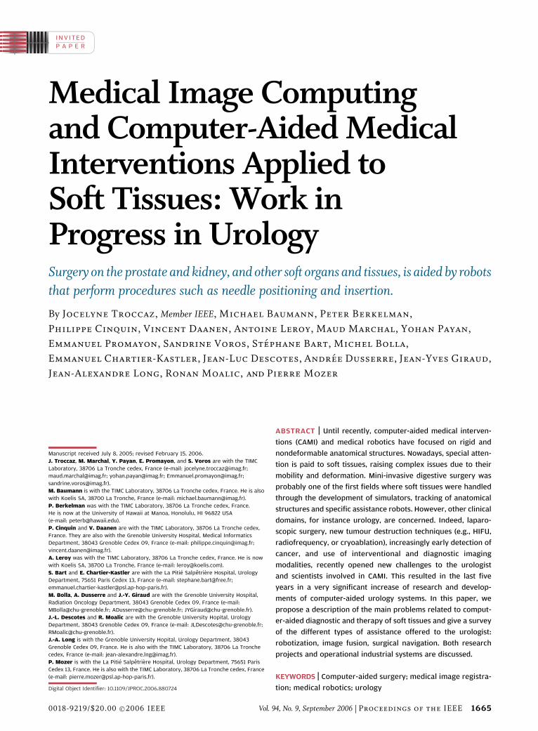
on the patient abdomen skin (see Fig. 2). Because the
robot is placed on the endoscope entry point, 3 DOF
(2 rotations and 1 translation) are sufficient to handle
the endoscope motions.
As compared to AESOP and to most of the other
systems which are positioned on the operating room (OR)
table, floor or ceiling, this very compact system follows thepatient motions and is very easy to install. It weights 625 g;
it is voice controlled and completely sterilizable. Interest-
ing evolutions of robotic endoscope holders deal with
automatically control of robots from image information in
order to track organs or instruments during the surgery
(see [54] for instance).
2) Tele-Surgery Robots: Based on the robotic endoscopeholders experience, instrument holders have naturally
been designed resulting in the so-called tele-surgery
robots. ZEUS, an evolution of the AESOP, is composed
of 3 separated 4 DOF arms (one endoscope holder and two
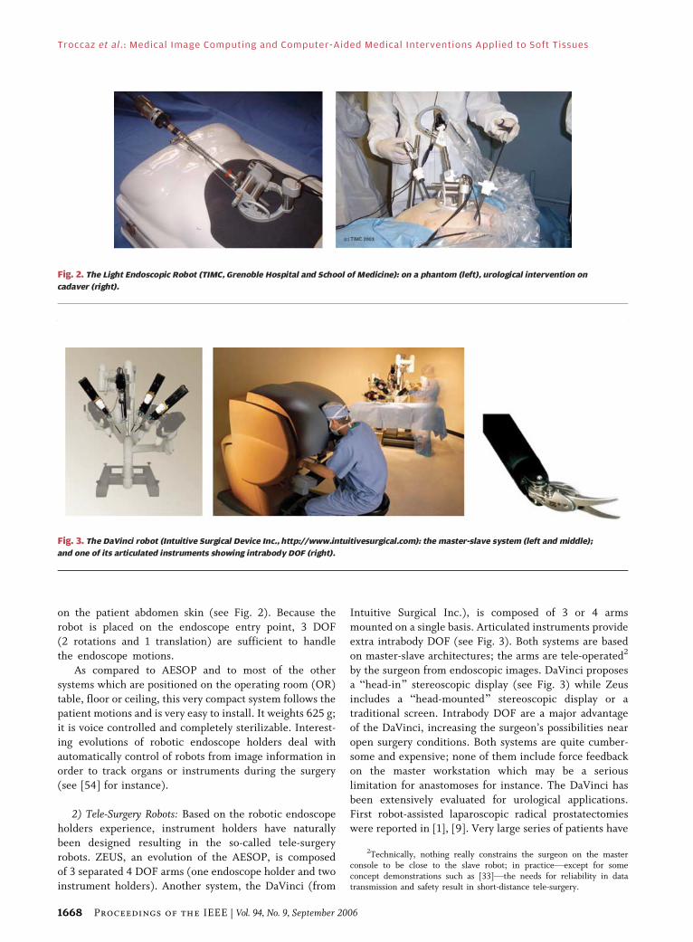
instrument holders). Another system, the DaVinci (from
Intuitive Surgical Inc.), is composed of 3 or 4 arms
mounted on a single basis. Articulated instruments provide
extra intrabody DOF (see Fig. 3). Both systems are based
on master-slave architectures; the arms are tele-operated2
by the surgeon from endoscopic images. DaVinci proposes
a Bhead-in[ stereoscopic display (see Fig. 3) while Zeus
includes a Bhead-mounted[ stereoscopic display or atraditional screen. Intrabody DOF are a major advantage
of the DaVinci, increasing the surgeon’s possibilities near
open surgery conditions. Both systems are quite cumber-
some and expensive; none of them include force feedback
on the master workstation which may be a serious
limitation for anastomoses for instance. The DaVinci has
been extensively evaluated for urological applications.
First robot-assisted laparoscopic radical prostatectomieswere reported in [1], [9]. Very large series of patients have
Fig. 2. The Light Endoscopic Robot (TIMC, Grenoble Hospital and School of Medicine): on a phantom (left), urological intervention on
cadaver (right).
Fig. 3. The DaVinci robot (Intuitive Surgical Device Inc., http://www.intuitivesurgical.com): the master-slave system (left and middle);
and one of its articulated instruments showing intrabody DOF (right).
2Technically, nothing really constrains the surgeon on the masterconsole to be close to the slave robot; in practiceVexcept for someconcept demonstrations such as [33]Vthe needs for reliability in datatransmission and safety result in short-distance tele-surgery.
Troccaz et al.: Medical Image Computing and Computer-Aided Medical Interventions Applied to Soft Tissues
1668 Proceedings of the IEEE | Vol. 94, No. 9, September 2006

since been operated: the Vattikuti Institute in the HenryFord Hospital of Detroit, USA, published in [34] a study
concerning more than 1100 cases. In this center,
laparoscopic prostatectomies started in October 2000
and the DaVinci assistance was introduced in March
2001. A study comparing conventional/laparoscopic/robot-
assisted laparoscopic procedures showed clear advantages
of the robotic series on many points including shorter
hospital stays, reduced pain, reduced blood loss, betterPSA control, reduced positive margins, better continence,
and less impotence. Another advantage of robot assistance
is a reduction of the learning curve for laparoscopic
procedures; [2] reports an improvement factor of about 10.
Laparoscopic radical prostatectomy is probably one of
the domains were the robotic clinical added-value was so
clearly demonstrated. Other applications of such robots to
urology are reported in full details in [51]. Each timecomplex dissections, microsurgery or intracorporal sutur-
ing are necessary, the robot may be a precious assistant.
Several research projects in the world aim at develop-
ing competitive smaller and/or cheaper solutions with
articulated intrabody instruments and endoscopes and
master station offering force feedback. Planning tools are
also developed in order to optimize the entry ports
positioning, enabling both target access and collision-freemotion of the robots (see for instance [14]).
B. Image-Guided RobotsMany gestures in urology are carried under interven-
tional radiology: the diagnostic or therapeutic tool is
moved under control of an imaging modality. Ultrasounds
or fluoroscopy enable continuous control: the operator can
see in real-time the tool position and the anatomy; CT orMRI allow asynchronous control: for instance, a needle is
positioned, a control image is taken and the needle po-
sition is corrected if necessary, and so on. This idea hasbeen exploited to control from medical images robots
performing simple tasks such as a linear tool insertion.
1) Prostate Biopsies and Brachytherapies: From a techni-
cal viewpoint prostate biopsies and brachytherapies (see
Fig. 4) are rather similar; they both consist in inserting
needles in the prostate, either for tissue sampling or for
radioactive seed placement, through transperineal ortransrectal access, under imaging controlVmost often
transrectal ultrasound imaging (TRUS).
However, each biopsy makes use of a single ultrasonic
(US) image in which the needle is visible while brachy-
therapy is based on a volume of images: often parallel axial
US images acquired every 5 mm. Brachytherapy is based
on a careful patient-specific dose planning while biopsies
are generally performed following a predefined globalscheme (for instance sextant or 11-core protocols). Needle
insertion is slow and manual during brachytherapies while
biopsies are very rapidly performed using a biospy gun. As
demonstrated by [25] in a different medical context,
increasing needle velocity results in minimizing the
displacement and deformation of the tissue. Thus auto-
mation may have a positive impact in terms of gesture
accuracy. Moreover, prostate brachytherapy is based onthe use of a template (a stereotactic grid) rigidly con-
nected to the US probe. This restrains needle trajectories
to lines parallel to the probe axis and results in potential
collisions of the needles with the pelvic bone. Again,
using a robot may enable various trajectory directions.
[55] proposes to use a general purpose 6 DOF robot for
needle positioning and insertion. [18] develops a special-
purpose robot mimicking the conventional procedure(trajectories parallel to the US probe axis); a rotational
DOF for the needle is added to reduce the needle flexion
Fig. 4. US-guided transrectal biopsy (left) and transperineal brachytherapy (right) operating principles.
From http://www.uropage.com/index.htm.
Troccaz et al.: Medical Image Computing and Computer-Aided Medical Interventions Applied to Soft Tissues
Vol. 94, No. 9, September 2006 | Proceedings of the IEEE 1669

during tissue penetration. Those systems are still labo-ratory test beds. [38] describes a preclinical evaluation of
a specific 9 DOF system (positioning platform plus
biopsy robot) for transperineal prostate biopsies. A three-
dimensional (3-D) prostate geometric model of the pros-
tate is approximated from series of close parallel US
images enabling planning of the biopsies. 2.5-mm accu-
racy is reported; those performances require very careful
patient preparation and US probe handling. None ofthese systems really considers prostate motion and de-
formation during the procedure.
Another approach consists in performing transrectal
prostate biopsies or brachytherapies with an intrarectal
robot under MRI control [47]. Although conventional MR
imaging (1.5 T with endorectal antenna and T2 sequence)
enables physicians to see precisely the prostate anatomy,
using such a modality for biopsies is probably restricted tothe few cases where US-guided biopsies are not possible or
not successful. Let us remind that in the United States
(respectively, in France) about 106 (respectively, 105)
series of diagnostic biopsies are performed each year). In
[47], conventional MRI is used. The robot is inserted in the
patient rectum and has three DOF to reach the target
defined on the MRI data: translation in the rectum, ro-
tation around its main axis and progression of the needle.Thanks to its design, the robot does not disturb the
magnetic field and includes two coils; one used as part of
the imaging sensor and the other one as a position sensor.
After validation on dogs, the robot is being clinically tested
for transrectal biopsies and brachytherapies [21]; 2-mm
accuracy is reported; this remaining error is probably
mainly due to the prostate motion and deformation during
needle insertion. [13] proposes another robot for prostatebiopsy or brachytherapy inside an open interventional MR
system. Interventional MRI (0.5 T) requires an additional
conventional MRI exam which makes the procedure even
more complex (see Section III-B1).
2) Percutaneous Renal Access: The purpose is to assist
percutaneous access to the kidney. Since 1996 a robot
named BPercutaneous Access of the KidneY[ (PAKY) isdeveloped by the Johns Hopkins groups (Baltimore, MD).
The robot has seven passive DOF used to position a 3 active
DOF structure (2 for orientation and 1 for translation of
the needle). Fluoroscopy is used for needle alignment and
control during insertion. During the procedure, the patient
is in apnoea in order to keep the kidney in a constant
position. [12] reports in vitro and in vivo experiments. In
[46], for 23 patients, no significant difference is reportedbetween the manual and robotic procedure in terms of
precision, rapidity, number of attempts, complications.
One advantage of the robotic procedure lies in the absence
of irradiation of the human operator. [7] proposes a visual
servoing approach from two fluoroscopic views enabling
the automatic placement of the needle to a given target
and entry point. This system was also applied to CT-
guided transperineal prostate biopsies through a singleentry point.
III . IMAGE-BASED UROLOGY
A. Image ProcessingMany papers propose tools to assist the segmentation of
urologic images especially for the prostate where TRUS
images have been paid close attention. Segmentation may
be two-dimensional (2-D), 2.5-D (the segmentation of a
given slice is used to help the segmentation of the
following parallel one), or 3-D. Most successful ap-
proaches make use of active contours and/or statistical
models. However, for 2-D images close to the prostateextremities, existing tools may not be robust enough due to
the poor quality of those images. Other works concern the
automatic segmentation of CT and MRI images of uro-
logical targets (kidney in particular). Because this problem
is very vast and not typical of interventional systems, no
details are given here. In [43] and [56] good reviews of
work concerning the image processing of prostate TRUS
images are presented.
B. Image FusionMR and US imaging are probably the most used
imaging modalities for prostate diagnosis and therapy. The
interventional nature of US is counterbalanced by their
traditional drawbacks: patient dependence, intraoperator
and interoperator variability, medium quality due to
speckle, artefacts, etc. Conventional MRI using external
or transrectal coils clearly show the prostate zonal anatomy
which is useful for biopsy planning, while open MRI (alsocalled iMRI for interventional MRI) enables near real-time
control. This is why several research groups implemented
fusion algorithm to benefit from complementary advan-
tages of these modalities. Other imaging modalities are
used such as CT imaging or histology sections; here also
image fusion may be very useful. This paragraph describes
different studies on multimodality fusion dedicated to
prostate imaging.
1) MRI/iMRI Fusion: The Surgical Planning Laboratory
(SPL) and Harvard Medical School have developed a
navigation system for transperineal prostate biopsies under
iMRI. Because of a lower intensity magnetic field, iMRI
does not clearly show the prostate anatomy. This is why a
preoperative MRI acquisition is performed (external pubic
antenna, 1.5 T, T2 FSE sequence) on which surgicalplanning is possible. Intraoperatively, iMRI data are col-
lected (external pubic antenna, 0.5 T, T2 FSE sequence)
enabling volume reconstruction. A localized stereotactic
grid similar to the one used for brachytherapy is calibrated
with respect to the MRI system and is used as a guiding
device. MRI and iMRI are registered using an intensity-
based elastic registration enabling transfer of the planning
Troccaz et al.: Medical Image Computing and Computer-Aided Medical Interventions Applied to Soft Tissues
1670 Proceedings of the IEEE | Vol. 94, No. 9, September 2006
data to the iMRI conditions. During the biopsy procedure,real-time FGR iMRI slices (acquisition time: 8 s per slice)
are obtained for biopsy tracking. The B3D Slicer[ de-
veloped by SPL enables computing from the iMRI T2-
volume the slice corresponding to the FGR one with
planning information added. [24] reports two successful
clinical cases. As mentioned previously, such an approach
is especially interesting for patients for which ultrasounds
have been unsuccessful (negative repeated US-guidedbiopsies with increasing PSA) or impossible. [10] describes
another solution for MRI/iMRI registration based on
elastic deformation of segmented data; a finite-element
model of the preoperative prostate is deformed using
isotropic linear deformations to match the intraoperative
prostate. Two regions of the prostate are considered with
different elasticity parameters: the central gland and
peripheral zone.
2) MRI/TRUS Fusion: In many cases, bringing the MRI
information to the US-guided procedures can be an in-
teresting alternative. [27] describes experiments con-
cerning the fusion of preoperative MRI data to TRUS
images for transperinal biopsies. Six fiducial points are
matched between the two modalities using a rigid
registration. From this transform, a composite image isproduced combining TRUS data and re-sliced matched
MRI data. No attention is paid to motion or deformation
that could occur between the two acquisitions or during
the procedure.
In one of the two main types of brachytherapy
protocols, the radiation plan is prepared in the OR and is
based on intraoperative TRUS acquisition and segmenta-
tion. One difficulty lies in this initial stage. [39] reportsthe evaluation of elastic surface registration between
TRUS and MRI data for prostate brachytherapy. Preop-
eratively, three orthogonal T2 TSE volumes are acquired
using an endorectal coil and the prostate contours are
segmented jointly on the three volumes. Intraoperatively,
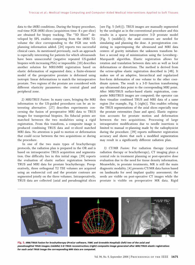
TRUS data are collected (axial and pseudosagittal slices
[see Fig. 5 (left)]). TRUS images are manually segmentedby the urologist as in the conventional procedure and this
results in a sparse intraoperative 3-D prostate model
[Fig. 5 (middle)]; the axial contours are needed for
simulating and planning the dose. A preregistration con-
sisting in superimposing the ultrasound and MRI data
centers of gravity initializes the unknown transform be-
fore a second step of minimization using the Levenberg–
Marquardt algorithm. Elastic registration allows forrotation and translation between data sets as well as local
deformations or distortions. The method is derived from
the octree-spline elastic registration described in [48]. It
makes use of an adaptive, hierarchical and regularized
free-form deformation of one volume to the other coor-
dinate system. The result is a 3-D function transforming
any ultrasound data point to the corresponding MRI point.
After MRI/TRUS surface-based elastic registration, com-posite MRI/TRUS images are computed; the operator can
then visualize combined TRUS and MRI data of a same
region [for example, Fig. 5 (right)]. This enables refining
the TRUS segmentations of the axial slices especially near
the prostate extremities (base and apex). Elastic registra-
tion accounts for prostate motion and deformation
between the two acquisitions. Processing of large
intraoperative modifications due to needle insertions islimited to manual re-planning made by the radiophysicist
during the procedure. [39] reports millimeter registration
accuracy and shows that such a modified segmentation
may result in a significantly different radiation plan.
3) CT/MR Fusion: For radiation therapy (external
radiation therapy or brachytherapy), CT imaging plays a
central role in treatment planning or post-operative doseevaluation due to the need for tissue density information.
Meanwhile, in prostate treatments, MR is still the main
diagnostic modality. [4] presents CT/MR data fusion based
on landmarks for seed implant quality assessment; the
seeds are visible on post-operative CT images while the
prostate is visible on preoperative MR data. Rigid
Fig. 5. IRM/TRUS fusion for brachytherapy (ProCur software, TIMC and Grenoble Hospital): (left) two of the axial and
pseudosagittal TRUS images; (middle) 3-D TRUS reconstruction; (right) composite image generated after MRI/TRUS elastic registration:
for each axial TRUS image the corresponding MRI data are computed.
Troccaz et al.: Medical Image Computing and Computer-Aided Medical Interventions Applied to Soft Tissues
Vol. 94, No. 9, September 2006 | Proceedings of the IEEE 1671
registration of CT and MR data is based on the use of a
urinary catheter going through the prostate and placed
before both MR and CT acquisitions making it visible on
both modalities. After registration of the catheter, seeds
visible on the CT data may be visualized with respect to the
MR prostate anatomy. The authors report an averageresidual error measured on each slice at the center of the
urethra of 1.2 mm for 11 patients and a maximum residual
error of 2.5 mm for one patient. [30] proposes to use a thin
plate spline (TPS) function to map segmented prostate
contours from MRI or magnetic resonance spectroscopic
imaging to CT data. In this paper, endorectal coils are used
for MR imaging which may deform the prostate as
compared to the CT acquisition situation; rigid registrationmay, thus, not be accurate enough. Obtained correspon-
dence index of 93.1 � 5% and centroid position of 0.56 �0.09 mm of the registered surface slices quantify the
registration accuracy and demonstrate the superiority of
TPS registration over rigid one.
4) Histology/Other Modality Fusion: Histology is the gold
standard of cancer diagnosis. Therefore, registeringhistological data to other modalities may be very fruitful.
After preparation and fixation in formalin of cancerous
prostates surgically removed from patients, 3 to 5-mm
blocks are generally cut in a pseudoaxial direction; then a
slice of 30- (to 50-) �m thickness is obtained from each
block and analyzed by a pathologist with a microscope for
detection and characterization of cancerous cells. As
proposed in [20] artificial markers are generally used inorder to build a 3-D reconstruction from those sparse data.
In [44], fused histological data from hundreds of
patients using elastic surface registration in order to build
a statistical atlas of cancer occurrence in the different
prostate zones and to optimize biopsy schemes based on
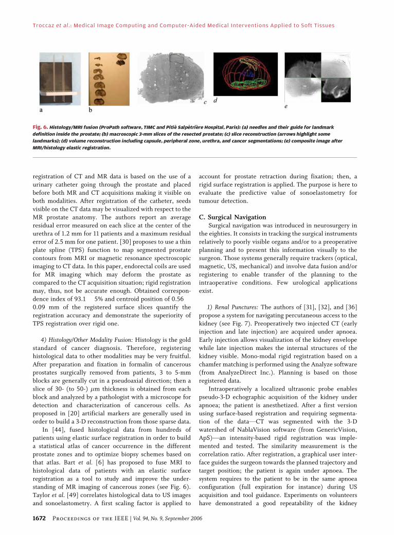
that atlas. Bart et al. [6] has proposed to fuse MRI to
histological data of patients with an elastic surface
registration as a tool to study and improve the under-standing of MR imaging of cancerous zones (see Fig. 6).
Taylor et al. [49] correlates histological data to US images
and sonoelastometry. A first scaling factor is applied to
account for prostate retraction during fixation; then, a
rigid surface registration is applied. The purpose is here to
evaluate the predictive value of sonoelastometry for
tumour detection.
C. Surgical NavigationSurgical navigation was introduced in neurosurgery in
the eighties. It consists in tracking the surgical instruments
relatively to poorly visible organs and/or to a preoperative
planning and to present this information visually to the
surgeon. Those systems generally require trackers (optical,
magnetic, US, mechanical) and involve data fusion and/or
registering to enable transfer of the planning to the
intraoperative conditions. Few urological applicationsexist.
1) Renal Punctures: The authors of [31], [32], and [36]
propose a system for navigating percutaneous access to the
kidney (see Fig. 7). Preoperatively two injected CT (early
injection and late injection) are acquired under apnoea.
Early injection allows visualization of the kidney envelope
while late injection makes the internal structures of thekidney visible. Mono-modal rigid registration based on a
chamfer matching is performed using the Analyze software
(from AnalyzeDirect Inc.). Planning is based on those
registered data.
Intraoperatively a localized ultrasonic probe enables
pseudo-3-D echographic acquisition of the kidney under
apnoea; the patient is anesthetized. After a first version
using surface-based registration and requiring segmenta-tion of the dataVCT was segmented with the 3-D
watershed of NablaVision software (from GenericVision,
ApS)Van intensity-based rigid registration was imple-
mented and tested. The similarity measurement is the
correlation ratio. After registration, a graphical user inter-
face guides the surgeon towards the planned trajectory and
target position; the patient is again under apnoea. The
system requires to the patient to be in the same apnoeaconfiguration (full expiration for instance) during US
acquisition and tool guidance. Experiments on volunteers
have demonstrated a good repeatability of the kidney
Fig. 6. Histology/MRI fusion (ProPath software, TIMC and Pitie Salpetriere Hospital, Paris): (a) needles and their guide for landmark
definition inside the prostate; (b) macroscopic 3-mm slices of the resected prostate; (c) slice reconstruction (arrows highlight some
landmarks); (d) volume reconstruction including capsule, peripheral zone, urethra, and cancer segmentations; (e) composite image after
MRI/histology elastic registration.
Troccaz et al.: Medical Image Computing and Computer-Aided Medical Interventions Applied to Soft Tissues
1672 Proceedings of the IEEE | Vol. 94, No. 9, September 2006

positioning under repeated apnoeas; this confirms literature
results. Validation of the developed system on real data and
on cadavers is promising. Real-time tracking of the kidney,
for instance using magnetic localization, by avoiding the
need for apnoea would greatly simplify the protocol;nevertheless the visual guidance may not be easy with a
mobile target. Robotic alternatives may, thus, have to be
considered for instance by synchronizing the robot to a
given stage of the respiratory cycle. The motion of the
kidney due to the needle insertion has not yet been studied.
Here again, robotic access may minimize induced motion.
The same framework was applied to the guidance of
stimulation electrode placement into the sacrum (S3 root)for incontinence treatment. CT/US surface-based registra-
tion of the pelvis originally developed for pelvic surgery
(see [16] and [50]) was successfully applied to this
application and validated though cadaver experiments
[32]. In this application, the bone position is tracked
thanks to an implanted rigid body.
2) Prostate Biopsies: The precise realization of prostatebiopsy schemes (for instance twelve biopsies regularly
distributed on the prostate gland) faces the difficulty of
using 2-D images to guide a 3-D action. The process, thus,
strongly depends on the surgeon’s ability to mentally
integrate successive images and trajectories in a 3-D space.
Because US-guided biopsies only detect 75% of the cancers
for the first series, assisting biopsies to guarantee that
samples are correctly and regularly acquired in the
targeted sites is an important objective. An exploratory
study using a navigation system (see Fig. 8) was developed
at TIMC. The US probe is localized using an optical system(Passive Polaris from Northern Digital Inc.) and the
executed trajectories are recorded in a fixed reference
system. Those recordings clearly demonstrate that the
prostate moves very significantly with the US probe
displacements; those displacements are necessary to orient
the needle, which is rigidly attached to the probe, towards
the targeted sites.
Current work deals with US-based real-time registra-tion enabling to represent the executed biopsies in a single
prostate reference frame despite prostate intraoperative
displacements.
IV. MODEL-BASED UROLOGY
As seen briefly in this paper, shape and/or appearance
models have been introduced as a priori information forimage processing. Statistical models concerning the oc-
currence of cancers in the different zones of the prostate
have also been constructed in order to optimize the biopsy
strategies by maximizing the ability to detect an existing
tumour. [45] reports a clinical evaluation of such an
optimized scheme.
Fig. 8. Prostate biopsy navigation (ProNavV1, TIMC, Grenoble Hospital)Vbiopsy recording: (left) localized intrarectal US probes
(2-D and 3-D)Vright) visualization interface presenting US images, prostate model and recorded trajectories in a fixed reference system.
Fig. 7. Navigated percutaneous access of the kidney (TIMC, Pitie-Salpetriere Hospital, Grenoble School of Medicine, PRAXIM):
(from left to right) scanner volume et segmented kidney; preoperative planning interface; US probe and puncture needle with their
optical localizers (passive Polaris from NDI, address); CT/US fusion; puncture guidance.
Troccaz et al.: Medical Image Computing and Computer-Aided Medical Interventions Applied to Soft Tissues
Vol. 94, No. 9, September 2006 | Proceedings of the IEEE 1673
In the context of medical interventions dealing with softtissues, much attention is paid to biomechanical models; the
purpose is in particular to better predict tissue motions and
deformations and tool interaction with the tissues. In a first
stage, these models were mostly developed for training
simulators especially in laparoscopic surgery. More recent-
ly, they are seen as a necessary input to the planning of a
surgery performed on soft tissues.
Some groups work on discrete models of the prostatecomplex environment; for instance [29] presents a sim-
ulator for rectal palpation of the prostate. The interactive
nature of such models enabling intraoperative re-planning
may be counterbalanced by difficult parameter identifica-
tion. Because of their theoretical background and the
ability to introduce rheological tissue parameters, finite-
element models are the most commonly used. Based on
DiMaio and Salcudean previous work (e.g., [19]), Goksel[23] proposes a brachytherapy simulator combining TRUS
image generation to a needle tissue interaction model.
Mohammed et al. [35] combines statistical and biome-
chanical models for evaluation of intraoperative prostate
deformation. Other groups work on steerable needle path
planner taking benefit of needle flexion to generate paths
avoiding anatomical obstacles [3].
Current limitations of planning approaches are due tothe hypothesis of a homogeneous tissue and, for patient-
specific planners, in the difficulty of determining in vivotissue physical parameters. Sonoelastometry, a very pro-
mising modality for the detection of cancers inside organs
[28], can also help for physical parameters identification.
V. CONCLUSION
As seen in this paper, many research projects are dedicated
to urological applications. Concerning target motion and
deformation, some partial solutions have been proposed.
Tele-operated robots enable adapting the surgical strategy
to the anatomical state based on the surgeon know-how.
Image-guided robots generally allow data acquisition justbefore the action to be executed; however, none of them,
in urology, is completely controlled by real-time data.
Image fusion can consider changes occurring between the
different acquisitions stages to be registered; however, no
system yet enables real-time fusion of intraoperative data
to preoperative data for tracking purpose. Regarding the
kidney, navigational assistance or robot actions are
performed on a stabilized organ (breathing is temporarilyheld); no tracking is available yet. Biomechanical models
are developed but none of them is yet used for guiding the
intervention.
A lot has been done but significant research efforts
must still be undertaken to fully consider a mobile and
deformable target such as the kidney or the prostate.
Intraoperative tracking based on intrabody markers (for
instance magnetic markers or other [11], [15]) or on real-time image processing is necessary. The development of
autonomous robots servo-ed to real-time intraoperative
data (see for instance [53]) raises very challenging ro-
bustness and safety issues since in this case the surgeon
will no longer be able to directly supervise the robot
actions. Finally, patient-specific modelling of organs me-
chanical properties is a key issue for predicting and
recognizing anatomical changes and to allow precise plan-ning. All those topics open very interesting and difficult
scientific tracks for the coming years with a very strong
clinical interest. h
Acknowledgment
Research projects of TIMC described in this paper
are/were supported by grants from the French Ministry ofHealth (PHRC 2003 Prostate-Echo, CIT PICAMI), the
French Ministry of Research and ANVAR (MMM project in
the RNTL Program), nonprofit organizations (Association
pour la Recherche contre le Cancer, Association Nationale
de la Recherche Technique), and by Praxim-Medivision.
REF ERENCE S
[1] C. C. Abbou, A. Hoznek, and L. Saloman,BRemote laparoscopic prostatectomy carriedout with a robot. Report of a case,[ Prog. Urol.,vol. 10, pp. 520–523, 2000.
[2] T. E. Alhering, D. Sharecky, D. Lee, andR. V. Clayman, BSuccessful transfer of opensurgical skills to a laparoscopic environmentusing a robotic interface: Initial experiencewith laparoscopic radical prostatectomy,[J. Urol., vol. 170, pp. 1738–1741, 2003.
[3] R. Alterovitz, K. Goldberg, and A. Okamura,BPlanning for steerable bevel-tip needleinsertion through 2D soft tissue withobstacles,[ in Proc. IEEE ICRA’05, 2005,pp. 1652–1657.
[4] R. J. Amdur, D. Gladstone, K. A. Leopold, andR. D. Harris, BProstate seed implant qualityassessment using MR and CT image fusion,[Int. J. Radiat. Oncol. Biol. Phys., vol. 43, no. 1,pp. 67–72, 1999.
[5] J. M. Balter, H. M. Sandler, K. Lam,R. L. Bree, A. S. Lichter, and R. Ten Haken,
BMeasurement of prostate movement overthe course of routine radiotherapy usingimplanted markers,[ Int. J. Radiat. Oncol. Biol.Phys., vol. 31, no. 1, pp. 113–118, 1995.
[6] S. Bart, P. Mozer, P. Hemar, G. Lenaour,E. Comperat, R. Renard-Penna,E. Chartier-Kastler, and J. Troccaz,BMRI-histology registration in prostatecancer,[ presented at the Surgetica’05,Chambery, France.
[7] B. Bascle, N. Navab, M. Loser, B. Geiger, andR. Taylor, BNeedle placement under X-rayfluoroscopy using perspective invariants,[ inProc. IEEE MMBIA’00 (Mathematical Methodsin Biomedical Image Analysis) Workshop, 2000.
[8] P. Berkelman, E. Boidard, P. Cinquin, andJ. Troccaz, BLER: The Light EndoscopeRobot,[ presented at the IEEE/RSJ Int. Conf.Intelligent Robots and Systems, Las Vegas,Oct. 27–31, 2003.
[9] J. Binder and W. Kramer, BRoboticallyassisted radical prostatectomy,[ BJU Int.,vol. 87, pp. 408–410, 2001.
[10] A. Bharata, M. Hirose, H. Hata,S. K. Warfield, M. Ferrant, K. H. Zhou,E. Suarez-Santana, J. Ruiz-Alzola, A. D’Amico,R. A. Cormack, R. Kikinis, F. A. Jolesz, andC. M. C. Tempany, BEvaluation ofthree-dimensional finite-element-baseddeformable registration of pre- andintra-operative prostate imaging,[ Med. Phys.,vol. 28, no. 12, pp. 2551–2560, 2001.
[11] I. Bricault, S. DiMaio, O. Clatz, S. Pujol,K. Vosburgh, and R. Kikinis, BComputer-assisted interventions on liver: Feasibility ofthe Fanchor needle_ technique for real-timetargeting of lesions with respiratory motion,[presented at the Surgetica’05, Chambery,France.
[12] J. A. Cadeddu, D. Stoianovici, R. N. Chen,R. G. Moore, and L. R. Kavoussi, BStereotacticmechanical percutaneous renal access,[J. Endourol., vol. 12, no. 2, pp. 121–125,Apr. 1998.
[13] K. Chinzei, N. Hata, A. Jolesz, and R. Kikinis,BSurgical assist robot for the active navigation
Troccaz et al.: Medical Image Computing and Computer-Aided Medical Interventions Applied to Soft Tissues
1674 Proceedings of the IEEE | Vol. 94, No. 9, September 2006

in the intraoperative MRI: Hardware designissues,[ in Proc. 2000 IEEE/RSJ Int.Conf. Intelligent Robots and Systems(IROS 2000), 2000, pp. 727–732.
[14] E. Coste-Maniere, L. Adhami, F. Mourgues,and O. Bantiche, BOptimal planning ofrobotically assisted heart surgery: Transferprecision in the operating room,[ Int. J. Robot.Res., vol. 23, no. 4/5, 2004.
[15] E. Coste-Maniere, D. Olender, W. Kilby, andR. A. Schulz, BRobotic whole bodystereotactic radiosurgery,[ Int. J. Med.Robot. Comput. Assist. Surg., vol. 1, no. 2,pp. 28–39, 2005.
[16] V. Daanen, J. Tonetti, and J. Troccaz, BA fullyautomated method for the delineation ofosseous interface in the ultrasound images,[in Proceedings of MICCAI’2004, vol. 3216,C. Barillot, D. R. Haynor, and P. Hellier, Eds.,2004, pp. 549–557.
[17] B. L. Davies, R. D. Hibberd, W. S. Ng,A. Timoney, and J. E. A. Wickham,BDevelopment of a surgeon robot forprostatectomies,[ J. Eng. Med., Proc. Inst.Mech. Eng., vol. 205, pp. 35–38, 2001.
[18] B. L. Davies, S. J. Harris, and E. Dibble,BBrachytherapyVAn example of a urologicalminimally invasive robotic procedure,[Int. J. Med. Robot. Comput. Assist. Surg., vol. 1,no. 1, pp. 88–96, 2004.
[19] S. P. DiMaio and S. E. Salcudean, BNeedleinsertion modelling and simulation,[ IEEETrans. Robot. Automat. (Special Issue onMedical Robotics), vol. 19, no. 5, pp. 864–875,Oct. 2003.
[20] L. Egevad, K. Engstrom, and C. Busch, BA newmethod for handling radical prostatectomiesenabling fresh tissue harvesting, wholemount sections, and landmarks foralignment of sections,[ J. Urol. Pathol., vol. 9,pp. 17–28, 1998.
[21] ‘‘From mini-invasive surgery to endocavitary/endoluminal interventionsVPart II: Nextgeneration of surgical robots and devices,’’Tutorial Given at MICCAI ’04, StMalo, 2004.[Online]. Available: http://www.miccai.irisa.fr/index2.php?menu=Exhibits_and_Workshops&page=Tutorials
[22] R. Ginhoux, J. Gangloff, M. de Mathelin,L. Soler, M. Arenas Sanchez, andJ. Marescaux, BActive filtering of physiologicalmotion in robotized surgery using predictivecontrol,[ IEEE Trans. Robotics, vol. 21, no. 1,pp. 67–79, Feb. 2005.
[23] O. Goksel, BUltrasound image and 3D finiteelement based tissue deformation simulatorfor prostate brachytherapy,[ Master thesis,UBC, Vancouver, BC, Canada, Dec. 2004.
[24] N. Hata, M. Jinzaki, D. Kacher, R. Cormak,D. Gering, A. Nabavi, S. G. Silverman,A. V. D’Amico, R. Kikinis, F. A. Jolesz, andC. M. C. Tempany, BMR imaging-guidedprostate biopsy with surgical navigationsoftware: Device validation and feasibility,[Radiology, vol. 220, pp. 263–268, 2001.
[25] M. Hervely, P. Dupont, and J. Triedman,BTrajectory optimization for dynamic needleinsertion,[ in Proc. 2005 IEEE ICRA Conf.,Barcelona, Spain, Apr. 2005, pp. 1658–1663.
[26] M. Hirose, A. Bharata, N. Hata,K. H. Zou, S. K. Warfield, R. A. Cormack,A. D’AMicao, R. Kikinis, F. A. Jolesz, andC. M. C. Temapny, BQuantitative MR imagingassessment of prostate gland deformationbefore and during MR imaging-guidedbrachytherapy,[ Acad. Radol, vol. 9,pp. 906–921, 2002.
[27] I. Kaplan, N. E. Oldenburg, P. Meskell,M. Blake, P. Church, and E. J. Holupka,BReal-time MRI-ultrasound image guided
stereotactic biopsy,[ Magn. Reson. Imag.,vol. 20, pp. 295–299, 2002.
[28] K. Konig, U. Scheipers, A. Pesavento,A. Lorenz, H. Ermert, and T. Senge, BInitialexperiences with real-time elastographyguided biopsies of the prostate,[ J. Urol.,vol. 174, pp. 115–177, 2005.
[29] Y. Kuroda, M. Nakao, T. Kuroda, H. Oyama,M. Komori, and T. Matsuda, BInteractionmodel between elastic objetcs for accuratehaptic display,[ presented at the ICAT’03,Tokyo, Dec. 2003.
[30] J. Lian, L. Xing, S. Hunjan, C. Dumoulin,J. Levin, A. Lo, R. Watkins, K. Rohling,R. Giaquinto, D. Kim, D. Spielman, andB. Daniel, BMapping of the prostate inendorectal coil-based MRI/MRSI and CT:A deformable registration and validationstudy,[ Med. Phys., vol. 31, no. 11,pp. 3087–3094, 2004.
[31] A. Leroy, P. Mozer, Y. Payan, and J. Troccaz,BRigid registration of freehand 3Dultrasound and CT-scan kidney images,[in Proc. MICCAI’2004, vol. 3216, C. Barillot,D. R. Haynor, and P. Hellier, Eds.,2004, pp. 837–844.
[32] A. Leroy, BMethodes de recalage scanner/echographie. Application a la navigationrenale des ponctions renales percutanees,[(in French), Ph.D. thesis, INPG, GrenobleFrance, Nov. 2004.
[33] J. Marescaux, J. Leroy, M. Gagner et al.,BTransatlantic robot-assisted telesurgery,[Nature, vol. 413, p. 379, 2001.
[34] M. Menon, A. Tewari, J. O. Peabody,A. Shrivastava, S. Kaul, A. Bhandari, andA. K. Hemal, BVattikuti Instituteprostatectomy, a technique of roboticradical prostatectomy for managementof localized carcinoma of the prostate:experience of over 1100 cases,[ Urol. Clin.N. Am., vol. 31, no. 4, pp. 701–717, 2004.
[35] A. Mohammed, C. Davatzikos, and R. Taylor,BA combined statistical and biomechanicalmodel for estimation of intra-operativeprostate deformation,[ in MICCAI’02,vol. 2489, Dohi and Kikinis, Eds., 2002,pp. 452–460.
[36] P. Mozer, A. Leroy, Y. Payan, J. Troccaz,E. Chartier-Kastler, and F. Richard,BComputer-assisted access to the kidney,[MRCAS J., vol. 2, no. 3, pp. 256–261, 2006.
[37] D. M. Parkin, BGlobal cancer statistics inthe year 2000,[ Lancet Oncol., vol. 2,pp. 533–543, 2001.
[38] L. Phee, D. Xiao, J. Yuen, C. F. Chan,C. H. Thng, C. Cheng, and W. S. Ng,BUltrasound guided robotic system fortransperineal biopsy of the prostate,[ in Proc.2005 IEEE ICRA Conf., Barcelona, Spain,Apr. 2005, pp. 1327–1332.
[39] C. Reynier, J. Troccaz, P. Fourneret,A. Dusserre, C. Gay-Jeune, J. L. Descotes,M. Bolla, and J.-Y. Giraud, BMRI/TRUS datafusion for prostate brachytherapy. Preliminaryresults,[ Med. Phys., vol. 31, no. 6,pp. 1568–1575, 2004.
[40] J. Sackier and Y. Wang, BRobotically assistedlaparoscopic surgery: From concept todevelopment,[ Surgical Endosc., no. 8,pp. 63–66, 1994.
[41] A. Sawada, K. Yoda, M. Kokubo, T. Kunieda,Y. Nagata, and M. Hiraoka, BA technique fornon invasive respiratory gated treatmentbased on a 3D realtime ultrasound imagecorrelation: A phantom study,[ Med. Phys.,vol. 31, no. 2, pp. 245–250, 2004.
[42] J. Shah, S. Mackay, T. Rockall, J. Vale, andA. Darzi, BFUrobotics_: Robots in urology,[BJU Int., vol. 88, pp. 313–320, 2001.
[43] F. Shao, K. V. Ling, W. S. Ng, andR. Y. Wu, BProstate boundary detection formultrasonography images,[ J. Ultrasound Med.,vol. 22, pp. 605–623, 2003.
[44] D. Shen, Z. Lao, J. Zeng et al., BA statisticalatlas of prostate cancer for optimal biopsy,[Proc. of MICCAI’2001, 2001, pp. 416–424.
[45] M. Schostak, F. Christoph, M. Schrader,M. Panick, A. Lingnau, and K. Miller,BProstate biopsyVPractical examinationof the adequacy of Chen’s virtual strategy,[(in German), Aktuelle Urol., vol. 36, no. 2,pp. 149–153, 2005.
[46] L. M. Su, D. Stoianovici, T. W. Jarrett,A. Patriciu, W. W. Roberts, J. A. Caddedu,S. Ravakumar, S. B. Solomon, andL. R. Kavoussi, BRobotic percutaneous accessto the kidney: Comparison with standardmanual access,[ J. Endourol., vol. 16, no. 7,pp. 471–475, 2002.
[47] R. C. Susil, A. Krieger, J. A. Derbyshire,A. Tanacs, L. L. Whitcomb, G. Fichtinger, andE. Atalar, BSystem for MR image-guidedprostate interventions: Canine study,[Radiology, vol. 228, no. 3, pp. 886–894, 2003.
[48] R. Szeliski and S. Lavallee, BMatching 3-Danatomical surfaces with non-rigiddeformations using octree-splines,[ Int. J.Comput. Vis., vol. 18, no. 2, pp. 171–196,1996.
[49] L. S. Taylor, B. C. Porter, G. Nadasdy,P. A. DiSantAgnese, D. Pasternack, Z. Wu,R. B. Baggs, D. J. Rubens, and K. J. Parker,BThree-dimensional registration of prostateimages from histology and ultrasound,[Ultrasound Med. Biol., vol. 30, no. 2,pp. 161–168, 2004.
[50] J. Tonetti, L. Carrat, S. Blendea, P. Merloz,J. Troccaz, S. Lavallee, and J. P. Chirossel,BClinical results of percutaneous perlvicsurgery. Computer assisted surgery usingultrasound compared to standardfluoroscopy,[ Comput.-Aided Surg., vol. 6,no. 4, pp. 204–211, 2001.
[51] BRobotic urologic surgery,[ Urol. Clin. N. Am.,vol. 31, no. 4, 2004, issue.
[52] M. Van Herk, A. Bruce, A. P. Guus Kroes,T. Shouman, A. Touw, and J. V. Lebseque,BQuantification of organ motion duringconformal radiotherapy of the prostate bythree dimensional image registration,[ Int. J.Radiat. Oncol. Biol. Phys., vol. 33, no. 5,pp. 1311–1320, 1995.
[53] M. A. Vitrani, G. Morel, and T. Ortmaier,BAutomatic guidance of a surgical instrumentwith ultrasound based visual serving,[ pre-sented at the IEEE Int. Conf. Robotics andAutomation, Barcelona, Spain, 2005.
[54] S. Voros, E. Orvain, J.-A. Long, andP. Cinquin, BAutomatic detection ofinstruments in laparoscopic images: A firststep towards high level command of robotizedendoscopic holders,[ presented at theIEEE/RAS-EMBS Int. Conf. BiomedicalRobotics and Biomechatronics (BioRob’06),Feb. 2006.
[55] Z. Wei, G. Wan, L. Gardi, G. Mills,D. Downey, and A. Fenster, BRobot-assisted3D-TRUS guided prostate brachytherapy:System integration and validation,[ Med.Phys., vol. 31, no. 3, pp. 539–548, 2004.
[56] Y. Zhu, S. Williams, and R. Zwiggelaar,BComputer technology in detection andstaging of prostate carcinoma: A review,[Med. Image Anal., vol. 10, no. 2, pp. 178–199,2006.
Troccaz et al.: Medical Image Computing and Computer-Aided Medical Interventions Applied to Soft Tissues
Vol. 94, No. 9, September 2006 | Proceedings of the IEEE 1675

ABOUT THE AUT HORS
Jocelyne Troccaz (Member, IEEE) was born in
1959. She received the Ph.D. degree in computer
science from the Institut National Polytechnique
de Grenoble, Grenoble, France, in 1986.
She is a CNRS Research Director since 1998.
Until 1990, her activity was in the field of
automatic robot programming for industrial and
spatial robotics. She moved to medical robotics in
1990. Since 1996, she is Director of the Computer
Assisted Medical Interventions group (35 people)
of the TIMC laboratory. Since 2006, she is vice-director of the TIMC
laboratory (230 people). Her personal research activity in the TIMC
laboratory of Grenoble is about the medical applications of robotics and
medical image processing. She is involved in several clinical collabora-
tions. She has over 150 publications including international patents. She
has been an invited speaker in several conferences. She is on the
organizing committees of a number of international conferences of her
former and current research domains. She has been a consultant for
robotics companies. She is Associate Editor for the Journal of Computer-
Aided Surgery. She is on the editorial board the Medical Image Analysis
Journal.
Dr. Troccaz is Associate Editor for the IEEE TRANSACTIONS ON ROBOTICS.
Michael Baumann is currently working towards
the Ph.D. degree at the TIMC/GMCAO laboratory,
Grenoble, France.
He received the M.Sc. degree in computer
science in 2004, from the Universitat Karlsruhe
TH, Karlsruhe, Germany, and the INSA de Lyon,
Lyon, France. His research interests include med-
ical image analysis, computer-assisted diagnosis
and treatment planning, and tracking systems.
Peter Berkelman received the B.S. and M.S.
degrees in mechanical engineering in 1992 from
the Massachusetts Institute of Technology, Cam-
bridge, MA, and the Ph.D. degree in robotics in
1999 from Carnegie Mellon University, Pittsburgh,
PA. He is currently an Assistant Professor in the
Department of Mechanical Engineering at the
University of Hawaii-Manoa, Honolulu HI. From
2001 to 2004 he was with the TIMC Laboratory,
Grenoble, France. His research interests include
surgical robotics and human-robot interfaces.
Philippe Cinquin was born in 1956. He received
the Ph.D. degree in applied mathematics.
He is a Medical Doctor and Professor of Medical
Informatics at Grenoble (France), in TIMC-IMAG, a
Research Unit of CNRS and Universite Joseph
Fourier, and heads the Department of Medical
Informatics of Grenoble’s University Hospital. In
1984, he launched a research team on Computer
Assisted Medical Interventions (CAMI). This activ-
ity led to an innovative approach in surgical
practice, thanks to the introduction of Information Technology in the
Operating Room. He is a member of the editorial board of Computer
Aided Surgery. He co-ordinated 4 European projects.
Dr. Cinquin Associate Editor of IEEE TRANSACTIONS ON MEDICAL IMAGING.
In 1999, he received the first Maurice E. Muller award for excellence in
Computer Assisted Orthopaedic Surgery and in 2003 the CNRS silver
award.
Vincent Daanen received the M.Sc. and Ph.D.
degrees in automatic control from the University
of Lille, Lille, France, in 1996 and 2001, respectively.
From 2002, he has been a Researcher in the
TIMC laboratory, Grenpble, France. His research
interests include medical image processing, multi-
modality imaging, registration and data fusion.
Antoine Leroy received the M.Eng. and M.Sc.
degrees in 2001 and the Ph.D. degree in computer-
assisted surgery from the INP University of
Grenoble, Grenoble, France, in 2004.
He is a Research Engineer in the TIMC labora-
tory, Grenoble, France, and until 2005 PRAXIM-
Medivision. In 2006, he set up his own company
named KOELIS.
Dr. Leroy specialized in urology
Maud Marchal received the M.S. degree from
Grenoble University, France, in 2003 and the
M.Eng. degree from the National Superior School
of Computer Science and Applied Mathematics,
Grenoble, France, in 2003. She is currently
working toward the Ph.D. degree at Grenoble
University.
Her research interests include soft tissue mod-
eling, biomechanics, and biomedical simulation.
Yohan Payan received the Ph.D. degree in
computer science and signal processing from the
Institut National Polytechnique of Grenoble,
Grenoble, France, in 1996.
Currently, he is Researcher at the French
National Research Institute (CNRS) in the TIMC
laboratory, with interests that include biomechan-
ics, computer-assisted surgery, signal processing,
and neurosciences.
Emmanuel Promayon received the M.S. degree
and Ph.D. degrees in computer science from the
University Joseph Fourier, Grenoble, France, in
1993 and 1997, respectively.
He is currently an Associate Professor of
Computer Science with University Joseph Fourier.
His research interests include biomechanics, soft
tissue modelling, and computer graphics.
Troccaz et al.: Medical Image Computing and Computer-Aided Medical Interventions Applied to Soft Tissues
1676 Proceedings of the IEEE | Vol. 94, No. 9, September 2006

Sandrine Voros graduated from the BEcole
Centrale de Nantes[ engineering school in 2002
with a computer science major. She received the
M.S. in medical informatics from Paris VI Univer-
sity, Paris, France, in 2003. She is is currently
working towards the Ph.D. degree in the GMCAO
team of the TIMC laboratory, Grenoble, France.
Her research interests include robotic assistance
in surgery and computer vision.
Stephane Bart received the M.S. degree in
biomedical engineering from the TIMC laboratory,
Grenoble, France, in 2004.
He is a Urology Resident, and is currently
working in the Urology Department of Pitie-
Salpetriere hospital in Paris, France.
Michel Bolla received the M.D. degree with a
radiation oncology specialty from the University
of Grenoble, Grenoble, France, in 1976.
Currently, he is a Professor of Radiation
Oncology in the Grenoble University Hospital
where he heads both the radiation oncology
department and the oncology federation. His
research interests are in mass screening, breast
cancer, and prostate cancer. He is on the editorial
board of several clinical journals among which
Radiotherapy and Oncology and the European Journal of Cancer. He
belongs to several national and international
scientific societies and is a very active member
of the EORTC.
Emmanuel Chartier-Kastler received the M.D.
and Ph.D. degrees from the University of Paris
(Paris VI and Paris V), Paris, France, in 1989 and
1996, respectively.
Currently he is working as a professor of
urology in the department of urology, Groupe
Hospitalier Pitie-Salpetriere and is head of the
voiding disorders and neuro-urology programme.
His research interests are focused on new tech-
nologies especially on high-intensity focused
ultrasounds and phase 2 and phase 3 clinical research protocols as
investigator and/or coordinator (BPH therapies, botulinum toxin An
application for incontinence and BPH, urethral prosthesis).
Jean-Luc Descotes received the M.D. and Ph.D.
degrees from Grenoble University, Grenoble,
France, in 1989 and 1997, respectively.
He has been a Professor in the Department of
Urology and Renal Transplant in Grenoble Univer-
sity Hospital, Grenoble, France, since 1997. His
surgical and clinical activity is focused on oncol-
ogy. His research interests concern robotics for
laparoscopic techniques, and image fusion tech-
niques for brachytherapy and prostatic biopsies in
prostate cancer. He is also involved in clinical studies for prostate,
bladder and kidney cancer.
Andree Dusserre is currently a Medical Physicist in the radiation
oncology department of the Grenoble University Hospital, Grenoble,
France.
Jean-Yves Giraud received the Ph.D. degree in
medical physics from Paul Sabatier University,
Toulouse, France, in 1991.
He was a Visiting Scientist at Harvard Medical
School, Joint Center for Radiation Therapy, De-
partment of Radiation Oncology, Cambridge, MA,
in 1997–1998. He is currently a Medical Physicist in
the Radiation Oncology Department of the Gre-
noble University Hospital, Grenoble, France.
Jean-Alexandre Long received the M.D. degree
from Grenoble University, Grenoble, France, in
2005. He received the M.Sc. degree in biomedical
engineering in 2006.
He is a Urologist Surgeon in the University
Hospital of Grenoble. His research interests are
computer-aided medical intervention applied to
prostate cancer.
Ronan Moalic received the M.D. degree as
Urologist Surgeon from St. Etienne University,
Saint-Etienne, France, in 2002.
Until 2005, he was involved in the research
projects concerning computer-guided biopsies
when he was in the Urology Department, Grenoble
University Hospital, Grenoble, France. He is cur-
rently working at Chambery Hospital, Chambery,
France.
Pierre Mozer received the M.D. degree as Urol-
ogist from Paris VI University, Paris, France, in
2002 and the M.S. degree from Grenoble Univer-
sity, Grenoble, France, in 2001. He is currently
working towards the Ph.D. degree in computer
aided surgery at the University of Grenoble.
His research interests include computer-aided
surgery in all fields of urology; especially for
kidney and prostate.
Troccaz et al.: Medical Image Computing and Computer-Aided Medical Interventions Applied to Soft Tissues
Vol. 94, No. 9, September 2006 | Proceedings of the IEEE 1677