Embed Size (px)
Citation preview

27/02/2015
1
Jocelyne TROCCAZ, DR CNRS Laboratoire TIMC-IMAG
Imaging for Computer-Assisted Medical Intervention
©TIMC-IMAG 2015
What is CAMI ?
• Assistance to help the clinician to use multi-modality data in order to plan, to simulate and to accurately and safely execute minimally-invasive diagnostic or therapeutic procedures
Digital patient
PERCEPTION
DECISION
ACTION + a priori knowledge (models, « maps », etc.)
SIMULATION
Guiding systems

27/02/2015
2
CAMI Historical perspective [Hounsfield]
[Leksell]
Zernov1890
[Bozzini]
Introduction to TIMC/GMCAO-CAMI team
• Created in 1985 –Team leaders : P. Cinquin (1985-96), J.Troccaz (1996-2013), Y.Payan
• First defended PhD in 1989 (S. Lavallée – robot for stereotactic neurosurgery)
• A few numbers
• about 90 defended Ph.D. theses and 10 HDR (Computer science, Applied Maths, Biomed. eng.), also M.D. theses
• About 45 people [11 permanent researchers (2 CNRS, 1 INSERM, 3 UJF, 3 UJF+Hospital+3 associated MDs]
• ≈ 20 journal and 20 international conferences per year
• More than 60 international patents, ≈ 10 startups created
• Several 105 operated patients

27/02/2015
3
Imaging
Sensors Modelling
Simulation
Registration and data fusion
Robotics
Navigation
From workbench to bedside and back
• Imaging: sensors, sampling, 3D reconstruction, segmentation, registration
• Modelling: statistical, biomechanical • Biomechanical simulation • Medical robotics • Human-Computer Interface for CAMI • Biomed. eng.: surgical navigation • Clinical evaluation
• CamiTK development and integration
platform http://camitk.imag.fr/
CAMI

27/02/2015
4
CAMI Applications
• From rigid structures… (1985-1995)
– Stereotactic neurosurgery, orthopedics-trauma, ENT, orthognatic surgery, dental implantology
– Rigid targets ± mobile – Pre-op planning – Easy tracking
• … to soft tissue (1990- …) – Radiotherapy, cranio-facial
surgery, cardiac, vascular, digestive surgery, urology, etc.
– Mobile and deformable target
– Need for modelling, updated planning
New issues
• Real-time acquisition, processing • Real-time data fusion and plan update • Real-time simulation • More models and a priori information
(biomechanics, statistics, clinical protocols) • Tracking abilities • Safety and reliability

27/02/2015
5
Prostate cancer • Most frequent cancer of men in the western
developed countries • In 2012 in France*: estimated 56841 new cases (1st
in men), 8876 deaths (3rd in men after lung and colon-rectum)
• Europe (EU-28)**: 345000 news cases, 72000 deaths
• Estimated worldwide 2012**: – 1 112000 new cases – 307000 deaths * Numbers from “Les cancers en France”. Edition 2013 **Numbers from World Health Organization
http://globocan.iarc.fr/
Prostate cancer (continued)
• Diagnosis: – Digital Rectal Examination (DRE) – Prostate Specific Antigen (PSA) – (MRI exam) – Histology of biopsy samples
• Treatments: – None / careful watching – Radical prostatectomy (open, laparoscopic): in France ≈30% – Radiotherapy (≈22%) – Adjuvant chemotherapy, hormonotherapy (≈23%) – Brachytherapy (≈5%) – Focal therapy (HiFU, cryotherapy, laser, etc.)

27/02/2015
6
Clinical expectations
• Improve diagnosis – Increase sensitivity and specificity of exams – Improve localization of cancer
• Take better decisions – Avoid over-treatment
• Improve treatments – Less undesired effects (urinary or rectal
incontinence, impotency) – Better control or cancer
Imaging the prostate
• Multi-parametric MRI • US (multi-parametric) • CT • Fluoroscopy • Endoscopy • Fluorescence • OCT • Etc.
[Beuvon et al 2014]

27/02/2015
7
The clinical /technical viewpoints
• Biopsy – Navigation
• US/US fusion • US/MRI fusion • Atlas-based
segmentation – Simulator
• Brachytherapy – Image processing
• Seed detection • MRI/US fusion • Atlas-based
segmentation – Robotics
• US-based • Needle steering
My choice of presentation: application-based

27/02/2015
8
UltraSound Guided Biopsy
• Reference examination for cancer diagnosis
• Histopathological analysis of samples, grading
• Sensitivity 60 to 80% - specificity 95% • False negative leads to repeated
biopsies • Most often: transrectal, US guided • In France (resp. USA) 105 (resp. 106)
biopsy series per year
Transrectal biopsies
• 2D transrectal ultrasound (TRUS) control
• Needle guide on the probe
longitudinal cut
TRUS probe with needle guide
Corresponding 2D US image with needle trajectory

27/02/2015
9
Biopsy targets
• 68% of cancer can be found in peripheral zone
• Prostate cancer is generally not visible in US images systematic targets (12-core
protocol)
+ specific target(s) when visible
coronal
plane
MRI
Computer-assisted prostate biopsy
• Difficulties of conventional protocol: – Guided by 2D images – Need for a mental 3D representation and transfer from a
plan – Unknown prostate motion and deformation
• Objectives of computer-assistance: – To localize precisely biopsy samples in the gland – To guide a biopsy toward a precise location (e.g. from
MRI)
• Approach based on 3D/3D non rigid image registration

27/02/2015
10
Tracking challenges • Probe Motion
• Prostate Motion
• Patient Motion
probe pressure required for image acquisition! strong deformations near probe head
no total anesthesia patient feels pain and moves!
cannot just track US beam in operational room coordinates
0°-30° 180°
probe used to place needle!
Prostate biopsy assistance: guidance to targets
Statistical/ systematic
targets
Suspicious lesions from
non-US modalities
Anatomical reference 3D US volume
transverse
sagittal
coronal
n
n-2
n-1
US control images standard: 2D our approach: 3D
Surface-based registration
Tracking
Locations of previous
biopsies Intensity-based registration

27/02/2015
11
Prostate biopsy assistance : 3D maps
control image
reference anatomy
interventional biopsy maps
cancer maps color-coded
Gleason-score
3D/3D TRUS non rigid image registration
• Image-based registration (no organ segmentation) • Construct a panorama reference volume (3 volumes
registered and fused) • Registration of intra-operative volumes
– Rigid plus elastic registration – Image-based (CR and SSD), multi-resolution – Use of kinematic model of probe movement – Use a model of probe related deformations
27/02/2015
12
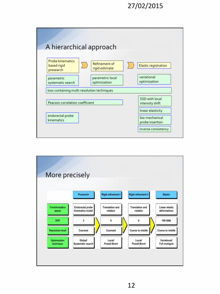
A hierarchical approach
Probe kinematics based rigid presearch
Refinement of rigid estimate
Elastic registration
Pearson correlation coefficient
parametric systematic search
SSD with local intensity shift
loss-containing multi-resolution techniques
endorectal probe kinematics
bio-mechanical probe insertion
parametric local optimization
variational optimization
inverse consistency
linear elasticity
More precisely

27/02/2015
13
Example u
nre
gis
tere
d
affi
ne
elas
tic
Registration evaluation
– 47 patients, 786 tracking volumes • 97.8% correctly registered (visual validation)
– 17 patients, 278 tracking volumes • Comparison to a gold standard computed using
manually segmented point fiducials (calcifications mainly)

27/02/2015
14
Recorded performances without assistance
Biopsym • Providing educational assistance based on
acquired data

27/02/2015
15
Biopsym originality
• Includes didactic material (collab with LES, Grenoble) – Specific exercises (US image
understanding, 3D representation, ability to target a quadrant or a MRI target)
– Related to relevant pieces of information – Two levels of guidance
• Quantitative evaluation of the trainee – Exercises proposed based on the trainee
performances and weaknesses
– US image reading by asking the user to select the different anatomical structures
– Prostate volume measurement – Estimation of the probability of positive biopsies
based on clinical data – Targeted biopsy
Seven types of exercises
MRI target
Volume measurement
1 2 3
4
5 6 7

27/02/2015
16
Learning path
Final evaluation
Initial evaluation B
High grade
Low grade
5 6 7
0
B
2
4
0
B
2
4
• Choosing the most appropriate exercises depending on user results
MCQ
Exercise 2
Exercise 4
Biopsy proc.
MCQ
Exercise 2
Exercise 4
Biopsy proc.
1 2 3 4
32
Performance assessment
A visual feedback about the real location of performed biopsy
sessions allowed improvement of the biopsy distribution
[P. Mozer et al., 2009 ]

27/02/2015
17
Biopsym evaluation
• First experimental evaluations with: – 8 non clinicians (PhD and master students):
reliability, face validity (realism judged by non experts) > ok
– 21 clinicians (14 medical students and 7 trained urologists): content validity (realism judged by experts), construct validity (scoring able to discriminate novice and expert)
• Modifications: score, probe mock-up, image real-time deformation
• Planned experiment: ability to transfer the acquired to skill to real patients
Prostate deformations
• 3D texture mapping and deformation (S. Selmi) – Simplified method – Shape memory model +
control points + voxels
• Complex biomechanical model (J.Sarrazin) – Patient specific – More predictive – Interactive time – MEF, mass-spring, other? – Phantom study done – Data acquisition on patients
interact Data base
visualize
undeformed
US volume
deformed
US volume
Probe
displacement
Clipping
Biomecha.
model

27/02/2015
18
US-guided prostate brachytherapy • Insert radioactive seeds into the
prostate through the perineum
US guidance
Dose planning from US images
• Planned dose: for instance 160Gy
• Dose constraints • Prostate: 160Gy<D90<180Gy and V100>85%
• Urethra: D30<240Gy
• Rectum: less than 1.3cc>160 Gy and D90<80Gy

27/02/2015
19
Image-guided brachy.
• MRI/US non rigid fusion – Surface based registration – Dosimetric evaluation (on 28
patients – PHRC Prostate-Echo) • Systematic underestimate of
US volume w.r.t. MRI • Overestimate of the delivered
dose • In average: volume -8,25% / D90
3% / V100 (160Gy) 3,91% – Development of semi-automated
atlas-based segmentation (atlas built from 36 exams of patients)
Post-implantation evaluation • Based on a CT exam performed one month after
seed implantation • No consideration of seed orientation
• Is it clinically important? Need for accurate seed localization and separation
As planned As implanted As considered in state of the art dose evaluation

27/02/2015
20
INSERM Dorgipro Project (UJF, CHUG, LPSC)
• Automatic detection and classification based on a priori information (seed volume, HU)
• Automatic separation of seeds – K-means – Modified k-means – Mixture of Gaussian
• Orientation given by PCA • Implemented in CamiTK • Evaluation on 2 phantoms and 14
patients (more than 1000 seeds) – Very good accuracy – Very fast – Very few false detections (1,8%)
• See N’Guyen 2015, IEEE TBME
Method cont’ed

27/02/2015
21
Impact on dose ?
• Mis-orientation of seeds
• Impact on dose
– No difference on DVH – Local inhomogeneities
(significant for about 25% of the volume)
• to be confirmed on a larger clinical study
n=1028
mean = 1.06°
sd = 21.1°
angle theta (degrees)
perc
enta
ge (%
)
5
10
15
-50 0 50
n=1028
mean = 0.81°
sd = 27.7°
> 3%
[-3% ; 3%]
< -3%
angle phi (degrees)
pe
rce
nta
ge
(%
)
5
10
15
-50 0 50
Sources of inaccuracy in seed positions
• The prostate moves and gets deformed due to: – Bladder or rectal filling – Patient leg position – Patient breathing – Ultrasound probe constraint – Needle penetration – Edema
• The needles may deflect

27/02/2015
22
Prosper robot
• Objectives: – Seeds implanted as planned – Suppress pubic arch conflict – Make it more rapidly if possible
• Architecture – Needle pre-positioning (5dofs) – Needle insertion (2dofs) – Automatic disengagement system in
case of collision with bone • Our solution to prostate motion
– Limiting US probe motion: 3D US – Rotate the needle – Prostate tracking using 3D/3D non
rigid registration
Accuracy evaluation

27/02/2015
23
Work in progress
• Improvement of trajectory correction – Biomechanical model – Needle steering (collaboration with
LIRMM – labex CAMI)
• Second version of prosper for use on patients (mid-end of 2015)
A pluri-disciplinary approach
SCIENCE
•modeling •image processing •robotics •simulation •etc.
INDUSTRY
•quality insurance •prototype design •large scale diffusion
CLINICS
•specifications •verifications •clinical validation
Regulations CAMI Projects
Ethical committees

27/02/2015
24
Industrial transfer of urology image processing tools
• Embedded in the Urostation® from Koelis (created 2007)
• Biopsy mapping in a product (EC, FDA approved)
• Early 2015: 30000 patients with 80 systems treated worldwide
• Integrates also – MRI/US image fusion – Semi-automatic image
segmentation from statistical atlas
Conclusion
• Several application fields for CAMI with strong potential clinical impact
• Opportunity for new imaging modalities, image processing, models, robotic or navigation assistance, training assistance
• Still a lot to do, to evaluate and to transfer to clinical routine
Specific acknowledgements: Grenoble University Hospital (urology, radiation oncology and radiology and
imaging departments) and La Pitié Salpétrière Hospital, Paris (urology department)
TIMC colleagues and students Funding agencies (ANR, Ministry of Health, INSERM, Région Rhône-Alpes, ANRT) KOELIS
27/02/2015
25
• N’Guyen G, Fouard C, Meneu F, Giraud JY, Troccaz J. Automatic 3D seed location and orientation detection in CT images for prostate brachytherapy. Proceedings of the IEEE ISBI’2014 (International Symposium on Biomedical Imaging), Beijing, May 2014
• Selmi S, Promayon E, Sarrazin J, Troccaz J. 3D Interactive Ultrasound Image Deformation for Realistic Prostate Biopsy SimulationProceedings of the 6th International Symposium on Biomedical Simulation, Strasbourg, 16-17 October, Springer, LNCS vol 8789, Fernando Bello and Stéphane Cotin (Editors), pp122-130, 2014
• Fiard G, Selmi SY, Promayon E, Vadcard L, Descotes JL, Troccaz J. Initial validation of a virtual reality learning environment for prostate biopsies: realism matters! Accepté pour publication dans Journal of Endourology, online first 22oct2013
• Fiard G, Hohn N, Descotes JL, Rambeaud JJ, Troccaz J, Long JA. Targeted MRI-guided prostate biopsies for the detection of prostate cancer: Initial clinical experience with real-time 3-dimensional transrectal ultrasound guidance and magnetic resonance/transrectal ultrasound image fusion. Urology, 2013:81(6):1372-8, online March 1013
• Hungr N, Baumann M, Long JA, Troccaz J . A 3D Ultrasound Robotic Prostate Brachytherapy System with Prostate Motion Tracking. IEEE Transactions on Robotics, 2012, 28(6) : 1382-1397
• Long JA, Hungr N, Baumann M, Descotes JL, Bolla M, Giraud JY, Rambeaud JJ, Troccaz J. Development of a Novel Robot for transperineal needle-based interventions: Focal Therapy, Brachytherapy and Prostate Biopsies. Journal of Urology, 2012, 188:1369-1374, online 20 août 2012
• Hungr N, Long JA, Beix V, Troccaz J. A realistic deformable prostate phantom for multi-modal imaging and needle-insertion procedures. Medical Physics, 2012, 39(4)-2031-2041
• Baumann M, Mozer P, Daanen V, Troccaz J. Prostate Biopsy Tracking with Deformation Estimation. Medical Image Analysis, 2012, 16(3):562-576, Online first May 2011
• Martin S., Troccaz J., Daanen V.. Automated Segmentation of the Prostate in 3D MR Images Using a Probabilistic Atlas and a Spatially Constrained Deformable Model. Medical Physics, 2010, 37(4) :1579-1590
• P. Mozer, M. Baumann, G. Chevreau, A. Moreau-Gaudry, S. Bart, R. Renard-Penna, E. Comperat, P. Conort, M.-O. Bitker, E. Chartier-Kastler, F. Richard, J. Troccaz. Mapping of Transrectal Ultrasound Prostate Biopsies: Quality Control and Learning Curve Assessment by Image Processing. Journal of Ultrasound in Medicine, 2009, 28(4):455-460
• V. Daanen, J. Gastaldo, J.Y. Giraud, P. Fourneret, J.L. Descotes, M. Bolla, D. Collomb, J. Troccaz. MRI/TRUS data fusion for brachytherapy. The International Journal of Medical Robotics and Computer-Assisted Surgery, Vol2, No.3, pp256-261, September 2006
Related publications