Embed Size (px)
Citation preview

University of Groningen
PET imaging of brain sex steroid hormone receptors and the role of estrogen in depressionKhayum, Mohamed Abdul
IMPORTANT NOTE: You are advised to consult the publisher's version (publisher's PDF) if you wish to cite fromit. Please check the document version below.
Publication date:2015
Link to publication in University of Groningen/UMCG research database
Citation for published version (APA):Khayum, M. A. (2015). PET imaging of brain sex steroid hormone receptors and the role of estrogen indepression. University of Groningen.
CopyrightOther than for strictly personal use, it is not permitted to download or to forward/distribute the text or part of it without the consent of theauthor(s) and/or copyright holder(s), unless the work is under an open content license (like Creative Commons).
The publication may also be distributed here under the terms of Article 25fa of the Dutch Copyright Act, indicated by the “Taverne” license.More information can be found on the University of Groningen website: https://www.rug.nl/library/open-access/self-archiving-pure/taverne-amendment.
Take-down policyIf you believe that this document breaches copyright please contact us providing details, and we will remove access to the work immediatelyand investigate your claim.
Downloaded from the University of Groningen/UMCG research database (Pure): http://www.rug.nl/research/portal. For technical reasons thenumber of authors shown on this cover page is limited to 10 maximum.
Download date: 22-02-2022

Chapter 6
Estrogen depletion, but not chronic mild stress,
induced depressive-like behavior and affected
brain glucose metabolism in female rats
Khayum MA, Doorduin J, Copray JCVM, Buwalda B, Koole M, den Boer JA, Dierckx
RAJO, de Vries EFJ
Submitted for publication at Hormones and Behavior

Chapter 6
116
Abstract
The decline in circulating estrogens during menopause increases the risk of
depression. The occurrence of depression in post-menopausal women is likely multi-
factorial in nature as only a minority suffers from depression. Stress could be the
additional trigger for inducing depression. We therefore investigated the effect of
estrogen depletion by ovariectomy and exposure to chronic mild stress (CMS) on
depressive-like behavior and brain glucose metabolism in rats. Ovariectomized rats
were divided into four groups: treatment with placebo or estradiol, and exposed to
CMS or not. Estradiol or placebo treatment was initiated directly after ovariectomy.
The six-week CMS protocol was started two weeks later. Immediately before and
after six weeks of CMS, rats underwent behavioral testing to assess depressive-like
behavior and an [18F]FDG PET scan. Placebo-treated rats showed increased weight
gain in the first two weeks after ovariectomy and more depressive-like behavior after
the CMS period than estradiol-treated rats. At both time points [18F]FDG PET
showed decreased glucose metabolism in brain regions of the basal ganglia, the
limbic system and regions related to cognition and emotion of placebo-treated rats,
as compared to estradiol-treated animals. In addition, an increased glucose
metabolism was found in connected (cortical) regions. Changes in body weight,
behavior and brain glucose metabolism were independent of CMS. In conclusion,
reduced levels of estrogen as a consequence of ovariectomy induced depressive-like
behavior and resulted in changes in glucose metabolism in brain areas involved in
emotion. Exposure to CMS did not enhance the depressive-like behavior in
ovariectomized rats.

Estrogen Depletion but not CMS Induced Depression & Brain Glucose Metabolism
117
Introduction
Estrogens are not only known for their role in the development of secondary sexual
and reproductive functions in females, but are also associated with mental disorders.
The decline in circulating estrogens during menopause increases the risk of
developing depression1, and estrogen replacement therapy was found to alleviate
symptoms of depression in post-menopausal women2,3. The occurrence of depression
in post-menopausal women is, however, not only dependent on the decline in
circulating estrogen levels, but is likely multi-factorial in nature as only a minority of
post-menopausal women suffers from depression4. An additional trigger is thought
to be required to induce the depression4. From demographic studies it was found
that negative life events can trigger depression, especially during the menopausal
transitional period5,6. These negative life events, which can act as stressors in
menopausal women, include higher body mass index, smoking, lack of sleep,
financial difficulties, marital problems, and health problems7,8.
Exposure of rodents to chronic mild stress (CMS) is a widely used model to
induce depressive-like behavior9. CMS involves continuous exposure to a variety of
mild stressors that can mimic negative life events, including periods of food and
water deprivation, temperature reductions and changes in cage mates. The CMS
protocol induces the core symptom of depression, namely anhedonia, which is the
inability of an individual to feel pleasure with things considered pleasurable.
Anhedonia is accompanied by alterations in noradrenergic, dopaminergic and
serotonergic neurotransmission10. CMS induced depressive-like behavior could be
reduced by chronic treatment with antidepressants9,10. We hypothesized that the
combination of negative life events, i.e. stressors, with declining estrogen levels may
culminate in a post-menopausal depression. The objective of this study was therefore
to determine if reduced circulating estrogen levels affect the susceptibility of female
rats to CMS. To this purpose, we investigated the effect of estrogen depletion by
ovariectomy and the effect of exposure to CMS for 6 weeks on depressive-like
behavior. Since estrogen and estrogen receptors play a role in maintaining glucose
homeostasis in the brain11 and glucose metabolism is altered in the brain of
depressed patients12, we also assessed the central effects of estrogen depletion and
CMS on brain glucose metabolism by positron emission tomography (PET) with the
tracer 2-[18F]fluoro-2-deoxyglucose ([18F]FDG).
Materials and methods
Animals
Female outbred Wistar rats (n=32, 9-12 weeks old, 200-250 g) were purchased from
Harlan (Horst, The Netherlands). The rats were housed individually in Macrolon
cages on a layer of wood shavings in a room with a constant temperature (21±2oC)
and a fixed 12 h light-dark regime. Standard laboratory chow and water were
available ad libitum. After arrival, the rats were allowed to acclimatize for at least 7
days. During the acclimatization period and throughout the study, all rats were
handled daily by the investigator. All experiments were approved by the Institutional

Chapter 6
118
Animal Care and Use Committee of the University of Groningen (IACUC-RuG),
Groningen, The Netherlands (study protocol: DEC 5842D).
Study design
All rats were ovariectomized and divided into two treatment groups, placebo and
estradiol, and further divided into two condition groups, control rats and rats
subjected to chronic mild stress for six weeks. In Figure 1 the study design is
represented in a time line of the experimental procedures.
On day 0 the rats were ovariectomized and either a placebo or an estradiol
releasing pellet was implanted (Figure 1). Rats were allowed to recover from the
surgery for 12 days. On day 12 post-ovariectomy (baseline), a sucrose consumption
test (SCT) was performed, followed by a [18F]FDG PET scan on day 13, an open field
test (OFT) on day 14 and the forced swim test (FST) on day 15 (pretest) and 16 (test).
From day 17 until day 59 (6 weeks), the rats were exposed to mild stressors according
to the CMS protocol (Table 1). Control animals were not exposed to these stressors.
During this 6-week period all rats underwent a weekly SCT (7 times). On day 60, all
rats were subjected to a [18F]FDG PET scan, followed by the OFT on day 61 and the
FST on day 62 (pretest) and 63 (test). Immediately after the FST, all rats were
sacrificed and the brain, uterus and adrenal glands were removed and weighed.
Figure 1: Study design. On day 0 rats were ovariectomized (OVX) and implanted with a placebo or
estradiol releasing pellet. After 12 days of recovery, rats were exposed to behavioral tests (SCT:
sucrose consumption test; OFT: open field test; and FST: forced swim test (pFST: pretest, tFST: test)),
to [18F]FDG positron emission tomography (PET) and to chronic mild stress (CMS).
Ovariectomy and estradiol replacement
Bilateral ovariectomy was performed under isoflurane anesthesia as previously
described Khayum et al 13 Both sides of the abdomen of the anesthetized rats were
shaved and sterilized with 70% ethanol. A small incision was made through the skin
and muscle layer of the abdomen to expose the ovaries. The blood vessels that supply
the ovaries were ligated with silk sutures and the ovaries with the associated fat pads
and a part of the uterus were removed. The muscle layer and the skin were then
closed with chromic sutures.
Immediately after ovariectomy, a placebo pellet or an estradiol-releasing pellet
(NC-111 Placebo, and NE-121 17ß-estradiol 2.25mg/pellet, 90 day release, Innovative

Estrogen Depletion but not CMS Induced Depression & Brain Glucose Metabolism
119
research of America, Florida, USA) was subcutaneously implanted in the neck of the
rat. In placebo-treated rats, circulating estrogen levels were reduced as a
consequence of ovariectomy, as is observed in post-menopausal women. In the
estradiol-treated rats, the pellets released 25 μg of estradiol per day with zero order
kinetics, resulting in preservation of physiological levels of estradiol in plasma.
After completion of the surgery, the rats were subcutaneously injected with
finadyne (2.5.mg/kg Flunixin, Schering-Plough N.V/S.A., Belgium) for pain relief.
Administration of finadyne was repeated 24 h after surgery.
Chronic mild stress
The chronic mild stress (CMS) protocol14 started at day 17 after ovariectomy and
consisted of 6 weekly cycles of exposure to different stressors (Table 1). The stressors
were: paired housing (each rat acted as a resident for six times and as an intruder for
six times), tilting of cages to 45 degrees, soiled cage, stroboscopic illumination (1500
watts, 2 flashes per second), and food and water deprivation. Control rats were
housed in a separate room, and were not exposed to these stressors. Before and
during the CMS protocol, rats were weighed every week to monitor body weight
changes.
Table 1: Schedule of the CMS protocol
Day Stressors
Sunday 10:00 h: Sucrose test, followed by normal housing
20:00 h: Paired housing for 14 hours
Monday 10:00 h: Tilting of the cages (45 degrees) for 10 hours
20:00 h: Soiled cage (250 ml of water was poured into the sawdust bedding) for 14 h
Tuesday 10:00 h: Cage cleaning, followed by water deprivation for 10 hours
20:00 h: Paired housing for 14 hours
Wednesday 10:00 h: Stroboscopic illumination in darkness for 10 hours
20:00 h: Food deprivation for 14 hours
Thursday 10:00 h: Tilting of the cages (45 degrees) for 10 hours
20:00 h: Cages were put back in straight position. Animals were allowed a period
without exposure to a stressor
Friday 10:00 h: Stroboscopic illumination in darkness for 10 hours
20:00 h: Soiled cage (250 ml of water was poured into the sawdust bedding) for 14 h
Saturday 10:00 h: Cage cleaning followed by no stress
20:00 h: Food and water deprivation for 14 hours
Sucrose consumption test
From day 2 until day 11, rats were habituated daily to the sucrose consumption test
(SCT) by putting two bottles in their home cages for 1h: one bottle contained normal
water and the other bottle 1% sucrose solution. After habituation, the rats were
subjected to the baseline SCT on day 12. Prior to the test, rats were deprived of food
and water for 14 h. The bottle with the 1% sucrose solution and the bottle with
normal water were placed at the same location in the home cage of the animals as
during habituation. After 1 h, the bottles were removed and the consumption of

Chapter 6
120
sucrose solution was measured by subtracting the weight of the bottle after the SCT
from the initial weight of the bottle. The sucrose consumption is expressed as grams
of sucrose solution consumed per hour. The SCT was performed on a fixed day of the
week at a fixed time (Sundays between 9 and 10 AM) and was repeated 6 times
during CMS protocol.
Open field test
The open field test was performed on day 14 and 61 post-ovariectomy. The rats were
placed in the center of a circular black arena (diameter of 80 cm, height of 40 cm).
Rats were allowed to explore the arena for a period of 5 min. During this period, the
behavior of the rats in the arena was videotaped for subsequent behavioral analysis.
After each open field experiment, the arena was cleaned with water and 70% alcohol,
and dried. Videos were analyzed using Ethovision XT 8 (Noldus Information
Technology, Wageningen, The Netherlands). The outcome parameters were the
distance travelled and the time spent in the center of the arena and the time spent at
the border of the arena. A reduction in the distance travelled or a reduction in the
time spent in the border zone of the open field reflects increased anxiety.
Forced swim test
The forced swim test (FST) was performed on day 15/16 and on day 62/63 post-
ovariectomy. On the first day of each test, the rats were subjected to a pretest FST.
The rat was individually placed in a cylindrical water tank (diameter of 20 cm and a
height of 40 cm) for 15 min. The water level in the tank was set at 30 cm to ensure
that the rats could swim or float without touching the bottom of the tank with
their hind limbs. The temperature of the water was maintained at 21±1 oC. After 15
min, the rats were taken out of the tank, dried with paper towels and returned to
their home cages. On the second day, the rats were subjected to the test FST by
placing the animals again in the cylindrical water tank for 5 min. After the test FST,
the rats were dried with paper towels and returned in their home cages. After every
test, the water from the tank was discarded; the tank was washed and filled with
fresh water for the next test. The entire pretest and test FST sessions were recorded
on video for subsequent analysis using Ethovision XT 8 software (Noldus
Information Technology, Wageningen, The Netherlands). For digital video analysis
of the behavior of the animal, the water tank was divided into two zones: the surface
zone and the diving zone. The surface zone was defined as the region between 10 cm
above the water surface until the depth equal to the vertical body length of the rat
below the water surface. The diving zone contained the whole area below the surface
zone. The outcome parameters were the time spent on swimming (horizontal
movement crossing all quadrants of the swim chamber), climbing (upward-directed
movements of the forepaws, usually along the side of the swim chamber or floating
(no additional activity observed other than that required to keep the rat’s head above
the water) in the surface zone.

Estrogen Depletion but not CMS Induced Depression & Brain Glucose Metabolism
121
[18F]FDG PET imaging
At day 13 and day 60 post-ovariectomy, the rats were subjected to an [18F]FDG PET
scan. Rats were placed in pre-warmed cages with a temperature of 30 °C for
environmental adaptation and minimization of [18F]FDG uptake in brown fat. After
30 min of adaptation, rats were anesthetized with isoflurane (5% induction and 2%
maintenance, in medical air) and injected with [18F]FDG via the tail vein (14.5±2.5
MBq). Hereafter, rats were returned to their pre-warmed cages to recover from
anesthesia. Forty min after tracer injection, the rats were again anesthetized with
isoflurane and placed in the small animal PET camera (Focus 220, Siemens Medical
Solutions, USA, Inc). Rats were positioned in a transaxial position with their heads in
the field of view. At 45 min after [18F]FDG injection, a static PET scan of 30 min was
acquired. During the PET scan, rats were kept under isoflurane anesthesia (2% in
medical air) and body temperature was maintained by heating pads. Eye salve was
used to prevent dehydration of the eyes. After completion of the emission scan, a
transmission scan with a 57Co point source was acquired for 515 seconds to allow for
correction of attenuation and scatter by tissue. List mode emission data was
iteratively reconstructed into a single frame image of 30 min (OSEM2D, 4 iterations,
and 16 subsets). PET data were normalized and corrected for attenuation, scatter,
random coincidences and radioactive decay.
PET image analysis
Whole-brain volume of interest (VOI) based analysis was performed to determine
global differences in [18F]FDG uptake, using Inveon 4.0 software (Siemens Medical
Solutions, USA, Inc). A VOI was drawn around the whole brain on a T2 MRI
template15 that was co-registered with the PET images. The VOI was then copied to
the PET images to determine the accumulation of radioactivity in the whole brain (in
Bq/cm3), which was converted into the standardized uptake value (SUV). The SUV
was defined as: [tissue activity concentration (Bq/cm3)]/[injected dose (Bq)/body
weight (g)]. It was assumed that 1 cm3 of brain tissue equals 1 g.
Voxel-based analysis was performed using SPM8 software (SPM; Wellcome
Department of Cognitive Neurology, University College London, UK), in combination
with an in-house toolbox that allows the visualization of the results over a rat ‘glass
brain’ (maximum intensity projection map), and to report the coordinates in Paxinos
space. [18F]FDG PET images were co-registered with a [18F]FDG rat brain template
that was developed according to the methodology described by Casteels et al. 16.
Extra-cerebral regions were removed from the PET images by the application of a
mask. The PET images were smoothed with a 1.2 mm isotropic Gaussian kernel.
Global brain uptake differences between animals were normalized by proportional
scaling relative to the mean whole brain [18F]FDG uptake.
Voxel-based analysis was performed using two-sample t-tests (paired within-
groups, and unpaired between groups). T-map data were interrogated at uncorrected
p<0.005 with an extent threshold of 200 voxels. Only clusters with a corrected
family-wise error p<0.05 were considered significant.

Chapter 6
122
Statistical analysis
All data are expressed as mean ± standard error of mean (SEM). Statistical analysis
was performed using IBM SPSS statistics 22 for Windows. Adrenal gland weight,
uterus weight and whole brain [18F]FDG uptake (SUV) were analyzed by univariate
analysis with condition (control or CMS) and treatment (estradiol or placebo) as
factors. The baseline measures, i.e. at day 12-17 post ovariectomy (before CMS), of
the body weight, SCT, OFT, FST and [18F]FDG PET were analyzed with a one-way
ANOVA, to compare the placebo-treated with the estradiol-treated ovariectomized
rats. The bodyweight and the SCT were analyzed by repeated measures ANOVA with
time, condition and treatment as factors, using the baseline measures as a covariate.
The post-CMS OFT, FST and [18F]FDG PET were analyzed by univariate analysis
with condition and treatment as factors, using the baseline measures as a covariate
measures. Significance was reached when the probability (p) was <0.05.
Results
Body weight
To assess changes in body weight as a consequence of ovariectomy and CMS, the
body weight gain from day 0 was determined at different time points (Figure 2). To
determine the effect of treatment, the body weight gain at day 10 and 17 post-
ovariectomy, i.e. before the start of CMS, was compared between groups. A
statistically significantly higher bodyweight gain was found for placebo-treated rats,
when compared to the estradiol-treated rats (p<0.001), both at day 10 (34.3±3.4 vs. -
13.6±1.8) and at day 17 (43.7±5.6 vs. -13.9±6.8) post-ovariectomy.
To determine the main effect of treatment (placebo or estradiol), condition
(control or CMS) and their interaction on the body weight gain post-CMS, the body
weight gain measure at day 17 was used as a covariate in the repeated measures
analysis. A statistically significant effect of time on body weight gain was observed
(F(1.8,49.2)=48.4, p<0.001), showing that there was an increase in body weight over
time. No statistically significant interaction of treatment and condition with the body
weight gain over time was observed. However, a statistically significant main effect of
treatment was observed for the body weight gain (F(1,27)=20.3, p<0.001), showing
that, independent of time, the placebo-treated rats had a higher body weight gain.
CMS did not affect the body weight gain, as no main effect of condition was found.
Uterus and adrenal gland weight
The weight of the uterus was determined as an indicator of changes in circulating
estrogen levels. The uterus weight of the control rats (not exposed to CMS) was
0.74±0.08 g for estradiol-treated rats and 0.22±0.03 g for placebo-treated rats. In
rats exposed to CMS, the weight of the uterus was 0.64±0.09 g for estradiol-treated
rats and 0.22±0.02 g for placebo-treated rats. Ovariectomy was found to cause a
statistically significant decrease in the uterus weight of placebo-treated rats, as
indicated by a main effect of treatment (F(1,27)=59.8, p<0.001). Neither a statistically
significant main effect of condition, nor an interaction between treatment and

Estrogen Depletion but not CMS Induced Depression & Brain Glucose Metabolism
123
condition was found (p>0.05), showing that CMS did not affect the weight of the
uterus.
Figure 2: Body weight gain of estradiol- and placebo-treated ovariectomized rats over time. Rats
were exposed to chronic mild stress (CMS) from day 17 until day 59. Control animals were not
exposed to CMS. Data are presented as mean ± SEM, *p<0.001 indicates a significant effect of time
and of treatment.
The weight of the adrenal glands was measured to determine the effect of stress
induced by the CMS protocol. Adrenal gland weight is indicative of chronic activation
of the hypothalamus-pituitary-adrenal (HPA) axis during stress. In control rats (not
exposed to CMS), the adrenal gland weight was 0.078±0.009 g for estradiol-treated
rats and 0.064±0.005 g for placebo-treated rats. In rats exposed to CMS, the weight
of the adrenal glands was 0.066±0.004 g for estradiol-treated rats and 0.068±0.006
g for placebo-treated rats. Neither treatment (placebo or estradiol) nor condition
(control or CMS) affected the weight of the adrenal glands, as no statistically
significant main and interaction effects were found.
Sucrose consumption test
The SCT was performed at baseline, i.e. at day 12 post-ovariectomy, and 7 times
during the CMS protocol (Figure 3). At baseline, no statistically significant difference
in sucrose solution consumption was found between placebo (9.0±1.4) and estradiol
(7.5±0.9) treated ovariectomized rats (p=0.368). Repeated measures analysis, using
the baseline measure as a covariate, revealed no significant differences in the sucrose
solution consumption in rats over time (F(4.3,115.2)=1.56, p=0.163). In addition, no
effect of treatment (placebo or estradiol) or condition (control or CMS) on the
sucrose consumption was observed.
Open field test
The results of the OFT at baseline and after CMS are presented in Figure 4. At
baseline, i.e. at day 14 post-ovariectomy, no statistically significant differences were
found between placebo- and estradiol-treated rats in the total distance travelled and

Chapter 6
124
in the distance travelled in the border zone or in the center zone (p>0.05). In
addition, no statistically significant differences were found in the time spent in the
center or at the border of the arena (data not shown). These findings indicate that the
reduced estrogen levels in ovariectomized rats did not affect open field behavior at
baseline.
Figure 3: Changes in sucrose consumption (SCT) of estradiol- and placebo-treated ovariectomized
rats at over time, during chronic mild stress (CMS). Control animals were not exposed to CMS. Data
are presented as mean ± SEM.
To assess if treatment (placebo or estradiol), condition (control or CMS) and their
interaction affected open field behavior, the distance travelled and time spent in the
center and the border zone were determined on day 61 post-ovariectomy, using the
baseline measure as a covariate. As for the baseline results, OFT at day 61 did not
reveal any statistically significant difference (p>0.05) in the distance travelled or the
time spent in any zone, showing that treatment and condition did not affect open
field behavior.
Figure 4: Open field test of estradiol- and placebo-treated ovariectomized rats at day 14 (baseline)
(A) and at day 61 (B). Rats were either exposed to mild chronic stress (CMS) from day 17 to 59 or were
not exposed to these stressors (control). Data are presented as mean ± SEM.

Estrogen Depletion but not CMS Induced Depression & Brain Glucose Metabolism
125
Forced swim test
The FST performed at baseline (day 16 post-ovariectomy) revealed that the reduced
levels of estradiol in placebo-treated ovariectomized rats did not affect the duration
of floating (p=0.802) and climbing (p=0.177) behavior (Figure 5A). At baseline, there
was a trend towards more swimming behavior in placebo-treated rats than in
estradiol-treated rats (12.8%, p=0.058).
Figure 5: Forced swim test behavior of estradiol- and placebo-treated ovariectomized rats at day 16
(baseline) (A) and at day 63 (B). Rats were either exposed to mild chronic stress (CMS) from day 17 to
59 or were not exposed to these stressors (control). Data are presented as mean ± SEM. Significant
main effect of treatment are indicated as *p<0.05 and **p<0.005.
The FST on day 63 was performed to determine if treatment (placebo or estradiol),
condition (control or CMS) and their interaction affected FST behavior, using the
baseline measure as a covariate (Figure 5B). The FST at day 63 post-ovariectomy
revealed a statistically significant main effect of treatment for floating (F(1,26)=11.6,
p=0.002), swimming (F(1,26)=6.0, p=0.021) and climbing (F(1,26)=14.3, p=0.001).
Ovariectomy increased floating behavior and decreased both swimming and climbing
behavior. No statistically significant main effect of condition (p>0.05) or an
interaction between treatment and condition (p>0.05) was found for any of the
behaviors, showing that CMS did not affect FST behavior.
Whole brain glucose metabolism
Whole brain [18F]FDG uptake (SUV) is shown in Figure 6A (baseline) and 6B (day 60
post-ovariectomy). No statistically significant differences in whole brain [18F]FDG
uptake between estradiol- and placebo-treated rats were found at baseline (p=0.145).
To determine the main effect of treatment (placebo or estradiol), condition (control
or CMS) and their interaction on whole brain [18F]FDG uptake, the uptake at day 13
was used as a covariate. No statistically significant effect of treatment, condition or
an interaction between treatment and condition on whole brain [18F]FDG uptake was
found at day 60 (p>0.05).

Chapter 6
126
Figure 6: Whole brain [18F]FDG uptake of estradiol- and placebo-treated ovariectomized rats at day
13 (baseline) (A) and at day 60 (B). Rats were either exposed to mild chronic stress (CMS) from day 17
to 59 or were not exposed to these stressors (control). Data are presented as mean ± SEM.
Voxel-based analysis of [18F]FDG PET scans
Voxel-based analysis of [18F]FDG PET scans revealed regions with significantly
increased (red) or decreased (blue) brain glucose metabolism, as presented in Figure
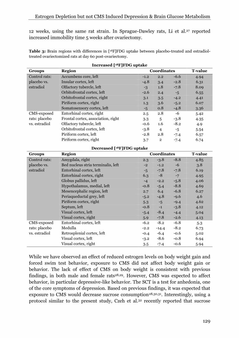
7. Regions of the brain with the highest T-value clusters are presented in Table 2 and
3. At two weeks after ovariectomy (baseline), placebo-treated rats displayed
statistically significantly increased glucose metabolism in the motor and entorhinal
cortex, as compared to estradiol-treated rats. In addition, statistically significantly
decreased glucose metabolism was observed in, amongst others, the thalamus,
periaqueductal grey (PAG), superior colliculus, caudate putamen and cortical regions
(Figure 7A, Table 2).
At day 60 post-ovariectomy, placebo-treated control rats (not exposed to
CMS) showed a significant increase in glucose metabolism in cortical regions and a
significant decrease in thalamus, hypothalamus, globus pallidus, amygdala,
periaqueductal grey, and several cortical regions, as compared to estradiol-treated
control rats (Figure 7B, Table 3). In contrast, placebo-treated rats that were exposed
to CMS showed increased glucose metabolism in several cortical regions and
decreased glucose metabolism in the medulla and several other cortical regions, as
compared to estradiol-treated rats exposed to CMS (Figure 7C, Table 3). Comparison
of placebo-treated control rats and placebo-treated rats exposed to CMS did not
reveal any statistically significant differences in glucose metabolism (Figure 7D).
Likewise, no statistically significant differences in glucose metabolism were found
between control estradiol-treated rats and estradiol-treated rats exposed to CMS
(Figure 7E, table 3).
Discussion
Reduction in circulating estrogens in post-menopausal women can result in the
development of depression. As not all post-menopausal women develop depression,
an additional trigger is likely required. Stress may be such a trigger and we therefore
investigated if chronic mild stress could lead to the development of depressive-like
behavior in ovariectomized rats. Our results showed that a reduction in circulating

Estrogen Depletion but not CMS Induced Depression & Brain Glucose Metabolism
127
estrogens affected body weight and brain glucose metabolism, and induced
depressive-like behavior. We did, however, not find any effect of chronic mild stress
on any of these outcome parameters.
The body weight gain of the female rats was found to be affected by both
ovariectomy and estradiol treatment. Healthy female Wistar rats with a bodyweight
of 200 to 250 grams gain about 1 gram of bodyweight per day. The placebo-treated
ovariectomized rats in this study gained about 44 grams of bodyweight in the first 10
days after ovariectomy, while the estradiol-treated ovariectomized rats lost about 14
grams of body weight in the same period.
Figure 7: Brain areas with significant effects on brain glucose metabolism: A) The effect of estradiol
depletion on brain glucose metabolism between placebo- and estradiol-treated rats at day 13 post-
ovariectomy (n=16). B and C) The effect of circulating estrogens on brain glucose metabolism in
control and CMS-exposed rats at day 60 post-ovariectomy (n=8). D and E) The effect of exposure to
CMS on brain glucose metabolism in placebo- and estradiol-treated rats (n=8).

Chapter 6
128
Table 2: Brain regions with differences in baseline [18F]FDG uptake (day 13) between placebo-treated
and estradiol-treated ovariectomized rats.
Increased [18F]FDG uptake (placebo vs. estradiol)
Region Coordinates T-value
Entorhinal cortex, right 4.7 -7.8 -7.8 7.24
Motor cortex, left -2.8 3.6 -0.6 7.26
Decreased [18F]FDG uptake (placebo vs. estradiol)
Region Coordinates T-value
Caudate putamen, left -4.2 0.6 -6.4 4.28
Cerebellum -0.6 -10.2 -6 5.19
Entorhinal cortex, left -4.8 0.4 -6.6 4.2
Entorhinal cortex, right 4.7 -1 -7.6 3.47
Insular cortex, left -5 0 -6.8 4.18
Periaqueductal grey 0.7 -6.4 -5.2 5.49
Pons, left -1.6 -9 -5.8 5.73
Superior colliculus, left -2.2 -8.9 -3.4 5.48
Thalamus, dorsal midline, right 1.3 -4 -4 6.9
Thalamus, dorsolateral, left -1.8 -3.8 -4.8 6.35
After those 10 days, the bodyweight gain was similar for all groups and comparable
to the normal gain in body weight of female Wistar rats. This finding of increased
bodyweight gain after ovariectomy is consistent with previous findings from animal
models, and is likely related to an increase in body fat17–19. Post-menopausal women
also often suffer from weight gain, but it has been consistently shown that this is a
consequence of aging rather than menopause19. However, the post-menopausal
decline in estrogens was associated with a more rapid increase in fat mass and the
redistribution of fat to the abdomen20,21. Hormone replacement therapy (estrogen
and progestin) was found to prevent this fat redistribution, although the effect was
small22.
To assess depressive-like behavior in placebo- or estradiol-treated
ovariectomized rats, the sucrose consumption, open field behavior and forced swim
test behavior were determined. Behavior at baseline, i.e. 16 days after ovariectomy
and before the start of CMS, was not affected by reduced levels of estrogens in the
placebo-treated ovariectomized rats. However, an effect of treatment was found on
forced swim test behavior at day 63. Reduced levels of estrogens in the placebo-
treated ovariectomized rats led to an increase in floating behavior and a decrease in
swimming and climbing behavior, confirming the role of estrogen in regulating
depressive-like behavior. The finding of increased floating behavior after
ovariectomy is consistent with previous studies that report an increase in immobility
time in both rats and mice23–25. Although the increased immobility time after
ovariectomy is consistently found, there are discrepancies in the time between
ovariectomy and the occurrence of depressive-like behavior, which could be related
to the rat strain used and the testing conditions. We did not find changes in floating
at day 16 post-ovariectomy, but did so at day 63. In contrast, Estrada-Camerena et
al.26 showed an increase in immobility at 1 week after ovariectomy, but not at 3 and
Estrogen Depletion but not CMS Induced Depression & Brain Glucose Metabolism
129
12 weeks, using the same rat strain. In Sprague-Dawley rats, Li et al.27 reported
increased immobility time 5 weeks after ovariectomy.
Table 3: Brain regions with differences in [18F]FDG uptake between placebo-treated and estradiol-
treated ovariectomized rats at day 60 post-ovariectomy.
Increased [18F]FDG uptake
Groups Region Coordinates T-value
Control rats:
placebo vs.
estradiol
Accumbens core, left -1.2 2.2 -6.6 4.94
Insular cortex, left -4.8 3.4 -2.8 6.31
Olfactory tubercle, left -3 1.8 -7.8 8.09
Orbitofrontal cortex, left -2.6 2.4 -5 6.55
Orbitofrontal cortex, right 3.1 3.5 -4.2 4.41
Piriform cortex, right 1.3 3.6 -5.2 6.07
Somatosensory cortex, left -5 0.8 -4.8 3.36
CMS-exposed
rats: placebo
vs. estradiol
Entorhinal cortex, right 2.5 2.8 -6 5.42
Frontal cortex, association, right 3.3 5 -3.8 4.35
Olfactory tubercle, left -0.6 1.6 -8.2 4.9
Orbitofrontal cortex, left -3.8 4 -5 5.54
Piriform cortex, left -2.8 2.8 -7.4 6.57
Piriform cortex, right 3.7 2 -7.4 6.74
Decreased [18F]FDG uptake
Groups Region Coordinates T-value
Control rats:
placebo vs.
estradiol
Amygdala, right 2.3 -3.8 -8.8 4.85
Bed nucleus stria terminalis, left -2 -1.2 -6 3.8
Entorhinal cortex, left -5 -7.8 -7.8 6.19
Entorhinal cortex, right 6.3 -8 -7 4.95
Globus pallidus, left -4 -2.2 -5.8 4.06
Hypothalamus, medial, left -0.8 -5.4 -8.8 4.69
Mesencephalic region, left 2.7 6.4 -6.8 6.27
Periaqueductal grey, left -5.2 -4.8 -9.6 4.6
Piriform cortex, right 5.3 -5 -9.4 4.62
Septum, left -0.8 -1 -3.8 4.12
Visual cortex, left -5.4 -8.4 -4.4 5.04
Visual cortex, right 5.9 -7.8 -2.6 4.13
CMS-exposed
rats: placebo
vs. estradiol
Entorhinal cortex, left -6.2 -8.2 -6.8 5.3
Medulla -2.2 -14.4 -8.2 6.73
Retrosplenial cortex, left -0.4 -6.4 -0.6 5.02
Visual cortex, left -3.2 -8.6 -0.8 6.94
Visual cortex, right 3.5 -7.4 -0.6 5.94
While we have observed an effect of reduced estrogen levels on body weight gain and
forced swim test behavior, exposure to CMS did not affect body weight gain or
behavior. The lack of effect of CMS on body weight is consistent with previous
findings, in both male and female rats28,29. However, CMS was expected to affect
behavior, in particular depressive-like behavior. The SCT is a test for anhedonia, one
of the core symptoms of depression. Based on previous findings, it was expected that
exposure to CMS would decrease sucrose consumption28,30,31. Interestingly, using a
protocol similar to the present study, Czeh et al.32 recently reported that sucrose

Chapter 6
130
consumption was decreased by 30% in about 45% of the male rats exposed to CMS,
but in none of the control rats. This finding confirmed earlier findings showing that
rats exposed to CMS segregated into two subgroups: a group sensitive to CMS
showing decreased sucrose consumption, and a group resilient to CMS with
unaffected sucrose consumption33. Apparently, not all rats are affected by CMS in a
similar way, which could explain the lack of an overall effect of CMS on sucrose
consumption found in our study. We observed that the majority (60%) of the female
rats showed a decrease in sucrose consumption between baseline and day 59,
independent of treatment (placebo or estradiol) or condition (control or CMS).
In addition to the lack of an effect of CMS on sucrose consumption, CMS did
not affect behavior in the OFT and in the FST either. The OFT can be used to
measure anxiety-related behavior by measuring the time the rat spends in the center
of the arena. Furthermore, decreased locomotor activity in the OFT can be used as a
measure of emotionality in rats34. Previous studies have shown decreased
exploration as a consequence of CMS, in both male and female rats29, but also
increased exploration has been reported30,35. A study by Pijlman et al.36 suggested
that activity in the open field is dependent on the type of stressor. Physical stress
caused a decrease in activity, whereas emotional stress caused an increase in activity.
Clearly, open field behavior is affected by many factors and it could be that the
physical stressors in our study were not severe enough to induce (additional)
depressive-like behavior that resulted in changes in open field activity. This is
supported by our finding that CMS did not affect FST behavior in placebo- or
estradiol-treated ovariectomized rats. In male rats, CMS has been shown to induce
an increase in the duration of immobility in the forced swim test, indicative of
depressive-like behavior37–39, although others did not find any effect40.
The lack of depressive-like behavior after CMS in this study could be related to
the stressors used in the CMS protocol. The placebo- or estradiol-treated
ovariectomized rats might not have experienced these to be stressful, or might have
habituated to the individual stressors. However, the CMS protocol used was based on
previous studies that did show an effect on sucrose consumption and FST behavior31.
Despite the fact that CMS was shown to induce depressive-like behavior in female
rats, there are other studies showing that the effects of CMS are less robust in female
rats, when compared to male rats. With regard to the SCT, sex differences (female
rats tend to drink more sucrose solution than male rats) could differentially affect the
measurement of anhedonia41. Indeed, Dalla et al. 31 found a smaller decrease in
sucrose consumption after CMS in normal cycling female rats than in male rats. Sex
differences have also been reported for the FST and locomotor activity in the OFT41.
In the FST, female rats were reported to cope better with the situation by displaying
more active behavior, while in the open field female rats tend to show lower
exploratory behavior than male rats after CMS. Overall, it is clear that there are sex
differences in coping with CMS, which could be related to circulating estrogen levels.
We observed that the decrease in circulating estrogens after ovariectomy
induced increased immobility in the FST. Additional exposure to CMS did not further
increase immobility. CMS did also not affect immobility in estradiol-treated rats,

Estrogen Depletion but not CMS Induced Depression & Brain Glucose Metabolism
131
which can be related to a protective effect of the physiological levels of circulating
estradiol. Thus, it appears that estrogens can protect against depressive-like
behavior, which is consistent with the findings that the risk of major depression in
women is higher during and immediately after menopause42, and that the year since
menopause was associated with depressive symptoms43.
In addition to behavioral changes, changes in circulating estrogens and
exposure to stressors can affect brain functioning. Brain functioning can be assessed
by determining changes in brain glucose metabolism. Estrogens play a role in
maintaining glucose homeostasis in the brain11 and glucose metabolism is altered in
the brain of depressed patients12. In our study, [18F]FDG PET at day 13 after
ovariectomy, i.e. before CMS, revealed mainly a decrease in brain metabolism in
different brain regions in placebo-treated rats, when compared to estrogen-treated
rats. These brain regions included part of the basal ganglia (i.e. thalamus and
caudate putamen), the superior colliculus, the insular cortex, the entorhinal cortex
and the periaqueductal grey (PAG). At day 60 post-ovariectomy, placebo-treated rats
not exposed to CMS revealed decreased brain metabolism in similar brain regions
when compared to estradiol-treated rats, in particular in the limbic system (i.e.
hypothalamus, septum, amygdala, piriform cortex and the entorhinal cortex), visual
cortex, mesencephalic region, bed nucleus of the stria terminalis (BNST) and PAG.
The majority of the brain regions with decreased glucose metabolism as a
consequence of reduced estrogen levels are involved in cognition and emotion. While
only the basal ganglia are affected at day 13, most of areas of the limbic systems show
reduced brain glucose metabolism at 60 days after ovariectomy, suggesting that the
effect of estrogen depletion on brain metabolism is increasing over time. This is
consistent with our finding of increased immobility in the forced swim test, i.e.
depressive-like behavior, at day 63, which was not observed at day 16 post-
ovariectomy. As some of the regions with reduced glucose metabolism (i.e.
mesencephalic region, caudate putamen and thalamus) are also involved in
controlling motor skills, it can be argued that the immobility is a consequence of a
general reduction in movement. Since open field behavior was not affected, this
explanation seems less plausible. Increased brain glucose metabolism was also
observed, mainly at day 60 after ovariectomy, in brain areas connecting to the basal
ganglia and limbic system. These areas include the olfactory tubercle, the
orbitofrontal cortex, the accumbens core and the somatosensory cortex. Why these
regions show increased metabolism is not clear, but it could be related to a partly
compensatory mechanism.
CMS had no effect on brain glucose metabolism, as no differences were found
between the PET scans made before and after CMS, using a within-subject design.
Differences in brain metabolism between placebo- and estradiol-treated rats at day
60 were similar for rats exposed to CMS and those not exposed to CMS; only an
effect of decreased estrogen levels was found. This finding is consistent with CMS
being unable to cause any behavioral change, in particular the inability to induce
depressive-like behavior. CMS was found to affect brain metabolism in male rats, as
Hu et al. 35 reported increased glucose metabolism in the auditory cortex and

Chapter 6
132
decreased metabolism in the inferior colliculus and piriform cortex. Likely,
differences in the experimental environment or gender differences do also play a role
in the differential effect of CMS on brain glucose metabolism.
Conclusion
Reduced levels of estrogen as a consequence of ovariectomy can induce depressive-
like behavior and changes in glucose metabolism in brain areas involved in emotion.
Only limited studies on the relation between circulating estrogen levels and brain
glucose metabolism have been performed in women. Additional studies in rats can
contribute to the understanding of the central effects of the decline in circulating
estrogen levels after menopause and hormonal replacement therapy. Exposure to
CMS neither induced depressive-like behavior nor potentiated the effect of reduced
estrogen levels on depressive-like behavior in ovariectomized rats. It is worthwhile to
determine if other stressors, which are more emotional than physical in nature, could
provoke depressive-like behavior or enhance the effect of estrogen depletion,
especially since gender differences exist in the stress response and women might be
more vulnerable to emotional stress.

Estrogen Depletion but not CMS Induced Depression & Brain Glucose Metabolism
133
References
1. Bryant C, Judd FK, Hickey M. Anxiety during the menopausal transition: A systematic review. J. Affect. Disord. 2012;139:141-148.
2. Zweifel JE, O’Brien WH. A meta-analysis of the effect of hormone replacement therapy upon depressed mood. Psychoneuroendocrinology 1997;22:189-212.
3. Whooley MA, Grady D, Cauley JA. Postmenopausal estrogen therapy and depressive symptoms in older women. J. Gen. Intern. Med. 2000;15:535-541.
4. Soares C. Mood disorders in midlife women: understanding the critical window and its clinical implications. Menopause (New York, NY) 2014;21(2):198-206.
5. Schmidt PJ, Murphy JH, Haq N, Rubinow DR, Danaceau MA. Stressful life events, personal losses, and perimenopause-related depression. Arch. Womens. Ment. Health 2004;7:19-26.
6. Cohen LS, Soares CN, Vitonis AF, Otto MW, Harlow BL. Risk for new onset of depression during the menopausal transition: the Harvard study of moods and cycles. Arch. Gen. Psychiatry 2006;63:385-390.
7. Freeman EW. Associations of depression with the transition to menopause. Menopause 2010;17:823-827.
8. Frey BN, Lord C, Soares CN. Depression during menopausal transition: a review of treatment strategies and pathophysiological correlates. Menopause Int. 2008;14:123-128.
9. Willner P. Chronic mild stress (CMS) revisited: Consistency and behavioural- neurobiological concordance in the effects of CMS. Neuropsychobiology 2005;52:90-110.
10. Willner P. Validity, reliability and utility of the chronic mild stress model of depression: A 10-year review and evaluation. Psychopharmacology (Berl). 1997;134:319-329.
11. Rettberg JR, Yao J, Brinton RD. Estrogen: A master regulator of bioenergetic systems in the brain and body. Front. Neuroendocrinol. 2014;35:8-30.
12. Su L, Cai Y, Xu Y, Dutt A, Shi S, Bramon E. Cerebral metabolism in major depressive disorder: a voxel-based meta-analysis of positron emission tomography studies. BMC Psychiatry 2014;14:321.
13. Khayum MA, de Vries EFJ, Glaudemans AWJM, Dierckx RAJO, Doorduin J. In vivo imaging of brain estrogen receptors in rats: a 16α-18F-fluoro-17β-estradiol PET study. J. Nucl. Med. 2014;55(3):481-7.
14. Dalla C, Antoniou K, Drossopoulou G, et al. Chronic mild stress impact: Are females more vulnerable? Neuroscience 2005;135:703-714.
15. Schweinhardt P, Fransson P, Olson L, Spenger C, Andersson JLR. A template for spatial normalisation of MR images of the rat brain. J. Neurosci. Methods 2003;129(2):105-13.
16. Casteels C, Vermaelen P, Nuyts J, et al. Construction and evaluation of multitracer small-animal PET probabilistic atlases for voxel-based functional mapping of the rat brain. J. Nucl. Med. 2006;47(11):1858-66.
17. Seidlová-Wuttke D, Prelle K, Fritzemeier KH, Wuttke W. Effects of estrogen receptor alpha- and beta-selective substances in the metaphysis of the tibia and on serum parameters of bone and fat tissue metabolism of ovariectomized rats. Bone 2008;43:849-855.
18. Stubbins RE, Najjar K, Holcomb VB, Hong J, Núñez NP. Oestrogen alters adipocyte biology and protects female mice from adipocyte inflammation and insulin resistance. Diabetes, Obes. Metab. 2012;14:58-66.
19. Davis SR, Castelo-Branco C, Chedraui P, et al. Understanding weight gain at menopause. Climacteric 2012;15(5):419-29.
20. Toth MJ, Tchernof A, Sites CK, Poehlman ET. Effect of menopausal status on body composition and abdominal fat distribution. Int. J. Obes. Relat. Metab. Disord. 2000;24:226-231.
21. Toth MJ, Tchernof A, Sites CK, Poehlman ET. Menopause-related changes in body fat distribution. Ann. N. Y. Acad. Sci. 2000;904:502-506.
22. Chen Z, Bassford T, Green SB, et al. Postmenopausal Hormone Therapy and Body Composition--a Substudy of the Estrogen plus Progestin Trial of the Women’s Health Initiative.Am J Clin Nut. 2005;82:651-656.
23. Kageyama A, Sakakibara H, Zhou W, et al. Genistein regulated serotonergic activity in the hippocampus of ovariectomized rats under forced swimming stress. Biosci. Biotechnol. Biochem. 2010;74:2005-2010.
24. Rachman IM, Unnerstall JR, Pfaff DW, Cohen RS. Estrogen alters behavior and forebrain c-fos expression in ovariectomized rats subjected to the forced swim test. Proc. Natl. Acad. Sci. U. S. A. 1998;95:13941-13946.
Chapter 6
134
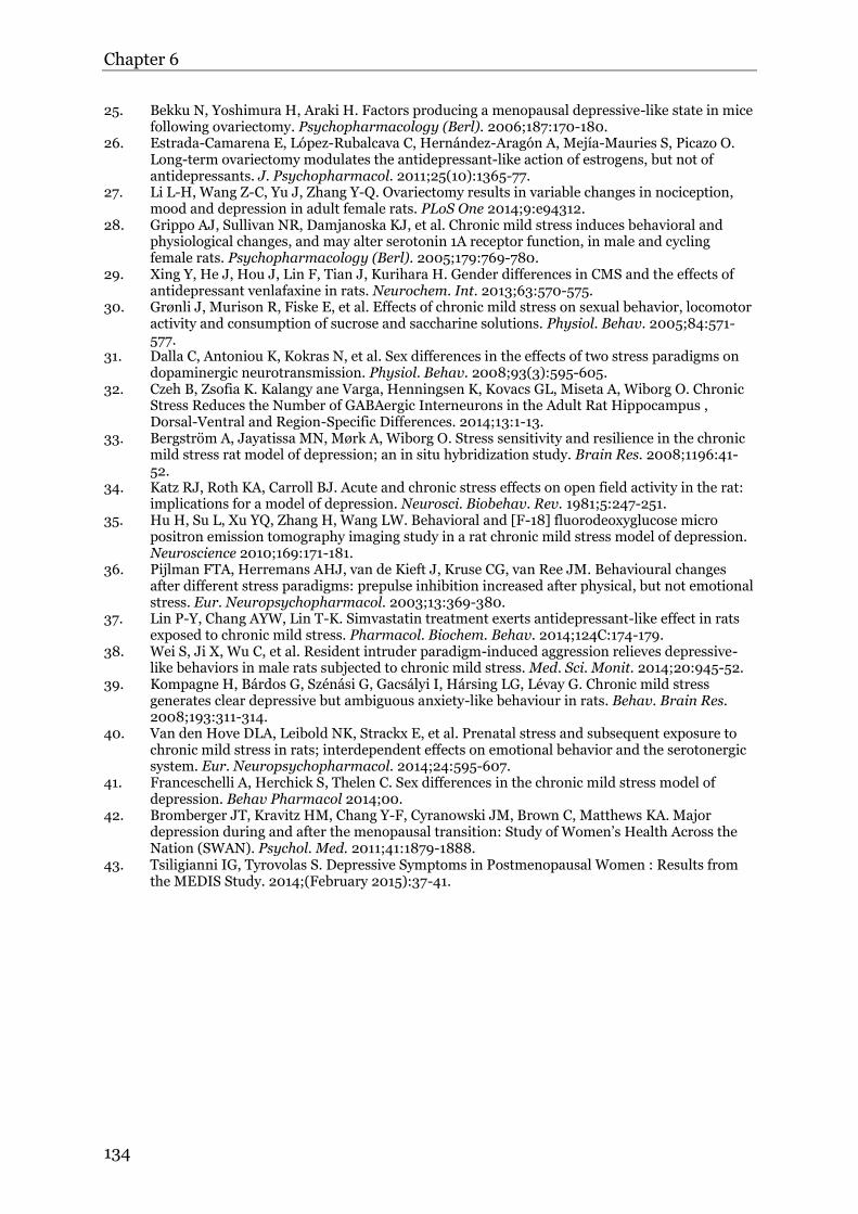
25. Bekku N, Yoshimura H, Araki H. Factors producing a menopausal depressive-like state in mice following ovariectomy. Psychopharmacology (Berl). 2006;187:170-180.
26. Estrada-Camarena E, López-Rubalcava C, Hernández-Aragón A, Mejía-Mauries S, Picazo O. Long-term ovariectomy modulates the antidepressant-like action of estrogens, but not of antidepressants. J. Psychopharmacol. 2011;25(10):1365-77.
27. Li L-H, Wang Z-C, Yu J, Zhang Y-Q. Ovariectomy results in variable changes in nociception, mood and depression in adult female rats. PLoS One 2014;9:e94312.
28. Grippo AJ, Sullivan NR, Damjanoska KJ, et al. Chronic mild stress induces behavioral and physiological changes, and may alter serotonin 1A receptor function, in male and cycling female rats. Psychopharmacology (Berl). 2005;179:769-780.
29. Xing Y, He J, Hou J, Lin F, Tian J, Kurihara H. Gender differences in CMS and the effects of antidepressant venlafaxine in rats. Neurochem. Int. 2013;63:570-575.
30. Grønli J, Murison R, Fiske E, et al. Effects of chronic mild stress on sexual behavior, locomotor activity and consumption of sucrose and saccharine solutions. Physiol. Behav. 2005;84:571-577.
31. Dalla C, Antoniou K, Kokras N, et al. Sex differences in the effects of two stress paradigms on dopaminergic neurotransmission. Physiol. Behav. 2008;93(3):595-605.
32. Czeh B, Zsofia K. Kalangy ane Varga, Henningsen K, Kovacs GL, Miseta A, Wiborg O. Chronic Stress Reduces the Number of GABAergic Interneurons in the Adult Rat Hippocampus , Dorsal-Ventral and Region-Specific Differences. 2014;13:1-13.
33. Bergström A, Jayatissa MN, Mørk A, Wiborg O. Stress sensitivity and resilience in the chronic mild stress rat model of depression; an in situ hybridization study. Brain Res. 2008;1196:41-52.
34. Katz RJ, Roth KA, Carroll BJ. Acute and chronic stress effects on open field activity in the rat: implications for a model of depression. Neurosci. Biobehav. Rev. 1981;5:247-251.
35. Hu H, Su L, Xu YQ, Zhang H, Wang LW. Behavioral and [F-18] fluorodeoxyglucose micro positron emission tomography imaging study in a rat chronic mild stress model of depression. Neuroscience 2010;169:171-181.
36. Pijlman FTA, Herremans AHJ, van de Kieft J, Kruse CG, van Ree JM. Behavioural changes after different stress paradigms: prepulse inhibition increased after physical, but not emotional stress. Eur. Neuropsychopharmacol. 2003;13:369-380.
37. Lin P-Y, Chang AYW, Lin T-K. Simvastatin treatment exerts antidepressant-like effect in rats exposed to chronic mild stress. Pharmacol. Biochem. Behav. 2014;124C:174-179.
38. Wei S, Ji X, Wu C, et al. Resident intruder paradigm-induced aggression relieves depressive-like behaviors in male rats subjected to chronic mild stress. Med. Sci. Monit. 2014;20:945-52.
39. Kompagne H, Bárdos G, Szénási G, Gacsályi I, Hársing LG, Lévay G. Chronic mild stress generates clear depressive but ambiguous anxiety-like behaviour in rats. Behav. Brain Res. 2008;193:311-314.
40. Van den Hove DLA, Leibold NK, Strackx E, et al. Prenatal stress and subsequent exposure to chronic mild stress in rats; interdependent effects on emotional behavior and the serotonergic system. Eur. Neuropsychopharmacol. 2014;24:595-607.
41. Franceschelli A, Herchick S, Thelen C. Sex differences in the chronic mild stress model of depression. Behav Pharmacol 2014;00.
42. Bromberger JT, Kravitz HM, Chang Y-F, Cyranowski JM, Brown C, Matthews KA. Major depression during and after the menopausal transition: Study of Women’s Health Across the Nation (SWAN). Psychol. Med. 2011;41:1879-1888.
43. Tsiligianni IG, Tyrovolas S. Depressive Symptoms in Postmenopausal Women : Results from the MEDIS Study. 2014;(February 2015):37-41.


](https://img.pdfslide.us/doc/110x75/556b0d2ad8b42ae47d8b4c69/3-sex-hormones12.jpg)