Embed Size (px)
Citation preview
MedTech and Diagnostics
Inventions represented at Danish IP Fair 2018MedTech and Diagnostics
This document contains one-pagers for all inventions within MedTech and Diagnosticspresented at the Danish IP Fair 2018. You can use this document to identify meetingpartners at the event. Each invention is marked with a unique ID at the top right of thepage. Use this ID to look up and book a meeting with the inventor(s) at the Danish IPFair website - www.dipfair.dk.
The document will be updated regularly in the period February-April, so ensure to re-visit the website for the newest version.
For further guidelines regarding meeting bookings please consult the menuMatchmaking on the website.
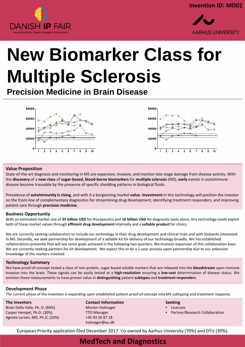
New Biomarker Class for
Multiple SclerosisPrecision Medicine in Brain Disease
MedTech and Diagnostics
Value PropositionState-of-the-art diagnosis and monitoring in MS are expensive, invasive, and monitor late-stage damage from disease activity. With the discovery of a new class of sugar-based, blood-borne biomarkers for multiple sclerosis (MS), early events in autoimmune disease become traceable by the presence of specific shedding patterns in biological fluids.
Prevalence of autoimmunity is rising, and with it a burgeoning market value. Investment in this technology will position the investor on the front-line of complementary diagnostics for streamlining drug development, identifying treatment responders, and improving patient care through precision medicine.
Business OpportunityWith an estimated market size of 25 billion USD for therapeutics and 16 billion USD for diagnostic tools alone, this technology could exploit both of these market values through efficient drug development internally and a sellable product for clinics.
We are currently seeking collaborators to include our technology in their drug development and clinical trials and with biobanks interested in MS. Secondly, we seek partnership for development of a sellable kit for delivery of our technology broadly. We has established collaborations presently that will see some goals achieved in the following two quarters. We envision expansion of this collaboration base. We are currently seeking partners for kit development. We expect this to be a 1-year process upon partnership due to our extension knowledge of the markers involved.
Technology SummaryWe have proof-of-concept tested a class of non-protein, sugar-based soluble markers that are released into the bloodstream upon immuneinvasion into the brain. These signals can be easily tested at a high-resolution ensuring a low-cost determination of disease status. Weenvision these measurements to have proven value in distinguishing patient subtypes and treatment responders.
Development PhaseThe current phase of the invention is expanding upon established patient proof-of-concept into MS subtyping and treatment response.
The inventorsBrian Della Valle, Ph. D. (60%)Casper Hempel, Ph.D. (30%)Agnete Larsen, MD, Ph.D. (10%)
Contact InformationMorten HolmagerTTO Manager+45 93 50 87 [email protected]
Seeking• Licensee• Partner/Research Collaboration
European Priority application filed December 2017. Co-owned by Aarhus University (70%) and DTU (30%).
Invention ID: MD01
Transforming epigenetic research and diagnostics - cutting edge DNA methylation detection technology for cancer research and diagnostics
MedTech and Diagnostics
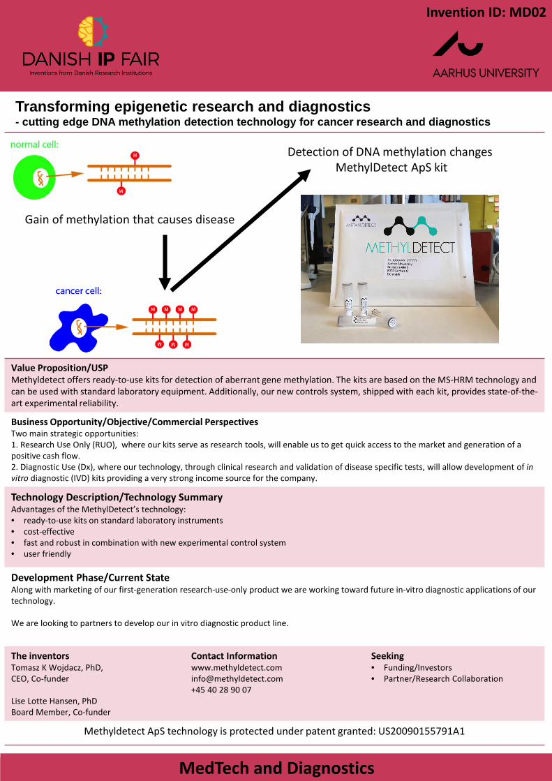
Value Proposition/USPMethyldetect offers ready-to-use kits for detection of aberrant gene methylation. The kits are based on the MS-HRM technology and can be used with standard laboratory equipment. Additionally, our new controls system, shipped with each kit, provides state-of-the-art experimental reliability.
Business Opportunity/Objective/Commercial PerspectivesTwo main strategic opportunities:1. Research Use Only (RUO), where our kits serve as research tools, will enable us to get quick access to the market and generation of a positive cash flow. 2. Diagnostic Use (Dx), where our technology, through clinical research and validation of disease specific tests, will allow development of in vitro diagnostic (IVD) kits providing a very strong income source for the company.
Technology Description/Technology SummaryAdvantages of the MethylDetect’s technology:• ready-to-use kits on standard laboratory instruments• cost-effective• fast and robust in combination with new experimental control system• user friendly
Development Phase/Current StateAlong with marketing of our first-generation research-use-only product we are working toward future in-vitro diagnostic applications of our technology.
We are looking to partners to develop our in vitro diagnostic product line.
The inventorsTomasz K Wojdacz, PhD,CEO, Co-funder
Lise Lotte Hansen, PhDBoard Member, Co-funder
Contact [email protected]+45 40 28 90 07
Seeking• Funding/Investors• Partner/Research Collaboration
Methyldetect ApS technology is protected under patent granted: US20090155791A1
Invention ID: MD02
Gain of methylation that causes disease
Detection of DNA methylation changes MethylDetect ApS kit
Home Kemo CareHow to reduce home and environmental impact of cytotoxic drugs after treatment.
MedTech and Diagnostics
Images and caption if relevant
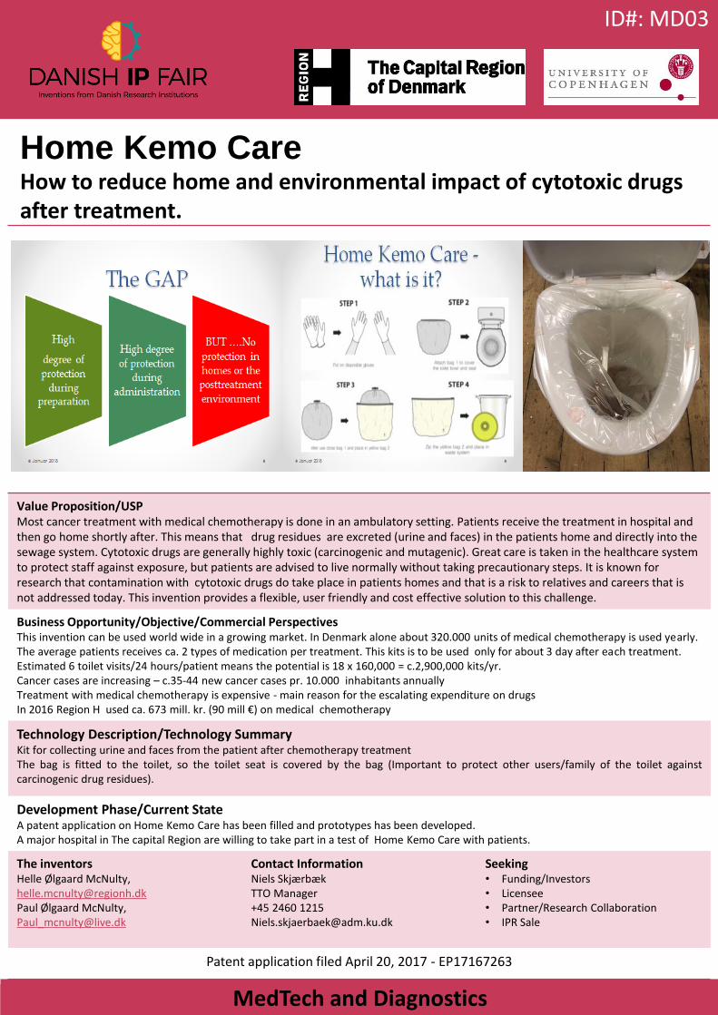
Value Proposition/USPMost cancer treatment with medical chemotherapy is done in an ambulatory setting. Patients receive the treatment in hospital and then go home shortly after. This means that drug residues are excreted (urine and faces) in the patients home and directly into the sewage system. Cytotoxic drugs are generally highly toxic (carcinogenic and mutagenic). Great care is taken in the healthcare system to protect staff against exposure, but patients are advised to live normally without taking precautionary steps. It is known forresearch that contamination with cytotoxic drugs do take place in patients homes and that is a risk to relatives and careers that is not addressed today. This invention provides a flexible, user friendly and cost effective solution to this challenge.
Business Opportunity/Objective/Commercial PerspectivesThis invention can be used world wide in a growing market. In Denmark alone about 320.000 units of medical chemotherapy is used yearly. The average patients receives ca. 2 types of medication per treatment. This kits is to be used only for about 3 day after each treatment. Estimated 6 toilet visits/24 hours/patient means the potential is 18 x 160,000 = c.2,900,000 kits/yr.Cancer cases are increasing – c.35-44 new cancer cases pr. 10.000 inhabitants annuallyTreatment with medical chemotherapy is expensive - main reason for the escalating expenditure on drugsIn 2016 Region H used ca. 673 mill. kr. (90 mill €) on medical chemotherapy
Technology Description/Technology SummaryKit for collecting urine and faces from the patient after chemotherapy treatmentThe bag is fitted to the toilet, so the toilet seat is covered by the bag (Important to protect other users/family of the toilet againstcarcinogenic drug residues).
Development Phase/Current StateA patent application on Home Kemo Care has been filled and prototypes has been developed.A major hospital in The capital Region are willing to take part in a test of Home Kemo Care with patients.
The inventorsHelle Ølgaard McNulty,[email protected] Ølgaard McNulty, [email protected]
Contact InformationNiels SkjærbækTTO Manager+45 2460 [email protected]
Seeking• Funding/Investors• Licensee• Partner/Research Collaboration• IPR Sale
Patent application filed April 20, 2017 - EP17167263
ID#: MD03
Blood Card and Vesicle-based Medical Tests
MedTech and Diagnostics
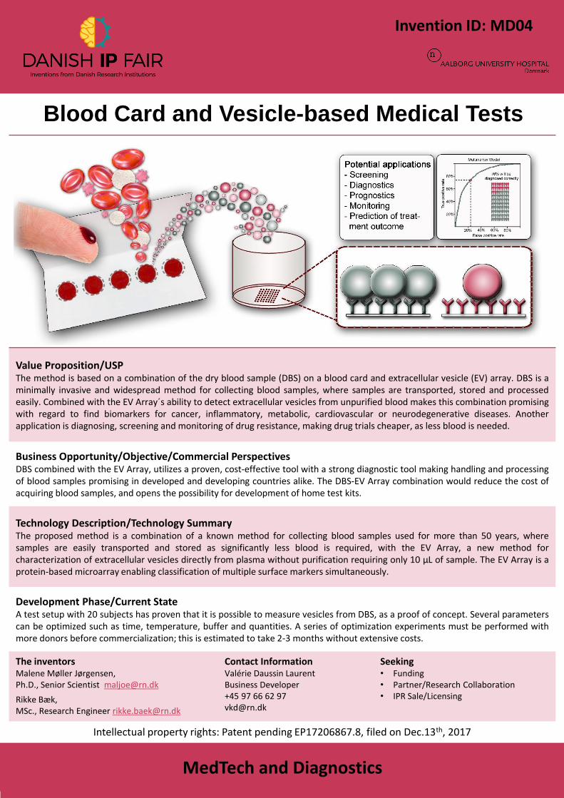
Value Proposition/USPThe method is based on a combination of the dry blood sample (DBS) on a blood card and extracellular vesicle (EV) array. DBS is aminimally invasive and widespread method for collecting blood samples, where samples are transported, stored and processedeasily. Combined with the EV Array´s ability to detect extracellular vesicles from unpurified blood makes this combination promisingwith regard to find biomarkers for cancer, inflammatory, metabolic, cardiovascular or neurodegenerative diseases. Anotherapplication is diagnosing, screening and monitoring of drug resistance, making drug trials cheaper, as less blood is needed.
Business Opportunity/Objective/Commercial PerspectivesDBS combined with the EV Array, utilizes a proven, cost-effective tool with a strong diagnostic tool making handling and processingof blood samples promising in developed and developing countries alike. The DBS-EV Array combination would reduce the cost ofacquiring blood samples, and opens the possibility for development of home test kits.
Technology Description/Technology SummaryThe proposed method is a combination of a known method for collecting blood samples used for more than 50 years, wheresamples are easily transported and stored as significantly less blood is required, with the EV Array, a new method forcharacterization of extracellular vesicles directly from plasma without purification requiring only 10 µL of sample. The EV Array is aprotein-based microarray enabling classification of multiple surface markers simultaneously.
Development Phase/Current StateA test setup with 20 subjects has proven that it is possible to measure vesicles from DBS, as a proof of concept. Several parameterscan be optimized such as time, temperature, buffer and quantities. A series of optimization experiments must be performed withmore donors before commercialization; this is estimated to take 2-3 months without extensive costs.
The inventorsMalene Møller Jørgensen,Ph.D., Senior Scientist [email protected]
Rikke Bæk,MSc., Research Engineer [email protected]
Contact InformationValérie Daussin LaurentBusiness Developer +45 97 66 62 [email protected]
Seeking• Funding• Partner/Research Collaboration• IPR Sale/Licensing
Intellectual property rights: Patent pending EP17206867.8, filed on Dec.13th, 2017
Invention ID: MD04

Method for detection and assessment of heart dyssynchrony- based on 2D echocardiography
MedTech and Diagnostics
Value PropositionHeart failure affects yearly on average 5-10% of the elderly population. Most severe cases are caused by left bundle branch block and are corrected by cardiac resynchronisation therapy (CRT). Every year, a thousand patients receive a CRT-implant in Denmark, while about 1/3 of the treatments are without benefit to the patient. So far, methods based on deformation of the heart have proven not robust enough for predicting the benefits of CRT's. Also, current 3D techniques are limited by significantly lower time-resolution. Interpolated 3D images aim to solve this problem.
Business Opportunity/Objective/Commercial PerspectivesThe potential commercial perspective is a more precise method for clinicians to evaluate cardiac failure mechanisms, dyssynchrony, and to assess the results of CRT. Calculation of used parameters are fast (< 1min on standard PC) and can be performed on 2D images recorded with hardware already present in the clinic.
Technology Description/Technology SummaryThe proposed method creates 3D heart deformation data from 2D scanning, using interpolated data from cardiac movement. Thisenables a quantitative and qualitative assessment of the heart’s deformation, with a higher time-resolution. The estimation of theheart’s dyssynchrony is made from a significantly larger area of the heart, giving a better representation of the dyssynchronouscontraction. The visual representation provides timing and contraction information in a dynamic 3D-perspective, allowing for betterassessment of patients who will benefit from CRT. The visualisation of the left ventricle is expected to aid in the optimal electrodeplacement of the CRT-unit during operation, as well as aid in the patient selection for CRT.
Development Phase/Current StatePreliminary results from 101 CRT patients show a sensitivity of 61% and a specificity on 82% The ability to predict an improvement inthe left ventricle is 90%, the ability to predict no improvement is 43%, and accuracy is 66%.
The inventorTomas ZarembaMD, Phd.Cardiology Department [email protected]
Contact InformationValérie Daussin LaurentBusiness Manager+45 97 66 62 [email protected]
Seeking• Funding• Research Collaboration• IPR Licensing or sale
Intellectual property rights: Patent pending CA2994617, AU2018200974, February, 9th 2018
Invention ID: MD05
MedTech and Diagnostics
Value Proposition/USPEV Array is an unique biomarker platform that can exploit the intercellular trafficking in various biofluids to provide a non-invasivemethod for diagnostics and prognostics.
Business Opportunity/Objective/Commercial PerspectivesThe apparent role of EVs in a vast number of biological processes, forms the basis of extending EV analysis beyond basic research and intoclinical and therapeutic context. The applications of this type of analysis include the areas of diagnostics and prognostics, as well as drugtherapy, regenerative medicine, and vaccines. The analysis of EVs could be incorporated as a screening tool and applied to confirm adiagnosis. Furthermore, the EV Array analysis has the potential to become an element in treatment surveillance and companiondiagnostics.
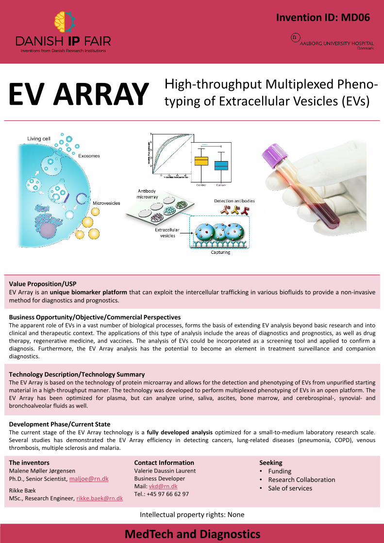
Technology Description/Technology Summary The EV Array is based on the technology of protein microarray and allows for the detection and phenotyping of EVs from unpurified startingmaterial in a high-throughput manner. The technology was developed to perform multiplexed phenotyping of EVs in an open platform. TheEV Array has been optimized for plasma, but can analyze urine, saliva, ascites, bone marrow, and cerebrospinal-, synovial- andbronchoalveolar fluids as well.
Development Phase/Current StateThe current stage of the EV Array technology is a fully developed analysis optimized for a small-to-medium laboratory research scale.Several studies has demonstrated the EV Array efficiency in detecting cancers, lung-related diseases (pneumonia, COPD), venousthrombosis, multiple sclerosis and malaria.
The inventorsMalene Møller Jørgensen
Ph.D., Senior Scientist, [email protected]
Rikke Bæk MSc., Research Engineer, [email protected]
Contact InformationValerie Daussin Laurent Business DeveloperMail: [email protected].: +45 97 66 62 97
Seeking• Funding• Research Collaboration• Sale of services
Intellectual property rights: None
EV ARRAY High-throughput Multiplexed Pheno-typing of Extracellular Vesicles (EVs)
Invention ID: MD06
A novel balloon-tipped catheter for continuos nerve block
- garanteed painfree after surgery
MedTech and Diagnostics
Images and caption if relevant
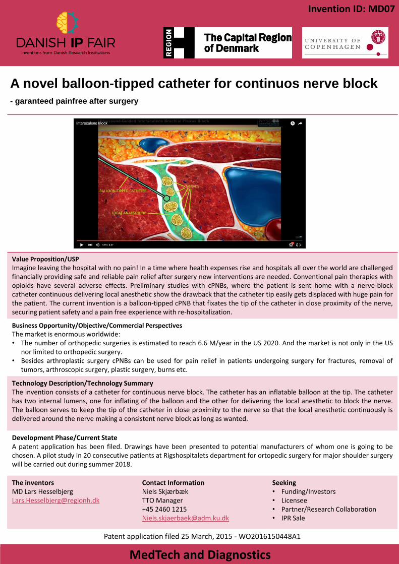
Value Proposition/USPImagine leaving the hospital with no pain! In a time where health expenses rise and hospitals all over the world are challengedfinancially providing safe and reliable pain relief after surgery new interventions are needed. Conventional pain therapies withopioids have several adverse effects. Preliminary studies with cPNBs, where the patient is sent home with a nerve-blockcatheter continuous delivering local anesthetic show the drawback that the catheter tip easily gets displaced with huge pain forthe patient. The current invention is a balloon-tipped cPNB that fixates the tip of the catheter in close proximity of the nerve,securing patient safety and a pain free experience with re-hospitalization.
Business Opportunity/Objective/Commercial PerspectivesThe market is enormous worldwide:• The number of orthopedic surgeries is estimated to reach 6.6 M/year in the US 2020. And the market is not only in the US
nor limited to orthopedic surgery.• Besides arthroplastic surgery cPNBs can be used for pain relief in patients undergoing surgery for fractures, removal of
tumors, arthroscopic surgery, plastic surgery, burns etc.
Technology Description/Technology SummaryThe invention consists of a catheter for continuous nerve block. The catheter has an inflatable balloon at the tip. The catheterhas two internal lumens, one for inflating of the balloon and the other for delivering the local anesthetic to block the nerve.The balloon serves to keep the tip of the catheter in close proximity to the nerve so that the local anesthetic continuously isdelivered around the nerve making a consistent nerve block as long as wanted.
Development Phase/Current StateA patent application has been filed. Drawings have been presented to potential manufacturers of whom one is going to bechosen. A pilot study in 20 consecutive patients at Rigshospitalets department for ortopedic surgery for major shoulder surgerywill be carried out during summer 2018.
The inventorsMD Lars [email protected]
Contact InformationNiels SkjærbækTTO Manager+45 2460 [email protected]
Seeking• Funding/Investors• Licensee• Partner/Research Collaboration• IPR Sale
Patent application filed 25 March, 2015 - WO2016150448A1
Invention ID: MD07
IBDetectTM:RNA-based
diagnosis of inflammatory
bowel diseaseA quick and cost-effective route to accurate diagnosis of ulcerative colitis and Crohn’s disease
MedTech and Diagnostics
Unique Selling PointIBDetectTM combines qPCR and machine learning technology to diagnose Inflammatory bowel disease (IBD) patients. It is ideal for the construction of a ready-to-use diagnostic kit. • It is cheap and easy to implement – qPCR equipment is already widely available at hospitals• It is quick to use – analysis can be completed within the same time frame as histological examination• It can increase the quality of IBD treatment without burdening the patient
ObjectiveIBD is a chronic disorder of the human gut, affecting 1 in 250 Europeans. The main types of IBD, ulcerative colitis (UC) and Crohn’s disease (CD), require different pharmaceutical and/or surgical treatments. Diagnosis is based on visual examination of the gut and histological examination of gut biopsies. Diagnosis is difficult: about 15% of patients are not classifiable, and the initial diagnosis is often wrong, which leads to additional costs for the healthcare provider and may harm the patient. Molecular methods to increase the certainty of diagnosis, like IBDetectTM, are needed.
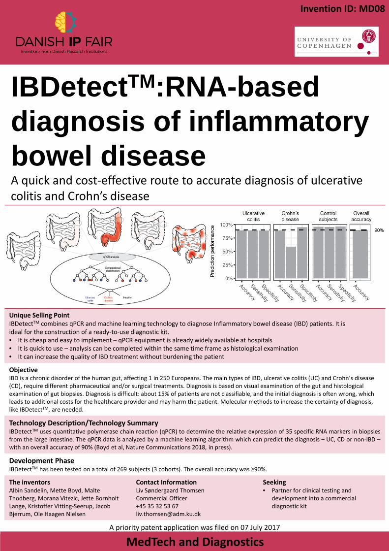
Technology Description/Technology SummaryIBDetectTM uses quantitative polymerase chain reaction (qPCR) to determine the relative expression of 35 specific RNA markers in biopsiesfrom the large intestine. The qPCR data is analyzed by a machine learning algorithm which can predict the diagnosis – UC, CD or non-IBD –with an overall accuracy of 90% (Boyd et al, Nature Communications 2018, in press).
Development PhaseIBDetectTM has been tested on a total of 269 subjects (3 cohorts). The overall accuracy was ≥90%.
The inventorsAlbin Sandelin, Mette Boyd, Malte Thodberg, Morana Vitezic, Jette BornholtLange, Kristoffer Vitting-Seerup, Jacob Bjerrum, Ole Haagen Nielsen
Contact InformationLiv Søndergaard ThomsenCommercial Officer+45 35 32 53 [email protected]
Seeking• Partner for clinical testing and
development into a commercial diagnostic kit
A priority patent application was filed on 07 July 2017
Invention ID: MD08
Knee Laxity in 3D- Accurate non-invasive assessment of knee laxity in 3D
MedTech and Diagnostics
Images and caption if relevant
Value Proposition/USPWe present the first ever technology capable of obtaining accurate non-invasive knee laxity in 3D. A technology that will greatlyimprove diagnostics, preoperative planning and rehabilitation.
Business Opportunity/Objective/Commercial PerspectivesAltered knee laxity occurs frequently after ligament injuries or joint replacements. For knee replacement this is the most common joint replacement, with increasing incidence. In US alone, knee replacements are expected to grow to 3.48 million annual procedures by 2030. However, current methods to assess joint laxity have several limitations, such as one-dimentionality, soft-tissue artifacts and no assessment of the loads applied during testing. These seriously limit their usability in the clinic.With our technology to accurately measure 3D joint laxity, surgeons, physiotherapists and researchers can objectively quantify 3D joint laxity and use it in the clinical management of the pathologies.
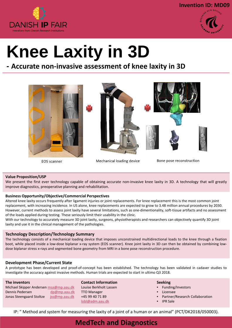
Technology Description/Technology SummaryThe technology consists of a mechanical loading device that imposes unconstrained multidirectional loads to the knee through a fixationboot, while placed inside a low-dose biplanar x-ray system (EOS scanner). Knee joint laxity in 3D can then be obtained by combining low-dose biplanar stress x-rays and segmented bone geometry from MRI in a bone pose reconstruction procedure.
Development Phase/Current StateA prototype has been developed and proof-of-concept has been established. The technology has been validated in cadaver studies toinvestigate the accuracy against invasive methods. Human trials are expected to start in ultimo Q3 2018.
The inventorsMichael Skipper Andersen [email protected] Pedersen [email protected] Steengaard Stoltze [email protected]
Contact InformationLouise Berkholt LassenTTO Manager+45 99 40 71 [email protected]
Seeking• Funding/Investors• Licensee• Partner/Research Collaboration• IPR Sale
IP: ” Method and system for measuring the laxity of a joint of a human or an animal” (PCT/DK2018/050003).
Invention ID: MD09

COMPACT X-SCISSORS DEVICE
MedTech and Diagnostics
Value Proposition/USPThe commercial potential of the Compact Joint for 3D Spherical Motion is particularly related to the combination of the• Scalability and Compactness of the joint minimizing the need for space around a center of motion, and the• High degree-of-freedom to design the joint to fit a specific purpose ( including the size of a person, when used in exoskeletons),
compared to other known joints, as the curvature and size of individual linkages can be optimized for the required movement, securing• High degree of control and precision, and enabling full range of motion, with three degrees of freedom.
Business Opportunity/Objective/Commercial PerspectivesParalysis in the upper extremity can result from traumatic injuries, congenital diseases, stroke, and aging in general. It can makeindependent life difficult and disable daily activities such as feeding and personal grooming. Exoskeletons can provide assistance andrehabilitation. The majority reject existing well performing devices because they are either bulky, heavy and impractical or do not have asolution for the shoulder complex. A compact solution that can fit and hide underneath clothing may drastically improve the acceptance ofthese devices and make them actually wearable. Enabling the impaired to achieve normal function and/or enables healthy people toperform heavy lifting, or endure loads for long periods without causing injuries.
Technology Description/Technology SummaryThe CXD (short for Compact X-scissors Device) is a spherical scissors mechanism capable of three rotations, thereby mimicking thebehaviour of a spherical joint. The mechanism moves on an imaginary sphere with a constant rotation centre and an arbitrary radiusdetermined by the design parameters. Since there is a void space within the mechanism, it is suitable for applications where the mechanicalparts surround a given object or workspace. The mechanism is particularly well suited to support anatomical, spherical joints such as theshoulder and the hip, thereby solving a problem that has been haunting the fields of orthotics and exoskeletons for decades.
Development Phase/Current StateProof-of-concept prototype made of aluminum and steel has been tested successfully as a highly compact shoulder joint in an upper body exoskeleton configuration. Watch the invention here: https://youtu.be/Pw_esFdwGmo
The inventorsMiguel N. Castro [email protected] Rasmussen [email protected] Skipper Andersen [email protected] Bai [email protected]
Contact InformationLars HalkjærTechnology Transfer Manager+45 9940 [email protected]
Seeking• Funding/Investors• Licensee• Partner/Research Collaboration
Patent Pending
Invention ID: MD10
Polyneedle- Less pain, 2 liquids and MR compatibility
MedTech and Diagnostics
Value Proposition/USP1. Perceived less pain when self injection is required. 2. Polymer needles can inject 2 or more reactive liquids through one needle without mixing the liquids before they are inside the
tissue3. Polymer needles make injections inside MR-scanner possible..
Business Opportunity/Objective/Commercial PerspectivesFast and more accurate diagnostics of cancer metastases will reduce surgery time and improve treatment. Simultaneous injection of 2 component drugs for cancer or diabetics treatment.
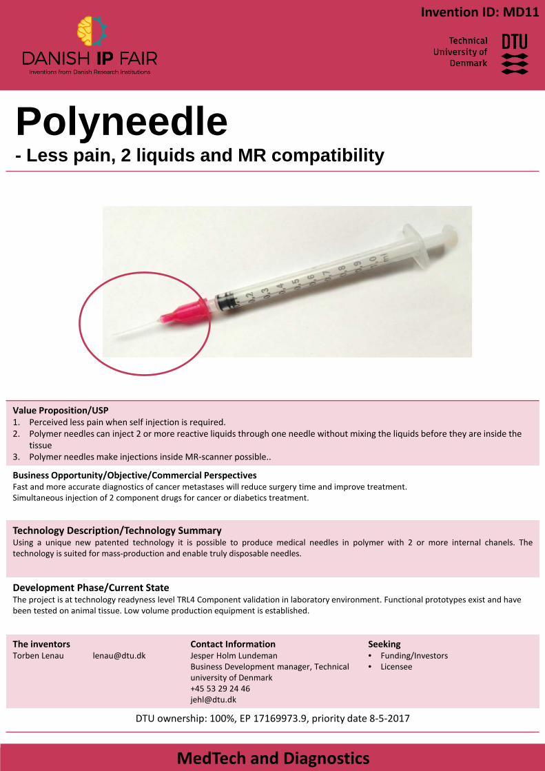
Technology Description/Technology SummaryUsing a unique new patented technology it is possible to produce medical needles in polymer with 2 or more internal chanels. Thetechnology is suited for mass-production and enable truly disposable needles.
Development Phase/Current StateThe project is at technology readyness level TRL4 Component validation in laboratory environment. Functional prototypes exist and have been tested on animal tissue. Low volume production equipment is established.
The inventorsTorben Lenau [email protected]
Contact InformationJesper Holm LundemanBusiness Development manager, Technical university of Denmark+45 53 29 24 [email protected]
Seeking• Funding/Investors• Licensee
DTU ownership: 100%, EP 17169973.9, priority date 8-5-2017
Invention ID: MD11
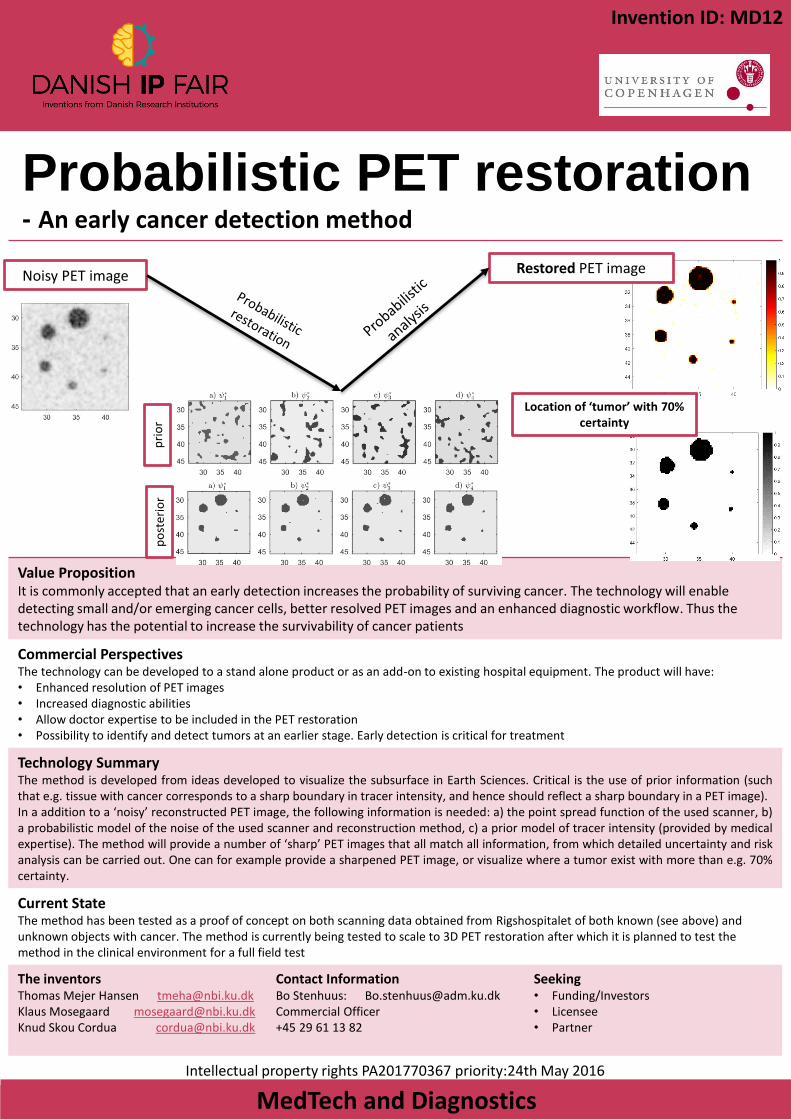
Probabilistic PET restoration- An early cancer detection method
MedTech and Diagnostics
Value PropositionIt is commonly accepted that an early detection increases the probability of surviving cancer. The technology will enable detecting small and/or emerging cancer cells, better resolved PET images and an enhanced diagnostic workflow. Thus the technology has the potential to increase the survivability of cancer patients
Commercial PerspectivesThe technology can be developed to a stand alone product or as an add-on to existing hospital equipment. The product will have:• Enhanced resolution of PET images• Increased diagnostic abilities• Allow doctor expertise to be included in the PET restoration• Possibility to identify and detect tumors at an earlier stage. Early detection is critical for treatment
Technology SummaryThe method is developed from ideas developed to visualize the subsurface in Earth Sciences. Critical is the use of prior information (suchthat e.g. tissue with cancer corresponds to a sharp boundary in tracer intensity, and hence should reflect a sharp boundary in a PET image).In a addition to a ‘noisy’ reconstructed PET image, the following information is needed: a) the point spread function of the used scanner, b)a probabilistic model of the noise of the used scanner and reconstruction method, c) a prior model of tracer intensity (provided by medicalexpertise). The method will provide a number of ‘sharp’ PET images that all match all information, from which detailed uncertainty and riskanalysis can be carried out. One can for example provide a sharpened PET image, or visualize where a tumor exist with more than e.g. 70%certainty.
Current StateThe method has been tested as a proof of concept on both scanning data obtained from Rigshospitalet of both known (see above) and unknown objects with cancer. The method is currently being tested to scale to 3D PET restoration after which it is planned to test the method in the clinical environment for a full field test
The inventorsThomas Mejer Hansen [email protected] Mosegaard [email protected] Skou Cordua [email protected]
Contact InformationBo Stenhuus: [email protected] Officer +45 29 61 13 82
Seeking• Funding/Investors• Licensee• Partner
Intellectual property rights PA201770367 priority:24th May 2016
Invention ID: MD12
Noisy PET image Restored PET image
pri
or
po
ster
ior
Location of ‘tumor’ with 70% certainty
Medical Brain Stimulation- a MRI compatible neurostimulator from 1.5T to 7.0T
MedTech and Diagnostics
Value Proposition/USPElectrical stimulation (Deep Brain Stimulation) is pharmacological therapy going digital. Neurostimulations delivers an adjustable stimulus to a specific target, thereby reducing systemic side effects as compared to pharmacological therapy. Neurostimulation is used to treat movement disorders (Parkinson’s Disease, tremor and Tourette’s syndrome), cardiac disease (use of pacemakers), and is the next big thing in treatment of other neurological disorders (Alzheimer and headache) as well as psychiatric disorders (depression, addiction and obsessive compulsive disorder).
Business Opportunity/Objective/Commercial PerspectivesThe apparatus is applicable for use in all MRI scanners (36.000 worldwide) as a combined disposable and non-disposable unit with software license opportunities. The market is huge and fast growing, which lend promise to large revenues. The applications are functional MRI scans of deep brain stimulation, spinal cord stimulation, transcutaneous nerve stimulation, peripheral nerve stimulation, cranial nerve stimulation, electrical muscle stimulation, cortical multi-electrode stimulation, retinal multi-electrode stimulation, gastric electrical stimulation therapy and cardiac stimulation (pacemakers).
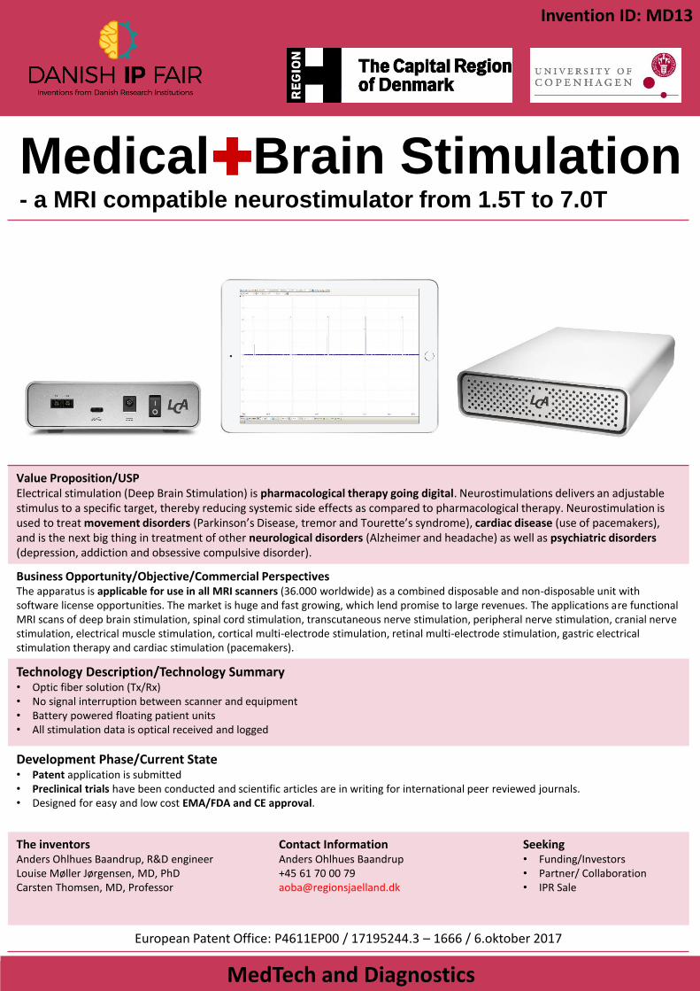
Technology Description/Technology Summary• Optic fiber solution (Tx/Rx)• No signal interruption between scanner and equipment• Battery powered floating patient units• All stimulation data is optical received and logged
Development Phase/Current State• Patent application is submitted• Preclinical trials have been conducted and scientific articles are in writing for international peer reviewed journals. • Designed for easy and low cost EMA/FDA and CE approval.
The inventorsAnders Ohlhues Baandrup, R&D engineerLouise Møller Jørgensen, MD, PhDCarsten Thomsen, MD, Professor
Contact InformationAnders Ohlhues Baandrup+45 61 70 00 [email protected]
Seeking• Funding/Investors• Partner/ Collaboration• IPR Sale
European Patent Office: P4611EP00 / 17195244.3 – 1666 / 6.oktober 2017
Invention ID: MD13

Value PropositionThe present technology enables acoustic monitoring of asthma by assessing correct inhaler use, drug administration and lung function with an add-on device for common asthma inhalers. When the inhaler is used the device generates sounds that are recorded by a smart phone allowing the user to assess the inhalation and track their condition. The add-on device and the inhaler are coupled forming a single device and is intended to provide simultaneous drug administration and asthma monitoring.
Business OpportunityThe team behind the invention is working on a product for asthma monitoring and is currently seeking funding for a spin-out company, Sonohaler. The global asthma therapy market is estimated at USD 22 billion and an upcoming smart inhaler market is predicted to grow to USD 1.6 billion by 2022. We see an immense opportunity for improving the lives of the +300 million asthma patients worldwide andreducing healthcare costs associated with hospitalization by better individual control of asthma and a reduction in asthma attacks.
Technology SummaryThe device can be used alone or designed to fit a specific inhaler or other devices. The device contains an acoustic element that produces asound when inhaling or exhaling through the device. The sounds generated can then be used to correlate with the flow rate producedallowing measurements. The sounds can be recorded and processed using software to allow further assessment of inhalation and inhaleruse. The technology can be used to monitor inhalation and drug administration giving an indication of lung health and treatment profile.
Development PhasePrototype devices have been produced and tested in combination with inhalers or as stand alone devices to administer medicine. Correlation between sound and flow rate has been established and software can be used analyze inhalation parameters. It is expected to test the powder flow performance of the product in the near future to demonstrate compatibility with inhaler drug administration. Software and algorithms are under development.
The inventorsAdam Bohr Johan Bøtker Henrik Jensen
Contact InformationPeter Stein NielsenCommercial Officer+45 21 64 74 [email protected]
Seeking• Funding/Investors• Partner/Research Collaboration
Intellectual property rights: Application no.: PA 2017 70492 Filing date: 23 June 2017
Acoustic device for asthma
monitoring
MedTech and Diagnostics
Invention ID: MD14
Value Proposition:Our calibrator technology provides highly improved solutions to two major problems in flow cytometry:
• Quantification: Current calibrator products can only determine very high numbers of surface protein (typically >2000 proteins) on cells and biological vesicles. However, a large proportion of e.g. protein-based biomarkers is expressed at much lower levels on these biological particles. The increased interest in finding protein biomarkers for understanding biology and various diseases needs proper technologies to detect and quantify these low abundant proteins across e.g. various flow cytometry facilities.
• Standardization: In order to compare flow cytometry data from different laboratories and across different flow cytometers proper standardization tools are highly needed. The lack of precise, accurate and uniform standardizations is an obstacle for development of new flow cytometry based assays. A more uniform and accurate measurement in flow cytometry will allow new development for both basal and applied research. This will enable a giant leap forward for development of flow cytometry based assays.
• Global calibration bead market is estimated to reach $213 M by year 2020
Business Opportunity/Objective/Commercial PerspectivesHealth care, diagnostics, flow cytometry, research
Technology DescriptionWe have developed novel calibrator technology for flow cytometry (FC) that relies on the fluorescence intensity of individualprecisely fluorophore-labeled particles. Labelling precision and low range (1-2000 fluorophores) allows quantification of flowcytometry output signal on a completely new level. FQuant allows precise quantification of low abundant antigens presented oncells, viruses, exosomes and microvesicles.
Development PhaseWe have successfully formed uniform medium sized particles and labeled these with 80 and 160 fluorophores. Thus, we have alreadyproved that we can form stable and well-defined particles that can be used as calibrators. We plan to increase the size of the particles to increase the degree of fluorophore-labeling and thereby form calibrators that cover a large dynamic range (50-2000 fluorophores) needed for proper commercial use. Patent application was filed with intend to create a company producing calibration bead kits.
The inventorsDenis Selnihhin, post.doc.Jens Bæk Simonsen, associate professor
Contact InformationEoin GalliganTTO [email protected]
Seeking• Funding/Investors• Licensee• Partner/Research Collaboration• IPR Sale
A European patent application has been filed (EP18154868)
MedTech and Diagnostics
Invention ID: MD15
New Startup: Moving from a qualitative to a quantitative description
Denis Selnihhin Jens Bæk Simonsen
FQuant is a startup company created by two scientists Denis Selnihhinand Jens Bæk Simonsen from Aarhus University and Danish TechnicalUniversity, respectively.
FQuant’s unique calibrators will elevate flow cytometry to an exact science
Device for measuring body part
movements and stretch
MedTech and Diagnostics
Value Proposition/USPMusculoskeletal disorders cost society approximately 36 billion annually and is a major cause of early retirement. The device is a new clinical diagnostics- and monitoring-equipment, that enables measuring the effect of therapy (e.g custom orthotics), minimizing human factor and ensuring an accurate diagnosis of fault position. A low cost solution with potential in the clinical-, sports-, and customer-specific shoes realm.
Business Opportunity/Objective/Commercial PerspectivesAs a diagnostic tool the device can assist the clinician in assessing the accurate position of the foot for diagnostic purposes. Shoe specialists can ensure that a costumer receives the best insole for the purpose, both in professional and commercial use. In sports the device can be used to optimize movement patterns during training and help with footwear selection
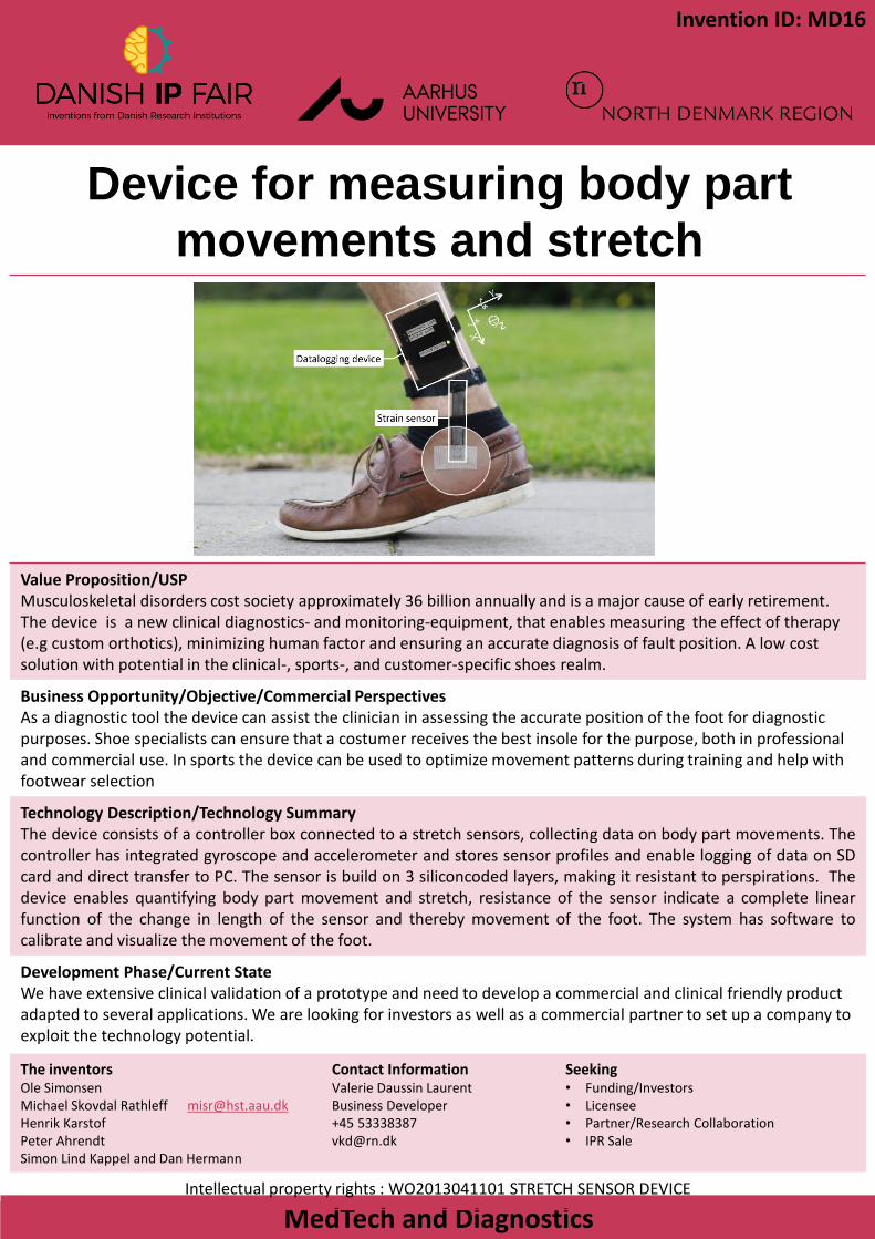
Technology Description/Technology SummaryThe device consists of a controller box connected to a stretch sensors, collecting data on body part movements. Thecontroller has integrated gyroscope and accelerometer and stores sensor profiles and enable logging of data on SDcard and direct transfer to PC. The sensor is build on 3 siliconcoded layers, making it resistant to perspirations. Thedevice enables quantifying body part movement and stretch, resistance of the sensor indicate a complete linearfunction of the change in length of the sensor and thereby movement of the foot. The system has software tocalibrate and visualize the movement of the foot.
Development Phase/Current StateWe have extensive clinical validation of a prototype and need to develop a commercial and clinical friendly product adapted to several applications. We are looking for investors as well as a commercial partner to set up a company to exploit the technology potential.
The inventorsOle SimonsenMichael Skovdal Rathleff [email protected] KarstofPeter Ahrendt Simon Lind Kappel and Dan Hermann
Contact InformationValerie Daussin LaurentBusiness Developer+45 [email protected]
Seeking• Funding/Investors• Licensee• Partner/Research Collaboration• IPR Sale
Intellectual property rights : WO2013041101 STRETCH SENSOR DEVICE
Invention ID: MD16
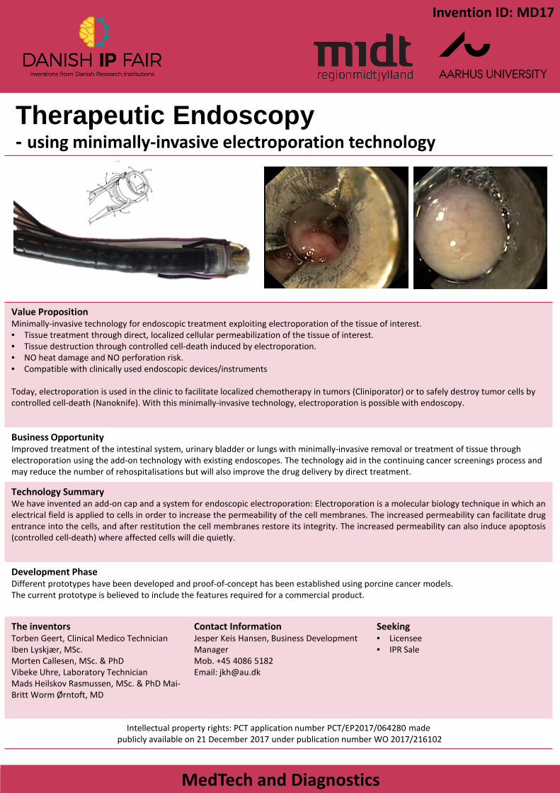
Therapeutic Endoscopy- using minimally-invasive electroporation technology
MedTech and Diagnostics
Value PropositionMinimally-invasive technology for endoscopic treatment exploiting electroporation of the tissue of interest. • Tissue treatment through direct, localized cellular permeabilization of the tissue of interest.• Tissue destruction through controlled cell-death induced by electroporation.• NO heat damage and NO perforation risk.• Compatible with clinically used endoscopic devices/instruments
Today, electroporation is used in the clinic to facilitate localized chemotherapy in tumors (Cliniporator) or to safely destroy tumor cells by controlled cell-death (Nanoknife). With this minimally-invasive technology, electroporation is possible with endoscopy.
Business OpportunityImproved treatment of the intestinal system, urinary bladder or lungs with minimally-invasive removal or treatment of tissue through electroporation using the add-on technology with existing endoscopes. The technology aid in the continuing cancer screenings process and may reduce the number of rehospitalisations but will also improve the drug delivery by direct treatment.
Technology SummaryWe have invented an add-on cap and a system for endoscopic electroporation: Electroporation is a molecular biology technique in which anelectrical field is applied to cells in order to increase the permeability of the cell membranes. The increased permeability can facilitate drugentrance into the cells, and after restitution the cell membranes restore its integrity. The increased permeability can also induce apoptosis(controlled cell-death) where affected cells will die quietly.
Development PhaseDifferent prototypes have been developed and proof-of-concept has been established using porcine cancer models.The current prototype is believed to include the features required for a commercial product.
The inventorsTorben Geert, Clinical Medico TechnicianIben Lyskjær, MSc.Morten Callesen, MSc. & PhDVibeke Uhre, Laboratory TechnicianMads Heilskov Rasmussen, MSc. & PhD Mai-Britt Worm Ørntoft, MD
Contact InformationJesper Keis Hansen, Business Development ManagerMob. +45 4086 5182 Email: [email protected]
Seeking• Licensee• IPR Sale
Intellectual property rights: PCT application number PCT/EP2017/064280 madepublicly available on 21 December 2017 under publication number WO 2017/216102
Invention ID: MD17
3D PRINTED ENDEGENOUS BONE IMPLANTS- 1. GENERATION IMPLANTS FROM PARTICLE3D
MedTech and Diagnostics
USPEndogenous material No need for bone grafting from patients. Minimizes complications for each patient and shortens hospital admission.Building block of bone Develops into living bone tissue over time.Patient specific and fitted Customized through 3D printing to fit each individual patient. Shortens surgery time and improves aesthetic outcomeDrug loadable pipeline The developed technology allows unique storage of therapeutic drugs. No other comparable product on market.
Commercial PerspectivesThe use of patient customized implants is growing fast yet current solutions are based on foreign permanent materials with high complication & re-surgery rates. We are uniquely able to provide patient customized implants that are both fully composed of resorbable and endogenous materials. This makes us well suited to enter a market of over 2.000.000 procedures worth $2.000.000.000 in revenue and growing 5% annually.
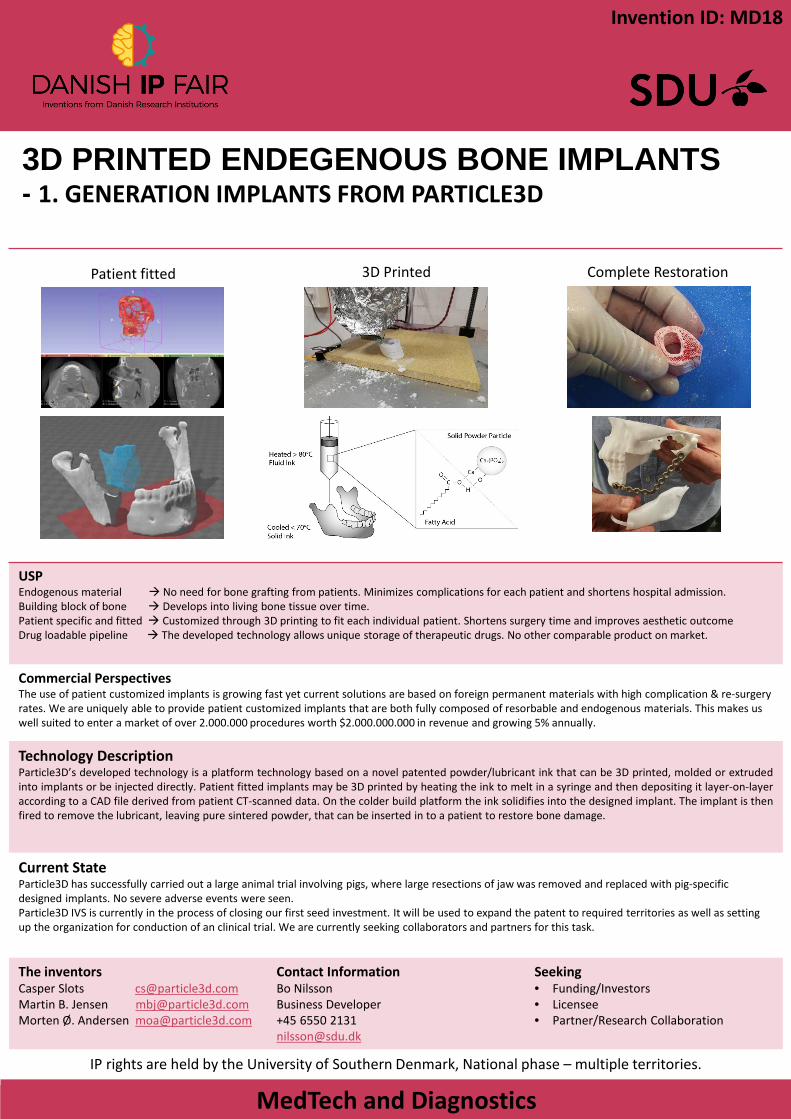
Technology DescriptionParticle3D’s developed technology is a platform technology based on a novel patented powder/lubricant ink that can be 3D printed, molded or extrudedinto implants or be injected directly. Patient fitted implants may be 3D printed by heating the ink to melt in a syringe and then depositing it layer-on-layeraccording to a CAD file derived from patient CT-scanned data. On the colder build platform the ink solidifies into the designed implant. The implant is thenfired to remove the lubricant, leaving pure sintered powder, that can be inserted in to a patient to restore bone damage.
Current StateParticle3D has successfully carried out a large animal trial involving pigs, where large resections of jaw was removed and replaced with pig-specific designed implants. No severe adverse events were seen. Particle3D IVS is currently in the process of closing our first seed investment. It will be used to expand the patent to required territories as well as setting up the organization for conduction of an clinical trial. We are currently seeking collaborators and partners for this task.
The inventorsCasper Slots [email protected] B. Jensen [email protected] Ø. Andersen [email protected]
Contact InformationBo NilssonBusiness Developer+45 6550 [email protected]
Seeking• Funding/Investors• Licensee• Partner/Research Collaboration
IP rights are held by the University of Southern Denmark, National phase – multiple territories.
Patient fitted 3D Printed Complete Restoration
Invention ID: MD18
PhotoRaman- Smartphone based molecular analyzer
MedTech and Diagnostics
Value Proposition/USPThe PhotoRaman optics are specially optimized for smartphone sensor, turning an everyday smartphone into a powerfull Ramanspectrograph. The device demonstrates sensitivity comparable with professional portable Raman analyzers. However, the sale price of PhotoRaman is expected to be 10 times cheaper (~ 8000dkk) in comparison with commercially available portable Raman devices.
Business Opportunity/Objective/Commercial PerspectivesThe portable Raman device market is estimated to $107M and is growing at about CARG 9%. Due to relatively low price of PhotoRaman we would like to implement it as a default sensor in smartphones via collaboration with mobile companies, which would highly expand the potential market. We believe that PhotoRaman will be of interest for the consumer market as a gadget to identify chemical composition of materials in everyday life: drugs, plastics, liquids, etc. On the other hand, the technology can compete with professional portable Raman devices. Therefore PhotoRaman can be used by in the pharmaceutical industry and by customs and police for drugs and narcotics identification in the already established market.
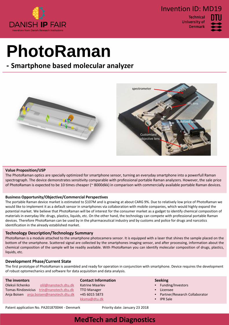
Technology Description/Technology SummaryPhotoRaman is a module attached to the smartphone photocamera sensor. It is equipped with a laser that shines the sample placed on thebottom of the smartphone. Scattered signal are collected by the smartphones imaging sensor, and after processing, information about thechemical composition of the sample will be readily available. With PhotoRaman you can identify molecular composition of drugs, plastics,liquids, etc.
Development Phase/Current StateThe first prototype of PhotoRaman is assembled and ready for operation in conjunction with smartphone. Device requires the development of robust optomechanics and software for data acquisition and data analysis.
The inventorsOleksii Ilchenko [email protected] Rindzevicius [email protected] Boisen [email protected]
Contact InformationKatrine MaarlevTTO Manager+45 6015 [email protected]
Seeking• Funding/Investors• Licensee• Partner/Research Collaborator• IPR Sale
Patent application No. PA201870044 - Denmark Priority date: January 23 2018
Invention ID: MD19

PreDiagnose- The next generation of bacterial detection
MedTech and Diagnostics
Value Proposition/USPOur technology is capable of instant microbial identification that is faster (60 sec vs several days), cheaper (up to 79% of savings per year) and more reliable than existing methods. The technology allows more frequent monitoring and/or a quicker diagnosis of early and intermittent infections by everyone, everywhere.
Business Opportunity/Objective/Commercial PerspectivesPatients infected with microorganisms are limited to late and poor diagnosis techniques. As a consequence, they are left to deal with a difficult treatment, poorer life quality and shorter life expectancy. We propose a time- and cost-effective fast point-of-care device able to detect early infections in a non-invasive manner.
Technology Description/Technology SummaryA point-of-care device with disposable sensing-array that can detect bacterial infections in patient samples immediately after infection. Anelectric signal is sent to the sample and a measured signal is analyzed using an algorithm to identify the presence of specific microbialorganisms. The sensor can be hooked to a smart phone and the results send to the health care provider.
Development Phase/Current StateThe patent behind our technology is in the PCT phase (PCT/EP2017/058614)We are currently expanding the library of microbials we are capable of detecting with the technology.
The inventorsFatima AlZahra’a [email protected]@prediagnose.dkWinnie E. Svendsen [email protected]
Contact InformationØrstads Plads 3442800 Kongens Lyngby
+45 4125 [email protected]
Seeking• Funding/Investors• Licensee• Partner/Research Collaboration• IPR Sale
Intellectual property rights: PCT/EP2017/058614 /WO 2017/178455/19 October 2017
Invention ID: MD20

PreDiagnose- The next generation of bacterial detection
MedTech and Diagnostics
Value Proposition/USPOur technology is capable of instant microbial identification that is faster (60 sec vs several days), cheaper (up to 79% of savings per year) and more reliable than existing methods. The technology allows more frequent monitoring and/or a quicker diagnosis of early and intermittent infections by everyone, everywhere.
Business Opportunity/Objective/Commercial PerspectivesPatients infected with microorganisms are limited to late and poor diagnosis techniques. As a consequence, they are left to deal with a difficult treatment, poorer life quality and shorter life expectancy. We propose a time- and cost-effective fast point-of-care device able to detect early infections in a non-invasive manner.
Technology Description/Technology SummaryA point-of-care device with disposable sensing-array that can detect bacterial infections in patient samples immediately after infection. Anelectric signal is sent to the sample and a measured signal is analyzed using an algorithm to identify the presence of specific microbialorganisms. The sensor can be hooked to a smart phone and the results send to the health care provider.
Development Phase/Current StateThe patent behind our technology is in the PCT phase (PCT/EP2017/058614)We are currently expanding the library of microbials we are capable of detecting with the technology.
The inventorsFatima AlZahra’a [email protected]@prediagnose.dkWinnie E. Svendsen [email protected]
Contact InformationØrstads Plads 3442800 Kongens Lyngby
+45 4125 [email protected]
Seeking• Funding/Investors• Licensee• Partner/Research Collaboration• IPR Sale
Intellectual property rights: PCT/EP2017/058614 /WO 2017/178455/19 October 2017
Invention ID: MD21
A Non-Invasive Light Therapy System for Potentially Slowing Down the Progression
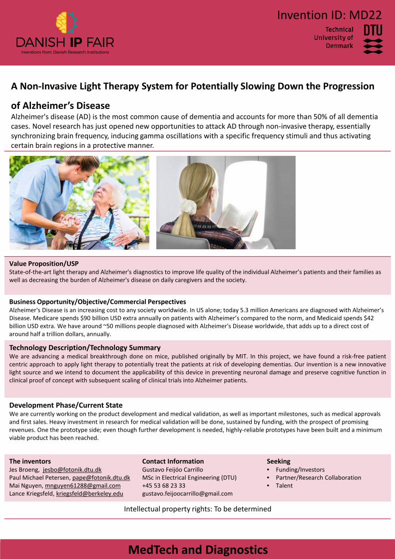
of Alzheimer’s DiseaseAlzheimer's disease (AD) is the most common cause of dementia and accounts for more than 50% of all dementia cases. Novel research has just opened new opportunities to attack AD through non-invasive therapy, essentially synchronizing brain frequency, inducing gamma oscillations with a specific frequency stimuli and thus activating certain brain regions in a protective manner.
MedTech and Diagnostics
Value Proposition/USPState-of-the-art light therapy and Alzheimer's diagnostics to improve life quality of the individual Alzheimer’s patients and their families as well as decreasing the burden of Alzheimer's disease on daily caregivers and the society.
Business Opportunity/Objective/Commercial PerspectivesAlzheimer's Disease is an increasing cost to any society worldwide. In US alone; today 5.3 million Americans are diagnosed with Alzheimer’s Disease. Medicare spends $90 billion USD extra annually on patients with Alzheimer’s compared to the norm, and Medicaid spends $42 billion USD extra. We have around ~50 millions people diagnosed with Alzheimer's Disease worldwide, that adds up to a direct cost of around half a trillion dollars, annually.
Technology Description/Technology SummaryWe are advancing a medical breakthrough done on mice, published originally by MIT. In this project, we have found a risk-free patientcentric approach to apply light therapy to potentially treat the patients at risk of developing dementias. Our invention is a new innovativelight source and we intend to document the applicability of this device in preventing neuronal damage and preserve cognitive function inclinical proof of concept with subsequent scaling of clinical trials into Alzheimer patients.
Development Phase/Current StateWe are currently working on the product development and medical validation, as well as important milestones, such as medical approvals and first sales. Heavy investment in research for medical validation will be done, sustained by funding, with the prospect of promising revenues. One the prototype side; even though further development is needed, highly-reliable prototypes have been built and a minimum viable product has been reached.
The inventorsJes Broeng, [email protected] Michael Petersen, [email protected] Nguyen, [email protected] Kriegsfeld, [email protected]
Contact InformationGustavo Feijóo CarrilloMSc in Electrical Engineering (DTU)+45 53 68 23 [email protected]
Seeking• Funding/Investors• Partner/Research Collaboration• Talent
Intellectual property rights: To be determined
Invention ID: MD22
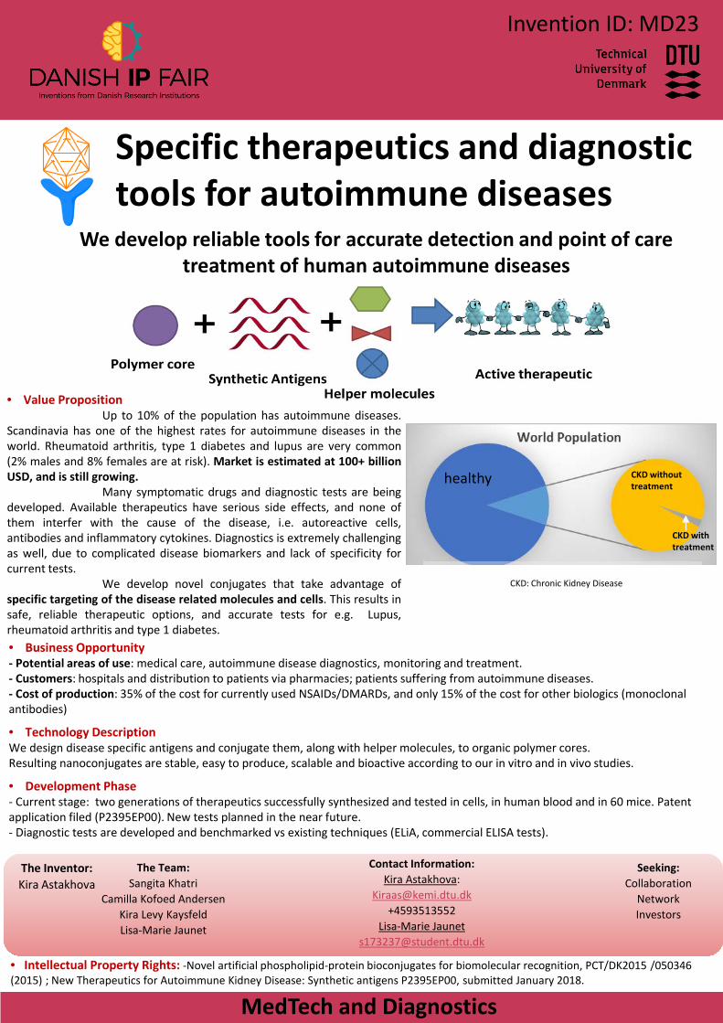
Specific therapeutics and diagnostic tools for autoimmune diseases
We develop reliable tools for accurate detection and point of caretreatment of human autoimmune diseases
• Value PropositionUp to 10% of the population has autoimmune diseases.
Scandinavia has one of the highest rates for autoimmune diseases in theworld. Rheumatoid arthritis, type 1 diabetes and lupus are very common(2% males and 8% females are at risk). Market is estimated at 100+ billionUSD, and is still growing.
Many symptomatic drugs and diagnostic tests are beingdeveloped. Available therapeutics have serious side effects, and none ofthem interfer with the cause of the disease, i.e. autoreactive cells,antibodies and inflammatory cytokines. Diagnostics is extremely challengingas well, due to complicated disease biomarkers and lack of specificity forcurrent tests.
We develop novel conjugates that take advantage ofspecific targeting of the disease related molecules and cells. This results insafe, reliable therapeutic options, and accurate tests for e.g. Lupus,rheumatoid arthritis and type 1 diabetes.• Business Opportunity- Potential areas of use: medical care, autoimmune disease diagnostics, monitoring and treatment.- Customers: hospitals and distribution to patients via pharmacies; patients suffering from autoimmune diseases. - Cost of production: 35% of the cost for currently used NSAIDs/DMARDs, and only 15% of the cost for other biologics (monoclonal antibodies)
• Technology DescriptionWe design disease specific antigens and conjugate them, along with helper molecules, to organic polymer cores. Resulting nanoconjugates are stable, easy to produce, scalable and bioactive according to our in vitro and in vivo studies.
• Development Phase- Current stage: two generations of therapeutics successfully synthesized and tested in cells, in human blood and in 60 mice. Patent application filed (P2395EP00). New tests planned in the near future.- Diagnostic tests are developed and benchmarked vs existing techniques (ELiA, commercial ELISA tests).
• Intellectual Property Rights: -Novel artificial phospholipid-protein bioconjugates for biomolecular recognition, PCT/DK2015 /050346 (2015) ; New Therapeutics for Autoimmune Kidney Disease: Synthetic antigens P2395EP00, submitted January 2018.
The Inventor:Kira Astakhova
The Team:Sangita Khatri
Camilla Kofoed AndersenKira Levy Kaysfeld Lisa-Marie Jaunet
Contact Information:Kira Astakhova:
[email protected]+4593513552
Lisa-Marie [email protected]
Seeking:Collaboration
NetworkInvestors
CKD: Chronic Kidney Disease
MedTech and Diagnostics
healthy CKD withouttreatment
CKD with treatment
Invention ID: MD23