Embed Size (px)
Citation preview
In�uence of Bio-Inspired Ag doped MoO3Nanoparticles in the Seedling Growth and InhibitoryAction Against Microbial OrganismsA. Nirmal Paul Raj
Research Department of Chemistry, St. John’s CollegeR. Biju Bennie
Research Department of Chemistry, St. John’s CollegeG. Alex Immanuel Xavier
Research Department of Chemistry, St. John’s CollegeC Joel
Research Department of Chemistry, St. John’s CollegeD. Abiya Chelliah
A�liated to Manonmaniam Sundaranar UniversityS. Hari Kengaram ( [email protected] )
PMT college https://orcid.org/0000-0003-4226-244X
Research Article
Keywords: Nanoparticles, MoO3, Band gap energy, Antimicrobial studies, Seed germination
Posted Date: June 7th, 2021
DOI: https://doi.org/10.21203/rs.3.rs-571730/v1
License: This work is licensed under a Creative Commons Attribution 4.0 International License. Read Full License

1
Influence of Bio-inspired Ag doped MoO3 nanoparticles in the
seedling growth and inhibitory action against microbial organisms
A. Nirmal Paul Raj1a, R. Biju Bennie1, G. Alex Immanuel Xavier1, C. Joel1b,*,
D. Abiya Chelliah2, S. Hari Kengaram3,*
1aResearch Scholar (Reg. No.18221272031022) & Assistant Professor, Research Department
of Chemistry, St. John’s College, Palayamkottai-627002, Tirunelveli, Tamil Nadu, India.
1bAssistant Professor, Research Department of Chemistry, St. John’s College, Palayamkottai-
627002, Tirunelveli, Tamil Nadu, India.
2Research Department of Botany, St. John’s College, Tirunelveli-627002, Tamil Nadu, India.
3Associate Professor, Department of Chemistry, PMT College, Melaneelithanallur-627953,
Tamil Nadu, India.
1,2,3Affiliated to Manonmaniam Sundaranar University, Abishekapatti, Tirunelveli-627012,
Tamil Nadu, India.
2
Abstract
Herein we report the hydrothermal synthesis, characterization and biological
applications of h-MoO3 and silver doped MoO3 nanoparticles (NPs). The phase formation of
the synthesized NPs was identified using X-ray diffraction studies and vibrational spectral
studies. The average crystallite size of the NPs tends to decrease as the dopant concentration
increases. The surface morphology and the elemental composition of the nanoparticles were
observed from SEM and EDAX analysis. The crystallite nature was obtained from HRTEM
images. The band gap energies obtained from UV-DRS spectra for h-MoO3 (3.26 eV) were
starting to decrease as the concentration of the dopant Ag increases (3.22-2.76eV). The
antibacterial activity of the prepared nanoparticles was tested against some gram positive and
gram negative bacterial strains viz., Staphylococcus aureus, Bacillus cereus and Citrobacter
koseri and Pseudomonas aeruginosa respectively. Also their seed germination properties
were studied on foxtail and finger millet seeds for a period of seven days.
Keywords: Nanoparticles; MoO3; Band gap energy; Antimicrobial studies; Seed germination
Corresponding authors:
Dr. S. Hari Kengaram
E-mail: [email protected]
Dr. C. Joel
E-mail: [email protected]
3
1. Introduction
Agriculture is considered to be the most vital and stable part due to the production of raw
materials for food and feed producing industries. In order to meet the demands of the growing
population, there is a necessity to enhance the existing food production. The deterioration of
natural resources viz., land, water and soil due to contamination claims the need for
agricultural development to be economic, viable and eco-friendly [1]. In the existing
situation, the agricultural field is prone to irregular climatic changes, contamination of
resources by various hazardous pollutants like fertilizers and pesticides which leads to the
elevation of food demands for the growing population [2]. Thus, it is known that there is a
need for increased crop production as well as better food security.
No doubt that the sustainable growth of agriculture totally depends on the new and
innovative techniques like nanotechnology. In agro-industrial sector, nanotechnology is
commonly used in the production of herbicides, fertilizers, fungicides, pesticides as well as
nano-sensor materials [3]. These advances can be used to overcome the future demands in
agriculture thereby increasing quality and yield of crops thus reducing the environmental
pollution due to chemicals and also protecting the vegetatation against environmental stresses
[4]. The of NPs serve a better application in agricultural filed by reducing the nutrient losses
to enhance their yields and minimizing the cost of production to obtain maximum output [5].
The nanoherbicides and nanopesticides used for the treatment of weeds and pests is
observed to significantly increase the crop production. Various types of polymeric and
inorganic nanoparticles are utilized as nanoherbicides. Nanomaterials having specific
antimicrobial properties aid in the prevention of microbial invasions. Using nanotechnology
researchers have designed a smart delivery system to release the nutrients to the targeted site
in a slow and controlled manner in order to tackle the nutrient deficiency in plants.
4
Among the transition metal oxides, molybdenum trioxide (MoO3) has attracted the
attention of the researchers greatly on account of its multifunctional structural, optical,
electronic properties along with better intercalation chemistry with unique chemical,
electrochemical, and catalytic properties. Molybdenum trioxide (MoO3), an n-type
semiconductor material with a wide band gap of 2.8-3.6 eV [6]. There are three common
phases of MoO3: a thermodynamically stable orthorhombic phase (α-MoO3) and two
metastable phases: monoclinic (β-MoO3) and hexagonal (h-MoO3) [7]. The hexagonal phase
of MoO3 is composed of the zigzag chains of MoO6 octahedrons connecting through the cis-
positions [8,9]. In addition, MoO3 was found to be an effective antimicrobial against
different bacterial strains [10]. Antimicrobial activity of MoO3 is related to their acidic
surface involving the formation of intermediate molybdic acid [11]. Also, Mo is responsible
for the conversion of nitrogen into ammonia (NH3) and important for the metabolism of
nitrogen and sulfur. However, the increased amount of Mo in plants leads to molybdenum
toxicity, causing yellowish leaves, reduced growth in seedlings, and increased concentrations
of anthocyanin [12].
Silver nanoparticles (AgNPs) are extensively used for their antimicrobial property against
a wide range of phytopathogens [13]. Silver nanoparticles can constantly release the silver
ions, which may be considered the mechanism behind their antimicrobial effect. Owing to the
electrostatic attraction and greater affinity towards sulfur proteins, silver ions can stick to the
cytoplasmic membrane of the cell and increase the permeability of the membrane leading to
disruption of the bacterial cell [14]. In recent times, silver nanoparticles have been involved
in enhancing seed germination, plant growth and improving photosynthetic quantum
efficiency and also serve as antimicrobial agents in treating plant diseases. There are many
reports showing that AgNPs with appropriate concentrations play a crucial role in enhancing
seed germination and plant growth [15].
5
Owing to the extensive applications of h-MoO3 and Ag nanoparticles, in this study, we
report the synthesis of Ag doped h-MoO3 nanoparticles and their characterization using
several analytical techniques. The antibacterial studies of these nanoparticles against
Staphylococcus aureus, Bacillus cereus, Citrobacter koseri and Pseudomonas aeruginosa
strains have been evaluated and also their seed germination properties on the seeds of foxtail
millet and finger millet seeds have been studied.
2. Materials and methods
2.1 Materials
All the chemicals used in this research work are of analytical grade. Ammonium
heptamolybdate tetrahydrate (AHM) was purchased from Sigma Aldrich and silver nitrate
from Loba Chemie. Deionised water was used throughout the entire research work.
2.2 Synthesis of MoO3 and silver doped MoO3 nanoparticles
For the synthesis of MoO3 nanoparticles, 2.43 g of AHM ([NH4]6Mo7.O24.4H2O]) was
dissolved in 10 mL of deionised water. After stirring for 15 mins, 10 mL of Con. HNO3 was
added slowly in the aqueous solution containing AHM. The reaction mixture was then
transferred to Teflon lined stainless steel autoclave (100 mL) and heated at 90 oC for 9 hours.
The system was then allowed to cool naturally to room temperature. The obtained precipitate
was collected by centrifugation and washed several times with deionised water and ethanol.
Finally the powder was dried in vacuum oven at 70 oC for 12 hours. Similar procedure was
followed for synthesizing Ag doped MoO3. During the reaction, silver nitrate was added
along with AHM. The synthesis was carried out for three different weight percentages (1%,
3% & 5%) of silver nitrate along with AHM. The synthesized nanoparticles MoO3, 1% Ag
doped MoO3, 3% Ag doped MoO3 and 5% Ag doped MoO3 which is mentioned as M, 1AM,
3AM and 5AM respectively. The formation of molybdenum trioxide from the precursor can
be described by the following endothermic reaction,
6
(NH4)6Mo7O244H2O 7MoO3(s) + 6NH3 (g) + 7H2O(g)
2.3 Characterization techniques
X-ray diffraction patterns of the synthesized samples were recorded on X’pert PRO
powder X-ray diffractometer in 2θ range of 20°–60° at a scan rate of 2° min-1 using Cu-
Kα X-ray radiation source. The surface morphologies of the samples were examined by using
field emission scanning electron microscope ((JSM-6700, JEOL, Japan). The elemental
compositions were obtained on Energy Dispersive X-ray spectrometer (RONTEC's EDX
system, Model QuanTax 200, Germany). The HRTEM images were obtained using High
Resolution Transmission Electron Microscope (JEM-2100 Plus, JEOL, Japan). The FTIR
spectra of the samples were recorded on SHIMADZU spectrometer having ATR facility by
pelletizing the samples with KBr in the frequency range of 400–4000 cm−1. FT Raman
spectra were recorded using 1064 nm line of Nd:YAG laser as excitation wavelength on an
EZ Raman, Enwave optronics, and USA IFS 66 V spectrometer. For recording UV–Vis
absorption spectra, a computer controlled JASCO V-530 dual beam spectrophotometer
coupled with DRS support was used.
2.4 Antimicrobial studies
Antimicrobial activity of the nanoparticles was carried out against four different
bacterial strains i.e. Staphylococcus aureus, Bacillus cereus, Citrobacter koseri and
Pseudomonas aeruginosa employing well diffusion method. It was performed by properly
sterilizing the Mueller Hinton media of agar. After solidification, the wells were cut using a
cork borer. The bacterial pathogens to be tested were swabbed onto the surface of Mueller
Hinton agar plates. The cut pieces of thin film of samples were placed on the well. The plates
were incubated at 37oC for 24 hours, and then the zone of inhibition was measured in
millimeters. Each antibacterial assay was performed in triplicate and mean values were
reported.
7
2.5 Seed Germination
The experimental treatments involved four samples M, 1AM, 3AM and 5AM NPs for
observing the germination of foxtail millet (Setaria italica (L.) P.Beauv.) and finger millet
(Eleusine coracana (L.) Gaertn.) seeds. Healthy seeds of uniform size were selected to
minimize the errors and were rinsed using water to clean them from chemicals and surface
impurities. After rinsing the seeds in running tap water, they were incubated in a 5% (v/v)
detergent solution for 5 min. Then, seeds were then immersed in 70% (v/v) ethanol solution
(1 min) and finally in NaCl (1.5% (v/v), 10 min). Finally, the seeds were washed twice
thoroughly in sterile distilled water. The experiment was performed under normal laboratory
conditions in natural light. Germination tests were done in petri dishes containing a Whatman
No-1 filter paper moistened with 5 ml of suspension or distilled water and each plate was
loaded with fifteen seeds with 5ml suspension of NPs with specific concentration. The
distilled water was utilized in control sample for the reference. The petri dishes were kept in a
dark place and the number of seeds germinated and other relevant parameters were recorded
on daily for a period of 7 days.
3. Results and discussion
3.1 X-Ray Diffraction studies
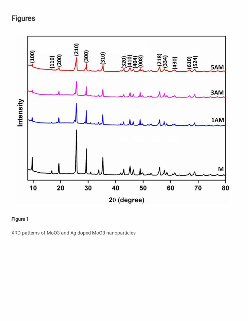
The crystal structure of MoO3 and Ag doped MoO3 were analyzed by powder XRD
measurements. The presence of sharp diffraction peaks is an indication of the high
crystallinity of the synthesized nanoparticles. The peak with maximum intensity was
observed at 25.6° and corresponds to the diffraction from (210) plane of MoO3. All the other
observed diffraction lines at 2θ = 19°, 29°, 36° and 45° were corresponds to the (200), (300),
(310), and (410) reflection lines of MoO3, respectively. All the observed diffraction peaks
have been indexed and matched well with standard data card (JCPDS-21-0569). It was found
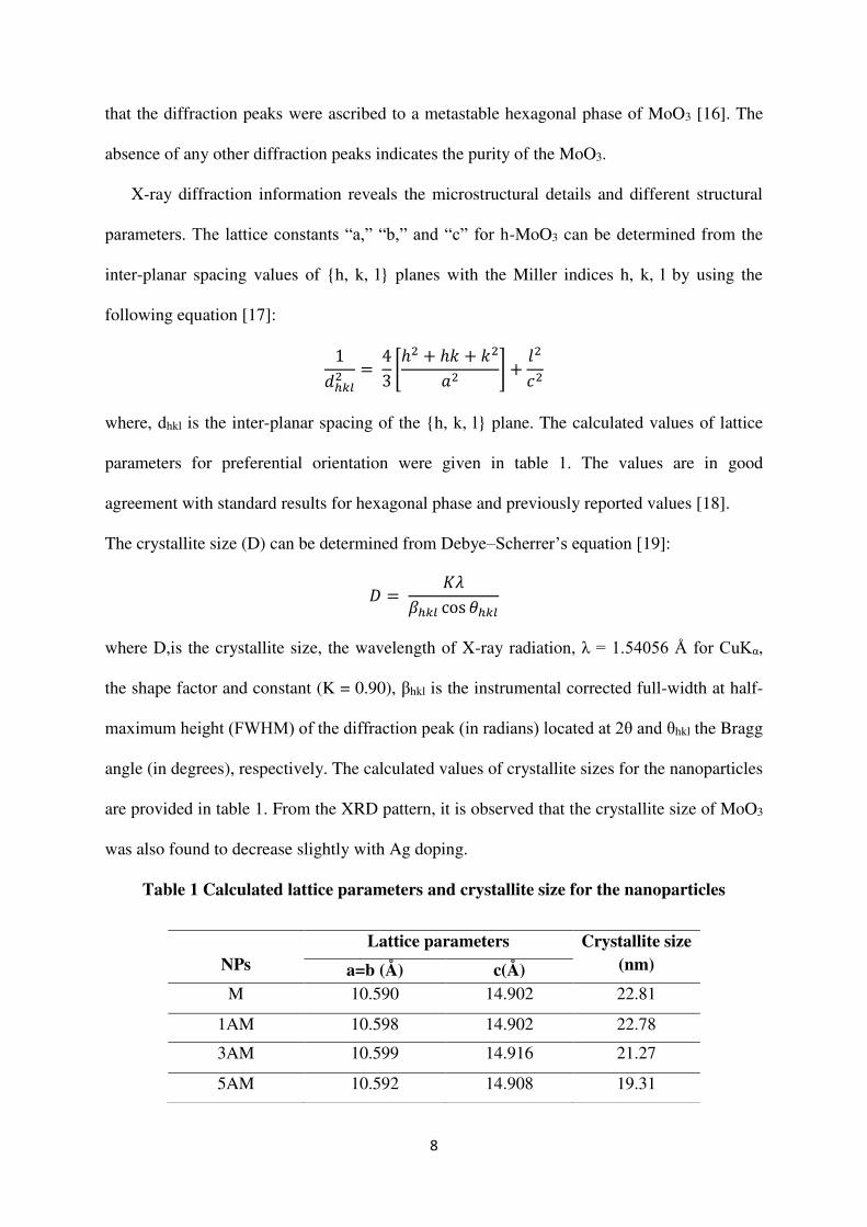
8
that the diffraction peaks were ascribed to a metastable hexagonal phase of MoO3 [16]. The
absence of any other diffraction peaks indicates the purity of the MoO3.
X-ray diffraction information reveals the microstructural details and different structural
parameters. The lattice constants “a,” “b,” and “c” for h-MoO3 can be determined from the
inter-planar spacing values of {h, k, l} planes with the Miller indices h, k, l by using the
following equation [17]: 1𝑑ℎ𝑘𝑙2 = 43 [ℎ2 + ℎ𝑘 + 𝑘2𝑎2 ] + 𝑙2𝑐2
where, dhkl is the inter-planar spacing of the {h, k, l} plane. The calculated values of lattice
parameters for preferential orientation were given in table 1. The values are in good
agreement with standard results for hexagonal phase and previously reported values [18].
The crystallite size (D) can be determined from Debye–Scherrer’s equation [19]:
𝐷 = 𝐾𝜆𝛽ℎ𝑘𝑙 cos 𝜃ℎ𝑘𝑙 where D,is the crystallite size, the wavelength of X-ray radiation, λ = 1.54056 Å for CuKα,
the shape factor and constant (K = 0.90), βhkl is the instrumental corrected full-width at half-
maximum height (FWHM) of the diffraction peak (in radians) located at 2θ and θhkl the Bragg
angle (in degrees), respectively. The calculated values of crystallite sizes for the nanoparticles
are provided in table 1. From the XRD pattern, it is observed that the crystallite size of MoO3
was also found to decrease slightly with Ag doping.
Table 1 Calculated lattice parameters and crystallite size for the nanoparticles
NPs
Lattice parameters Crystallite size (nm) a=b (Å) c(Å)
M 10.590 14.902 22.81
1AM 10.598 14.902 22.78
3AM 10.599 14.916 21.27
5AM 10.592 14.908 19.31
9
Fig. 1 XRD patterns of MoO3 and Ag doped MoO3 nanoparticles
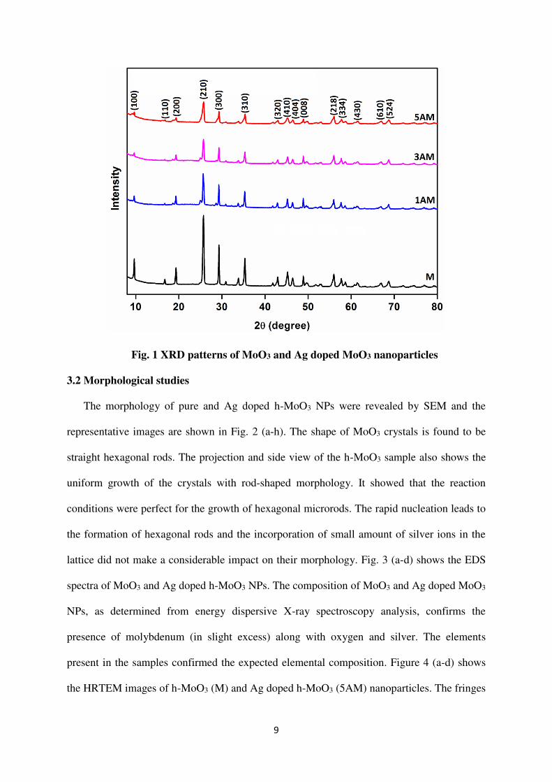
3.2 Morphological studies
The morphology of pure and Ag doped h-MoO3 NPs were revealed by SEM and the
representative images are shown in Fig. 2 (a-h). The shape of MoO3 crystals is found to be
straight hexagonal rods. The projection and side view of the h-MoO3 sample also shows the
uniform growth of the crystals with rod-shaped morphology. It showed that the reaction
conditions were perfect for the growth of hexagonal microrods. The rapid nucleation leads to
the formation of hexagonal rods and the incorporation of small amount of silver ions in the
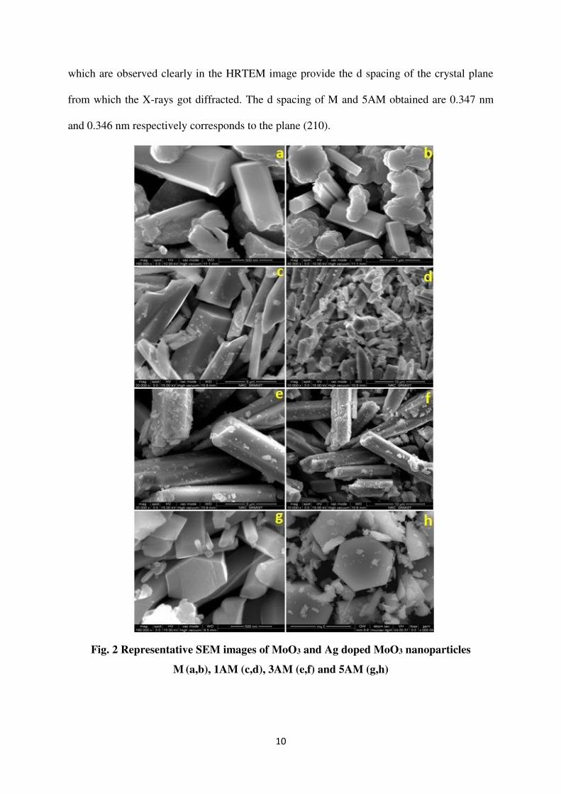
lattice did not make a considerable impact on their morphology. Fig. 3 (a-d) shows the EDS
spectra of MoO3 and Ag doped h-MoO3 NPs. The composition of MoO3 and Ag doped MoO3
NPs, as determined from energy dispersive X-ray spectroscopy analysis, confirms the
presence of molybdenum (in slight excess) along with oxygen and silver. The elements
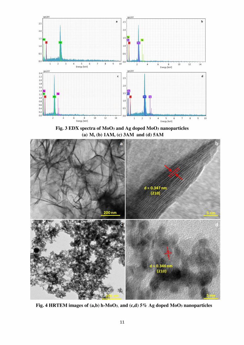
present in the samples confirmed the expected elemental composition. Figure 4 (a-d) shows
the HRTEM images of h-MoO3 (M) and Ag doped h-MoO3 (5AM) nanoparticles. The fringes
10
which are observed clearly in the HRTEM image provide the d spacing of the crystal plane
from which the X-rays got diffracted. The d spacing of M and 5AM obtained are 0.347 nm
and 0.346 nm respectively corresponds to the plane (210).
Fig. 2 Representative SEM images of MoO3 and Ag doped MoO3 nanoparticles
M (a,b), 1AM (c,d), 3AM (e,f) and 5AM (g,h)
11
Fig. 3 EDX spectra of MoO3 and Ag doped MoO3 nanoparticles (a) M, (b) 1AM, (c) 3AM and (d) 5AM
Fig. 4 HRTEM images of (a,b) h-MoO3, and (c,d) 5% Ag doped MoO3 nanoparticles
12
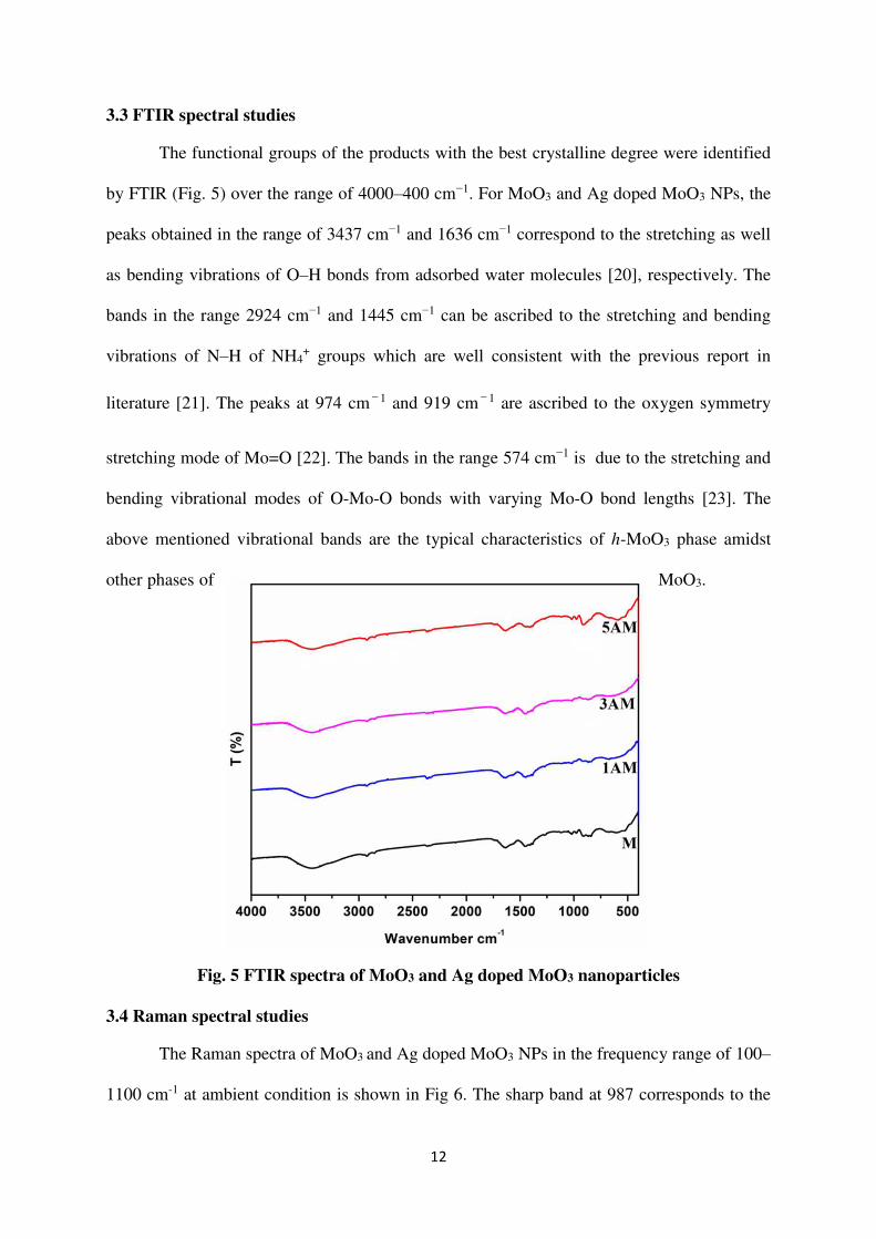
3.3 FTIR spectral studies
The functional groups of the products with the best crystalline degree were identified
by FTIR (Fig. 5) over the range of 4000–400 cm−1. For MoO3 and Ag doped MoO3 NPs, the
peaks obtained in the range of 3437 cm−1 and 1636 cm−1 correspond to the stretching as well
as bending vibrations of O–H bonds from adsorbed water molecules [20], respectively. The
bands in the range 2924 cm−1 and 1445 cm−1 can be ascribed to the stretching and bending
vibrations of N–H of NH4+ groups which are well consistent with the previous report in
literature [21]. The peaks at 974 cm−1 and 919 cm−1 are ascribed to the oxygen symmetry
stretching mode of Mo=O [22]. The bands in the range 574 cm−1 is due to the stretching and
bending vibrational modes of O-Mo-O bonds with varying Mo-O bond lengths [23]. The
above mentioned vibrational bands are the typical characteristics of h-MoO3 phase amidst
other phases of MoO3.
Fig. 5 FTIR spectra of MoO3 and Ag doped MoO3 nanoparticles
3.4 Raman spectral studies
The Raman spectra of MoO3 and Ag doped MoO3 NPs in the frequency range of 100–
1100 cm-1 at ambient condition is shown in Fig 6. The sharp band at 987 corresponds to the
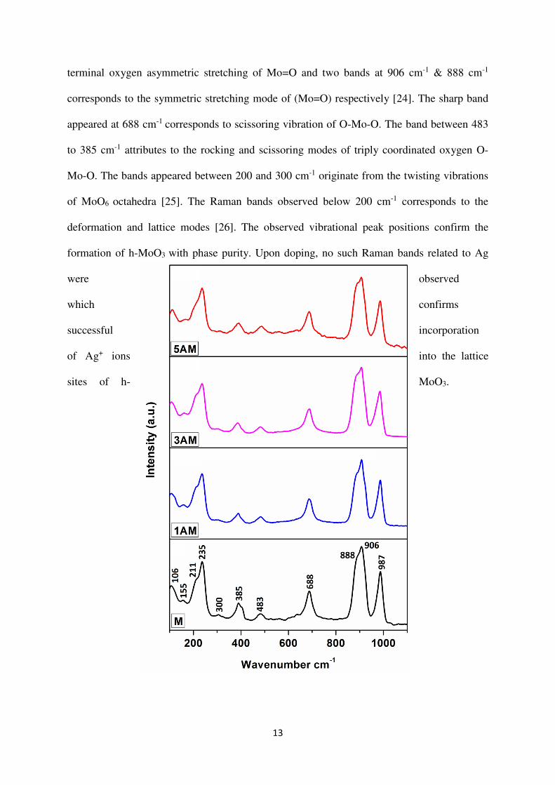
13
terminal oxygen asymmetric stretching of Mo=O and two bands at 906 cm-1 & 888 cm-1
corresponds to the symmetric stretching mode of (Mo=O) respectively [24]. The sharp band
appeared at 688 cm-1 corresponds to scissoring vibration of O-Mo-O. The band between 483
to 385 cm-1 attributes to the rocking and scissoring modes of triply coordinated oxygen O-
Mo-O. The bands appeared between 200 and 300 cm-1 originate from the twisting vibrations
of MoO6 octahedra [25]. The Raman bands observed below 200 cm-1 corresponds to the
deformation and lattice modes [26]. The observed vibrational peak positions confirm the
formation of h-MoO3 with phase purity. Upon doping, no such Raman bands related to Ag
were observed
which confirms
successful incorporation
of Ag+ ions into the lattice
sites of h- MoO3.
14
Fig. 6 Raman spectra of MoO3 and Ag doped MoO3 nanoparticles
3.5 UV-DRS spectra
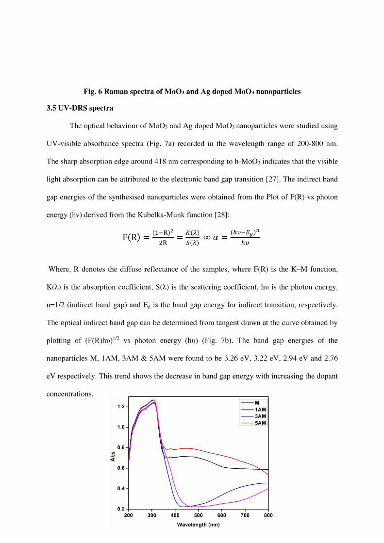
The optical behaviour of MoO3 and Ag doped MoO3 nanoparticles were studied using
UV-visible absorbance spectra (Fig. 7a) recorded in the wavelength range of 200-800 nm.
The sharp absorption edge around 418 nm corresponding to h-MoO3 indicates that the visible
light absorption can be attributed to the electronic band gap transition [27]. The indirect band
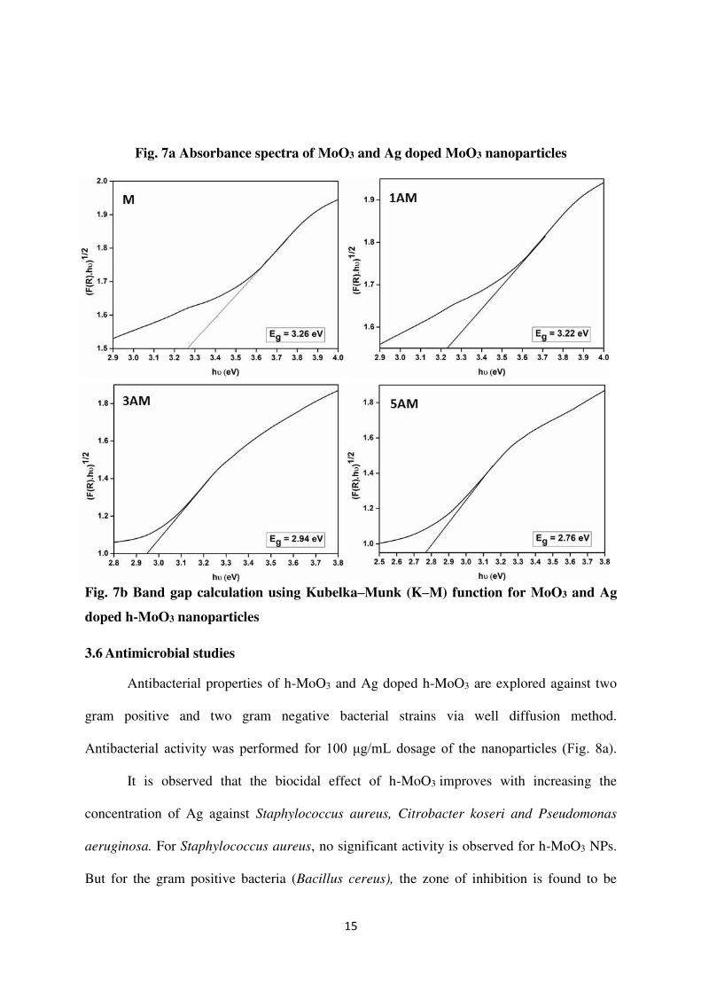
gap energies of the synthesised nanoparticles were obtained from the Plot of F(R) vs photon
energy (hν) derived from the Kubelka-Munk function [28]: F(R) = (1−R)22R = 𝐾(𝜆)𝑆(𝜆) ∞ 𝛼 = (ℎυ−𝐸𝑔)𝑛ℎυ Where, R denotes the diffuse reflectance of the samples, where F(R) is the K–M function,
K(λ) is the absorption coefficient, S(λ) is the scattering coefficient, hυ is the photon energy,
n=1/2 (indirect band gap) and Eg is the band gap energy for indirect transition, respectively.
The optical indirect band gap can be determined from tangent drawn at the curve obtained by
plotting of (F(R)hυ)1/2 vs photon energy (hυ) (Fig. 7b). The band gap energies of the
nanoparticles M, 1AM, 3AM & 5AM were found to be 3.26 eV, 3.22 eV, 2.94 eV and 2.76
eV respectively. This trend shows the decrease in band gap energy with increasing the dopant
concentrations.
15
Fig. 7a Absorbance spectra of MoO3 and Ag doped MoO3 nanoparticles
Fig. 7b Band gap calculation using Kubelka–Munk (K–M) function for MoO3 and Ag
doped h-MoO3 nanoparticles
3.6 Antimicrobial studies
Antibacterial properties of h-MoO3 and Ag doped h-MoO3 are explored against two
gram positive and two gram negative bacterial strains via well diffusion method.
Antibacterial activity was performed for 100 μg/mL dosage of the nanoparticles (Fig. 8a).
It is observed that the biocidal effect of h-MoO3 improves with increasing the
concentration of Ag against Staphylococcus aureus, Citrobacter koseri and Pseudomonas
aeruginosa. For Staphylococcus aureus, no significant activity is observed for h-MoO3 NPs.
But for the gram positive bacteria (Bacillus cereus), the zone of inhibition is found to be
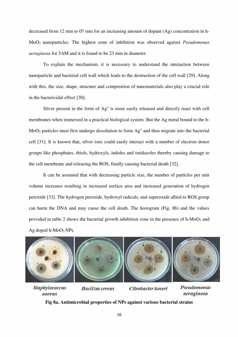
16
decreased from 12 mm to 07 mm for an increasing amount of dopant (Ag) concentration in h-
MoO3 nanoparticles. The highest zone of inhibition was observed against Pseudomonas
aeruginosa for 5AM and it is found to be 23 mm in diameter.
To explain the mechanism, it is necessary to understand the interaction between
nanoparticle and bacterial cell wall which leads to the destruction of the cell wall [29]. Along
with this, the size, shape, structure and composition of nanomaterials also play a crucial role
in the bactericidal effect [30].
Silver present in the form of Ag+ is more easily released and directly react with cell
membranes when immersed in a practical biological system. But the Ag metal bound to the h-
MoO3 particles must first undergo dissolution to form Ag+ and then migrate into the bacterial
cell [31]. It is known that, silver ions could easily interact with a number of electron donor
groups like phosphates, thiols, hydroxyls, indoles and imidazoles thereby causing damage to
the cell membrane and releasing the ROS, finally causing bacterial death [32].
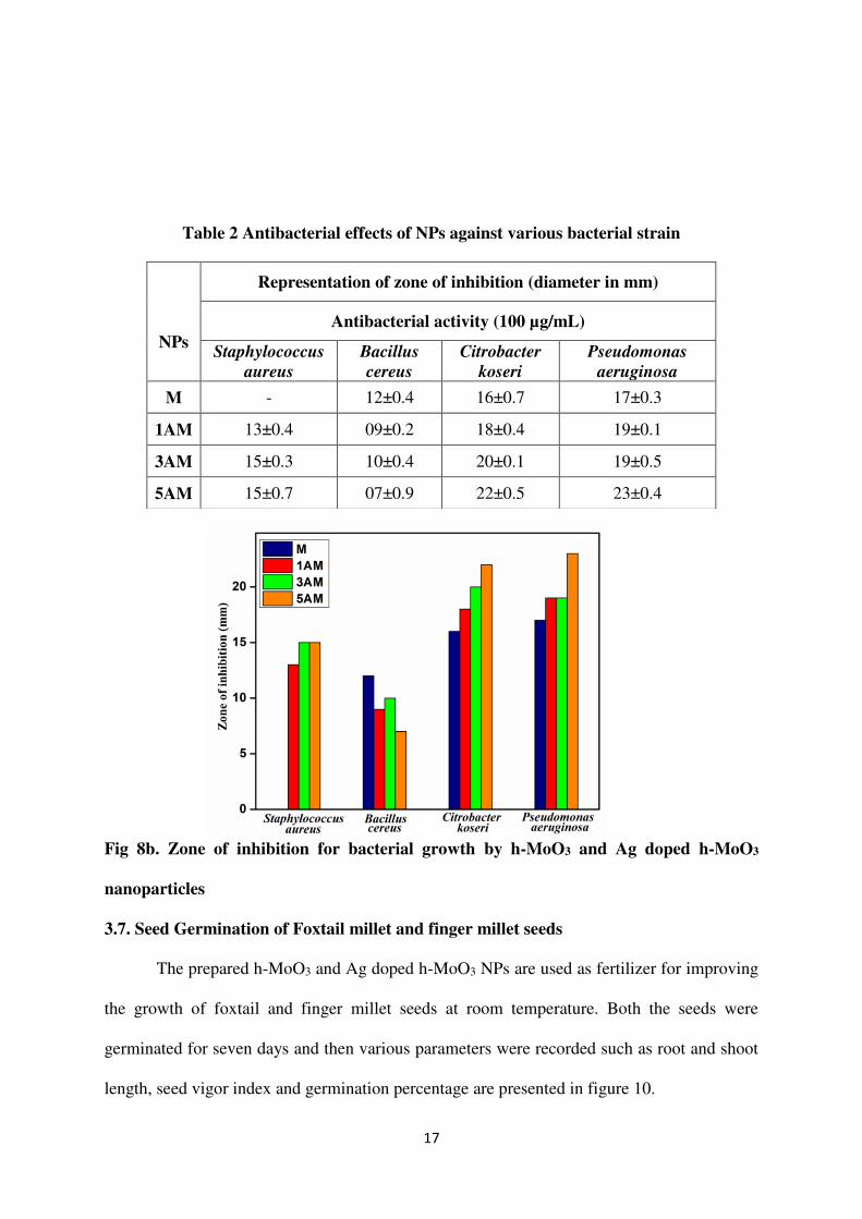
It can be assumed that with decreasing particle size, the number of particles per unit
volume increases resulting in increased surface area and increased generation of hydrogen
peroxide [33]. The hydrogen peroxide, hydroxyl radicals, and superoxide allied to ROS group
can harm the DNA and may cause the cell death. The histogram (Fig. 8b) and the values
provided in table 2 shows the bacterial growth inhibition zone in the presence of h-MoO3 and
Ag doped h-MoO3 NPs.
Fig 8a. Antimicrobial properties of NPs against various bacterial strains
17
Table 2 Antibacterial effects of NPs against various bacterial strain
Fig 8b. Zone of inhibition for bacterial growth by h-MoO3 and Ag doped h-MoO3
nanoparticles
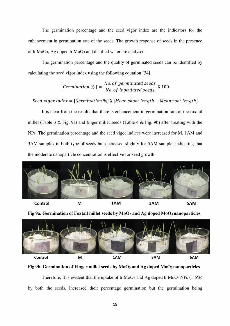
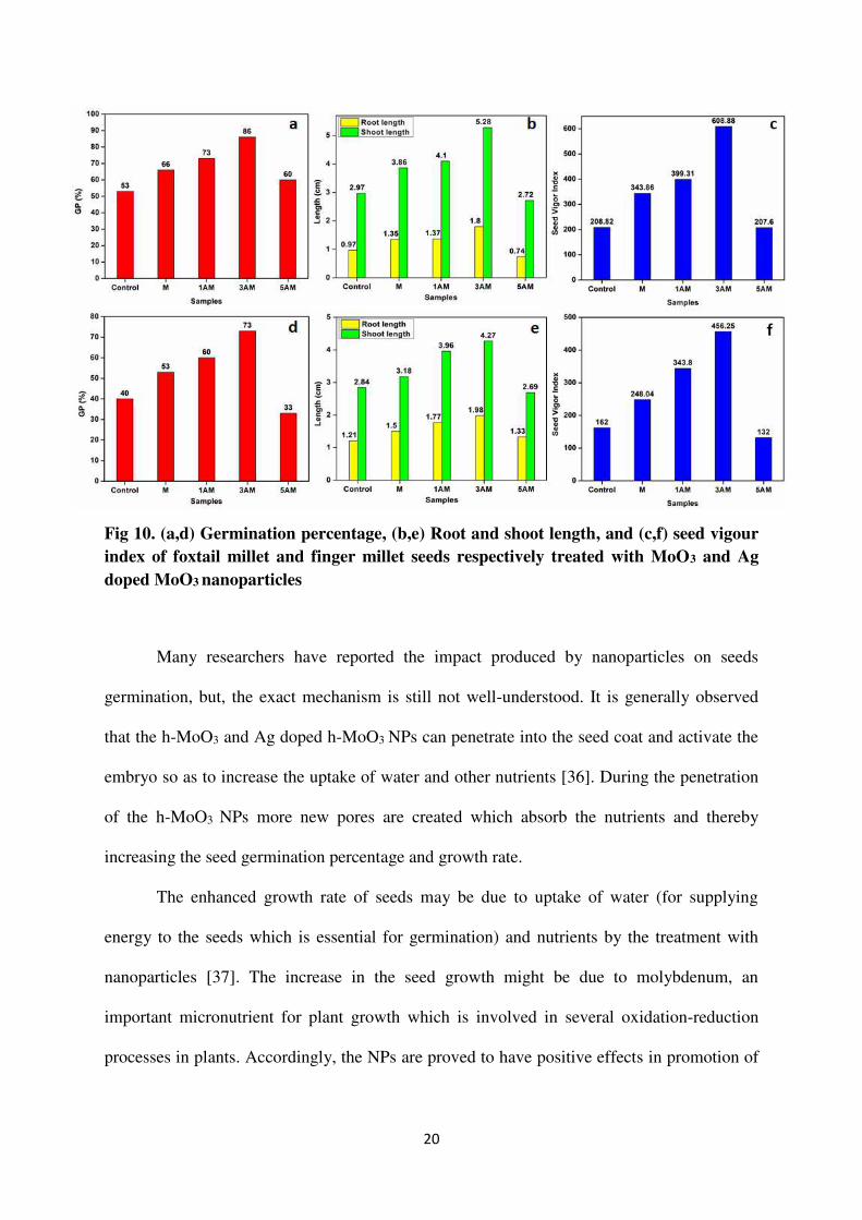
3.7. Seed Germination of Foxtail millet and finger millet seeds
The prepared h-MoO3 and Ag doped h-MoO3 NPs are used as fertilizer for improving
the growth of foxtail and finger millet seeds at room temperature. Both the seeds were
germinated for seven days and then various parameters were recorded such as root and shoot
length, seed vigor index and germination percentage are presented in figure 10.
NPs
Representation of zone of inhibition (diameter in mm)
Antibacterial activity (100 µg/mL)
Staphylococcus
aureus
Bacillus
cereus
Citrobacter
koseri
Pseudomonas
aeruginosa
M - 12±0.4 16±0.7 17±0.3
1AM 13±0.4 09±0.2 18±0.4 19±0.1
3AM 15±0.3 10±0.4 20±0.1 19±0.5
5AM 15±0.7 07±0.9 22±0.5 23±0.4
18
The germination percentage and the seed vigor index are the indicators for the
enhancement in germination rate of the seeds. The growth response of seeds in the presence
of h-MoO3, Ag doped h-MoO3 and distilled water are analysed.
The germination percentage and the quality of germinated seeds can be identified by
calculating the seed vigor index using the following equation [34].
[𝐺𝑒𝑟𝑚𝑖𝑛𝑎𝑡𝑖𝑜𝑛 % ] = 𝑁𝑜. 𝑜𝑓 𝑔𝑒𝑟𝑚𝑖𝑛𝑎𝑡𝑒𝑑 𝑠𝑒𝑒𝑑𝑠𝑁𝑜. 𝑜𝑓 𝑖𝑛𝑜𝑐𝑢𝑙𝑎𝑡𝑒𝑑 𝑠𝑒𝑒𝑑𝑠 X 100
𝑆𝑒𝑒𝑑 𝑣𝑖𝑔𝑜𝑟 𝑖𝑛𝑑𝑒𝑥 = [𝐺𝑒𝑟𝑚𝑖𝑛𝑎𝑡𝑖𝑜𝑛 %] X [𝑀𝑒𝑎𝑛 𝑠ℎ𝑜𝑜𝑡 𝑙𝑒𝑛𝑔𝑡ℎ + 𝑀𝑒𝑎𝑛 𝑟𝑜𝑜𝑡 𝑙𝑒𝑛𝑔𝑡ℎ] It is clear from the results that there is enhancement in germination rate of the foxtail
millet (Table 3 & Fig. 9a) and finger millet seeds (Table 4 & Fig. 9b) after treating with the
NPs. The germination percentage and the seed vigor indices were increased for M, 1AM and
3AM samples in both type of seeds but decreased slightly for 5AM sample, indicating that
the moderate nanoparticle concentration is effective for seed growth.
Fig 9a. Germination of Foxtail millet seeds by MoO3 and Ag doped MoO3 nanoparticles
Fig 9b. Germination of Finger millet seeds by MoO3 and Ag doped MoO3 nanoparticles
Therefore, it is evident that the uptake of h-MoO3 and Ag doped h-MoO3 NPs (1-5%)
by both the seeds, increased their percentage germination but the germination being
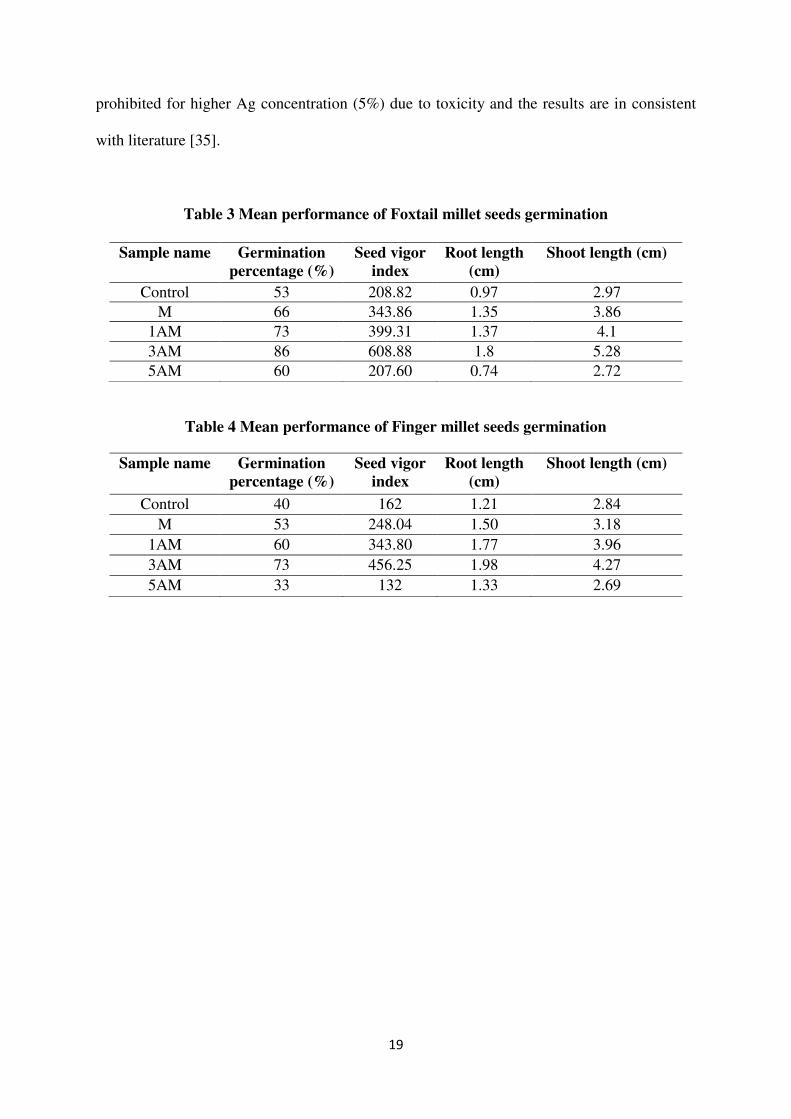
19
prohibited for higher Ag concentration (5%) due to toxicity and the results are in consistent
with literature [35].
Table 3 Mean performance of Foxtail millet seeds germination
Table 4 Mean performance of Finger millet seeds germination
Sample name Germination percentage (%)
Seed vigor index
Root length (cm)
Shoot length (cm)
Control 53 208.82 0.97 2.97
M 66 343.86 1.35 3.86
1AM 73 399.31 1.37 4.1
3AM 86 608.88 1.8 5.28
5AM 60 207.60 0.74 2.72
Sample name Germination percentage (%)
Seed vigor index
Root length (cm)
Shoot length (cm)
Control 40 162 1.21 2.84
M 53 248.04 1.50 3.18
1AM 60 343.80 1.77 3.96
3AM 73 456.25 1.98 4.27
5AM 33 132 1.33 2.69
20
Fig 10. (a,d) Germination percentage, (b,e) Root and shoot length, and (c,f) seed vigour index of foxtail millet and finger millet seeds respectively treated with MoO3 and Ag doped MoO3 nanoparticles
Many researchers have reported the impact produced by nanoparticles on seeds
germination, but, the exact mechanism is still not well-understood. It is generally observed
that the h-MoO3 and Ag doped h-MoO3 NPs can penetrate into the seed coat and activate the
embryo so as to increase the uptake of water and other nutrients [36]. During the penetration
of the h-MoO3 NPs more new pores are created which absorb the nutrients and thereby
increasing the seed germination percentage and growth rate.
The enhanced growth rate of seeds may be due to uptake of water (for supplying
energy to the seeds which is essential for germination) and nutrients by the treatment with
nanoparticles [37]. The increase in the seed growth might be due to molybdenum, an
important micronutrient for plant growth which is involved in several oxidation-reduction
processes in plants. Accordingly, the NPs are proved to have positive effects in promotion of
21
root and shoot formation, and also accumulation of plant biomass from seeds in most of the
crops compared to the untreated one [38].
Silver based nanoparticles are commonly used in the food and agriculture application
at specified concentrations due to their well-known antimicrobial activity and root
regeneration property. The highest concentration tested for Ag doped NPs (higher
concentrations induce oxidative stress in plant cells) was not found to be effective for plant
growth but had a robust antimicrobial effect, which would be beneficial in during seed
germination period. The silver doped NPs at moderate concentrations significantly enhance
seed germination, germination %, seed vigour index, root length and shoot length of seeds.
Even higher concentrations of Ag doped NPs have led to a severe decrease in the growth
parameters of seeds which were associated with accumulation of Ag in the stems and roots
[39].
4. Conclusion
The h-MoO3 and different concentrations of Ag doped h-MoO3 NPs were successfully
synthesized by autoclave mediated hydrothermal route. Powder XRD analysis shows that the
Ag doped MoO3 NPs crystallize in meta-stable hexagonal structure with (210) preferred
orientation. Further the FTIR, Raman and SEM-EDX studies confirms the formation of phase
pure h-MoO3 and Ag doped h-MoO3 NPs. The morphology of the synthesized h-MoO3 and Ag
doped h-MoO3 shows the rod like structure with hexagonal cross section. From HRTEM
analysis, the crystallite size was found to decrease slightly for the Ag doped h-MoO3
compared to pure h-MoO3 NPs. From the UV-DRS spectra, the indirect band gap energy was
found to decrease from 3.26 eV to 2.76 eV as the concentration of Ag increases and the
22
reason may be due to the Ag doping can cause great deviations in the electronic structure of
the host material. The synthesized h-MoO3 and Ag doped h-MoO3 NPs exhibit good
inhibitory action against four bacterial strains viz. Staphylococcus aureus, Bacillus cereus,
Citrobacter koseri and Pseudomonas aeruginosa. The h-MoO3 and Ag doped h-MoO3 NPs
displayed tremendous germination growth of foxtail and finger millet seeds upto 86% and
73%, respectively, and therefore can be employed as a promising fertilizer in the plant
growth. Because of the toxicity, 5% Ag doped h-MoO3 NPs study had little negative effect on
seedling growth for both the seeds, but shows the maximum zone of inhibition against all the
bacterial strains.
Declarations
The authors declare no conflict of interest.
Acknowledgments
The authors thank the management of St. John’s College, Tirunelveli, Tamil Nadu
also being our research centre for providing the lab facilities to carry out this research work.
We would like to acknowledge SRM University, Chennai, Tamil Nadu, India for providing
PXRD and SEM with EDX spectral analysis. We thank Dr.C. Vethi, VOC College, Tuticorin,
for recording FTIR and UV-DRS spectra. Also we would like to thank the Department of
Nanotechnology, Noorul Islam centre for higher Education, Thuckalay, Kanniyakumari,
Tamil Nadu-629180, India for recording Raman spectra.
References
[1] R. Prasad, A. Bhattacharyya, Q.D. Nguyen, Nanotechnology in sustainable agriculture:
recent developments, challenges, and perspectives, Front. Microbiol. 8 (2017) 1014.
[2] D. Mittal, G. Kaur, P. Singh, K. Yadav, S.A. Ali, Nanoparticle-based sustainable
agriculture and food science: recent advances and future outlook, Front. Nanotechnol. 2
(2020).
23
[3] I. Iavicoli, V. Leso, D.H. Beezhold, A.A. Shvedova, Nanotechnology in agriculture:
Opportunities, toxicological implications, and occupational risks, Toxicol. Appl.
Pharmacol. 329 (2017) 96-111.
[4] R. Liu, R. Lal, Potentials of engineered nanoparticles as fertilizers for increasing
agronomic productions, Sci. Total Environ. 514 (2015) 131-139.
[5] M. Usman, M. Farooq, A. Wakeel, A. Nawaz, S.A. Cheema, H. ur Rehman, M.
Sanaullah, Nanotechnology in agriculture: Current status, challenges and future opportunities,
Sci. Total Environ. 721 (2020) 137778.
[6] A. Manivel, G.J. Lee, C.Y. Chen, J.H. Chen, S.H. Ma, T.L. Horng, J.J. Wu, Synthesis of
MoO3 nanoparticles for azo dye degradation by catalytic ozonation, Mater. Res. Bull. 62
(2015)184-191.
[7] C.V. Ramana, I.B. Troitskaia, V.V. Atuchin, M. Ramos, D. Ferrer, Electron microscopy
characterization of hexagonal molybdenum trioxide (MoO3) nanorods, J. Vac. Sci. Technol.
A 28 (2010) 726-729.
[8] J. Song, X. Ni, L. Gao, H. Zheng, Synthesis of metastable h-MoO3 by simple chemical
precipitation, Mater. Chem. Phys. 102 (2007) 245-248.
[9] L. Zhou, L. Yang, P. Yuan, J. Zou, Y. Wu, C. Yu, α-MoO3 nanobelts: a high performance
cathode material for lithium ion batteries, J. Phys. Chem. C 114 (2010) 21868-21872.
[10] C. Chaves-Lopez, H.N. Nguyen, R.C. Oliveira, E.T. Nadres, A. Paparella, D.F.
Rodrigues, A morphological, enzymatic and metabolic approach to elucidate apoptotic-like
cell death in fungi exposed to h-and α-molybdenum trioxide nanoparticles, Nanoscale, 10
(2018) 20702-20716.
[11] C. Zollfrank, K. Gutbrod, P. Wechsler, J.P. Guggenbichler, Antimicrobial activity of
transition metal acid MoO3 prevents microbial growth on material surfaces, Mater. Sci.
Eng. C 32 (2012) 47-54.
24
[12] M. Imran, X. Sun, S. Hussain, U. Ali, M.S. Rana, F. Rasul, C.X. Hu, Molybdenum-
induced effects on nitrogen metabolism enzymes and elemental profile of winter wheat
(Triticum aestivum L.) under different nitrogen sources, Int. J. Mol. Sci. 20 (2019) 3009.
[13] M.R. Bindhu, M. Umadevi, G.A. Esmail, N.A. Al-Dhabi, M.V. Arasu, Green synthesis
and characterization of silver nanoparticles from Moringa oleifera flower and assessment of
antimicrobial and sensing properties, J. Photochem. Photobiol. B 205 (2020) 111836.
[14] I.X. Yin, J. Zhang, I.S. Zhao, M.L. Mei, Q. Li, C.H. Chu, The antibacterial mechanism
of silver nanoparticles and its application in dentistry, Int. J. Nanomedicine 15 (2020) 2555-
2562.
[15] M.S. Sadak, Impact of silver nanoparticles on plant growth, some biochemical aspects,
and yield of fenugreek plant (Trigonella foenum-graecum), Bull. Natl. Res. Cent. 43 (2019)
1-6.
[16] A. Chithambararaj, A.C. Bose, Investigation on structural, thermal, optical and sensing
properties of meta-stable hexagonal MoO3 nanocrystals of one dimensional structure,
Beilstein J. Nanotechnol. 2 (2011) 585-592.
[17] K.V. Alex, A.R. Jayakrishnan, A.S. Ibrahim, K. Kamakshi, J.P.B. Silva, K.C. Sekhar,
M.J.M. Gomes, Substrate temperature induced effect on microstructure, optical and
photocatalytic activity of ultrasonic spray pyrolysis deposited MoO3 thin films, Mater.
Res. Express 6 (2019) 066421.
[18] A. Chithambararaj, N.S. Sanjini, S. Velmathi, A.C. Bose, Preparation of h-MoO3 and α-
MoO3 nanocrystals: comparative study on photocatalytic degradation of methylene blue
under visible light irradiation, Phys. Chem. Chem. Phys. 15 (2013) 14761-14769.
[19] S.K. Sen, S. Dutta, M.R. Khan, M.S. Manir, S. Dutta, A. Al Mortuza, M.A.
Hakim, Characterization and Antibacterial Activity Study of Hydrothermally Synthesized h-
MoO3 Nanorods and α-MoO3 Nanoplates, BioNanoScience 9 (2019) 873-882.
25
[20] K. Nakamoto, Infrared Spectra of Inorganic and Coordination Compounds, John Wiley
and Son. Inc. New york, London 1963.
[21] L. Fang, Y. Shu, A. Wang, T. Zhang, Template-free synthesis of molybdenum oxide-
based hierarchical microstructures at low temperatures, J. Cryst. Growth 310 (2008) 4593-
4600.
[22] K. Kurleto, F. Tielens, J. Handzlik, Isolated Molybdenum (VI) and Tungsten (VI) Oxide
Species on Partly Dehydroxylated Silica: A Computational Perspective, J. Phys. Chem. C 124
(2020) 3002-3013.
[23] P. Thangasamy, V. Shanmugapriya, M. Sathish, One-dimensional growth of hexagonal
rods of metastable h-MoO3 using one-pot, rapid and environmentally benign supercritical
fluid processing, Physica E Low Dimens. Syst. Nanostruct. 99 (2018) 189-193.
[24] J.V. Silveira, J.A. Batista, G.D. Saraiva, J. Mendes Filho, A.G. Souza Filho, S. Hu, X.
Wang, Temperature dependent behavior of single walled MoO3 nanotubes: A Raman
spectroscopy study, Vib. Spectrosc. 54 (2010) 179-183.
[25] J.V.B. Moura, J.V. Silveira, J.G. da Silva Filho, A.G. Souza Filho, C. Luz-Lima, P.T.C.
Freire, Temperature-induced phase transition in h-MoO3: Stability loss mechanism uncovered
by Raman spectroscopy and DFT calculations, Vib. Spectrosc. 98 (2018) 98-104.
[26] C.C. Zhang, L. Zheng, Z.M. Zhang, R.C. Dai, Z.P. Wang, J.W. Zhang, Z.J. Ding, Raman
studies of hexagonal MoO3 at high pressure, Phys. Status Solidi B 248 (2011)1119-1122.
[27] A. Chithambararaj, N.S. Sanjini, A.C. Bose, S. Velmathi, Flower-like hierarchical h-
MoO3: new findings of efficient visible light driven nano photocatalyst for methylene blue
degradation, Catal. Sci. Technol. 3 (2013) 1405-1414.
[28] M. Paul, M. Dhanasekar, S.V. Bhat, Silver doped h-MoO3 nanorods for
sonophotocatalytic degradation of organic pollutants in ambient sunlight, Appl. Surf. Sci. 418
(2017) 113-118.
26
[29] K.S. Siddiqi, A. Rahman, H.A. Tajuddin, Azamal Husen, Properties of zinc oxide
nanoparticles and their activity against microbes, Nanoscale Res. Lett, 13 (2018) 1-13.
[30] H. Li, Q. Cui, B. Feng, J. Wang, X. Lu, J. Weng, Antibacterial activity of TiO2
nanotubes: influence of crystal phase, morphology and Ag deposition, Appl. Surf. Sci. 284
(2013) 179-183.
[31] Z.M. Xiu, Q.B. Zhang, H.L. Puppala, V.L. Colvin, P.J. Alvarez, Negligible particle-
specific antibacterial activity of silver nanoparticles, Nano Lett. 12 (2012) 4271-4275.
[32] A.U. Mirza, A. Kareem, S.A. Nami, M.S. Khan, S. Rehman, S.A. Bhat, N. Nishat,
Biogenic synthesis of iron oxide nanoparticles using Agrewia optiva and Prunus persica
phyto species: characterization, antibacterial and antioxidant activity, J. Photochem.
Photobiol. B 185 (2018) 262-274.
[33] N. Sharma, J. Kumar, S. Thakur, S. Sharma, V. Shrivastava, Antibacterial study of silver
doped zinc oxide nanoparticles against Staphylococcus aureus and Bacillus subtilis, Drug
Invent. Today 5 (2013) 50-54.
[34] M. Rafique, J. Jahangir, B.A.Z. Amin, M.B. Tahir, G. Nabi, M.I. Khan, I.
Sadaf, Investigation of photocatalytic and seed germination effects of TiO2 nanoparticles
synthesized by Melia azedarach L. Leaf extract, J Inorg Organomet Polym Mater. 2 (2019)
2133-2144.
[35] A. Elizabath, V. Bahadur, P. Misra, V.M. Prasad, T. Thomas, Effect of different
concentrations of iron oxide and zinc oxide nanoparticles on growth and yield of carrot
(Daucus carota L.), J. Pharmacogn. Phytochem. 6 (2017) 1266-1269.
[36] M.I. Khan, N. Fatima, M. Shakil, M.B. Tahir, K.N. Riaz, M. Rafique, K.
Mahmood, Investigation of in-vitro antibacterial and seed germination properties of green
synthesized pure and nickel doped ZnO nanoparticles, Physica B Condens. 601 (2021)
412563.
27
[37] N. Savithramma, S. Ankanna, G. Bhumi, Effect of nanoparticles on seed germination
and seedling growth of Boswellia ovalifoliolata an endemic and endangered medicinal tree
taxon, Nano Vision, 2 (2012) 61-68.
[38] S.A. Osman, D.M. Salama, M.E. Abd El-Aziz, E.A. Shaaban, M.S. Abd Elwahed, The
influence of MoO3-NPs on agro-morphological criteria, genomic stability of DNA,
biochemical assay, and production of common dry bean (Phaseolus vulgaris L.), Plant
Physiol. Biochem. 151 (2020) 77-87.
[39] R. Prażak, A. Święciło, A. Krzepiłko, S. Michałek, M. Arczewska, Impact of Ag
Nanoparticles on Seed Germination and Seedling Growth of Green Beans in Normal and
Chill Temperatures, Agriculture, 10 (2020) 312.
Figures
Figure 1
XRD patterns of MoO3 and Ag doped MoO3 nanoparticles
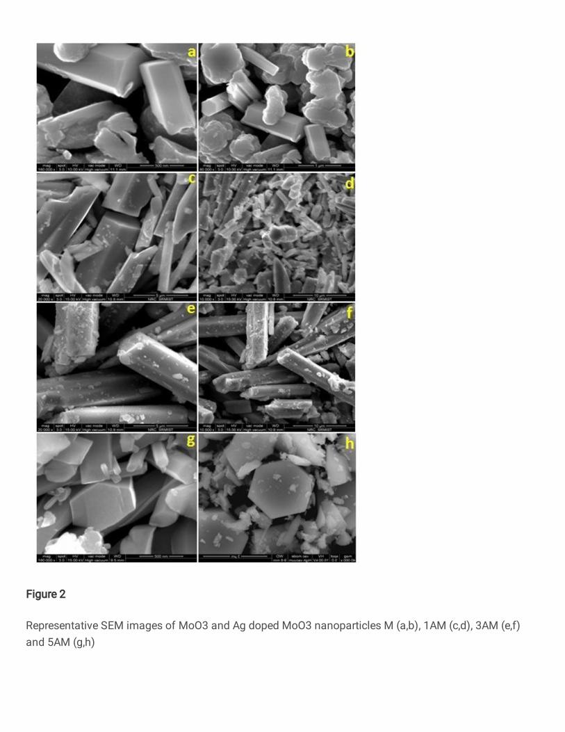
Figure 2
Representative SEM images of MoO3 and Ag doped MoO3 nanoparticles M (a,b), 1AM (c,d), 3AM (e,f)and 5AM (g,h)
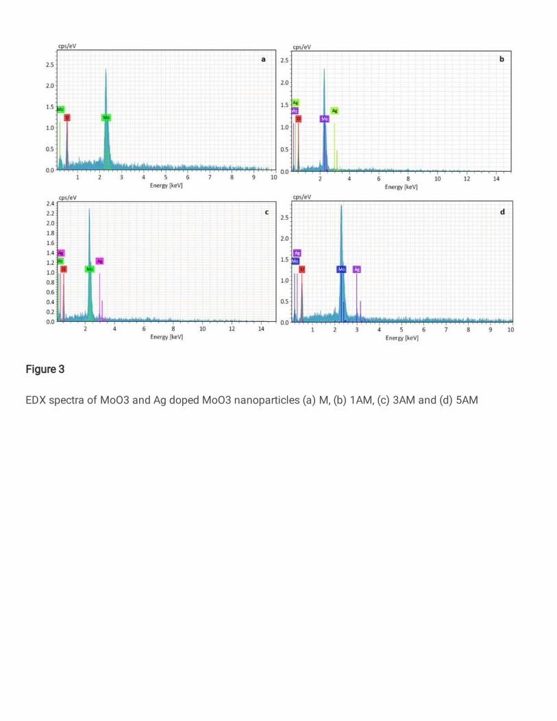
Figure 3
EDX spectra of MoO3 and Ag doped MoO3 nanoparticles (a) M, (b) 1AM, (c) 3AM and (d) 5AM
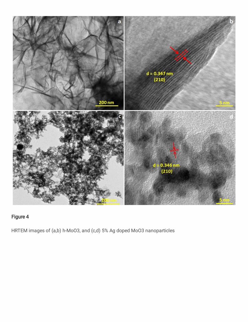
Figure 4
HRTEM images of (a,b) h-MoO3, and (c,d) 5% Ag doped MoO3 nanoparticles
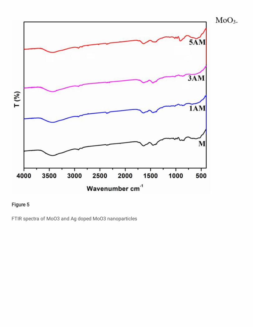
Figure 5
FTIR spectra of MoO3 and Ag doped MoO3 nanoparticles
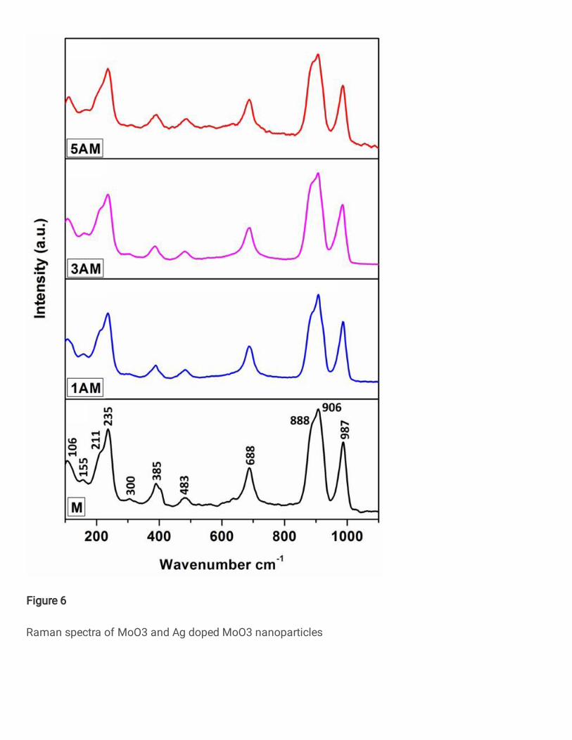
Figure 6
Raman spectra of MoO3 and Ag doped MoO3 nanoparticles
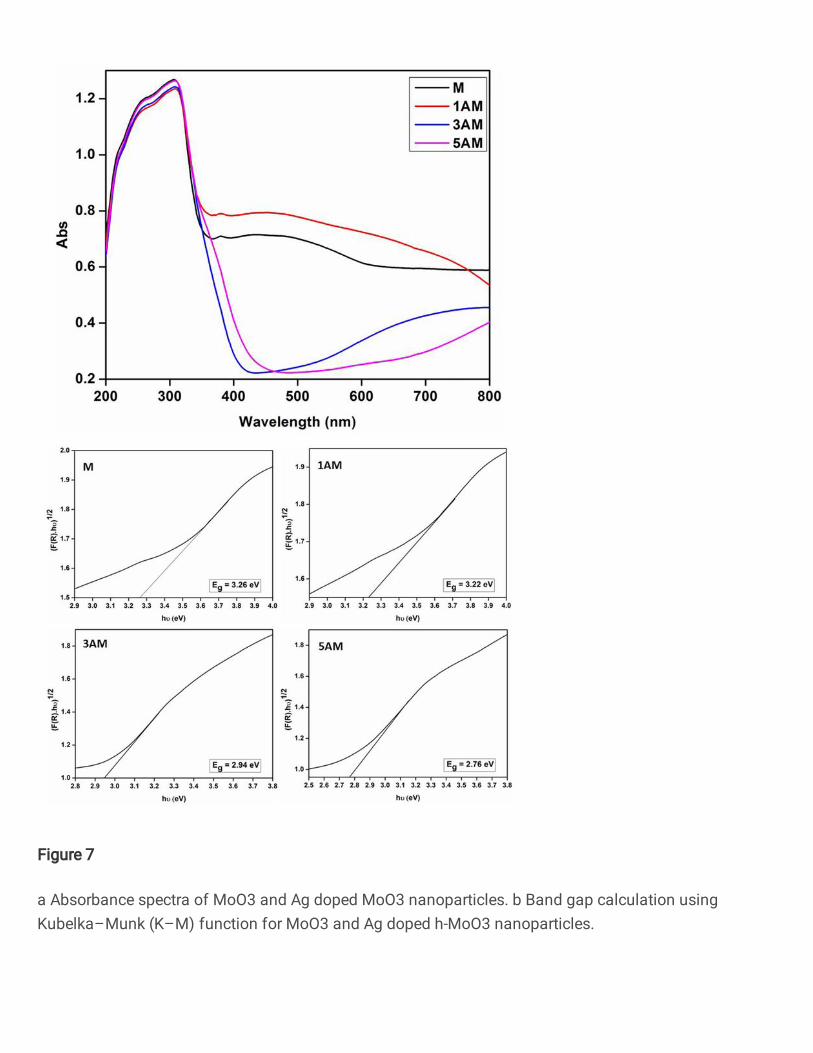
Figure 7
a Absorbance spectra of MoO3 and Ag doped MoO3 nanoparticles. b Band gap calculation usingKubelka–Munk (K–M) function for MoO3 and Ag doped h-MoO3 nanoparticles.
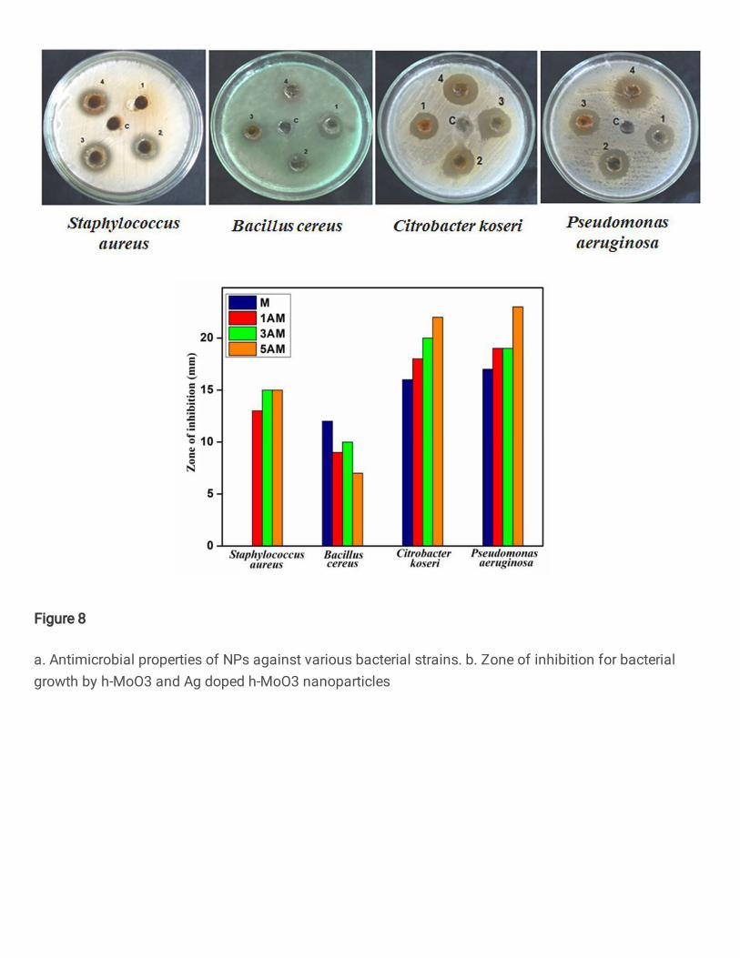
Figure 8
a. Antimicrobial properties of NPs against various bacterial strains. b. Zone of inhibition for bacterialgrowth by h-MoO3 and Ag doped h-MoO3 nanoparticles
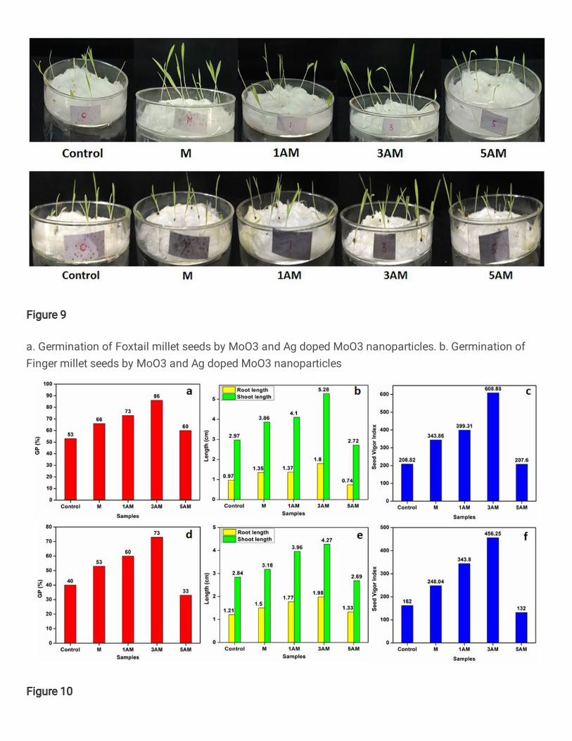
Figure 9
a. Germination of Foxtail millet seeds by MoO3 and Ag doped MoO3 nanoparticles. b. Germination ofFinger millet seeds by MoO3 and Ag doped MoO3 nanoparticles
Figure 10

(a,d) Germination percentage, (b,e) Root and shoot length, and (c,f) seed vigour index of foxtail millet and�nger millet seeds respectively treated with MoO3 and Ag doped MoO3 nanoparticles