Embed Size (px)
Citation preview
Human body in context Unit 1: The nervous system and the brain.
M.Àngels Hernández Sierra IES Valldemossa 2008
1
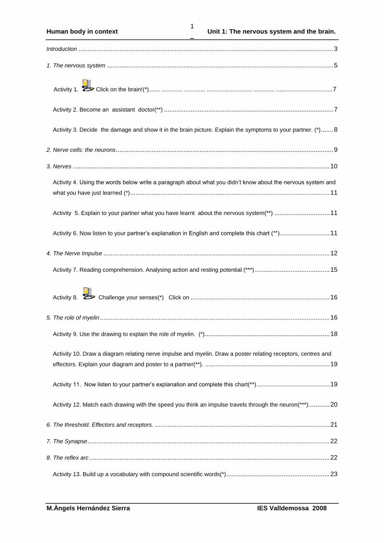
Introduction ............................................................................................................................................... 3
1. The nervous system ............................................................................................................................... 5
Activity 1. Click on the brain!(*)....... ............. ............. ............................ ............. ...................................7
Activity 2. Become an assistant doctor(**) ............................................................................................... 7
Activity 3. Decide the damage and show it in the brain picture. Explain the symptoms to your partner. (*) ....... 8
2. Nerve cells: the neurons .......................................................................................................................... 9
3. Nerves ................................................................................................................................................ 10
Activity 4. Using the words below write a paragraph about what you didn‟t know about the nervous system and
what you have just learned (*) ................................................................................................................ 11
Activity 5. Explain to your partner what you have learnt about the nervous system(**) ............................... 11
Activity 6. Now listen to your partner‟s explanation in English and complete this chart (**) ............................ 11
4. The Nerve Impulse ............................................................................................................................... 12
Activity 7. Reading comprehension. Analysing action and resting potential (***) .......................................... 15
Activity 8. Challenge your senses(*) Click on .............................................................................. 16
5. The role of myelin ................................................................................................................................. 16
Activity 9. Use the drawing to explain the role of myelin. (*) ...................................................................... 18
Activity 10. Draw a diagram relating nerve impulse and myelin. Draw a poster relating receptors, centres and
effectors. Explain your diagram and poster to a partner(**). ...................................................................... 19
Activity 11. Now listen to your partner‟s explanation and complete this chart(**) ......................................... 19
Activity 12. Match each drawing with the speed you think an impulse travels through the neuron(***). ........... 20
6. The threshold. Effectors and receptors. .................................................................................................. 21
7. The Synapse ........................................................................................................................................ 22
8. The reflex arc ....................................................................................................................................... 22
Activity 13. Build up a vocabulary with compound scientific words(*) .......................................................... 23
Human body in context Unit 1: The nervous system and the brain.
M.Àngels Hernández Sierra IES Valldemossa 2008
2
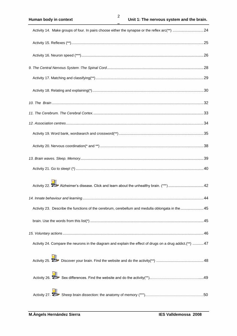
Activity 14. Make groups of four. In pairs choose either the synapse or the reflex arc(**) ............................ 24
Activity 15. Reflexes (**) ........................................................................................................................ 25
Activity 16. Neuron speed (***) ............................................................................................................... 26
9. The Central Nervous System :The Spinal Cord ........................................................................................ 28
Activity 17. Matching and classifying(**) .................................................................................................. 29
Activity 18. Relating and explaining(*) ..................................................................................................... 30
10. The Brain .......................................................................................................................................... 32
11. The Cerebrum. The Cerebral Cortex. .................................................................................................... 33
12. Association centres ............................................................................................................................. 34
Activity 19. Word bank, wordsearch and crossword(**) ............................................................................. 35
Activity 20. Nervous coordination(* and **) .............................................................................................. 38
13. Brain waves. Sleep. Memory................................................................................................................ 39
Activity 21. Go to sleep! (*) .................................................................................................................... 40
Activity 22. Alzheimer‟s disease. Click and learn about the unhealthy brain. (***) ................................ 42
14. Innate behaviour and learning .............................................................................................................. 44
Activity 23. Describe the functions of the cerebrum, cerebellum and medulla oblongata in the ..................... 45
brain. Use the words from this list(*) ....................................................................................................... 45
15. Voluntary actions ................................................................................................................................ 46
Activity 24. Compare the neurons in the diagram and explain the effect of drugs on a drug addict.(**) .......... 47
Activity 25. Discover your brain. Find the website and do the activity(**) ........................................... 48
Activity 26. Sex differences. Find the website and do the activity(**)..………………………………….....49
Activity 27. Sheep brain dissection: the anatomy of memory (***)………………………………………….50
Human body in context Unit 1: The nervous system and the brain.
M.Àngels Hernández Sierra IES Valldemossa 2008
3
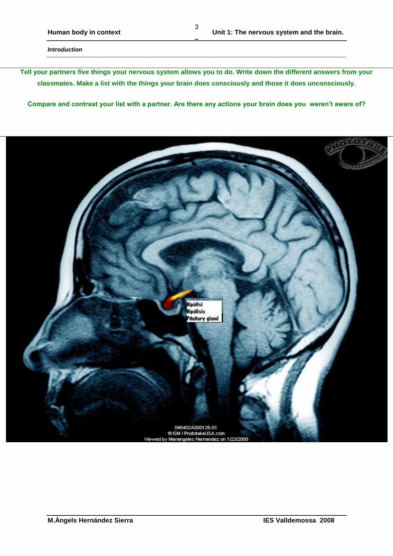
Introduction
Tell your partners five things your nervous system allows you to do. Write down the different answers from your
classmates. Make a list with the things your brain does consciously and those it does unconsciously.
Compare and contrast your list with a partner. Are there any actions your brain does you weren’t aware of?
Human body in context Unit 1: The nervous system and the brain.
M.Àngels Hernández Sierra IES Valldemossa 2008
4
Human body in context Unit 1: The nervous system and the brain.
M.Àngels Hernández Sierra IES Valldemossa 2008
5
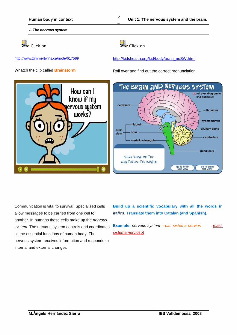
1. The nervous system
Click on
http://www.zimmertwins.ca/node/617589
Whatch the clip called Brainstorm
Click on
http://kidshealth.org/kid/body/brain_noSW.html
Roll over and find out the correct pronunciation.
Communication is vital to survival. Specialized cells
allow messages to be carried from one cell to
another. In humans these cells make up the nervous
system. The nervous system controls and coordinates
all the essential functions of human body. The
nervous system receives information and responds to
internal and external changes
Build up a scientific vocabulary with all the words in
italics. Translate them into Catalan (and Spanish).
Example: nervous system = cat. sistema nerviós (cast.
sistema nervioso)
Human body in context Unit 1: The nervous system and the brain.
M.Àngels Hernández Sierra IES Valldemossa 2008
6
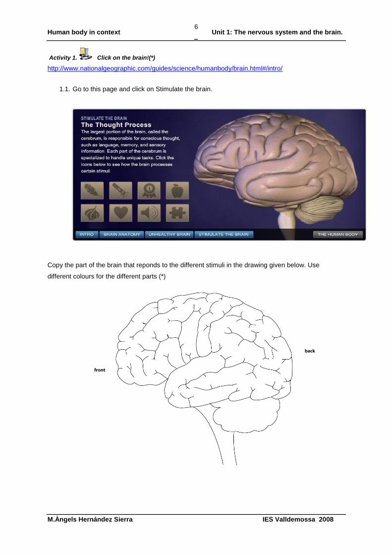
Activity 1. Click on the brain!(*)
http://www.nationalgeographic.com/guides/science/humanbody/brain.html#/intro/
1.1. Go to this page and click on Stimulate the brain.
Copy the part of the brain that reponds to the different stimuli in the drawing given below. Use
different colours for the different parts (*)
Human body in context Unit 1: The nervous system and the brain.
M.Àngels Hernández Sierra IES Valldemossa 2008
7
Activity 2. Become an assistant doctor(**)
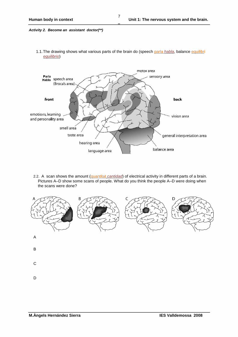
1.1. The drawing shows what various parts of the brain do (speech parla habla, balance equilibri
equilibrio)
2.2. A scan shows the amount (quantitat cantidad) of electrical activity in different parts of a brain.
Pictures A–D show some scans of people. What do you think the people A–D were doing when
the scans were done?
A
B
C
D
Human body in context Unit 1: The nervous system and the brain.
M.Àngels Hernández Sierra IES Valldemossa 2008
8
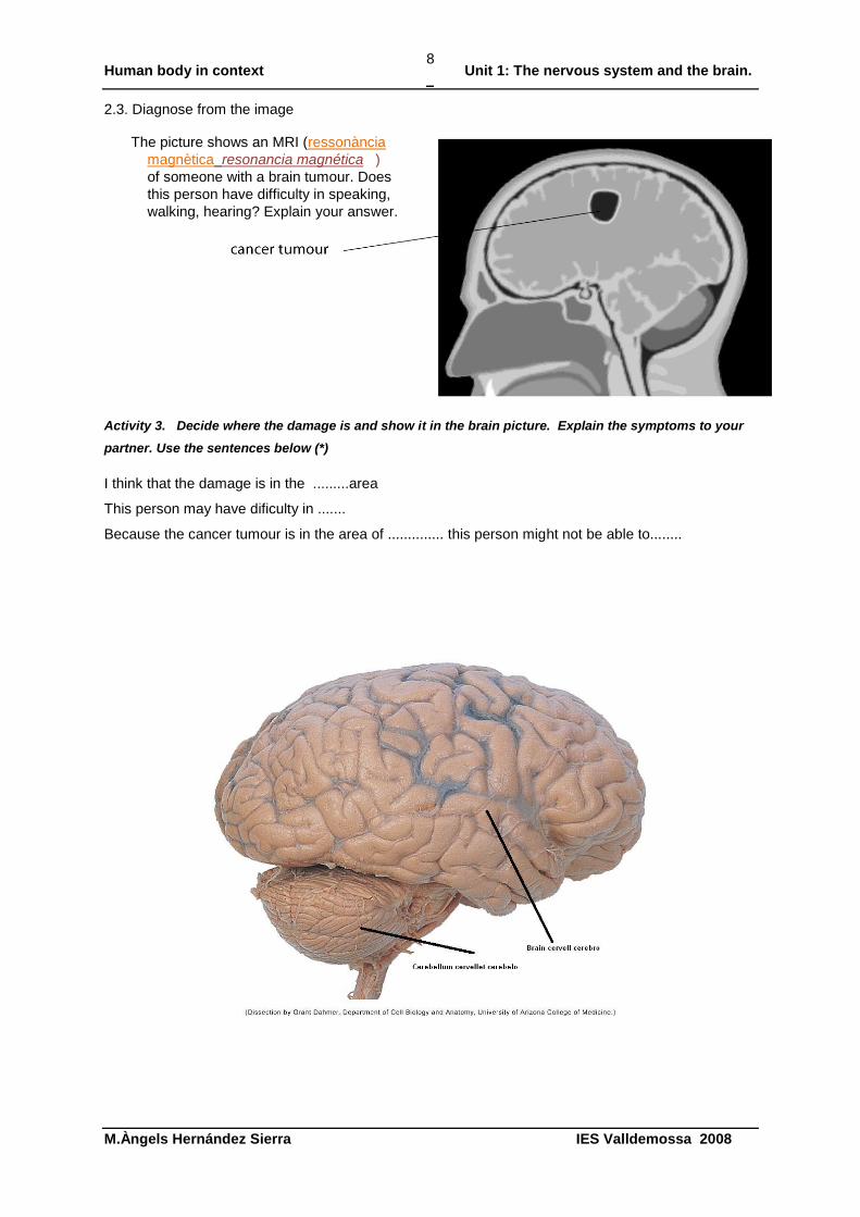
2.3. Diagnose from the image
Activity 3. Decide where the damage is and show it in the brain picture. Explain the symptoms to your
partner. Use the sentences below (*)
I think that the damage is in the .........area
This person may have dificulty in .......
Because the cancer tumour is in the area of .............. this person might not be able to........
The picture shows an MRI (ressonància
magnètica resonancia magnética )
of someone with a brain tumour. Does
this person have difficulty in speaking,
walking, hearing? Explain your answer.
Human body in context Unit 1: The nervous system and the brain.
M.Àngels Hernández Sierra IES Valldemossa 2008
9
1. Nerve cells: the neurons
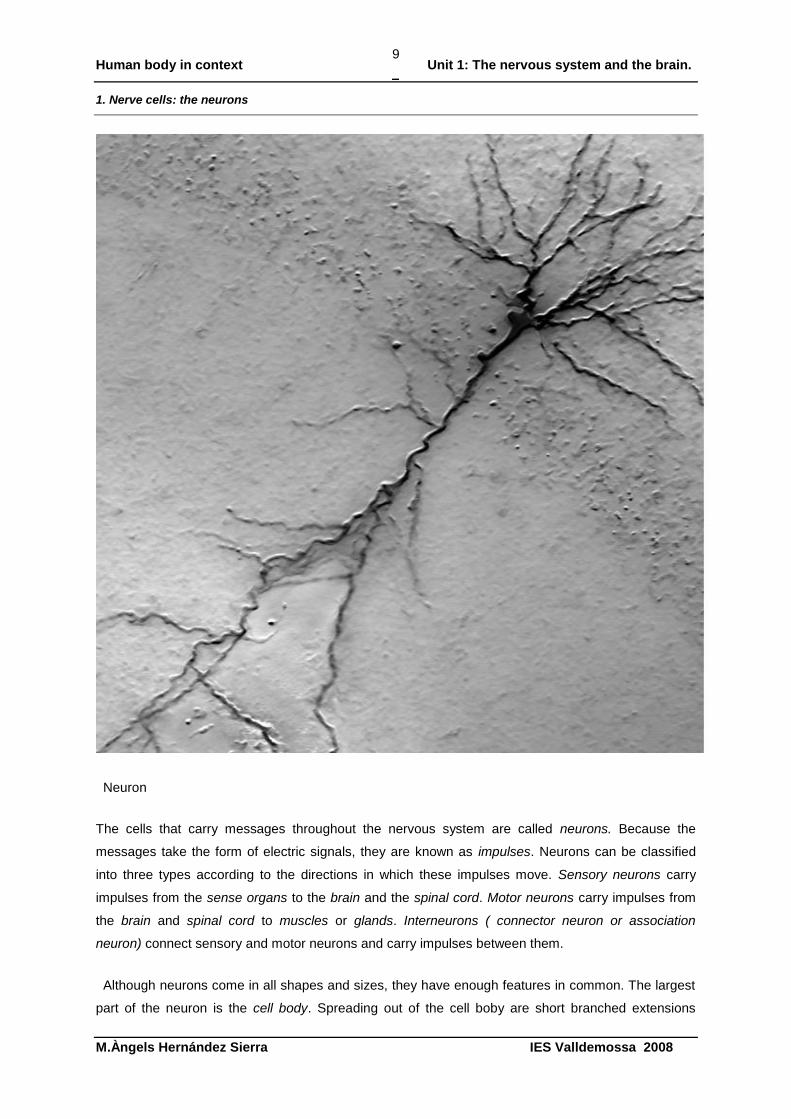
Neuron
The cells that carry messages throughout the nervous system are called neurons. Because the
messages take the form of electric signals, they are known as impulses. Neurons can be classified
into three types according to the directions in which these impulses move. Sensory neurons carry
impulses from the sense organs to the brain and the spinal cord. Motor neurons carry impulses from
the brain and spinal cord to muscles or glands. Interneurons ( connector neuron or association
neuron) connect sensory and motor neurons and carry impulses between them.
Although neurons come in all shapes and sizes, they have enough features in common. The largest
part of the neuron is the cell body. Spreading out of the cell boby are short branched extensions
Human body in context Unit 1: The nervous system and the brain.
M.Àngels Hernández Sierra IES Valldemossa 2008
10
called dendrites. Dendrites carry impulses from the environment or from other neurons toward the cell
body. The long fiber that carries impulses away from the cell body is called axon or nerve fibre. The
axon ends in a series of small branches called axon terminals.Neurons may have dozens or even
hundreds of dendrites but usually only one axon.
2. Nerves
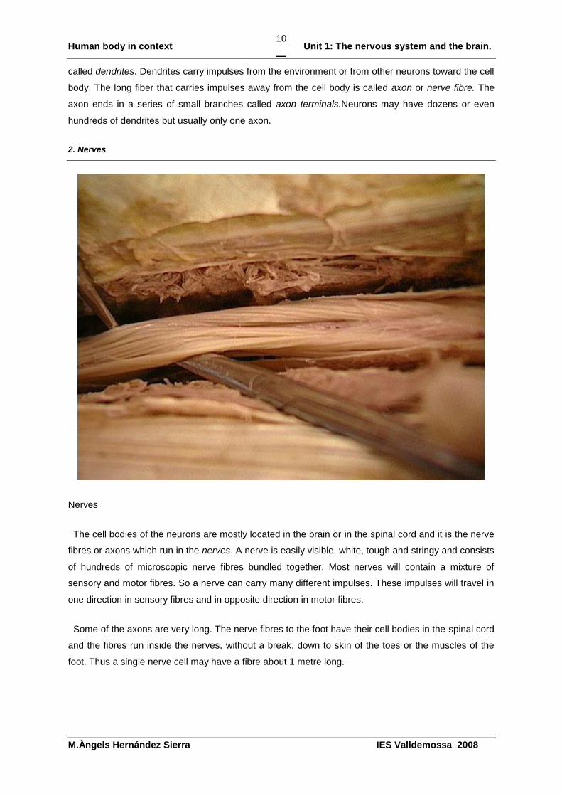
Nerves
The cell bodies of the neurons are mostly located in the brain or in the spinal cord and it is the nerve
fibres or axons which run in the nerves. A nerve is easily visible, white, tough and stringy and consists
of hundreds of microscopic nerve fibres bundled together. Most nerves will contain a mixture of
sensory and motor fibres. So a nerve can carry many different impulses. These impulses will travel in
one direction in sensory fibres and in opposite direction in motor fibres.
Some of the axons are very long. The nerve fibres to the foot have their cell bodies in the spinal cord
and the fibres run inside the nerves, without a break, down to skin of the toes or the muscles of the
foot. Thus a single nerve cell may have a fibre about 1 metre long.
Human body in context Unit 1: The nervous system and the brain.
M.Àngels Hernández Sierra IES Valldemossa 2008
11
Activity 4. Using the words below write a paragrahph about what you didn’t know about the nervous
system and what you have just learned (*)
Before I began to read about the nervous system I thought that…
First of all…
I found out that…
I have learnt several interesting facts that help me
As I read more ……….. I found out that…
Additionally…
Following this…
Consequently…
Activity 5. Explain to your partner using your paragraph what you have learnt so far about the nervous
system(**)
Activity 6. Now listen to your partner’s explanation in English and complete this chart (**)
YES/NO
Identifies main nervous system parts
Introduces new scientific words
Speaks clearly
Expresses his/her ideas using English words
Links information
Uses English connectors to link information
Human body in context Unit 1: The nervous system and the brain.
M.Àngels Hernández Sierra IES Valldemossa 2008
12
3. The Nerve Impulse
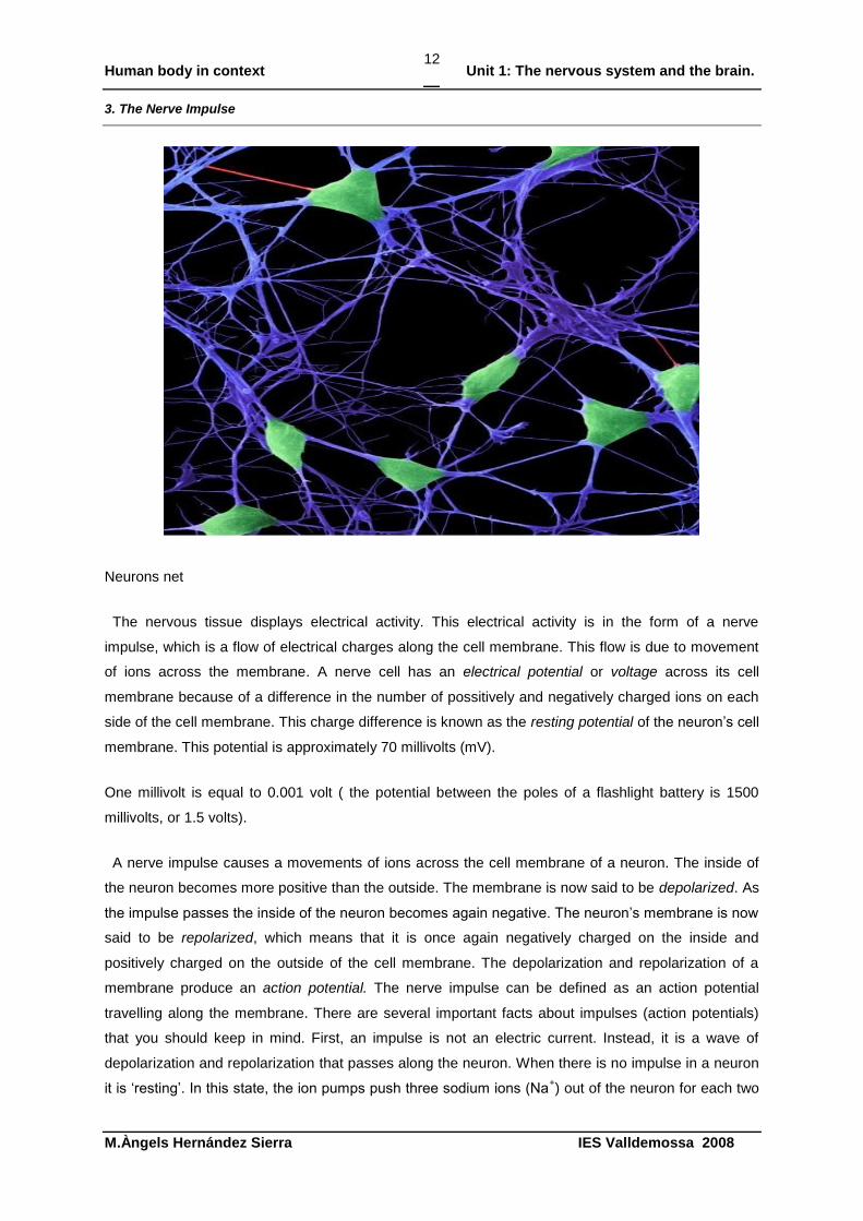
Neurons net
The nervous tissue displays electrical activity. This electrical activity is in the form of a nerve
impulse, which is a flow of electrical charges along the cell membrane. This flow is due to movement
of ions across the membrane. A nerve cell has an electrical potential or voltage across its cell
membrane because of a difference in the number of possitively and negatively charged ions on each
side of the cell membrane. This charge difference is known as the resting potential of the neuron‟s cell
membrane. This potential is approximately 70 millivolts (mV).
One millivolt is equal to 0.001 volt ( the potential between the poles of a flashlight battery is 1500
millivolts, or 1.5 volts).
A nerve impulse causes a movements of ions across the cell membrane of a neuron. The inside of
the neuron becomes more positive than the outside. The membrane is now said to be depolarized. As
the impulse passes the inside of the neuron becomes again negative. The neuron‟s membrane is now
said to be repolarized, which means that it is once again negatively charged on the inside and
positively charged on the outside of the cell membrane. The depolarization and repolarization of a
membrane produce an action potential. The nerve impulse can be defined as an action potential
travelling along the membrane. There are several important facts about impulses (action potentials)
that you should keep in mind. First, an impulse is not an electric current. Instead, it is a wave of
depolarization and repolarization that passes along the neuron. When there is no impulse in a neuron
it is „resting‟. In this state, the ion pumps push three sodium ions (Na+) out of the neuron for each two
Human body in context Unit 1: The nervous system and the brain.
M.Àngels Hernández Sierra IES Valldemossa 2008
13
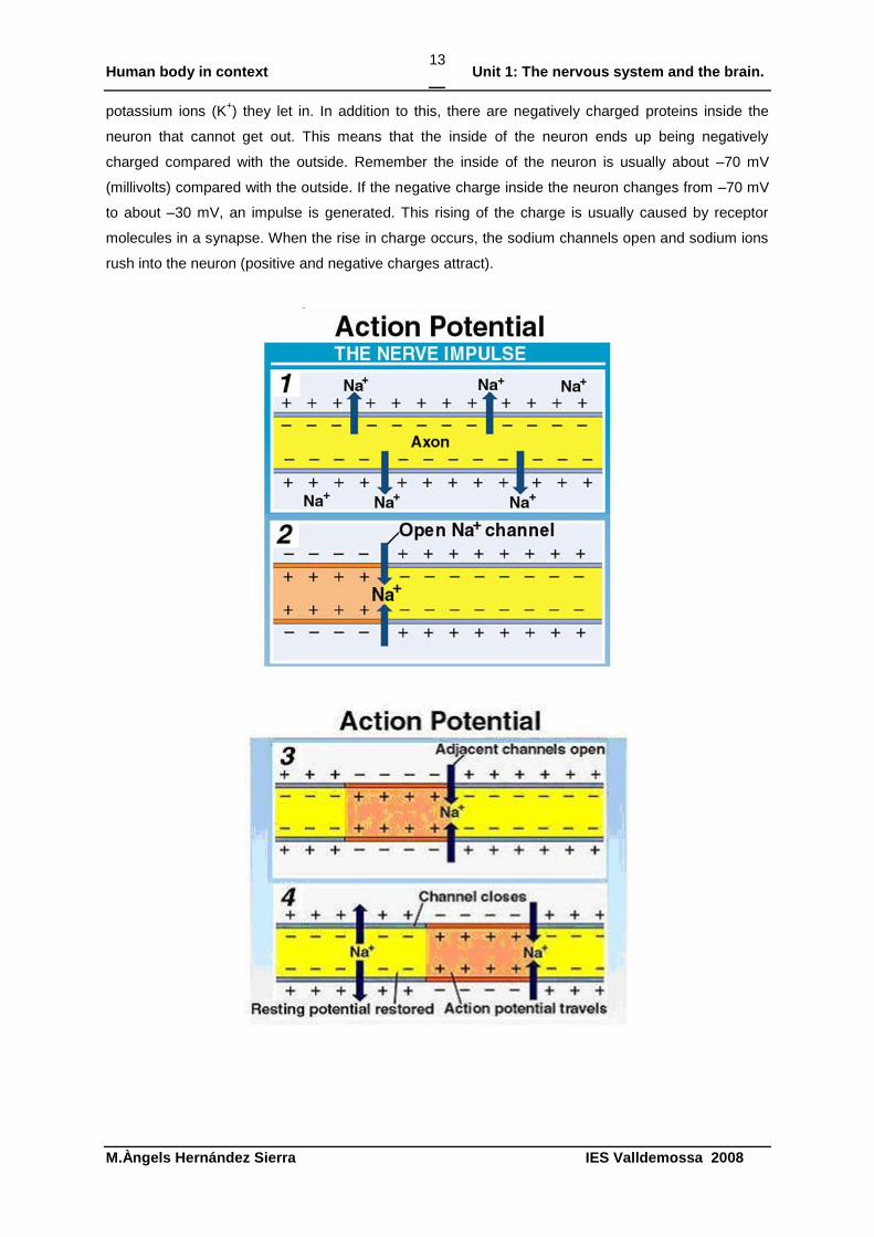
potassium ions (K+) they let in. In addition to this, there are negatively charged proteins inside the
neuron that cannot get out. This means that the inside of the neuron ends up being negatively
charged compared with the outside. Remember the inside of the neuron is usually about –70 mV
(millivolts) compared with the outside. If the negative charge inside the neuron changes from –70 mV
to about –30 mV, an impulse is generated. This rising of the charge is usually caused by receptor
molecules in a synapse. When the rise in charge occurs, the sodium channels open and sodium ions
rush into the neuron (positive and negative charges attract).
Human body in context Unit 1: The nervous system and the brain.
M.Àngels Hernández Sierra IES Valldemossa 2008
14
Second, an impulse is much slower than an electric current. Electric current move almost
instantaneously, whereas action potentials usually travel at speeds ranging from 10 centimetres per
second to 1 metre per second. Third, unlike an electric current, the strength of an impulse is always
the same- there is either an impulse in response to a stimulus or there is not. The impulse is self-
propagating, that is , an impulse at any point on the membrane causes an impulse to the next point
along the membrane, but it can move only in one direction. The impulse is said to be unidirectional.
The nerve fibres do not carry sensations like pain or cold. These sensations are felt only when a
nerve impulse reaches the brain. All nerve impulses are similar; there is no difference between nerve
impulses from the eyes, ears or hands.
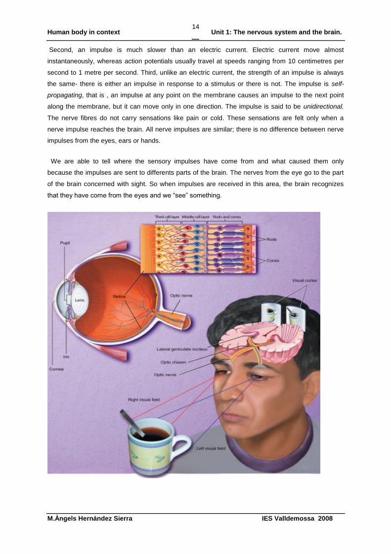
We are able to tell where the sensory impulses have come from and what caused them only
because the impulses are sent to differents parts of the brain. The nerves from the eye go to the part
of the brain concerned with sight. So when impulses are received in this area, the brain recognizes
that they have come from the eyes and we “see” something.
Human body in context Unit 1: The nervous system and the brain.
M.Àngels Hernández Sierra IES Valldemossa 2008
15
Activity 7. Reading comprehension. Analysing action and resting potential (***)
1 State (anomena nombra) two reasons why the inside of a resting axon is negatively charged
compared with the outside.
-
-
2 Why do Na+
ions enter the neuron when the sodium channels open?
…………………………………………………………………………………………………………………
……………………………………………..
3 What happens to the charge inside the axon when repolarisation occurs?
……………………………………………..
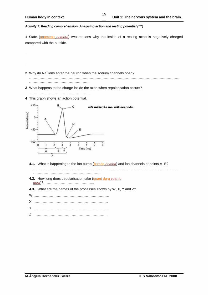
4 This graph shows an action potential.
4.1. What is happening to the ion pump (bomba bomba) and ion channels at points A–E?
………………………………………………………………………………………………………………
………………………………………………….
4.2. How long does depolarisation take (quant dura cuanto
dura)?……………………………………..
4.3. What are the names of the processes shown by W, X, Y and Z?
W ………………………………………………………..
X ……………………………………………………….
Y ………………………………………………………..
Z ………………………………………………………..
Human body in context Unit 1: The nervous system and the brain.
M.Àngels Hernández Sierra IES Valldemossa 2008
16
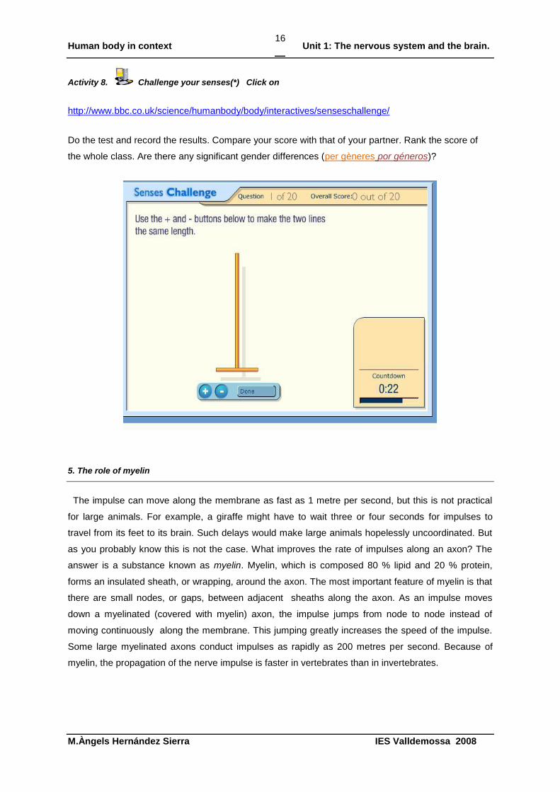
Activity 8. Challenge your senses(*) Click on
http://www.bbc.co.uk/science/humanbody/body/interactives/senseschallenge/
Do the test and record the results. Compare your score with that of your partner. Rank the score of
the whole class. Are there any significant gender differences (per gèneres por géneros)?
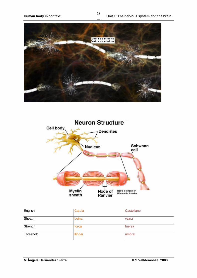
5. The role of myelin
The impulse can move along the membrane as fast as 1 metre per second, but this is not practical
for large animals. For example, a giraffe might have to wait three or four seconds for impulses to
travel from its feet to its brain. Such delays would make large animals hopelessly uncoordinated. But
as you probably know this is not the case. What improves the rate of impulses along an axon? The
answer is a substance known as myelin. Myelin, which is composed 80 % lipid and 20 % protein,
forms an insulated sheath, or wrapping, around the axon. The most important feature of myelin is that
there are small nodes, or gaps, between adjacent sheaths along the axon. As an impulse moves
down a myelinated (covered with myelin) axon, the impulse jumps from node to node instead of
moving continuously along the membrane. This jumping greatly increases the speed of the impulse.
Some large myelinated axons conduct impulses as rapidly as 200 metres per second. Because of
myelin, the propagation of the nerve impulse is faster in vertebrates than in invertebrates.
Human body in context Unit 1: The nervous system and the brain.
M.Àngels Hernández Sierra IES Valldemossa 2008
17
English Català Castellano
Sheath beina vaina
Strengh força fuerza
Threshold llindar umbral
Human body in context Unit 1: The nervous system and the brain.
M.Àngels Hernández Sierra IES Valldemossa 2008
18
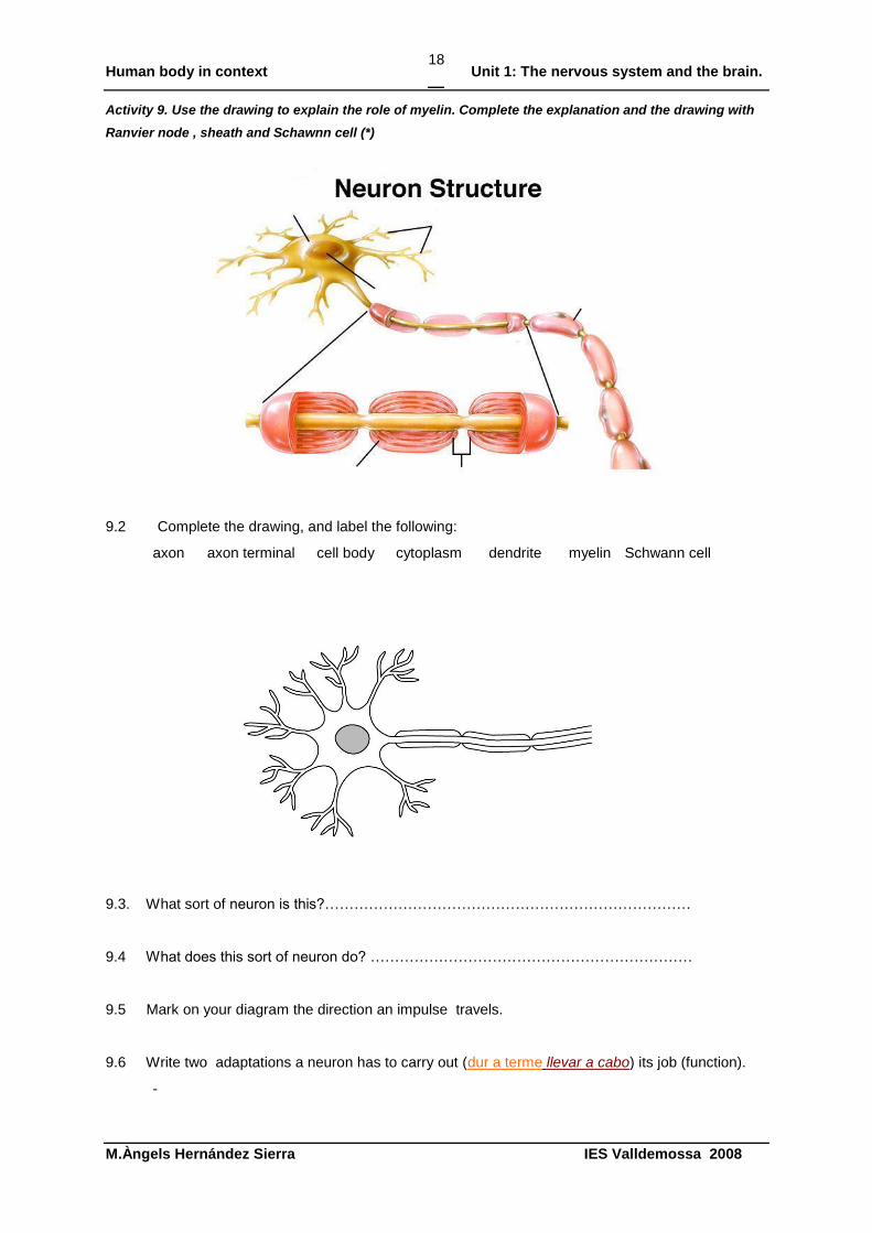
Activity 9. Use the drawing to explain the role of myelin. Complete the explanation and the drawing with
Ranvier node , sheath and Schawnn cell (*)
9.2 Complete the drawing, and label the following:
axon axon terminal cell body cytoplasm dendrite myelin Schwann cell
9.3. What sort of neuron is this?…………………………………………………………………
9.4 What does this sort of neuron do? …………………………………………………………
9.5 Mark on your diagram the direction an impulse travels.
9.6 Write two adaptations a neuron has to carry out (dur a terme llevar a cabo) its job (function).
-
Human body in context Unit 1: The nervous system and the brain.
M.Àngels Hernández Sierra IES Valldemossa 2008
19
-
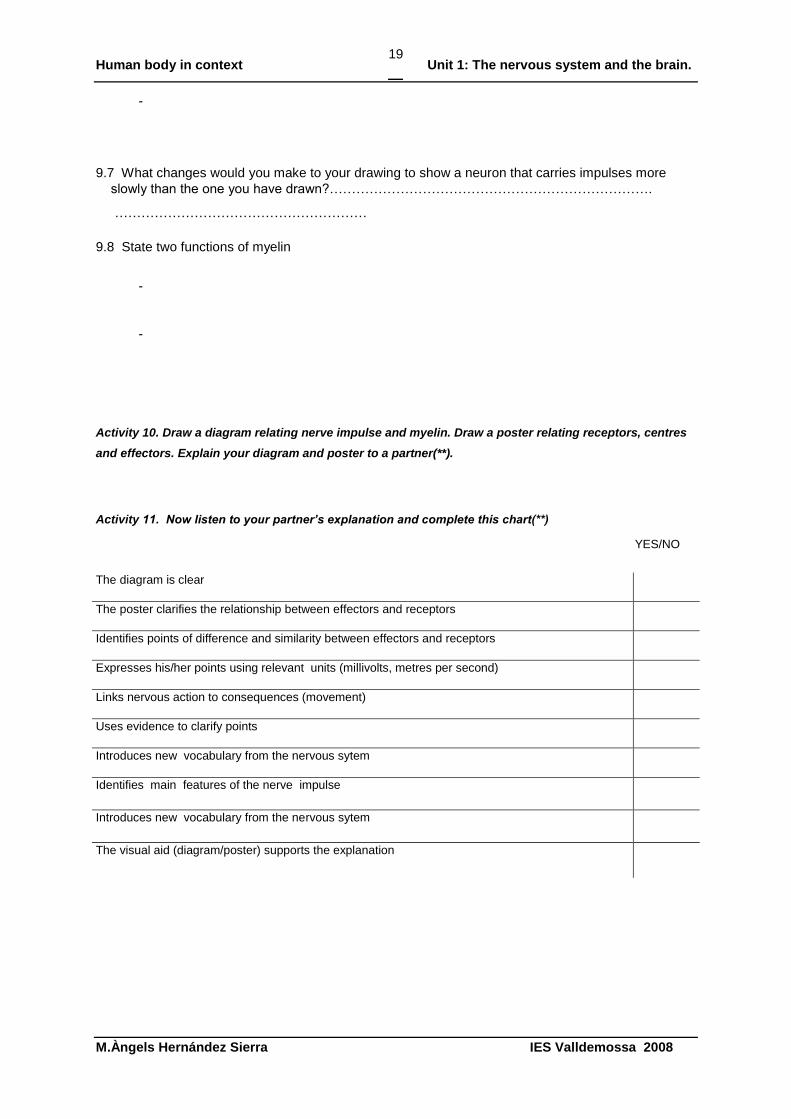
9.7 What changes would you make to your drawing to show a neuron that carries impulses more
slowly than the one you have drawn?……………………………………………………………….
…………………………………………………
9.8 State two functions of myelin
-
-
Activity 10. Draw a diagram relating nerve impulse and myelin. Draw a poster relating receptors, centres
and effectors. Explain your diagram and poster to a partner(**).
Activity 11. Now listen to your partner’s explanation and complete this chart(**)
YES/NO
The diagram is clear
The poster clarifies the relationship between effectors and receptors
Identifies points of difference and similarity between effectors and receptors
Expresses his/her points using relevant units (millivolts, metres per second)
Links nervous action to consequences (movement)
Uses evidence to clarify points
Introduces new vocabulary from the nervous sytem
Identifies main features of the nerve impulse
Introduces new vocabulary from the nervous sytem
The visual aid (diagram/poster) supports the explanation
Human body in context Unit 1: The nervous system and the brain.
M.Àngels Hernández Sierra IES Valldemossa 2008
20
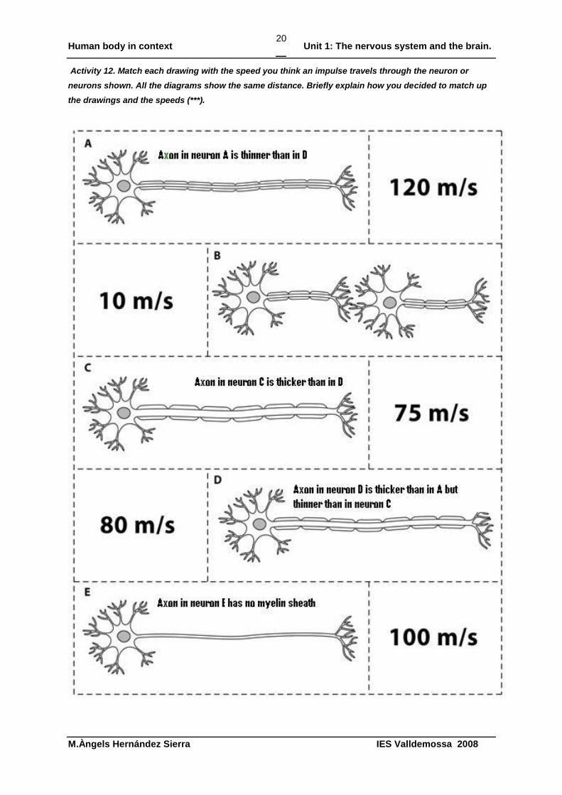
Activity 12. Match each drawing with the speed you think an impulse travels through the neuron or
neurons shown. All the diagrams show the same distance. Briefly explain how you decided to match up
the drawings and the speeds (***).
Human body in context Unit 1: The nervous system and the brain.
M.Àngels Hernández Sierra IES Valldemossa 2008
21
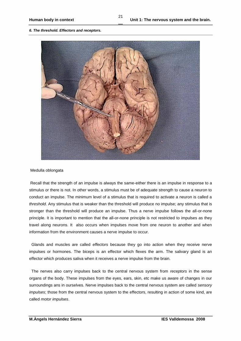
6. The threshold. Effectors and receptors.
Medulla oblongata
Recall that the strength of an impulse is always the same-either there is an impulse in response to a
stimulus or there is not. In other words, a stimulus must be of adequate strength to cause a neuron to
conduct an impulse. The minimum level of a stimulus that is required to activate a neuron is called a
threshold. Any stimulus that is weaker than the threshold will produce no impulse; any stimulus that is
stronger than the threshold will produce an impulse. Thus a nerve impulse follows the all-or-none
principle. It is important to mention that the all-or-none principle is not restricted to impulses as they
travel along neurons. It also occurs when impulses move from one neuron to another and when
information from the environment causes a nerve impulse to occur.
Glands and muscles are called effectors because they go into action when they receive nerve
impulses or hormones. The biceps is an effector which flexes the arm. The salivary gland is an
effector which produces saliva when it receives a nerve impulse from the brain.
The nerves also carry impulses back to the central nervous system from receptors in the sense
organs of the body. These impulses from the eyes, ears, skin, etc make us aware of changes in our
surroundings ans in ourselves. Nerve impulses back to the central nervous system are called sensory
impulses; those from the central nervous system to the effectors, resulting in action of some kind, are
called motor impulses.
Human body in context Unit 1: The nervous system and the brain.
M.Àngels Hernández Sierra IES Valldemossa 2008
22
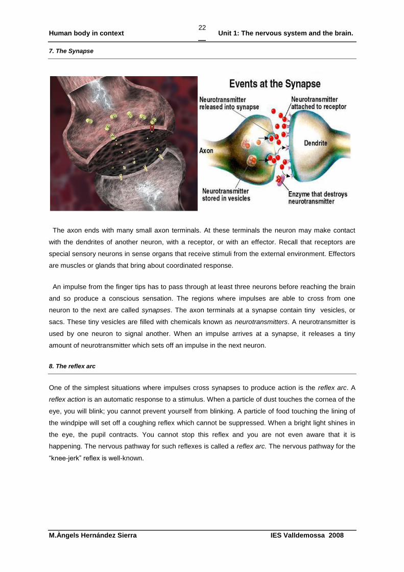
7. The Synapse
The axon ends with many small axon terminals. At these terminals the neuron may make contact
with the dendrites of another neuron, with a receptor, or with an effector. Recall that receptors are
special sensory neurons in sense organs that receive stimuli from the external environment. Effectors
are muscles or glands that bring about coordinated response.
An impulse from the finger tips has to pass through at least three neurons before reaching the brain
and so produce a conscious sensation. The regions where impulses are able to cross from one
neuron to the next are called synapses. The axon terminals at a synapse contain tiny vesicles, or
sacs. These tiny vesicles are filled with chemicals known as neurotransmitters. A neurotransmitter is
used by one neuron to signal another. When an impulse arrives at a synapse, it releases a tiny
amount of neurotransmitter which sets off an impulse in the next neuron.
8. The reflex arc
One of the simplest situations where impulses cross synapses to produce action is the reflex arc. A
reflex action is an automatic response to a stimulus. When a particle of dust touches the cornea of the
eye, you will blink; you cannot prevent yourself from blinking. A particle of food touching the lining of
the windpipe will set off a coughing reflex which cannot be suppressed. When a bright light shines in
the eye, the pupil contracts. You cannot stop this reflex and you are not even aware that it is
happening. The nervous pathway for such reflexes is called a reflex arc. The nervous pathway for the
“knee-jerk” reflex is well-known.
Human body in context Unit 1: The nervous system and the brain.
M.Àngels Hernández Sierra IES Valldemossa 2008
23
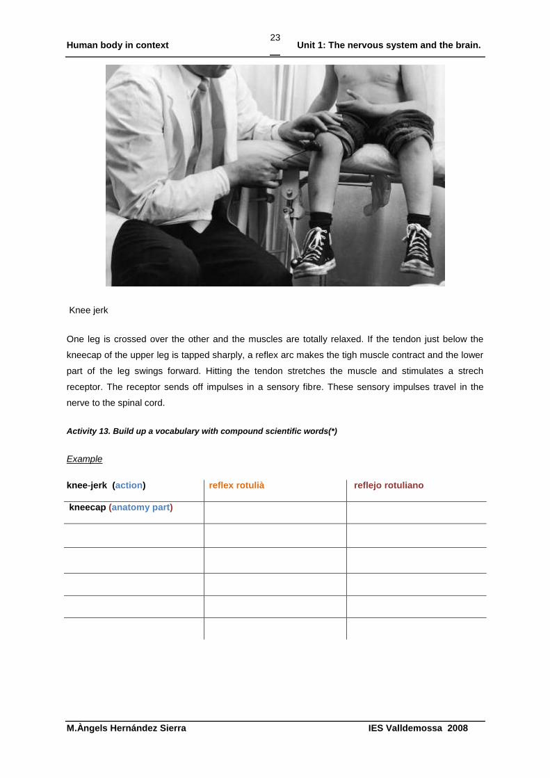
Knee jerk
One leg is crossed over the other and the muscles are totally relaxed. If the tendon just below the
kneecap of the upper leg is tapped sharply, a reflex arc makes the tigh muscle contract and the lower
part of the leg swings forward. Hitting the tendon stretches the muscle and stimulates a strech
receptor. The receptor sends off impulses in a sensory fibre. These sensory impulses travel in the
nerve to the spinal cord.
Activity 13. Build up a vocabulary with compound scientific words(*)
Example
knee-jerk (action) reflex rotulià reflejo rotuliano
kneecap (anatomy part)
Human body in context Unit 1: The nervous system and the brain.
M.Àngels Hernández Sierra IES Valldemossa 2008
24
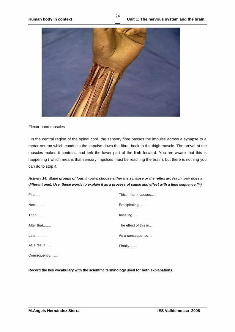
Flexor hand muscles
In the central region of the spinal cord, the sensory fibre passes the impulse across a synapse to a
motor neuron which conducts the impulse down the fibre, back to the thigh muscle. The arrival at the
muscles makes it contract, and jerk the lower part of the limb forward. You are aware that this is
happening ( which means that sensory impulses must be reaching the brain), but there is nothing you
can do to stop it.
Activity 14. Make groups of four. In pairs choose either the synapse or the reflex arc (each pair does a
different one). Use these words to explain it as a process of cause and effect with a time sequence.(**)
First.....
Next.........
Then.........
After that........
Later..........
As a result……
Consequently…….
This, in turn, causes…..
Precipitating……..
Initiating…..
The effect of this is….
As a consequence…
Finally........
Record the key vocabulary with the scientific terminology used for both explanations.
Human body in context Unit 1: The nervous system and the brain.
M.Àngels Hernández Sierra IES Valldemossa 2008
25
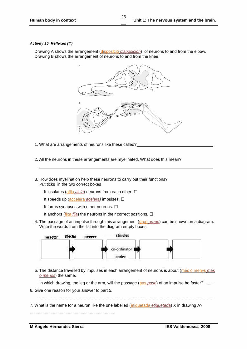
Activity 15. Reflexes (**)
Drawing A shows the arrangement (disposició disposición) of neurons to and from the elbow.
Drawing B shows the arrangement of neurons to and from the knee.
1. What are arrangements of neurons like these called?
2. All the neurons in these arrangements are myelinated. What does this mean?
3. How does myelination help these neurons to carry out their functions?
Put ticks in the two correct boxes
It insulates (aïlla aisla) neurons from each other.
It speeds up (accelera acelera) impulses.
It forms synapses with other neurons.
It anchors (fixa fija) the neurons in their correct positions.
4. The passage of an impulse through this arrangement (grup grupo) can be shown on a diagram.
Write the words from the list into the diagram empty boxes.
5. The distance travelled by impulses in each arrangement of neurons is about (més o menys más
o menos) the same.
In which drawing, the leg or the arm, will the passage (pas paso) of an impulse be faster? ........
6. Give one reason for your answer to part 5.
………………………………………………………………………………………………………………
7. What is the name for a neuron like the one labelled (etiquetada etiquetada) X in drawing A?
..........................................................................
Human body in context Unit 1: The nervous system and the brain.
M.Àngels Hernández Sierra IES Valldemossa 2008
26
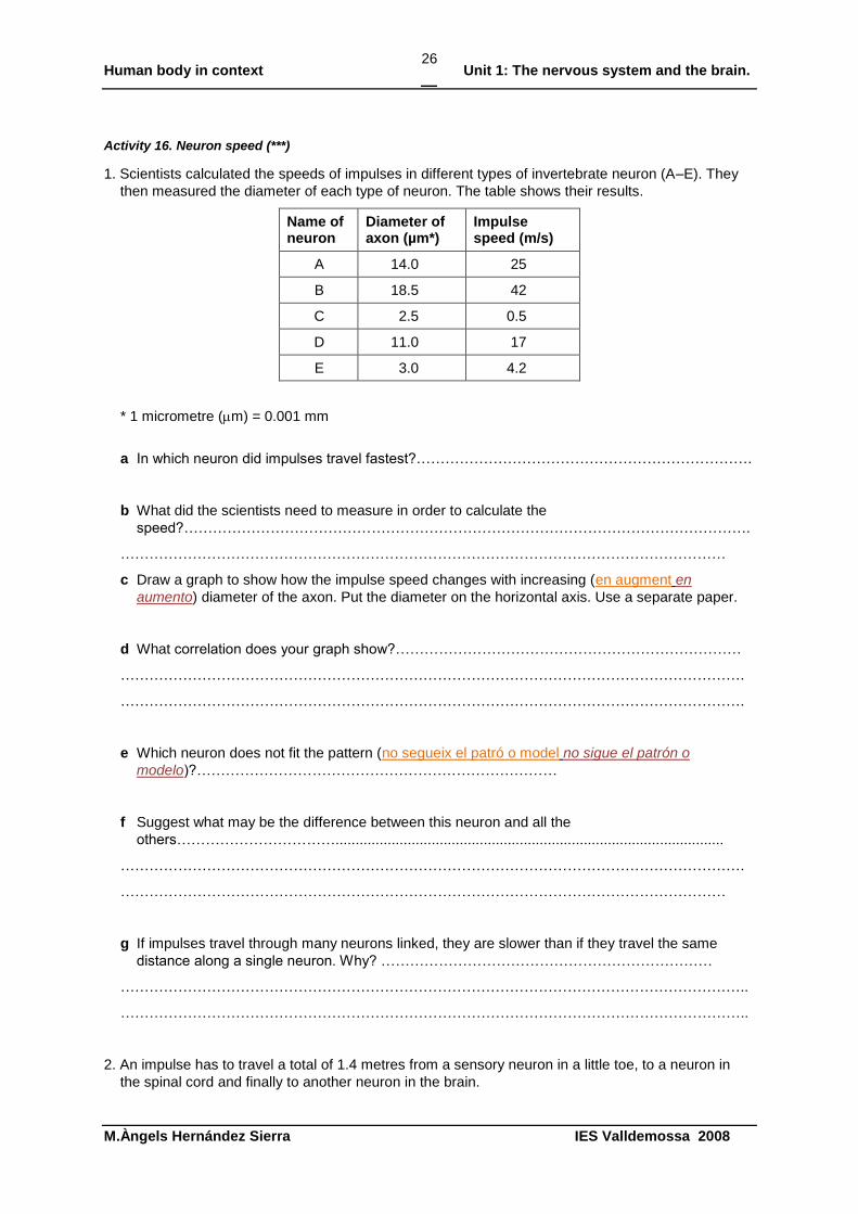
Activity 16. Neuron speed (***)
1. Scientists calculated the speeds of impulses in different types of invertebrate neuron (A–E). They
then measured the diameter of each type of neuron. The table shows their results.
Name of neuron
Diameter of axon (µm*)
Impulse speed (m/s)
A 14.0 25
B 18.5 42
C 2.5 0.5
D 11.0 17
E 3.0 4.2
* 1 micrometre (m) = 0.001 mm
a In which neuron did impulses travel fastest?…………………………………………………………….
b What did the scientists need to measure in order to calculate the
speed?……………………………………………………………………………………………………….
………………………………………………………………………………………………………………
c Draw a graph to show how the impulse speed changes with increasing (en augment en
aumento) diameter of the axon. Put the diameter on the horizontal axis. Use a separate paper.
d What correlation does your graph show?………………………………………………………………
………………………………………………………………………………………………………………….
………………………………………………………………………………………………………………….
e Which neuron does not fit the pattern (no segueix el patró o model no sigue el patrón o
modelo)?…………………………………………………………………
f Suggest what may be the difference between this neuron and all the
others…………………………….................................................................................................
………………………………………………………………………………………………………………….
………………………………………………………………………………………………………………
g If impulses travel through many neurons linked, they are slower than if they travel the same
distance along a single neuron. Why? ……………………………………………………………
…………………………………………………………………………………………………………………..
…………………………………………………………………………………………………………………..
2. An impulse has to travel a total of 1.4 metres from a sensory neuron in a little toe, to a neuron in
the spinal cord and finally to another neuron in the brain.
Human body in context Unit 1: The nervous system and the brain.
M.Àngels Hernández Sierra IES Valldemossa 2008
27
The time for the impulse to cross each synapse is 0.5 milliseconds.
The impulse travels at a mean speed of 100 m/s.
How long does the impulse take? Write all the calculations here
………………………………………………………………………….
…………………………………………………………………………………………………………………
........................................................................................................................................................
..................................................................................................................................................................
3. Explain how a neurotransmitter causes an impulse to cross a synapse by writing three sentences.
Each sentence must contain the words shown below. Beware some words are not in the process
order
impulse, vesicles, neurotransmitter
..........................................................................................................................................................
..........................................................................................................................................................
..........................................................................................................................................................
..........................................................................................................................................................
..........................................................................................................................................................
..........................................................................................................................................................
synapse, released, gap
..........................................................................................................................................................
..........................................................................................................................................................
..........................................................................................................................................................
..........................................................................................................................................................
..........................................................................................................................................................
..........................................................................................................................................................
receptor, bond, impulse
..........................................................................................................................................................
..........................................................................................................................................................
..........................................................................................................................................................
..........................................................................................................................................................
..........................................................................................................................................................
..........................................................................................................................................................
Human body in context Unit 1: The nervous system and the brain.
M.Àngels Hernández Sierra IES Valldemossa 2008
28
9. The Central Nervous System :The Spinal Cord
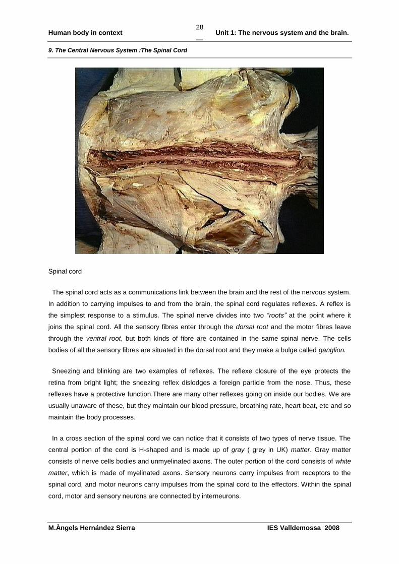
Spinal cord
The spinal cord acts as a communications link between the brain and the rest of the nervous system.
In addition to carrying impulses to and from the brain, the spinal cord regulates reflexes. A reflex is
the simplest response to a stimulus. The spinal nerve divides into two “roots” at the point where it
joins the spinal cord. All the sensory fibres enter through the dorsal root and the motor fibres leave
through the ventral root, but both kinds of fibre are contained in the same spinal nerve. The cells
bodies of all the sensory fibres are situated in the dorsal root and they make a bulge called ganglion.
Sneezing and blinking are two examples of reflexes. The reflexe closure of the eye protects the
retina from bright light; the sneezing reflex dislodges a foreign particle from the nose. Thus, these
reflexes have a protective function.There are many other reflexes going on inside our bodies. We are
usually unaware of these, but they maintain our blood pressure, breathing rate, heart beat, etc and so
maintain the body processes.
In a cross section of the spinal cord we can notice that it consists of two types of nerve tissue. The
central portion of the cord is H-shaped and is made up of gray ( grey in UK) matter. Gray matter
consists of nerve cells bodies and unmyelinated axons. The outer portion of the cord consists of white
matter, which is made of myelinated axons. Sensory neurons carry impulses from receptors to the
spinal cord, and motor neurons carry impulses from the spinal cord to the effectors. Within the spinal
cord, motor and sensory neurons are connected by interneurons.
Human body in context Unit 1: The nervous system and the brain.
M.Àngels Hernández Sierra IES Valldemossa 2008
29
Activity 17. Matching and classifying(**)
1 Match the definition with a word from the list: brain, neurotransmitter, neuron, axon, impulse,
nerves, synapse, dendrite, myelin, threshold.
a. progressive wave of biochemically generated energy that travels along a nerve fibre or
muscle and stimulates or inhibits activity
b. a branched extension of a nerve cell neuron that receives electrical signals from other
neurons and conducts those signals to the cell body
c. a bundle of fibres forming a network that transmits messages in the form of impulses between
the brain or spinal cord and the body's organs
d. a material made up of protein and fats that surrounds some nerve cells in concentric layers,
insulating adjacent nerve fibres and enabling transmission of nerve impulses
e. the smallest detectable sensation
f. a cell, usually consisting of a cell body, axon, and dendrites, that transmits nerve impulses
and is the basic functional unit of the nervous system
g. the controlling centre of the nervous system in vertebrates, connected to the spinal cord and
enclosed in the cranium
h. an extension of a nerve cell, similar in shape to a thread, that transmits impulses outwards
from the cell body
i. a junction between two nerve cells, where the tip of a nerve fibre almost touches another cell
in order to transmit signals
j. a chemical that carries messages between different nerve cells or between nerve cells and
muscles
2 Classify the previous words into processes and anatomical parts. Compare your list with your
partner.
Example
Processes Anatomical parts
letter a letter b
Human body in context Unit 1: The nervous system and the brain.
M.Àngels Hernández Sierra IES Valldemossa 2008
30
Activity 18. Relating and explaining(*)
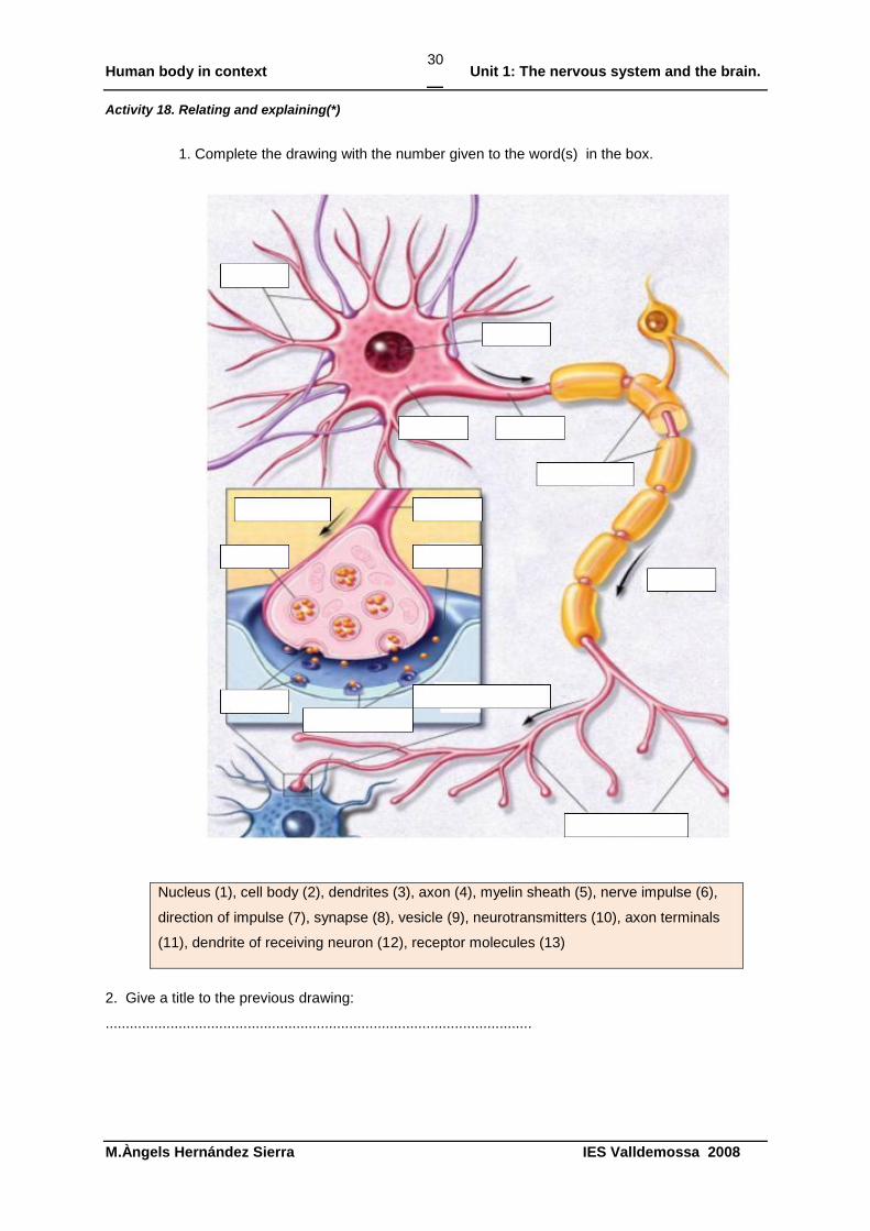
1. Complete the drawing with the number given to the word(s) in the box.
Nucleus (1), cell body (2), dendrites (3), axon (4), myelin sheath (5), nerve impulse (6),
direction of impulse (7), synapse (8), vesicle (9), neurotransmitters (10), axon terminals
(11), dendrite of receiving neuron (12), receptor molecules (13)
2. Give a title to the previous drawing:
.........................................................................................................

Human body in context Unit 1: The nervous system and the brain.
M.Àngels Hernández Sierra IES Valldemossa 2008
31
3. Are those drawings below processes or anatomical parts of the nervous system?
:
drawing A
drawing B
Explain to your partner the reasons for your choice.
Human body in context Unit 1: The nervous system and the brain.
M.Àngels Hernández Sierra IES Valldemossa 2008
32
10. The Brain
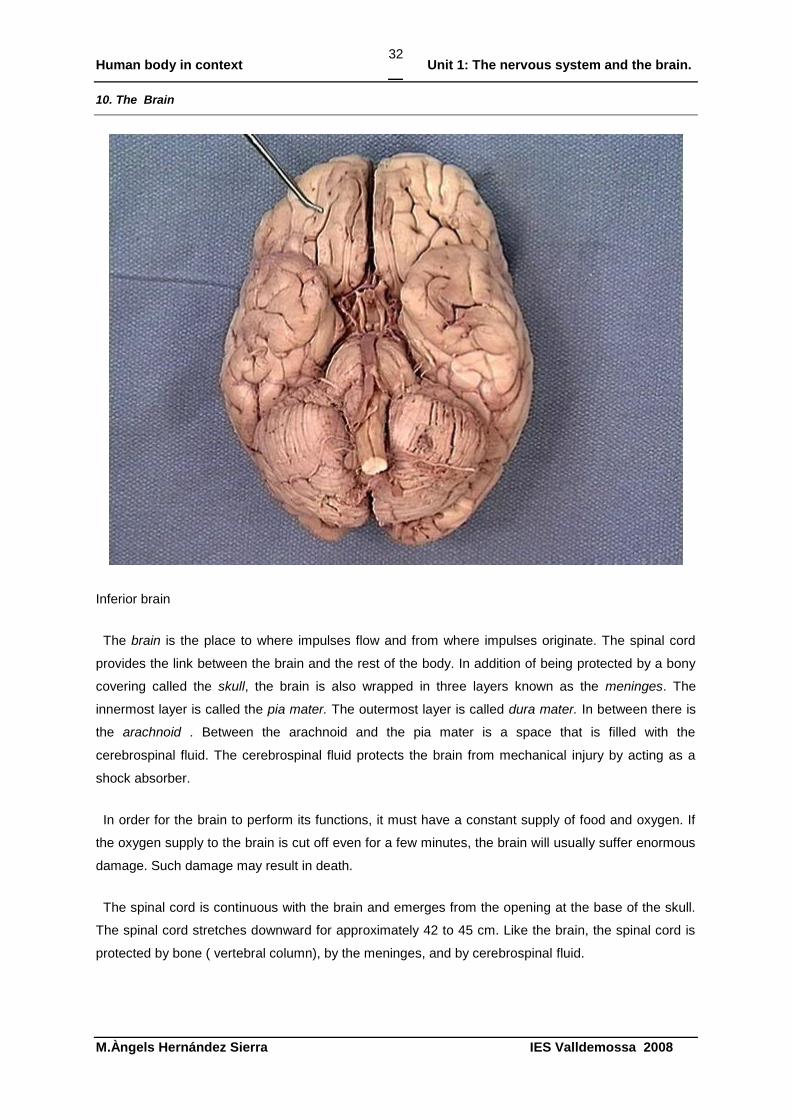
Inferior brain
The brain is the place to where impulses flow and from where impulses originate. The spinal cord
provides the link between the brain and the rest of the body. In addition of being protected by a bony
covering called the skull, the brain is also wrapped in three layers known as the meninges. The
innermost layer is called the pia mater. The outermost layer is called dura mater. In between there is
the arachnoid . Between the arachnoid and the pia mater is a space that is filled with the
cerebrospinal fluid. The cerebrospinal fluid protects the brain from mechanical injury by acting as a
shock absorber.
In order for the brain to perform its functions, it must have a constant supply of food and oxygen. If
the oxygen supply to the brain is cut off even for a few minutes, the brain will usually suffer enormous
damage. Such damage may result in death.
The spinal cord is continuous with the brain and emerges from the opening at the base of the skull.
The spinal cord stretches downward for approximately 42 to 45 cm. Like the brain, the spinal cord is
protected by bone ( vertebral column), by the meninges, and by cerebrospinal fluid.
Human body in context Unit 1: The nervous system and the brain.
M.Àngels Hernández Sierra IES Valldemossa 2008
33
11. The Cerebrum. The Cerebral Cortex.
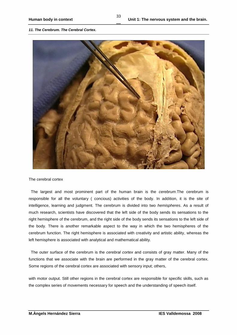
The cerebral cortex
The largest and most prominent part of the human brain is the cerebrum.The cerebrum is
responsible for all the voluntary ( concious) activities of the body. In addition, it is the site of
intelligence, learning and judgment. The cerebrum is divided into two hemispheres. As a result of
much research, scientists have discovered that the left side of the body sends its sensations to the
right hemisphere of the cerebrum, and the right side of the body sends its sensations to the left side of
the body. There is another remarkable aspect to the way in which the two hemispheres of the
cerebrum function. The right hemisphere is associated with creativity and artistic ability, whereas the
left hemisphere is associated with analytical and mathematical ability.
The outer surface of the cerebrum is the cerebral cortex and consists of gray matter. Many of the
functions that we associate with the brain are performed in the gray matter of the cerebral cortex.
Some regions of the cerebral cortex are associated with sensory input; others,
with motor output. Still other regions in the cerebral cortex are responsible for specific skills, such as
the complex series of movements necessary for speech and the understanding of speech itself.
Human body in context Unit 1: The nervous system and the brain.
M.Àngels Hernández Sierra IES Valldemossa 2008
34
For some time, scientists believed that many functions of the body were controlled by specific
regions of the cerebral cortex. In recent years the story has turned out to be quite a bit more complex.
You might think that sensory neurons are connected directly to the appropiate part of the sensory
cortex. This is not the case. There is not direct connection. Instead, sensory neurons synapse in the
spinal cord, and neurons located in the spinal cord carry the impulse to the sensory cortex of the
cerebrum.
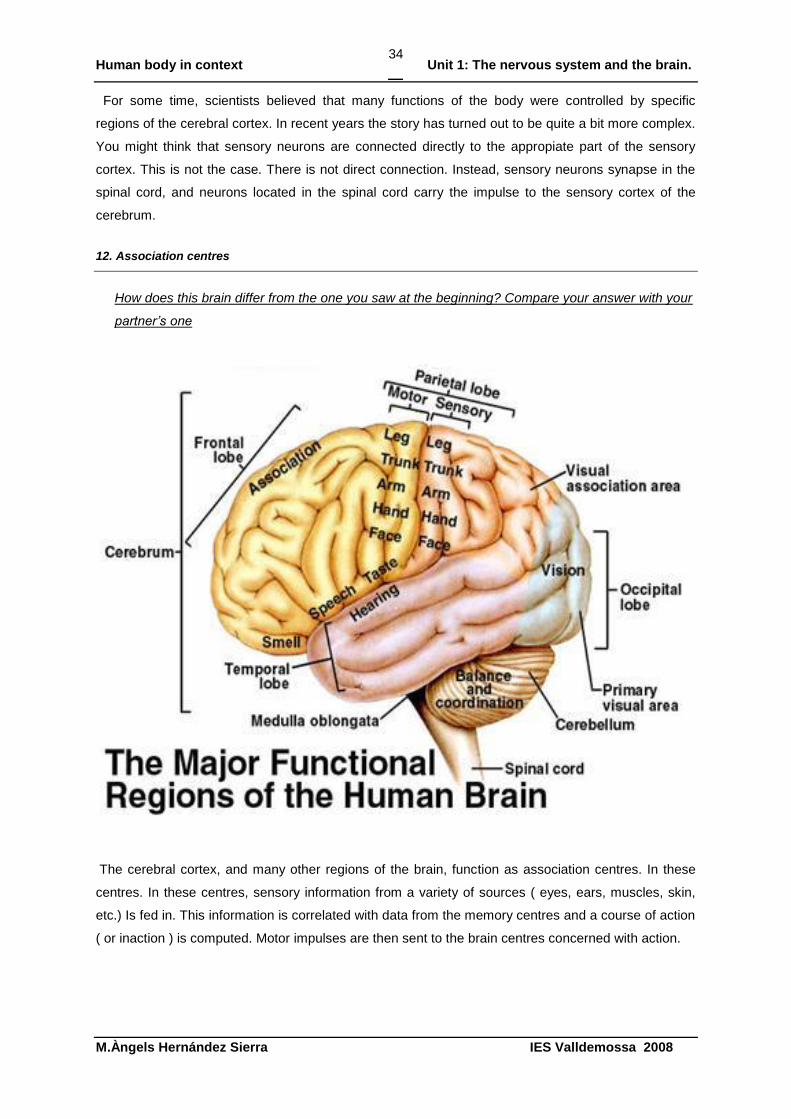
12. Association centres
How does this brain differ from the one you saw at the beginning? Compare your answer with your
partner’s one
The cerebral cortex, and many other regions of the brain, function as association centres. In these
centres. In these centres, sensory information from a variety of sources ( eyes, ears, muscles, skin,
etc.) Is fed in. This information is correlated with data from the memory centres and a course of action
( or inaction ) is computed. Motor impulses are then sent to the brain centres concerned with action.
Human body in context Unit 1: The nervous system and the brain.
M.Àngels Hernández Sierra IES Valldemossa 2008
35
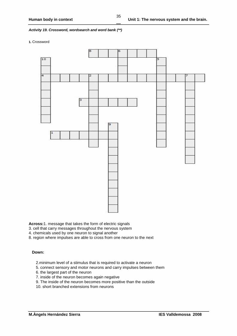
Activity 19. Crossword, wordsearch and word bank (**)
1. Crossword
Across:1. message that takes the form of electric signals 3. cell that carry messages throughout the nervous system 4. chemicals used by one neuron to signal another 8. region where impulses are able to cross from one neuron to the next
Down:
2.minimum level of a stimulus that is required to activate a neuron
5. connect sensory and motor neurons and carry impulses between them
6. the largest part of the neuron
7. inside of the neuron becomes again negative
9. The inside of the neuron becomes more positive than the outside
10. short branched extensions from neurons
Human body in context Unit 1: The nervous system and the brain.
M.Àngels Hernández Sierra IES Valldemossa 2008
36
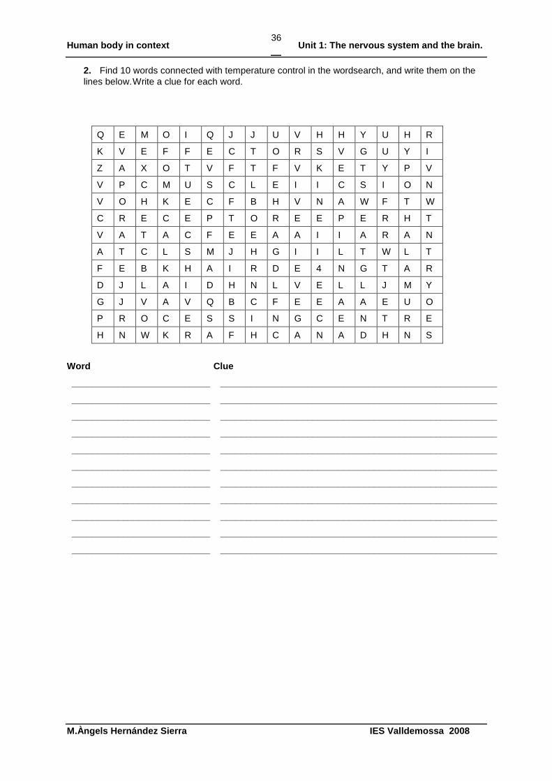
2. Find 10 words connected with temperature control in the wordsearch, and write them on the
lines below. Write a clue for each word.
Q E M O I Q J J U V H H Y U H R
K V E F F E C T O R S V G U Y I
Z A X O T V F T F V K E T Y P V
V P C M U S C L E I I C S I O N
V O H K E C F B H V N A W F T W
C R E C E P T O R E E P E R H T
V A T A C F E E A A I I A R A N
A T C L S M J H G I I L T W L T
F E B K H A I R D E 4 N G T A R
D J L A I D H N L V E L L J M Y
G J V A V Q B C F E E A A E U O
P R O C E S S I N G C E N T R E
H N W K R A F H C A N A D H N S
Word Clue
___________________________ ______________________________________________________
___________________________ ______________________________________________________
___________________________ ______________________________________________________
___________________________ ______________________________________________________
___________________________ ______________________________________________________
___________________________ ______________________________________________________
___________________________ ______________________________________________________
___________________________ ______________________________________________________
___________________________ ______________________________________________________
___________________________ ______________________________________________________
___________________________ ______________________________________________________
Human body in context Unit 1: The nervous system and the brain.
M.Àngels Hernández Sierra IES Valldemossa 2008
37
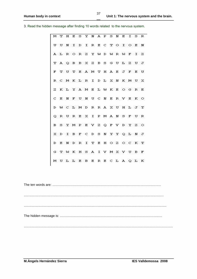
3. Read the hidden message after finding 10 words related to the nervous system.
The ten words are: ......................................................................................................................
........................................................................................................................................................
...........................................................................................................................................................
The hidden message is: ...............................................................................................................
..................................................................................................................................................................
Human body in context Unit 1: The nervous system and the brain.
M.Àngels Hernández Sierra IES Valldemossa 2008
38
Activity 20. Nervous coordination(* and **)
1. Look at the sentences below and then fill in the table.
a Iker sees in a TV guide that there is something good on TV and he switches the TV on with the
remote control.
b Anna turned the CD player off when she realised she was listening to one of her father‟s CDs.
Sentence What is the stimulus?
Where are the receptor cells for this stimulus?
What is the response? Where are the effector cells for this response?
a
b
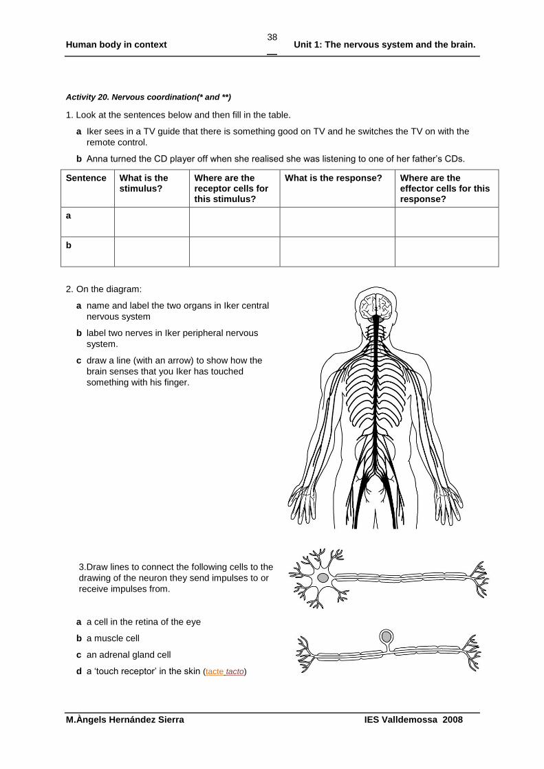
2. On the diagram:
a name and label the two organs in Iker central
nervous system
b label two nerves in Iker peripheral nervous
system.
c draw a line (with an arrow) to show how the
brain senses that you Iker has touched
something with his finger.
3.Draw lines to connect the following cells to the
drawing of the neuron they send impulses to or
receive impulses from.
a a cell in the retina of the eye
b a muscle cell
c an adrenal gland cell
d a „touch receptor‟ in the skin (tacte tacto)
Human body in context Unit 1: The nervous system and the brain.
M.Àngels Hernández Sierra IES Valldemossa 2008
39
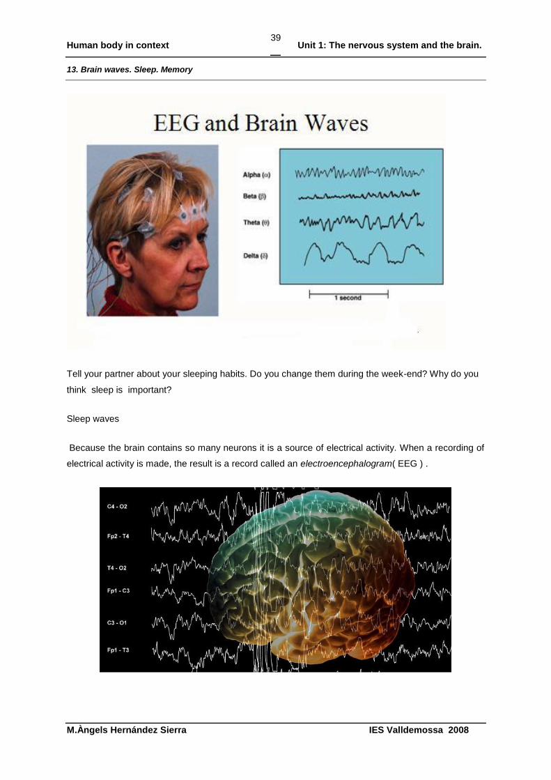
13. Brain waves. Sleep. Memory
Tell your partner about your sleeping habits. Do you change them during the week-end? Why do you
think sleep is important?
Sleep waves
Because the brain contains so many neurons it is a source of electrical activity. When a recording of
electrical activity is made, the result is a record called an electroencephalogram( EEG ) .
Human body in context Unit 1: The nervous system and the brain.
M.Àngels Hernández Sierra IES Valldemossa 2008
40
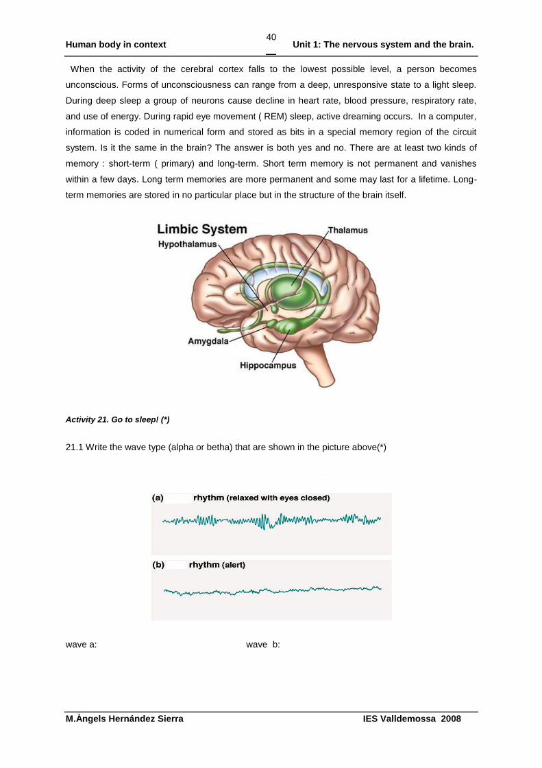
When the activity of the cerebral cortex falls to the lowest possible level, a person becomes
unconscious. Forms of unconsciousness can range from a deep, unresponsive state to a light sleep.
During deep sleep a group of neurons cause decline in heart rate, blood pressure, respiratory rate,
and use of energy. During rapid eye movement ( REM) sleep, active dreaming occurs. In a computer,
information is coded in numerical form and stored as bits in a special memory region of the circuit
system. Is it the same in the brain? The answer is both yes and no. There are at least two kinds of
memory : short-term ( primary) and long-term. Short term memory is not permanent and vanishes
within a few days. Long term memories are more permanent and some may last for a lifetime. Long-
term memories are stored in no particular place but in the structure of the brain itself.
Activity 21. Go to sleep! (*)
21.1 Write the wave type (alpha or betha) that are shown in the picture above(*)
wave a: wave b:
Human body in context Unit 1: The nervous system and the brain.
M.Àngels Hernández Sierra IES Valldemossa 2008
41
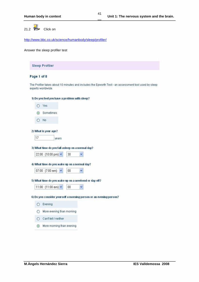
21.2 Click on
http://www.bbc.co.uk/science/humanbody/sleep/profiler/
Answer the sleep profiler test
Human body in context Unit 1: The nervous system and the brain.
M.Àngels Hernández Sierra IES Valldemossa 2008
42
Activity 22. Alzheimer’s disease. Click and learn about the unhealthy brain. (***)
http://www.nationalgeographic.com/guides/science/humanbody/brain.html#/brainUnhealthy/
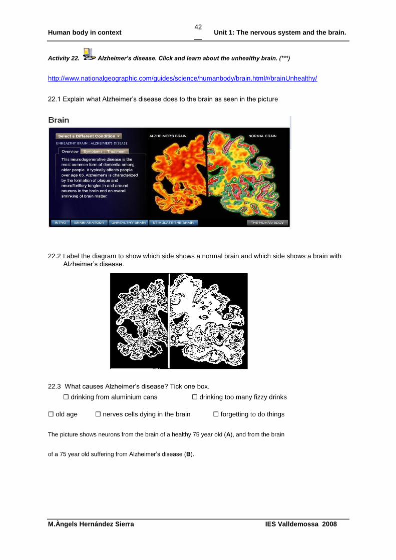
22.1 Explain what Alzheimer‟s disease does to the brain as seen in the picture
22.2 Label the diagram to show which side shows a normal brain and which side shows a brain with
Alzheimer‟s disease.
22.3 What causes Alzheimer‟s disease? Tick one box.
drinking from aluminium cans drinking too many fizzy drinks
old age nerves cells dying in the brain forgetting to do things
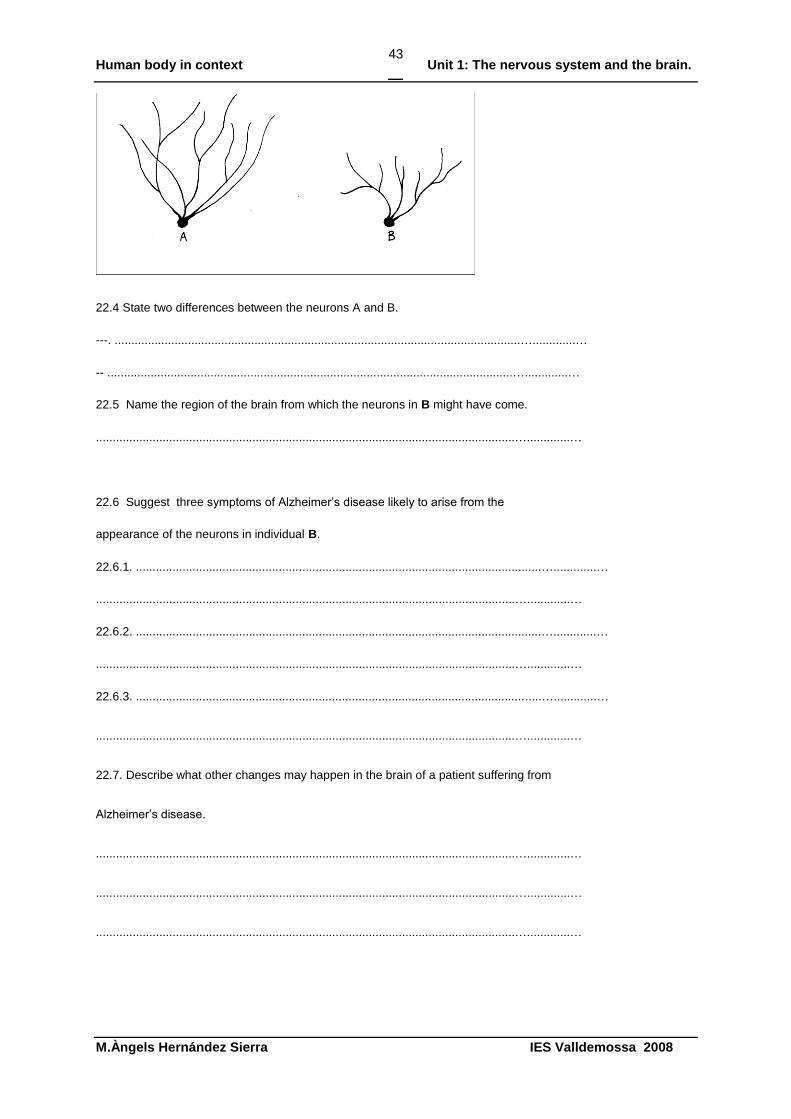
The picture shows neurons from the brain of a healthy 75 year old (A), and from the brain
of a 75 year old suffering from Alzheimer‟s disease (B).
Human body in context Unit 1: The nervous system and the brain.
M.Àngels Hernández Sierra IES Valldemossa 2008
43
22.4 State two differences between the neurons A and B.
---. ..........................................................................................................................….............…
-- ..........................................................................................................................….............…
22.5 Name the region of the brain from which the neurons in B might have come.
..............................................................................................................................….............…
22.6 Suggest three symptoms of Alzheimer‟s disease likely to arise from the
appearance of the neurons in individual B.
22.6.1. ..........................................................................................................................….............…
..............................................................................................................................….............…
22.6.2. ..........................................................................................................................….............…
..............................................................................................................................….............…
22.6.3. ..........................................................................................................................….............…
..............................................................................................................................….............…
22.7. Describe what other changes may happen in the brain of a patient suffering from
Alzheimer‟s disease.
..............................................................................................................................….............…
..............................................................................................................................….............…
..............................................................................................................................….............…
Human body in context Unit 1: The nervous system and the brain.
M.Àngels Hernández Sierra IES Valldemossa 2008
44
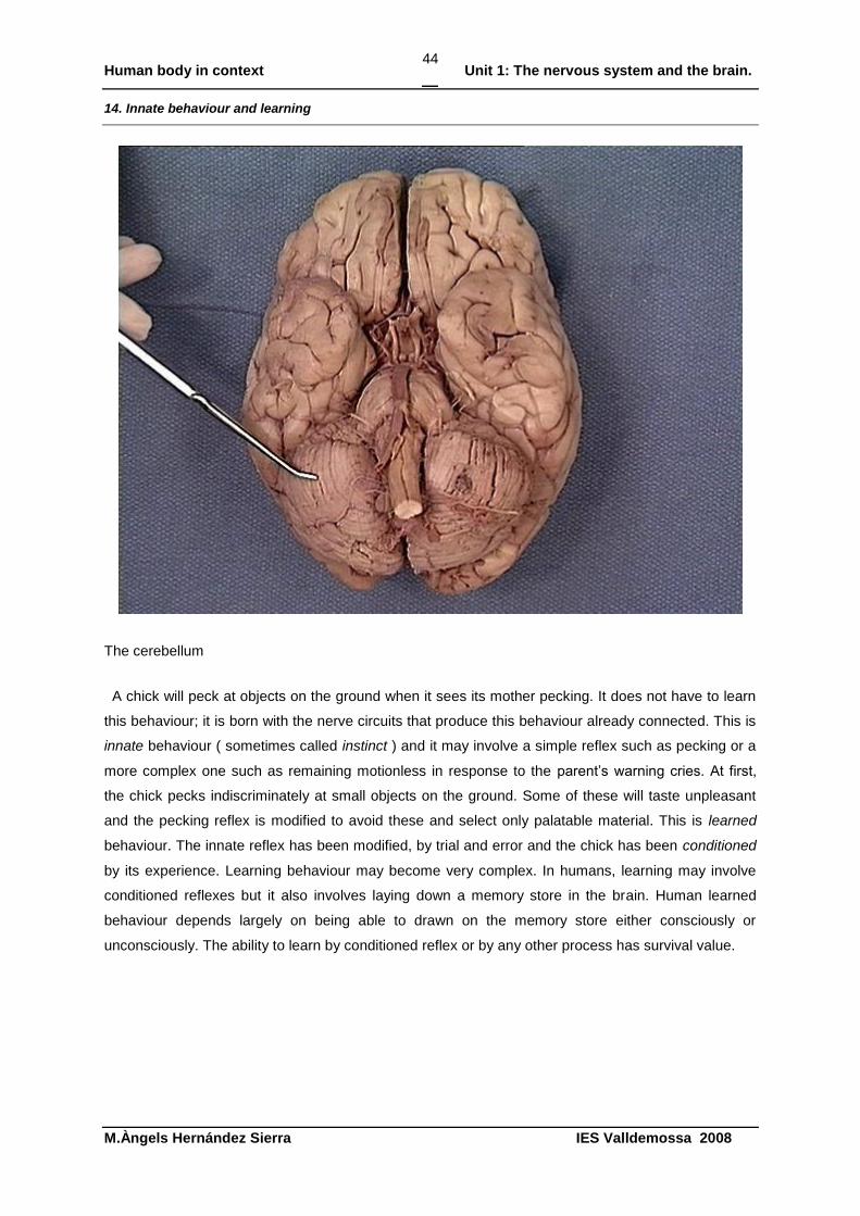
14. Innate behaviour and learning
The cerebellum
A chick will peck at objects on the ground when it sees its mother pecking. It does not have to learn
this behaviour; it is born with the nerve circuits that produce this behaviour already connected. This is
innate behaviour ( sometimes called instinct ) and it may involve a simple reflex such as pecking or a
more complex one such as remaining motionless in response to the parent‟s warning cries. At first,
the chick pecks indiscriminately at small objects on the ground. Some of these will taste unpleasant
and the pecking reflex is modified to avoid these and select only palatable material. This is learned
behaviour. The innate reflex has been modified, by trial and error and the chick has been conditioned
by its experience. Learning behaviour may become very complex. In humans, learning may involve
conditioned reflexes but it also involves laying down a memory store in the brain. Human learned
behaviour depends largely on being able to drawn on the memory store either consciously or
unconsciously. The ability to learn by conditioned reflex or by any other process has survival value.
Human body in context Unit 1: The nervous system and the brain.
M.Àngels Hernández Sierra IES Valldemossa 2008
45
Activity 23. Describe the functions of the cerebrum, cerebellum and medulla oblongata in the brain. Use
the words from this list(*)
To begin with…
It starts by/with…I want to describe…
There are different functions …
The location of………. To better understand…….. it is necessary to examine …
There are several reasons for this, the first
One function of…
It is believed/understood that..
After that…
Influenced by…
So, now you can see…
In conclusion the facts show
Consequently…
Finally…
..............................................................................................................................….............…
..............................................................................................................................….............…
..............................................................................................................................….............…
..............................................................................................................................….............…
..............................................................................................................................….............…
..............................................................................................................................….............…
..............................................................................................................................….............…
..............................................................................................................................….............…
..............................................................................................................................….............…
..............................................................................................................................….............…
Human body in context Unit 1: The nervous system and the brain.
M.Àngels Hernández Sierra IES Valldemossa 2008
46
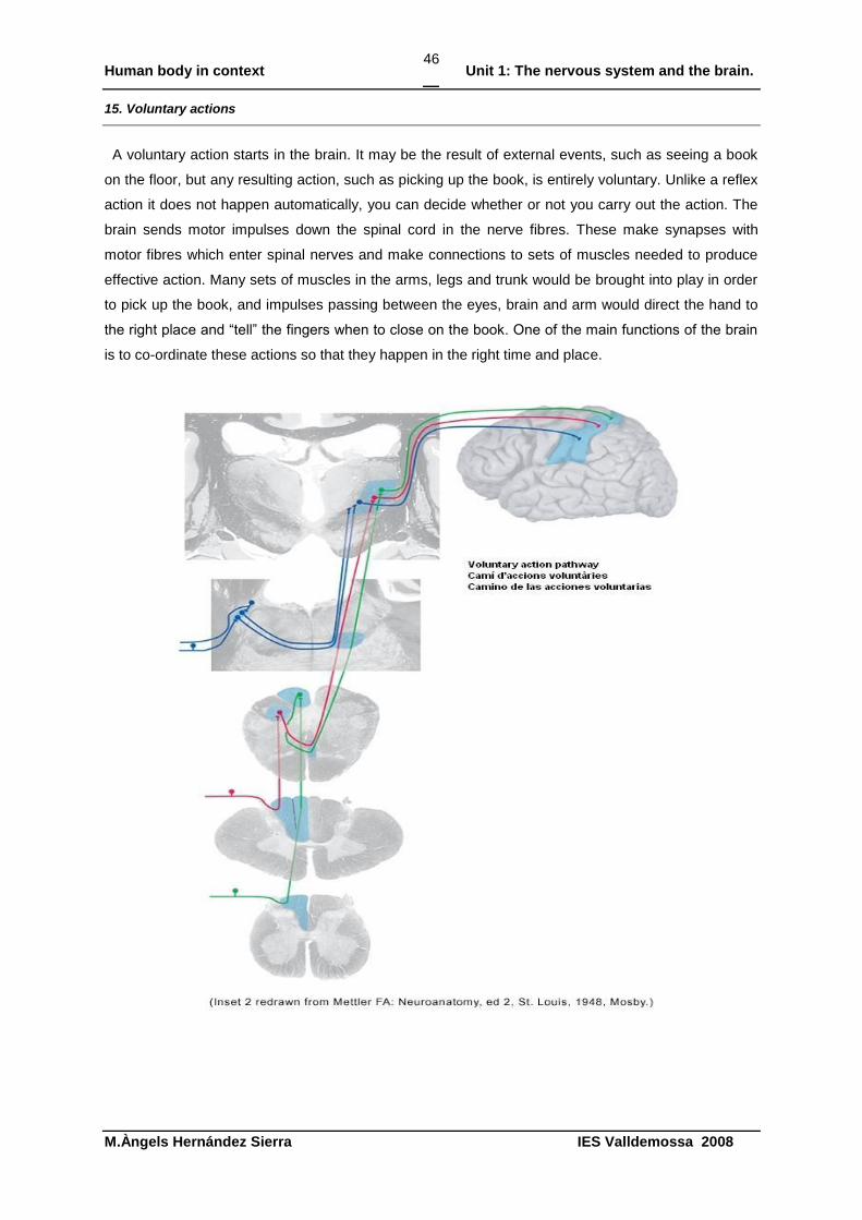
15. Voluntary actions
A voluntary action starts in the brain. It may be the result of external events, such as seeing a book
on the floor, but any resulting action, such as picking up the book, is entirely voluntary. Unlike a reflex
action it does not happen automatically, you can decide whether or not you carry out the action. The
brain sends motor impulses down the spinal cord in the nerve fibres. These make synapses with
motor fibres which enter spinal nerves and make connections to sets of muscles needed to produce
effective action. Many sets of muscles in the arms, legs and trunk would be brought into play in order
to pick up the book, and impulses passing between the eyes, brain and arm would direct the hand to
the right place and “tell” the fingers when to close on the book. One of the main functions of the brain
is to co-ordinate these actions so that they happen in the right time and place.
Human body in context Unit 1: The nervous system and the brain.
M.Àngels Hernández Sierra IES Valldemossa 2008
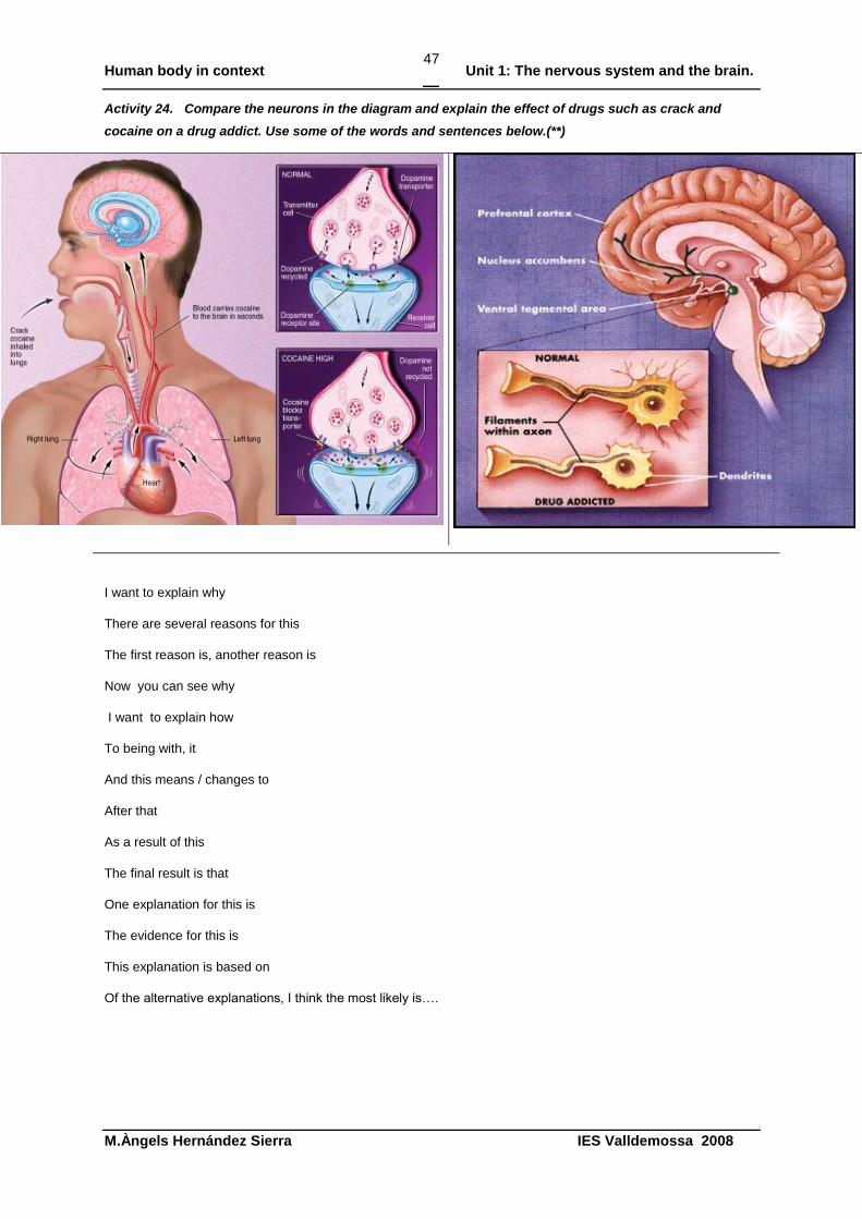
47
Activity 24. Compare the neurons in the diagram and explain the effect of drugs such as crack and
cocaine on a drug addict. Use some of the words and sentences below.(**)
I want to explain why
There are several reasons for this
The first reason is, another reason is
Now you can see why
I want to explain how
To being with, it
And this means / changes to
After that
As a result of this
The final result is that
One explanation for this is
The evidence for this is
This explanation is based on
Of the alternative explanations, I think the most likely is….
Human body in context Unit 1: The nervous system and the brain.
M.Àngels Hernández Sierra IES Valldemossa 2008
48
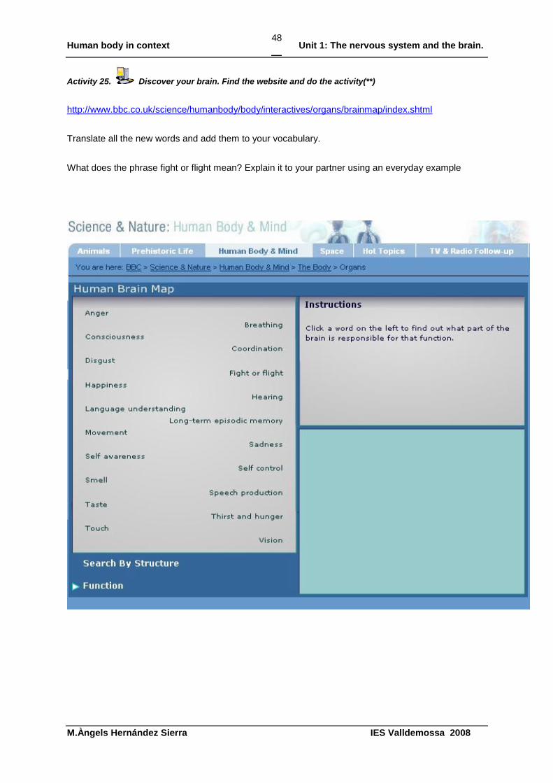
Activity 25. Discover your brain. Find the website and do the activity(**)
http://www.bbc.co.uk/science/humanbody/body/interactives/organs/brainmap/index.shtml
Translate all the new words and add them to your vocabulary.
What does the phrase fight or flight mean? Explain it to your partner using an everyday example
Human body in context Unit 1: The nervous system and the brain.
M.Àngels Hernández Sierra IES Valldemossa 2008
49
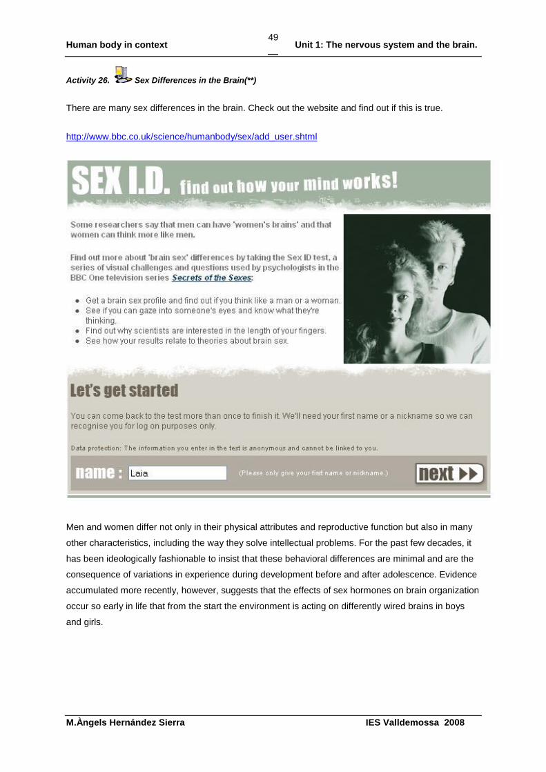
Activity 26. Sex Differences in the Brain(**)
There are many sex differences in the brain. Check out the website and find out if this is true.
http://www.bbc.co.uk/science/humanbody/sex/add_user.shtml
Men and women differ not only in their physical attributes and reproductive function but also in many
other characteristics, including the way they solve intellectual problems. For the past few decades, it
has been ideologically fashionable to insist that these behavioral differences are minimal and are the
consequence of variations in experience during development before and after adolescence. Evidence
accumulated more recently, however, suggests that the effects of sex hormones on brain organization
occur so early in life that from the start the environment is acting on differently wired brains in boys
and girls.
Human body in context Unit 1: The nervous system and the brain.
M.Àngels Hernández Sierra IES Valldemossa 2008
50
Activity 27. Sheep brain dissection: the anatomy of memory
http://www.exploratorium.edu/memory/braindissection/
Watch the videos and learn from the dissection where the memory is stored
Human body in context Unit 1: The nervous system and the brain.
M.Àngels Hernández Sierra IES Valldemossa 2008
51
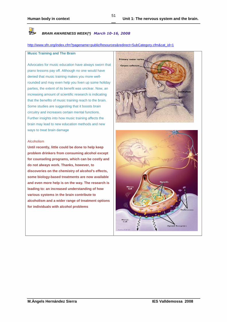
BRAIN AWARENESS WEEK(*) March 10-16, 2008
http://www.sfn.org/index.cfm?pagename=publicResources&redirect=SubCategory.cfm&cat_id=1
Music Training and The Brain
Advocates for music education have always sworn that
piano lessons pay off. Although no one would have
denied that music training makes you more well-
rounded and may even help you liven up some holiday
parties, the extent of its benefit was unclear. Now, an
increasing amount of scientific research is indicating
that the benefits of music training reach to the brain.
Some studies are suggesting that it boosts brain
circuitry and increases certain mental functions.
Further insights into how music training affects the
brain may lead to new education methods and new
ways to treat brain damage
Alcoholism
Until recently, little could be done to help keep
problem drinkers from consuming alcohol except
for counseling programs, which can be costly and
do not always work. Thanks, however, to
discoveries on the chemistry of alcohol's effects,
some biology-based treatments are now available
and even more help is on the way. The research is
leading to: an increased understanding of how
various systems in the brain contribute to
alcoholism and a wider range of treatment options
for individuals with alcohol problems
Human body in context Unit 1: The nervous system and the brain.
M.Àngels Hernández Sierra IES Valldemossa 2008
52
Adult Neurogenesis
For more than a century, medical science firmly
believed that our brain could not repair itself and
that we were born with all the brain cells we would
ever have. That belief has changed. Over the last
20 years, research has shown that neurogenesis,
the creation of new brain cells, actually occurs in the
adult human. For decades, scientists believed that
brain cells of the central nervous system could not
regrow following damage due to trauma such as
head injury or disorders such as Alzheimer's
disease. Scientists recently have discovered a
whole family of proteins called neurotrophic factors.
These proteins play a crucial role in the
development and survival of nerve cells, or neurons,
and in supporting adult neurons to keep them
healthy throughout life
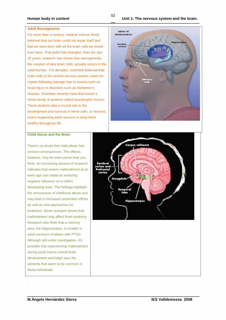
Child Abuse and the Brain
There's no doubt that child abuse has
serious consequences. The effects,
however, may be even worse than you
think. An increasing amount of research
indicates that severe maltreatment at an
early age can create an enduring
negative influence on a child's
developing brain. The findings highlight
the seriousness of childhood abuse and
may lead to increased prevention efforts
as well as new approaches for
treatment. Some research shows that
maltreatment may affect brain anatomy.
Research also finds that a memory
area, the hippocampus, is smaller in
adult survivors of abuse with PTSD.
Although still under investigation, it's
possible that experiencing maltreatment
during youth harms overall brain
development and helps spur the
ailments that seem to be common in
these individuals
.
Human body in context Unit 1: The nervous system and the brain.
M.Àngels Hernández Sierra IES Valldemossa 2008
53
.
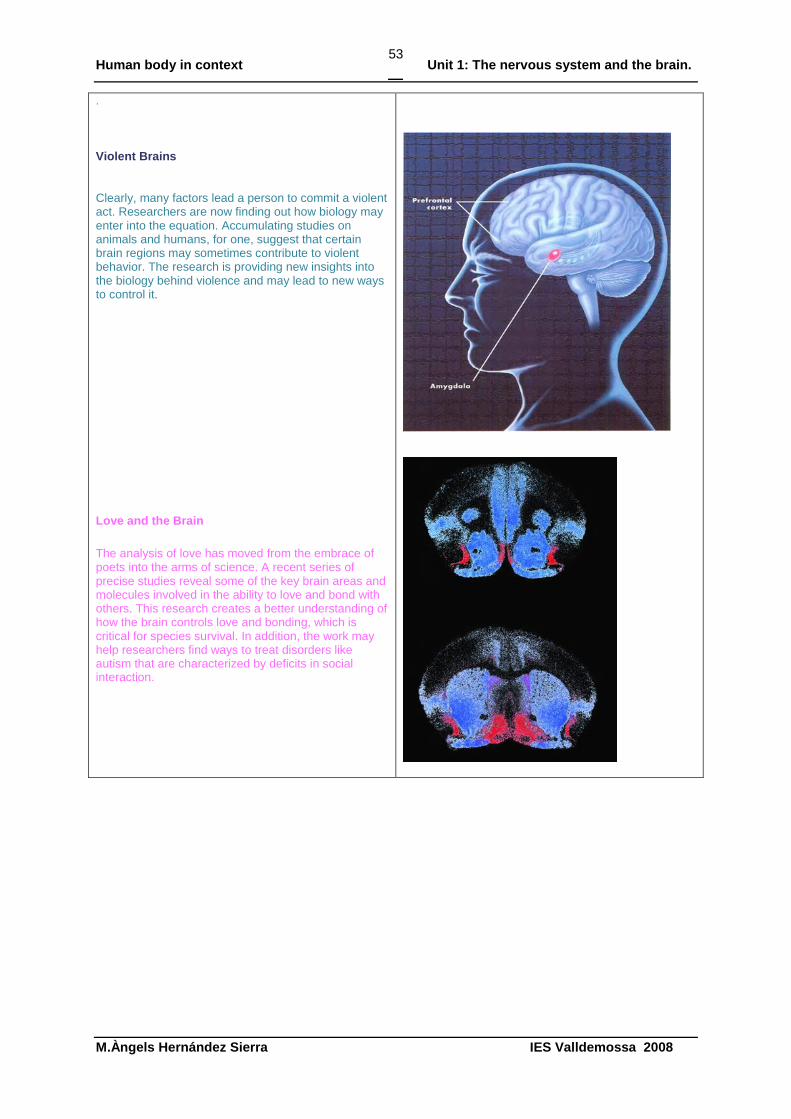
Violent Brains
Clearly, many factors lead a person to commit a violent act. Researchers are now finding out how biology may enter into the equation. Accumulating studies on animals and humans, for one, suggest that certain brain regions may sometimes contribute to violent behavior. The research is providing new insights into the biology behind violence and may lead to new ways to control it.
Love and the Brain
The analysis of love has moved from the embrace of poets into the arms of science. A recent series of precise studies reveal some of the key brain areas and molecules involved in the ability to love and bond with others. This research creates a better understanding of how the brain controls love and bonding, which is critical for species survival. In addition, the work may help researchers find ways to treat disorders like autism that are characterized by deficits in social interaction.