Embed Size (px)
Citation preview
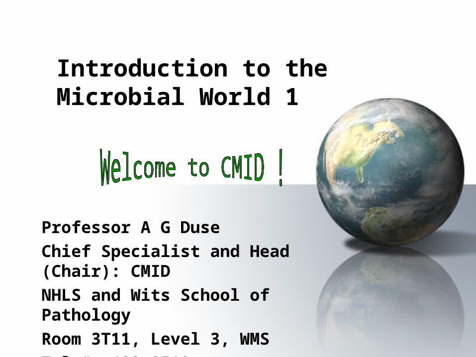
Introduction to the Microbial World 1
Professor A G Duse
Chief Specialist and Head (Chair): CMID
NHLS and Wits School of Pathology
Room 3T11, Level 3, WMS
Tel #: 489 8510
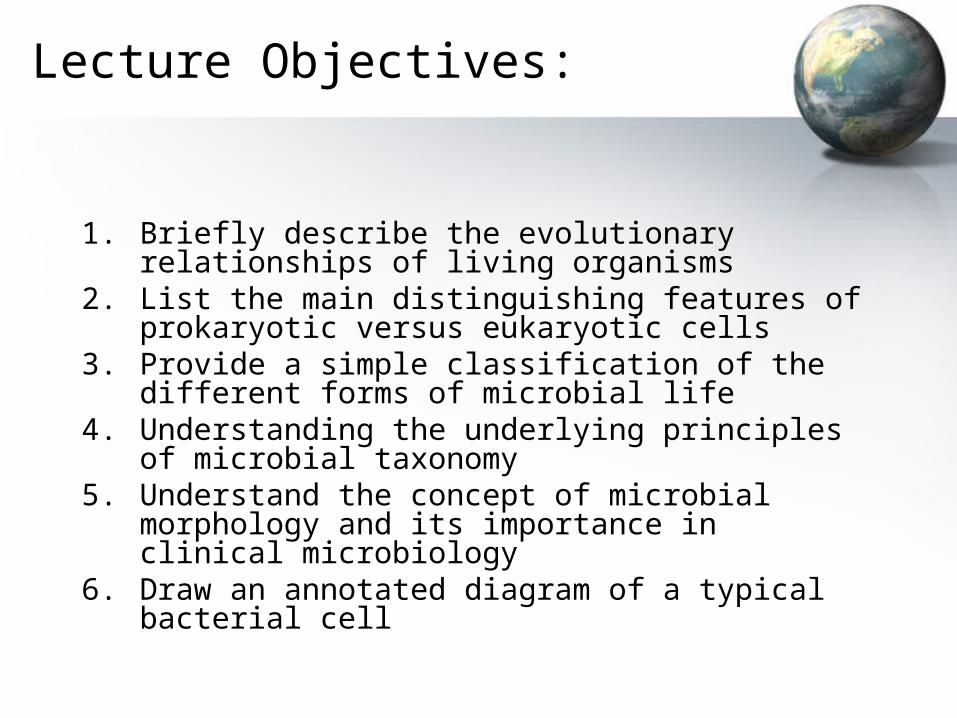
Lecture Objectives:
1. Briefly describe the evolutionary relationships of living organisms
2. List the main distinguishing features of prokaryotic versus eukaryotic cells
3. Provide a simple classification of the different forms of microbial life
4. Understanding the underlying principles of microbial taxonomy
5. Understand the concept of microbial morphology and its importance in clinical microbiology
6. Draw an annotated diagram of a typical bacterial cell
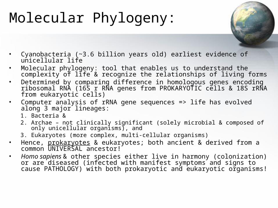
Molecular Phylogeny:
• Cyanobacteria (~3.6 billion years old) earliest evidence of unicellular life• Molecular phylogeny: tool that enables us to understand the complexity of
life & recognize the relationships of living forms• Determined by comparing difference in homologous genes encoding
ribosomal RNA (16S r RNA genes from PROKARYOTIC cells & 18S rRNA from eukaryotic cells)
• Computer analysis of rRNA gene sequences => life has evolved along 3 major lineages:1. Bacteria &2. Archae – not clinically significant (solely microbial & composed of only unicellular
organisms), and3. Eukaryotes (more complex, multi-cellular organisms)
• Hence, prokaryotes & eukaryotes; both ancient & derived from a common UNIVERSAL ancestor!
• Homo sapiens & other species either live in harmony (colonization) or are diseased (infected with manifest symptoms and signs to cause PATHOLOGY) with both prokaryotic and eukaryotic organisms!
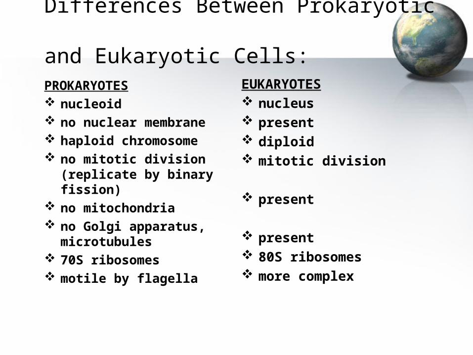
Differences Between Prokaryotic and Eukaryotic Cells:
PROKARYOTES nucleoid no nuclear membrane haploid chromosome no mitotic division (replicate
by binary fission) no mitochondria no Golgi apparatus,
microtubules 70S ribosomes motile by flagella
EUKARYOTES nucleus present diploid mitotic division
present
present 80S ribosomes more complex
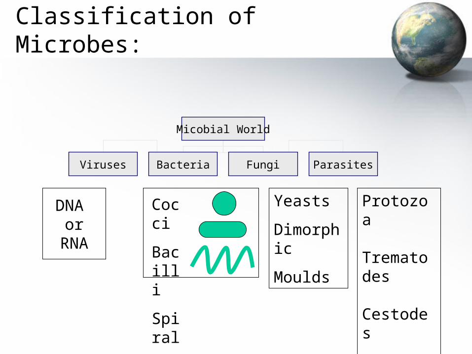
Classification of Microbes:
DNA or
RNA
Cocci
Bacilli
Spiral
Protozoa
Trematodes
Cestodes
Nematodes
Yeasts
Dimorphic
Moulds
Micobial World
Viruses Bacteria Fungi Parasites
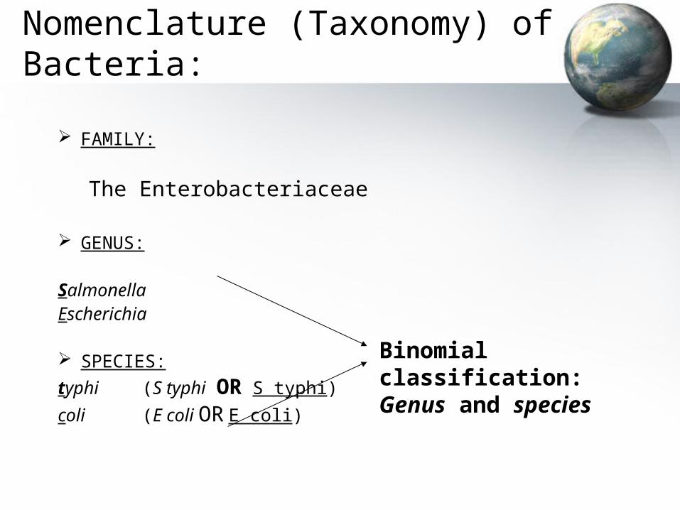
Nomenclature (Taxonomy) of Bacteria:
FAMILY:
The Enterobacteriaceae
GENUS:
Salmonella Escherichia
SPECIES:typhi (S typhi OR S typhi)
coli(E coli OR E coli)
Binomial classification: Genus and species
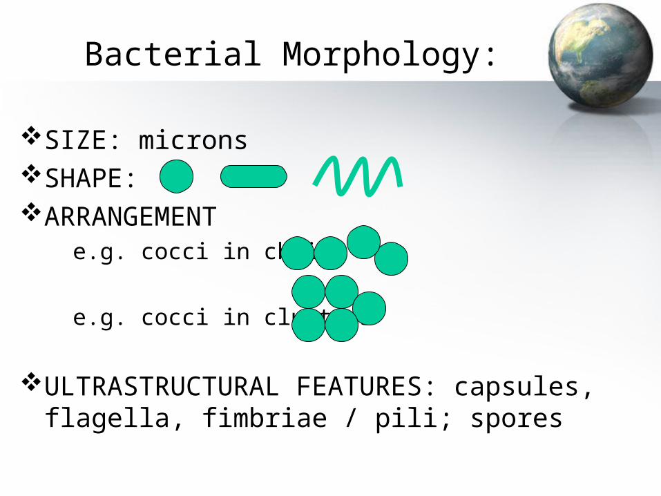
Bacterial Morphology:
SIZE: micronsSHAPE: ARRANGEMENT
e.g. cocci in chains
e.g. cocci in clusters
ULTRASTRUCTURAL FEATURES: capsules, flagella, fimbriae / pili; spores
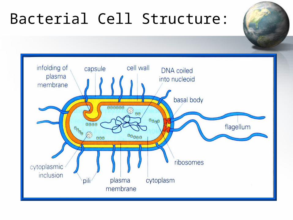
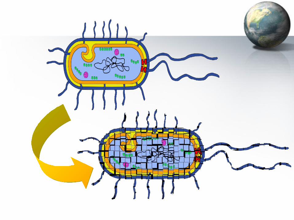
Bacterial Cell Structure:

Introduction to the Microbial World 2
Professor A G Duse
Chief Specialist and Head (Chair): CMID
NHLS and Wits School of Pathology
Room 3T11, Level 3, WMS
Tel #: 489 8510
Lecture Objectives:
• Using a bacterial cell as an example, describe the ultra-structural features of bacteria & discuss both their laboratory & clinical relevance
• Discuss the role of bacterial cell components in disease causation (pathogenesis)
• Classify bacteria according to their morphology, aero-tolerance, and staining reactions with particular emphasis on Gram and acid-fast stains
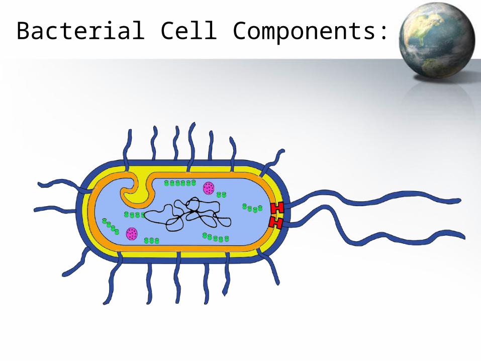
Bacterial Cell Components:
Bacterial Cell Components:
Capsule (mostly polysaccharide): antiphagocytic; antigenic/ immunogenic
Flagella (proteinaceous): locomotion; antigenic; ? Immune evasion Fimbriae/pili: adherence Cell wall (cytoskeleton = peptidoglycan): rigidity & shape;
protection against osmotic pressure - prevention of lysis; antigenic Cytoplasmic membrane: cell respiration; cell precursor synthesis Intracytoplasmic inclusions; DNA; ribosomes Spores: protect species of genera Bacillus and Clostridium from
unfavourable conditions
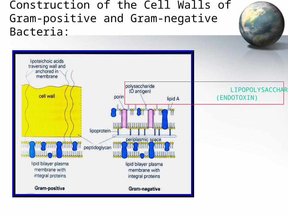
Construction of the Cell Walls of Gram-positive and Gram-negative Bacteria:
LIPOPOLYSACCHARIDE (ENDOTOXIN)
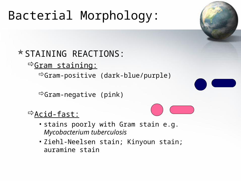
Bacterial Morphology:
STAINING REACTIONS:Gram staining:
Gram-positive (dark-blue/purple)
Gram-negative (pink)
Acid-fast:• stains poorly with Gram stain e.g. Mycobacterium
tuberculosis
• Ziehl-Neelsen stain; Kinyoun stain; auramine stain
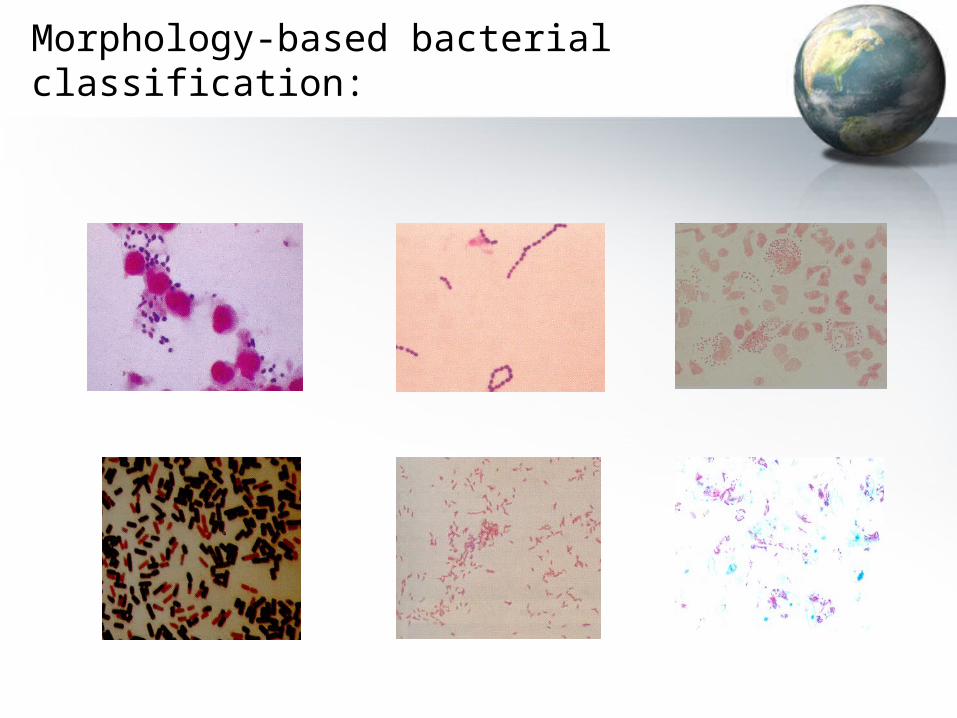
Morphology-based bacterial classification:
Man versus Microbes:
Professor A G Duse
Chief Specialist and Head (Chair): CMID
NHLS and Wits School of Pathology
Room 3T11, Level 3, WMS
Tel #: 489 8510
Objectives:
• Understand the concept of infectious disease causation• Discuss the interactions between hosts, microbes and the
environment• Describe the concepts of true virulence versus opportunism• List, and using appropriate examples, discuss the 7 major
challenges that a microbe must overcome to cause infection• Illustrate all of the above by briefly discussing the recent
outbreak caused by Rift Valley Fever virus in Kenya (Dec 2006-Jan 2007)
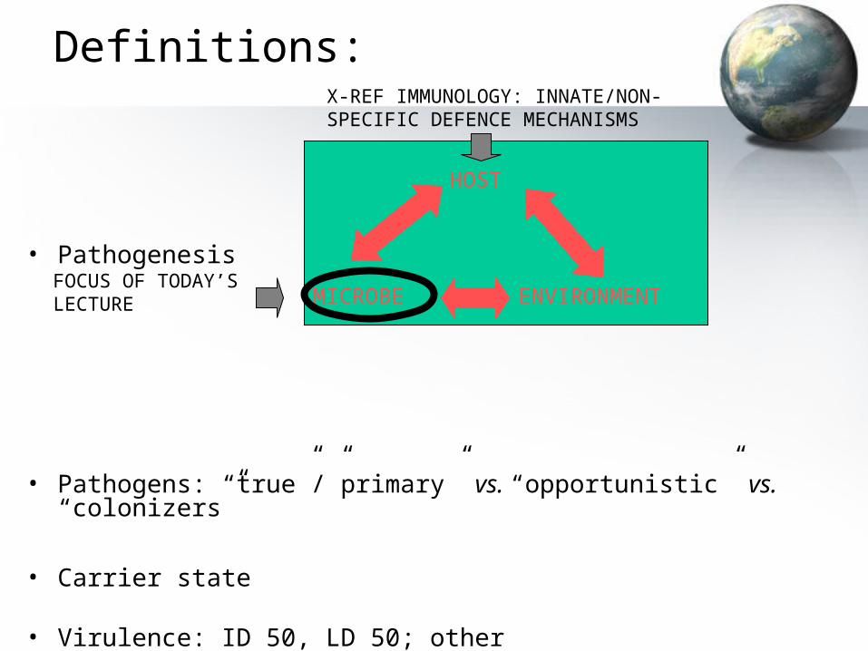
Definitions:
• Pathogenesis
• Pathogens: “true”/”primary” vs. “opportunistic” vs. “colonizers”
• Carrier state
• Virulence: ID 50, LD 50; other
HOST
MICROBE ENVIRONMENTFOCUS OF TODAY’S LECTURE
X-REF IMMUNOLOGY: INNATE/NON-SPECIFIC DEFENCE MECHANISMS
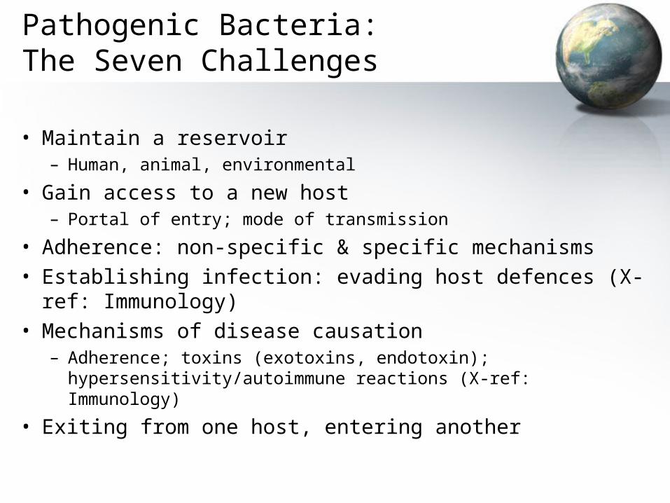
Pathogenic Bacteria:The Seven Challenges
• Maintain a reservoir– Human, animal, environmental
• Gain access to a new host– Portal of entry; mode of transmission
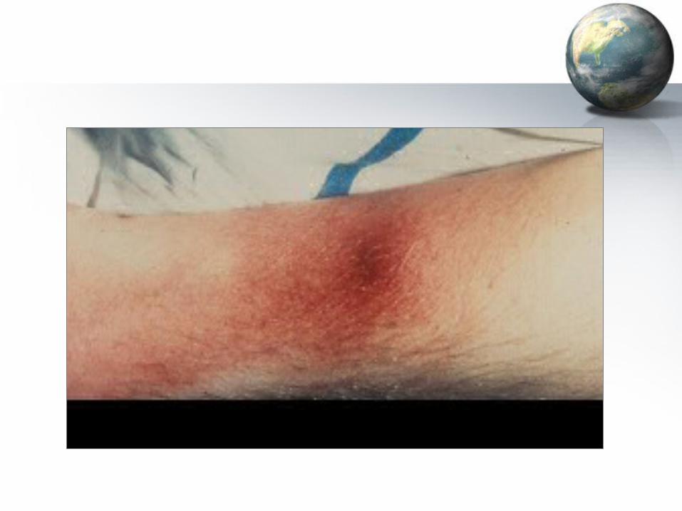
• Adherence: non-specific & specific mechanisms• Establishing infection: evading host defences (X-ref: Immunology)• Mechanisms of disease causation
– Adherence; toxins (exotoxins, endotoxin); hypersensitivity/autoimmune reactions (X-ref: Immunology)
• Exiting from one host, entering another

Portals Of Entry:
• Respiratory tract
• Gastrointestinal tract
• Genitourinary tract
• Skin and mucous membranes
Modes Of Transmission:
• By respiratory droplets; droplet nuclei• Airborne (other than above)• Faecal-oral• By direct body contact• By fomites• Parenteral• By arthropod vectors
Remember:
• Adherence; + / -
• Invasion; +/ -
• Toxin production– Differences between exotoxins & endotoxins; + / -
• Hypersensitivity reactions

Pathogenic Bacteria:The Seven Challenges
• Maintain a reservoir– Human, animal, environmental
• Gain access to a new host– Portal of entry; mode of transmission
• Adherence: non-specific & specific mechanisms• Establishing infection: evading host defences (X-ref: Immunology)• Mechanisms of disease causation
– Adherence; toxins (exotoxins, endotoxin); hypersensitivity/autoimmune reactions (X-ref: Immunology)
• Exiting from one host, entering another
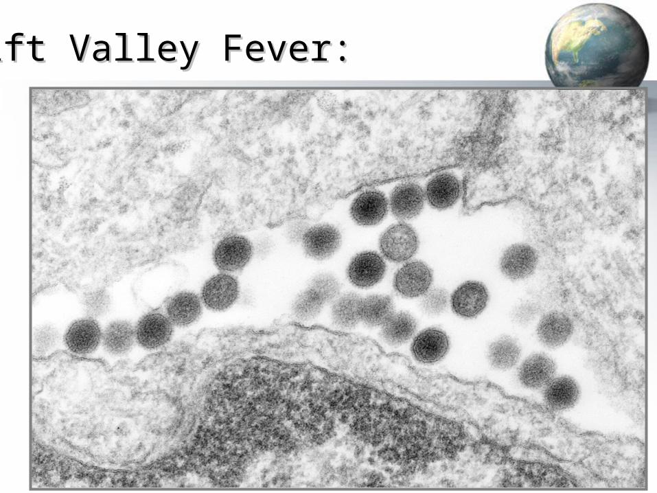
Rift Valley Fever:Rift Valley Fever:
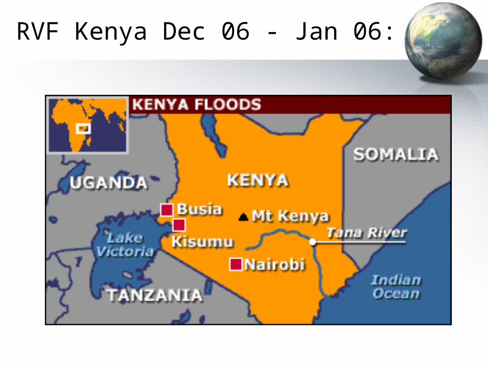
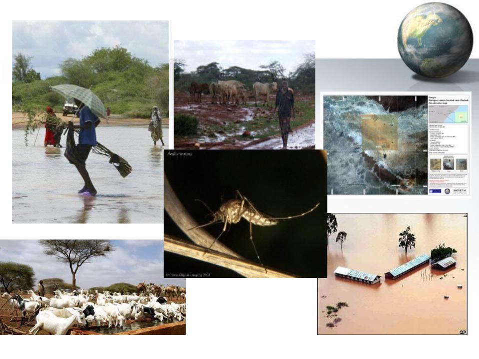
RVF Kenya Dec 06 - Jan 06:
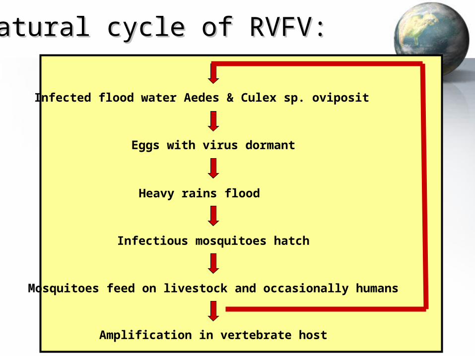
Natural cycle of RVFV:Natural cycle of RVFV:
Infected flood water Aedes & Culex sp. oviposit
Eggs with virus dormant
Heavy rains flood
Infectious mosquitoes hatch
Mosquitoes feed on livestock and occasionally humans
Amplification in vertebrate host
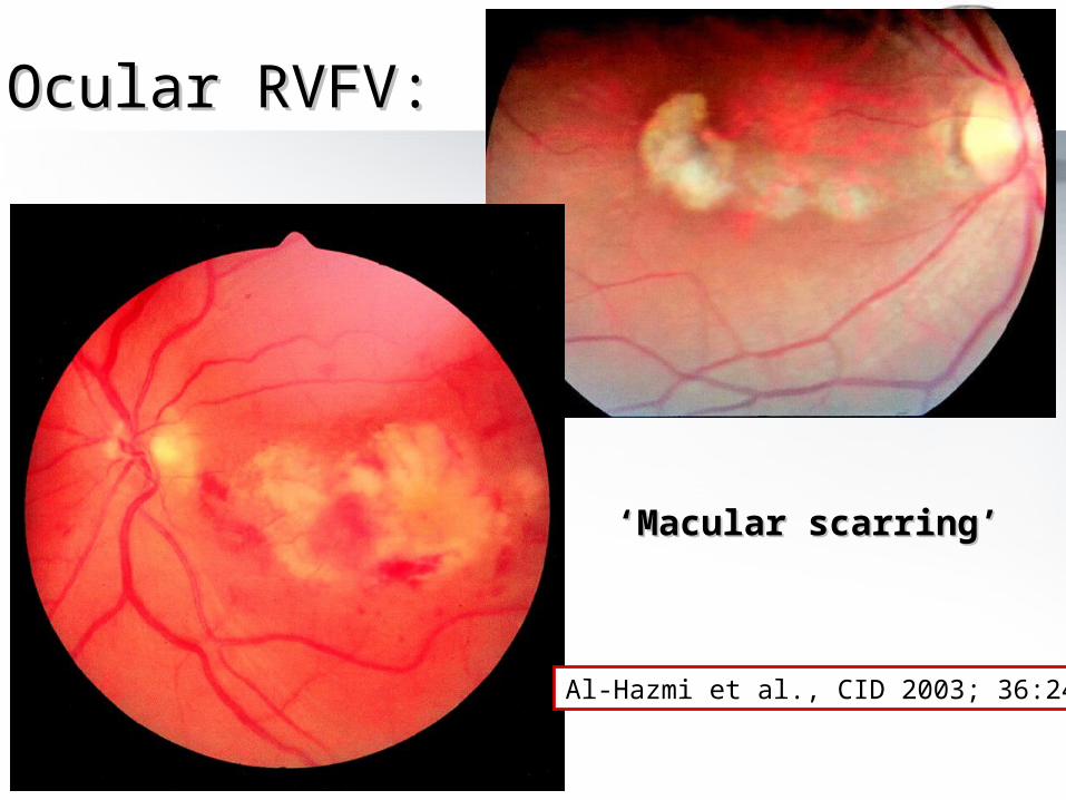
Ocular RVFV:Ocular RVFV:
Al-Hazmi et al., CID 2003; 36:245
‘‘Macular scarring’Macular scarring’

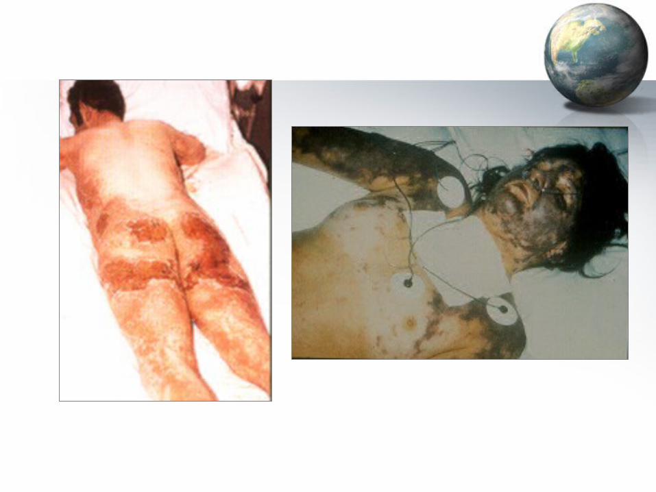
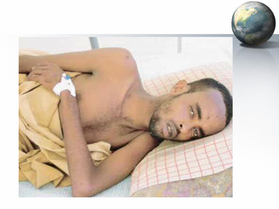
Hemorrhagic RVF:Hemorrhagic RVF:

Epizootic cycle of RVFV:Epizootic cycle of RVFV:
Livestock (mainly sheep, cattle, goat) infected by vector
Incubation followed by viremia and illness
Feeding by uninfected vectors
Extrinsic incubation
Newly infectious vector
Slaughter, abortion, or necropsy
Aerosol or contact infectionof humans

• Many mosquitoes world-wide can be vectors in the lab and in the field at blood meal virus concentrations found in sheep, cattle, humans
• We know it can cause epidemics when vertebrates that develop high viremias are present (sheep, cattle, goat)
• Mortality in domestic livestock and subsequent trade interruption have severe economic impact
• Humans may visit endemic/epidemic areas and return to their homes within an incubation period (e.g., tourist with retinopathy)
• Both arthropod and direct transmission from blood efficient (concern: bioterrorism and aerosols)
• Established epidemics in new territory: Egypt, Saudi, Yemen
RVFV – a worldwide concernRVFV – a worldwide concern

THANK YOU !