Embed Size (px)
Citation preview
Introduction to Emulsion Technology, Emulsifiers and Stability
By: Dr. Lisa Zychowski
1. Emulsions
An emulsion is a dispersed system consisting of at least two immiscible liquid phases. The
dispersed liquid is usually in the form of droplets and is referred to as the dispersed,
discontinuous, or internal phase. The liquid surrounding these droplets is the continuous or
external phase (McClements, 2015). These dispersions can be of two main formats, oil-in-
water (o/w) or water-in-oil (w/o); most food systems are o/w emulsions, such as milk, soup, or
salad dressing, but there are some instances of w/o, such as margarine or butter (Dickinson,
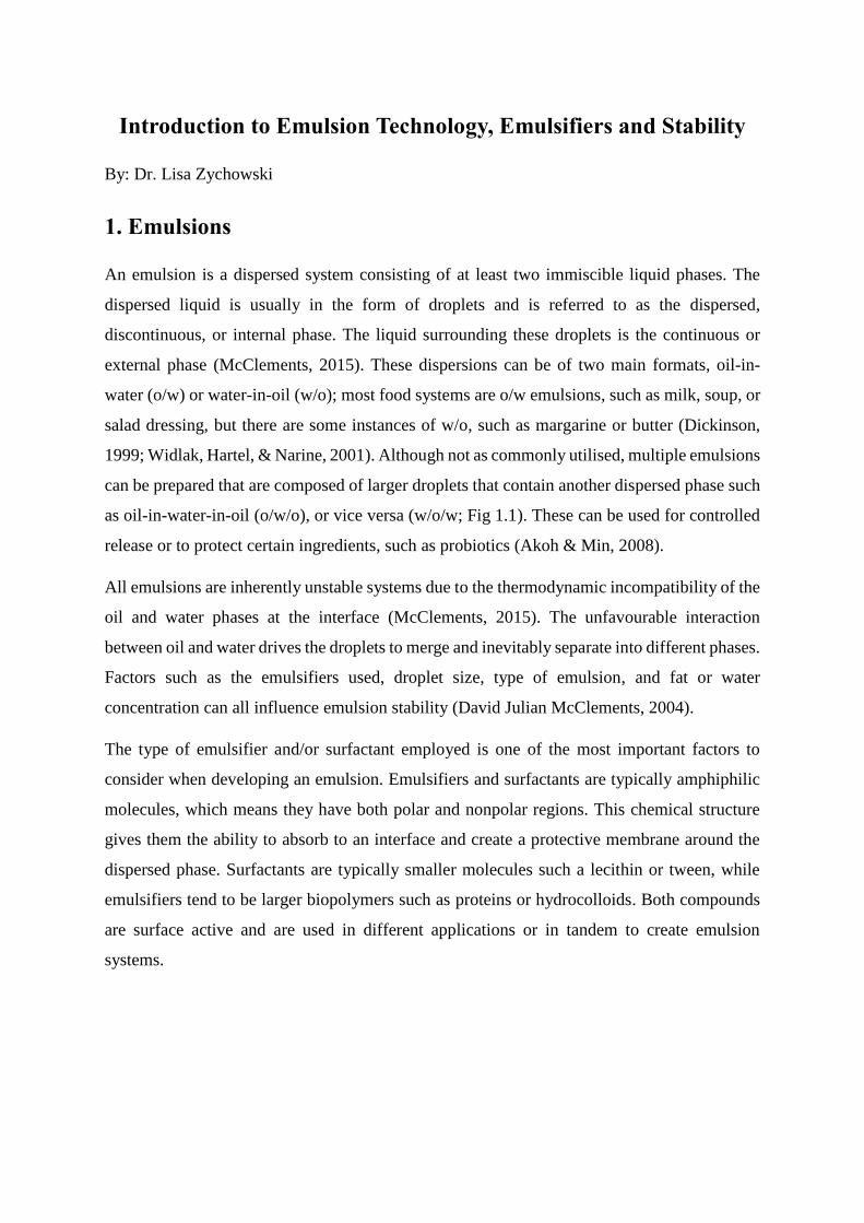
1999; Widlak, Hartel, & Narine, 2001). Although not as commonly utilised, multiple emulsions
can be prepared that are composed of larger droplets that contain another dispersed phase such
as oil-in-water-in-oil (o/w/o), or vice versa (w/o/w; Fig 1.1). These can be used for controlled
release or to protect certain ingredients, such as probiotics (Akoh & Min, 2008).
All emulsions are inherently unstable systems due to the thermodynamic incompatibility of the
oil and water phases at the interface (McClements, 2015). The unfavourable interaction
between oil and water drives the droplets to merge and inevitably separate into different phases.
Factors such as the emulsifiers used, droplet size, type of emulsion, and fat or water
concentration can all influence emulsion stability (David Julian McClements, 2004).
The type of emulsifier and/or surfactant employed is one of the most important factors to
consider when developing an emulsion. Emulsifiers and surfactants are typically amphiphilic
molecules, which means they have both polar and nonpolar regions. This chemical structure
gives them the ability to absorb to an interface and create a protective membrane around the
dispersed phase. Surfactants are typically smaller molecules such a lecithin or tween, while
emulsifiers tend to be larger biopolymers such as proteins or hydrocolloids. Both compounds
are surface active and are used in different applications or in tandem to create emulsion
systems.
1.1 Emulsion formation and stability
When the immiscible phases of an emulsion are mixed, they generally separate, as this is the
most thermodynamically stable state. Thus, in order to mix the liquids, a mechanical force is
required to combine the two phases into an emulsion. Often a two-step process, emulsion
formation requires mixing by means of high shear mixing, homogenisation, membrane
processing, and/or ultrasonication. In emulsion formation a course emulsion is first created to
mix the two phases. Afterwards, high shear is applied to reduce the size of the dispersed
droplets (Dickinson, 1999).
The quantity and time of the shear applied influences the size and stability of the dispersed
droplets (Damodaran, Parkin, & Fennema, 2007). For example, in a study by Desrumaux and
Marcand (2002), the average size of an emulsion droplet before final homogenisation (coarse
emulsion) was 30 µm. When 50 MPa of pressure was applied to the emulsion from
homogenisation, the average emulsion droplet size decreased to 0.7 µm, and with a higher
homogenisation pressure (250 MPa) the droplet size decreased further to 0.25 µm. While shear
is needed to create an emulsion to prevent the droplets from merging during formation, a
Figure 1.1 The two-main single (top) and double emulsion varieties (bottom).
sufficient quantity of an emulsifier must also be present to absorb onto the droplet surface. The
emulsifier will interact with both the dispersed and continuous phase to create a barrier between
the two liquids. This barrier could consist of one or more emulsifiers but needs to be effective
in preventing the dispersed droplets from interacting (D. J. McClements, 2004; Palanuwech &
Coupland, 2003).
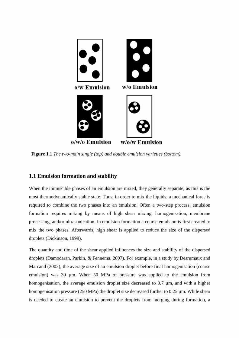
Droplet interactions can result in several different types of emulsion destabilisation, such as
coalescence, Oswald ripening, flocculation, phase inversion, sedimentation, and/or creaming
(Fig. 1.2). Coalescence is the process by which two droplets merge during contact to form a
larger droplet (Damodaran, Parkin, & Fennema, 2007). Partial coalescence can also occur and
is normally seen in food emulsions when a crystallised fat crystal from one droplet pierces the
lipid phase of another droplet (Dickinson, 1999). Flocculation is the process in which droplets
make contact but do not merge. Oswald ripening can occur when both phases are not
completely immiscible and there are different droplet sizes present in the system; here, larger
droplets will form at the expense of smaller droplets (Damodaran, Parkin, & Fennema, 2007).
Creaming, sedimentation, and phase inversion also result in noticeable physical changes in the
emulsion structure and, in food emulsions can result in the end of shelf-life. Food emulsion
instability can also come in the form of chemical degradation of the dispersed phase, such as
lipid oxidation or microbial processes (David Julian McClements, 2004).
Figure 1.2 Physical mechanisms of emulsion destabilisation adapted from McClements,
2007.
1.2 Emulsifiers and surfactants
During emulsion formation, emulsifiers and/or surfactants present in the formulation will move
to the oil/water interface and align. These surface-active molecules are typically amphiphiles,
with polar and non-polar portions of the molecule and are able to interact with both emulsion
phases (Hill, 1998).
The smaller compounds, known as surfactants, have a greater mobility than emulsifiers and
tend to dominate the emulsion interface initially. Surfactants can help produce small droplets
during emulsion formation, but are not normally used to provide long-term stability to an
emulsion. Examples of surfactants utilised in the food industry are tweens, lecithin
(phospholipids), and mono-and di-glycerides (Akoh & Min, 2008). Emulsifiers, on the other
hand, impart long-term stability to an emulsion system, although they typically take longer to
reach an emulsion interface, as they tend to be larger molecules than surfactants. Commonly
utilised emulsifiers in the food industry include various proteins and hydrocolloids, such as
whey, caseinates, or modified starches (Foegeding & Davis, 2011).
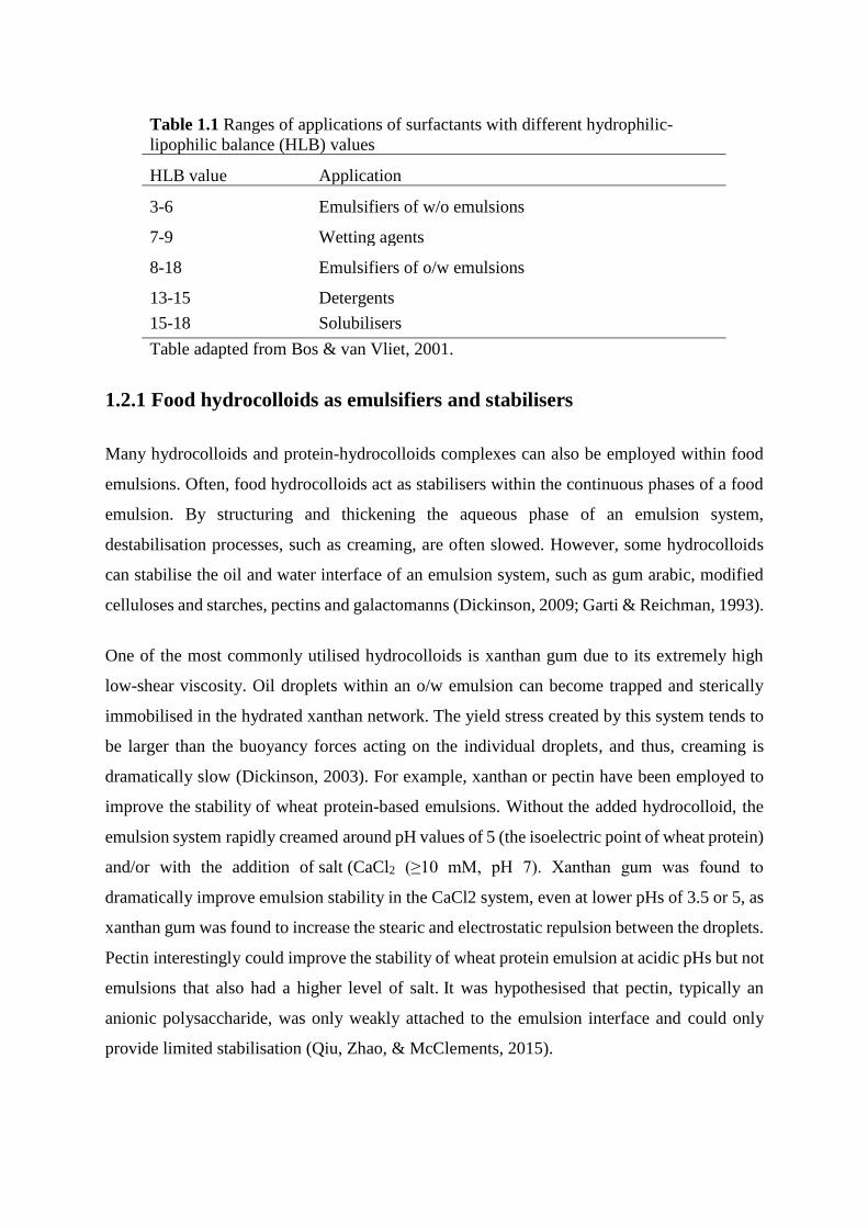
Non-ionic surface-active compounds are categorised by their hydrophilic-lipophilic balance
(HLB). The calculation of HLB is as follows:
HLB = 20(𝑀ℎ
𝑀) (1.1)
where Mh = molar mass of the hydrophilic portion of the molecules and M = molecular mass
of the whole molecule (Griffin, 1954; O' Dwyer, 2012). HLB values can be used to predict the
surface activity of a compound and are useful when formulating an emulsion. For example in
an o/w emulsion it would be advantageous to select a surface-active compound that has a higher
HLB value (8-18; Table 1.1), as the compound needs to be dispersible within the continuous
phase but still be able to interact with the oil droplets (Bos & van Vliet, 2001).
1.2.1 Food hydrocolloids as emulsifiers and stabilisers
Many hydrocolloids and protein-hydrocolloids complexes can also be employed within food
emulsions. Often, food hydrocolloids act as stabilisers within the continuous phases of a food
emulsion. By structuring and thickening the aqueous phase of an emulsion system,
destabilisation processes, such as creaming, are often slowed. However, some hydrocolloids
can stabilise the oil and water interface of an emulsion system, such as gum arabic, modified
celluloses and starches, pectins and galactomanns (Dickinson, 2009; Garti & Reichman, 1993).
One of the most commonly utilised hydrocolloids is xanthan gum due to its extremely high
low-shear viscosity. Oil droplets within an o/w emulsion can become trapped and sterically
immobilised in the hydrated xanthan network. The yield stress created by this system tends to
be larger than the buoyancy forces acting on the individual droplets, and thus, creaming is
dramatically slow (Dickinson, 2003). For example, xanthan or pectin have been employed to
improve the stability of wheat protein-based emulsions. Without the added hydrocolloid, the
emulsion system rapidly creamed around pH values of 5 (the isoelectric point of wheat protein)
and/or with the addition of salt (CaCl2 (≥10 mM, pH 7). Xanthan gum was found to
dramatically improve emulsion stability in the CaCl2 system, even at lower pHs of 3.5 or 5, as
xanthan gum was found to increase the stearic and electrostatic repulsion between the droplets.
Pectin interestingly could improve the stability of wheat protein emulsion at acidic pHs but not
emulsions that also had a higher level of salt. It was hypothesised that pectin, typically an
anionic polysaccharide, was only weakly attached to the emulsion interface and could only
provide limited stabilisation (Qiu, Zhao, & McClements, 2015).
Table 1.1 Ranges of applications of surfactants with different hydrophilic-
lipophilic balance (HLB) values
HLB value Application
3-6 Emulsifiers of w/o emulsions
7-9 Wetting agents
8-18 Emulsifiers of o/w emulsions
13-15 Detergents
15-18 Solubilisers
Table adapted from Bos & van Vliet, 2001.
In order to stabilise the interface of an emulsion, a hydrocolloid needs to possess both
hydrophobic and hydrophilic regions. This is why proteins are often employed, as many are
flexible molecules with an amphiphilic structure (D. J. McClements, 2004). However,
hydrocolloids can be utilised and are often useful when the conditions of the emulsion system
are unfavourable (i.e. extreme temperatures, pHs, ionic strength, calcium ion concentration
ect.). For instance, casein-proteins tend to be highly susceptible to destabilisation by
acidification of the aqueous medium, hence the cheese making process. Whey proteins, on the
other hand, are sensitive to changes in heat. In a study performed by Chanami and McClements
(2002), emulsions were stabilised by either gum arabic, modified starch or whey protein and
were studied under different pHs (3 to 9), CaCl2 salt concentrations (0 to 25
mM), or temperatures (30°C to 90°C). The stability of whey protein emulsions
was significantly influenced by all of the chosen variables, as the main stabilising
mechanism of whey protein is electrostatic repulsion. The hydrocolloid-based emulsions,
however, were stabilised via stearic repulsion, and thus, were not greatly affected by the
changes in pH, salt or temperature. As hydrocolloids can add a thick stabilising layer to an oil
and water interface; they can in doing so, prevent aggregation over a large range of
unfavourable conditions (Dickinson, 2009).
Hydrocolloids can also be used with different proteins to create complexes at an oil and water
interface. Typically, the two compounds are bound by either electrostatic or
covalent forces. Covalent linkages can be formed by chemically binding the two compounds
directly, i.e. amine bond between gelatine and high-methoxyl pectin (Diftis, Pirzas, &
Kiosseoglou, 2005). Conjugation of the two compounds can also occur enzymatically or by
directly heating the protein and hydrocolloid together. Once bound, the complex is more
surface-active than the hydrocolloid alone. Thus, less hydrocolloid is required and the stearic
layer from the hydrocolloid provides stability for the protein under extreme environments
(Dickinson, 2009). While effective, care must be taken as an excessive quantity of the
hydrocolloid can cause gelation in the aqueous phase of the emulsion, while too little can cause
depletion flocculation among the oil droplets (Dickinson, 2003).
In general, it is extremely important to consider the physiochemical properties of any given
emulsion system, as these factors determine the type of emulsifier that should be employed. As
food products vary dramatically in processing conditions, formulation, employed pHs and salt
content; often hydrocolloids can be utilised to stabilise these systems, either in the aqueous
phase or directly at the interface.
1.3 Methods to measure emulsion stability
The perceivable quality attributes of an emulsion system are strongly influenced by their
physicochemical characteristics such as droplet size, charge, droplet-droplet interactions and
pH. If destabilisation occurs, obvious sensory and bulk physicochemical defects can be
detected. In a food emulsion system, emulsion destabilisation often results in the end of shelf-
life and, thus, it is crucial to quantify how and why changes are occurring within the food
system (Dickinson, 1999).
To characterise a colloidal food system multiple different analytical instruments are used to
quantify emulsion characteristics, i.e., droplet size, morphology, and charge. It can also be
equally important to quantify changes in the continuous phase, such as crystallisation or
changes in viscosity. Often, combinations of techniques are used to elucidate the main
mechanisms behind emulsion destabilisation, which in the future could aid in the creation of
emulsions with improved physio-chemical properties.
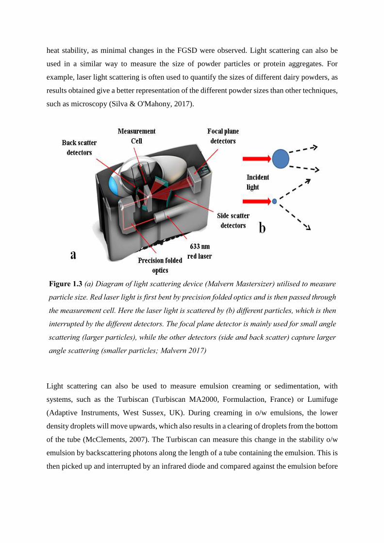
1.2.1 Light Scattering
The size of the dispersed droplets is a major factor in quantifying the stability of an emulsion
system. Droplet size is often evaluated using light-scattering techniques, which are related to
the intensity of light from a laser. Larger particles will scatter light at narrow angles, as opposed
to the smaller particles that scatter light at wider angles (Fig. 1.3b). This information is
processed by a laser diffraction instrument such as a Mastersizer (Malvern Instruments Ltd.,
Malvern, UK; Fig. 1.3a) and the range of particle sizes present in the sample is calculated
(Sprow, 1967). The most commonly utilized information from the Malvern is the D(4,3) value,
which is the volume-based mean diameter, and the particle size distribution. This average is
based on the volume of the dispersed phase instead of the number of particles. The effect of
this is to increase the sensitivity of larger particles (McClements, 2007).
Light scattering is often used to measure emulsion droplet sizes and can be used when
evaluating the response of emulsions to different treatments. In a study by Drapala et al. (2016),
sunflower oil emulsions with whey protein and maltodextrins were evaluated for heat stability.
Light scattering data showed changes in the fat globule size distribution (FGSD) after heat
treatments and demonstrated that whey proteins conjugated with maltodextrins had the best
heat stability, as minimal changes in the FGSD were observed. Light scattering can also be
used in a similar way to measure the size of powder particles or protein aggregates. For
example, laser light scattering is often used to quantify the sizes of different dairy powders, as
results obtained give a better representation of the different powder sizes than other techniques,
such as microscopy (Silva & O'Mahony, 2017).
Light scattering can also be used to measure emulsion creaming or sedimentation, with
systems, such as the Turbiscan (Turbiscan MA2000, Formulaction, France) or Lumifuge
(Adaptive Instruments, West Sussex, UK). During creaming in o/w emulsions, the lower
density droplets will move upwards, which also results in a clearing of droplets from the bottom
of the tube (McClements, 2007). The Turbiscan can measure this change in the stability o/w
emulsion by backscattering photons along the length of a tube containing the emulsion. This is
then picked up and interrupted by an infrared diode and compared against the emulsion before
Figure 1.3 (a) Diagram of light scattering device (Malvern Mastersizer) utilised to measure
particle size. Red laser light is first bent by precision folded optics and is then passed through
the measurement cell. Here the laser light is scattered by (b) different particles, which is then
interrupted by the different detectors. The focal plane detector is mainly used for small angle
scattering (larger particles), while the other detectors (side and back scatter) capture larger
angle scattering (smaller particles; Malvern 2017)
storage. A LUMiFuge functions in a similar way but uses centrifugation to accelerate the aging
process (McClements, 2015).
1.2.2 Zeta potential
Depending on the system, electrostatic charge (zeta potential, ζ) can also give an indication of
colloid stability. The ζ-potential is the most stable when the pH of the system is far from the
isoelectric point of the compounds present at the emulsion interface. Other factors such a
concentration of salts can also influence the ζ-potential and, thus, it important to establish the
electrostatic charge of the emulsifier (Drapala, 2016). ζ-potential information is also useful in
understanding the general behaviour of proteins and other dispersible compounds at various
pHs and conditions (Hunter, 2013).
To measure ζ-potential, an instrument such as the Zetasizer (Malvern Instruments Ltd.,
Malvern, UK) is used, which sends an electrical current through a diluted colloidal sample. The
electrical potential of the samples at the “shear plane” can give an indication of the
attraction/repulsion forces present on the dispersed droplet (McClements, 2007). Often this is
used, along with other techniques such as light scattering and microscopy, to gauge the
mechanism of emulsion destabilisation in a system.
For example, ζ-potential was utilised in a study evaluating the emulsion stability in a high salt
(CaCl2) system with lactoferrin and/or β-casein emulsions. ζ-potential decreased in emulsions
with a mixture of lactoferrin and β-casein or β-casein alone at the interface but no change was
observed with CaCl2 addition in emulsions with just lactoferrin. This information was
combined with light scattering data, which showed that emulsions made with lactoferrin or
both β-casein and lactoferrin had no significant change in particle size, while emulsions with
β-casein alone had a large increase in emulsion droplet size. From this information the authors
were able to conclude that lactoferrin can protect β-casein-stabilised emulsions from calcium-
induced flocculation, as it electrostatically interacts with the negative phosphates groups on β-
casein and can block calcium phosphate interactions (McCarthy, Kelly, O'Mahony, & Fenelon,
2014).
1.2.3 Confocal laser scanning microscopy
Confocal laser scanning microscopy (CLSM) offers a unique opportunity to visualise different
emulsion components, such as proteins, surfactants, and lipids. This is achieved through
specific fluorescent probes emitting different wavelengths of light from pre-labelled samples.
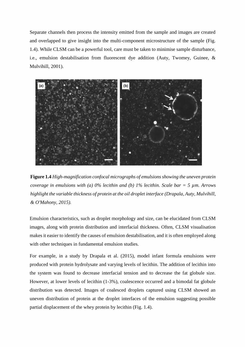
Separate channels then process the intensity emitted from the sample and images are created
and overlapped to give insight into the multi-component microstructure of the sample (Fig.
1.4). While CLSM can be a powerful tool, care must be taken to minimise sample disturbance,
i.e., emulsion destabilisation from fluorescent dye addition (Auty, Twomey, Guinee, &
Mulvihill, 2001).
Emulsion characteristics, such as droplet morphology and size, can be elucidated from CLSM
images, along with protein distribution and interfacial thickness. Often, CLSM visualisation
makes it easier to identify the causes of emulsion destabilisation, and it is often employed along
with other techniques in fundamental emulsion studies.
For example, in a study by Drapala et al. (2015), model infant formula emulsions were
produced with protein hydrolysate and varying levels of lecithin. The addition of lecithin into
the system was found to decrease interfacial tension and to decrease the fat globule size.
However, at lower levels of lecithin (1-3%), coalescence occurred and a bimodal fat globule
distribution was detected. Images of coalesced droplets captured using CLSM showed an
uneven distribution of protein at the droplet interfaces of the emulsion suggesting possible
partial displacement of the whey protein by lecithin (Fig. 1.4).
Figure 1.4 High-magnification confocal micrographs of emulsions showing the uneven protein
coverage in emulsions with (a) 0% lecithin and (b) 1% lecithin. Scale bar = 5 µm. Arrows
highlight the variable thickness of protein at the oil droplet interface (Drapala, Auty, Mulvihill,
& O'Mahony, 2015).
1.2.4 Cryogenic scanning electron microscopy
Cryogenic scanning electron microscopy (Cryo-SEM) is another useful technique for directly
visualising the microstructure of emulsions. As emulsions are typically liquid systems, the
sample is first rapidly frozen using liquid nitrogen. After freeze-fracturing, the sample is placed
under a vacuum, sublimated, and coated with a conductive metal such as platinum. The sample
can be imaged in the frozen hydrated state using a focused electron beam. During imaging,
primary electrons penetrate the sample and incident electron scattered from the sample are
interrupted by detectors as images.
Similar to CLSM, Cyro-SEM can be a helpful tool in visualising structure but expertise is
needed to minimise ice crystal development in high-moisture systems, such as o/w emulsions
(Auty, Twomey, Guinee, & Mulvihill, 2001; Kelly, 2015). Cryo-SEM is often useful for
visualising crystal development in emulsion systems, as this is difficult to see using CLSM. In
a study by Norton et al. (2009), cocoa-butter emulsion systems were successfully imaged using
Cyro-SEM, showed that in tempered emulsions a smooth layer of crystalline fat on the surface
aided in emulsion stability. Fat-based Pickering emulsions, that exploit crystalline fat for
interfacial stability, are often also characterised by Cryo-SEM (Frasch-Melnik, Norton, &
Spyropoulos, 2010; Frasch-Melnik, Spyropoulos, & Norton, 2010).
1.4 Interfacial tension
When emulsifiers or surfactants align and interact at an emulsion interface, a decrease in the
free energy of the system occurs. This decrease in free energy makes emulsion formation
possible and can be quantified through interfacial tension measurements (Kim & Burgess,
2001). Interfacial tension is commonly utilised to quantify the surface activity of a compound.
Traditionally performed with a Langmuir tray, interfacial tension measurements have become
possible with optical devices, du Noüy rings, and Wilhelmy plates. The Wilhelmy plate (Krüss
GmbH, Hamburg, Germany) is useful for emulsion systems as dynamic interfacial tension can
be recorded over time. Thus, it is possible to quantify how quickly a compound can move to
the interface, changes in interfacial tension over time, and if the compound is stable at the
interface (Drapala, 2016).
The Wilhelmy plate is a thin vertical plate, which first operates by making contact with the
heaviest (higher density) immiscible phase. The second phase (typically the lighter oil phase)
is then added to cover the plate and the plate begins to pull against the interface (Fig. 1.10).
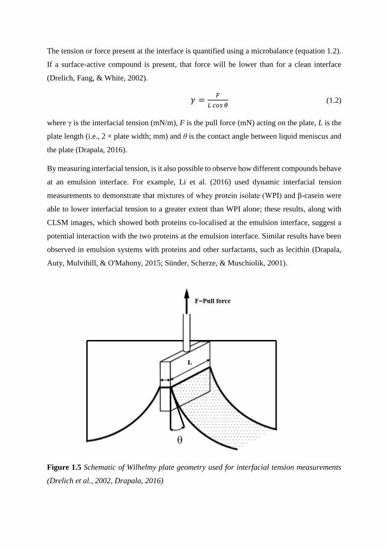
The tension or force present at the interface is quantified using a microbalance (equation 1.2).
If a surface-active compound is present, that force will be lower than for a clean interface
(Drelich, Fang, & White, 2002).
𝛾 =𝐹
𝐿 𝑐𝑜𝑠 𝜃 (1.2)
where γ is the interfacial tension (mN/m), F is the pull force (mN) acting on the plate, L is the
plate length (i.e., 2 × plate width; mm) and θ is the contact angle between liquid meniscus and
the plate (Drapala, 2016).
By measuring interfacial tension, is it also possible to observe how different compounds behave
at an emulsion interface. For example, Li et al. (2016) used dynamic interfacial tension
measurements to demonstrate that mixtures of whey protein isolate (WPI) and β-casein were
able to lower interfacial tension to a greater extent than WPI alone; these results, along with
CLSM images, which showed both proteins co-localised at the emulsion interface, suggest a
potential interaction with the two proteins at the emulsion interface. Similar results have been
observed in emulsion systems with proteins and other surfactants, such as lecithin (Drapala,
Auty, Mulvihill, & O'Mahony, 2015; Sünder, Scherze, & Muschiolik, 2001).
Figure 1.5 Schematic of Wilhelmy plate geometry used for interfacial tension measurements
(Drelich et al., 2002, Drapala, 2016)
References
Akoh, C. C., & Min, D. B. (2008). Food Lipids: Chemistry, Nutrition, and Biotechnology: CRC
press.
Auty, M. A. E., Twomey, M., Guinee, T. P., & Mulvihill, D. M. (2001). Development and
application of confocal scanning laser microscopy methods for studying the distribution
of fat and protein in selected dairy products. Journal of Dairy Research, 68(3), 417-
427.
Bos, M. A., & van Vliet, T. (2001). Interfacial rheological properties of adsorbed protein layers
and surfactants: a review. Advances in Colloid and Interface Science, 91(3), 437-471.
Damodaran, S., Parkin, K. L., & Fennema, O. R. (2007). Fennema's food chemistry. Boca
Raton, FL: CRC press.
Desrumaux, A., & Marcand, J. (2002). Formation of sunflower oil emulsions stabilized by
whey proteins with high-pressure homogenization (up to 350 MPa): effect of pressure
on emulsion characteristics. International Journal of Food Science & Technology,
37(3), 263-269.
Dickinson, E. (1999). Food Emulsions and Foams. Witney, UK: Elsevier Applied Science
Publishers Ltd.
Dickinson, E. (2003). Hydrocolloids at interfaces and the influence on the properties of
dispersed systems. Food hydrocolloids, 17(1), 25-39.
Dickinson, E. (2009). Hydrocolloids as emulsifiers and emulsion stabilizers. Food
hydrocolloids, 23(6), 1473-1482.
Diftis, N. G., Pirzas, T. A., & Kiosseoglou, V. D. (2005). Emulsifying properties of gelatin
conjugated to pectin under alkaline conditions. Journal of the Science of Food and
Agriculture, 85(5), 804-808.
Drapala, K. P. (2016). Processing and Stability of Infant Formula-Based Emulsions as Affected
by Emulsifier Type. University College Cork.
Drapala, K. P., Auty, M. A. E., Mulvihill, D. M., & O'Mahony, J. A. (2015). Influence of
lecithin on the processing stability of model whey protein hydrolysate‐based infant
formula emulsions. International Journal of Dairy Technology, 68(3), 322-333.
Drelich, J., Fang, C., & White, C. (2002). Measurement of interfacial tension in fluid-fluid
systems. Encyclopedia of Surface and Colloid Science, 3, 3158-3163.
Foegeding, E. A., & Davis, J. P. (2011). Food protein functionality: A comprehensive
approach. Food Hydrocolloids, 25(8), 1853-1864.
Frasch-Melnik, S., Norton, I. T., & Spyropoulos, F. (2010). Fat-crystal stabilised w/o
emulsions for controlled salt release. Journal of Food Engineering, 98(4), 437-442.
Frasch-Melnik, S., Spyropoulos, F., & Norton, I. T. (2010). W 1/O/W 2 double emulsions
stabilised by fat crystals–formulation, stability and salt release. Journal of Colloid and
Interface Science, 350(1), 178-185.
Garti, N., & Reichman, D. (1993). Hydrocolloids as food emulsifiers and stabilizers. Food
structure, 12(4), 3.
Griffin, W. C. (1954). Calculation of HLB values of non-ionic surfactants. Journal of Cosmetic
Science, 5, 249-256.
Hill, S. E. (1998). Functional Properties of Food Macromolecules. In S. E. L. Hill, D.A.;
Mitchell, J.R. (Ed.), Food Emulsions and Foams). Gaithersburg, MD: Aspen
Publishers.
Hunter, R. J. (2013). Zeta potential in colloid science: principles and applications (Vol. 2).
London, UK: Academic press.
Kelly, G. M. (2015). The effects of formulation and processing on surface characteristics and
functional properties of dairy powders. University College of Cork.
Kim, H., & Burgess, D. J. (2001). Prediction of interfacial tension between oil mixtures and
water. Journal of Colloid and Interface Science, 241(2), 509-513.
Malvern, I. (2017). The Masterclass 1: Laser Diffraction Explained. In Webinars).
McCarthy, N. A., Kelly, A. L., O'Mahony, J. A., & Fenelon, M. A. (2014). Sensitivity of
emulsions stabilised by bovine β-casein and lactoferrin to heat and CaCl 2. Food
Hydrocolloids, 35, 420-428.
McClements, D. J. (2004). Food emulsions: principles, practices, and techniques: CRC press.
McClements, D. J. (2004). Protein-stabilized emulsions. Current Opinion in Colloid &
Interface Science, 9(5), 305-313.
McClements, D. J. (2007). Critical review of techniques and methodologies for
characterization of emulsion stability. Critical Reviews in Food Science and Nutrition,
47(7), 611-649.
McClements, D. J. (2015). Food emulsions: principles, practices, and techniques. Boca Raton,
FL: CRC press.
O' Dwyer, S. (2012). Stabilisation of Omega-3 Oils in Food and Emulsion Systems University
of Limerick.
Palanuwech, J., & Coupland, J. N. (2003). Effect of surfactant type on the stability of oil-in-
water emulsions to dispersed phase crystallization. Colloids and Surfaces A:
Physicochemical and Engineering Aspects, 223(1), 251-262.
Qiu, C., Zhao, M., & McClements, D. J. (2015). Improving the stability of wheat protein-
stabilized emulsions: Effect of pectin and xanthan gum addition. Food Hydrocolloids,
43, 377-387.
Silva, J. V. C., & O'Mahony, J. A. (2017). Flowability and wetting behaviour of milk protein
ingredients as influenced by powder composition, particle size and microstructure.
International Journal of Dairy Technology, 70(2), 277-286.
Sprow, F. (1967). Distribution of drop sizes produced in turbulent liquid—liquid dispersion.
Chemical Engineering Science, 22(3), 435-442.
Sünder, A., Scherze, I., & Muschiolik, G. (2001). Physico-chemical characteristics of oil-in-
water emulsions based on whey protein–phospholipid mixtures. Colloids and Surfaces:
Biointerfaces, 21(1), 75-85.
Widlak, N., Hartel, R. W., & Narine, S. (2001). Crystallization and solidification properties of
lipids. Champaign, IL: The American Oil Chemists Society.