Embed Size (px)
Citation preview
5/14/2016
1
Digestive System
Introduction
• Every cell requires a constant energy source
– Ingested food is complex
• Modification is needed to utilize
Introduction
• Digestive system is a tube
– Gastrointestinal tract
• Specialized regions
– Mouth
– Pharynx
– Esophagus
– Stomach
– Small intestine
– Large intestine
Figure 23.1
Mouth (oral cavity)
Tongue
Esophagus
Liver
Gallbladder
Anus
DuodenumJejunumIleum
Small
intestine
Parotid glandSublingual glandSubmandibular
gland
Salivary
glands
Pharynx
Stomach
Pancreas
(Spleen)
Transverse colonDescending colonAscending colonCecumSigmoid colonRectumVermiform appendixAnal canal
Large
intestine
Introduction
• Digestive processes
1. Ingestion
2. Propulsion
3. Mechanical digestion
4. Chemical digestion
5. Absorption
6. Defecation
Figure 23.2
Food
Ingestion
Propulsion
Esophagus
Stomach
PharynxMechanicaldigestion
Chemicaldigestion
• Chewing (mouth)• Churning (stomach)• Segmentation(small intestine)
Smallintestine Largeintestine
Defecation Anus
Feces
Bloodvessel
Lymphvessel
Absorption
• Swallowing(oropharynx)
• Peristalsis(esophagus,stomach,small intestine,large intestine)
Mainly H2O
5/14/2016
2
Introduction
• Histology of the alimentary canal
– Four basic layers (tunics)
• Tunica mucosa – innermost layer
– Protection and absorption
– Epithelium and connective tissue
• Tunica submucosa
– Connective tissue
– Binds tube together
• Tunica muscularis (externa)
– Double layer of muscle
• Tunica serosa
– Single layer of epithelium and connective tissue
– Forms the visceral peritoneum
Figure 23.6
Glands in submucosa
Submucosa
Lumen
Mucosa-associated
lymphoid tissue
Duct of gland outside
alimentary canal
Gland in mucosa
NerveArteryVein
Lymphatic
vessel Mesentery
Intrinsic nerve plexuses
• Myenteric nerve plexus• Submucosal nerve plexus
Mucosa
• Epithelium• Lamina propria• Muscularis
mucosae
Muscularis
externa
• Longitudinal
muscle • Circular muscleSerosa
• Epithelium• Connective
tissue
Introduction
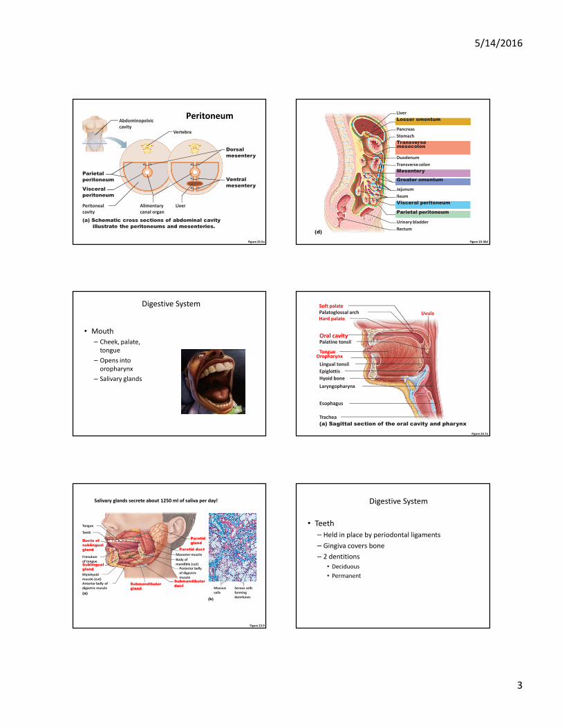
• Peritoneum
– Membrane that lines abdominal cavity
– Serous membrane – remember, that means 2
layers!
• Parietal peritoneum = outer layer (attached to
abdominal wall)
• Visceral peritoneum = inner layer (wrapped around
visceral organs)
• Space in between = peritoneal cavity
Introduction
• Mesentery
– Folds in the peritoneum
– Attached to intestinal tract
– Encapsulate blood vessels, nerves, fat stores that
supply the intestine
Introduction
• Omenta
– Folds in peritoneum
– Connect stomach to another organ
• Examples:
– Lesser omentum connects
stomach to liver
– Greater omentum connects
stomach to colon
Lesser
omentum
5/14/2016
3
Figure 23.5a
Peritoneal
cavity
Parietal
peritoneum
Visceral
peritoneum
Ventral
mesentery
Abdominopelvic
cavity
Dorsal
mesentery
Vertebra
Alimentary
canal organ
(a) Schematic cross sections of abdominal cavity
illustrate the peritoneums and mesenteries.
Liver
Peritoneum
Figure 23.30d
(d)
Pancreas
Liver
Lesser omentum
Stomach
Duodenum
Transversemesocolon
Greater omentum
Mesentery
Jejunum
Visceral peritoneum
Urinary bladder
Transverse colon
Ileum
Parietal peritoneum
Rectum
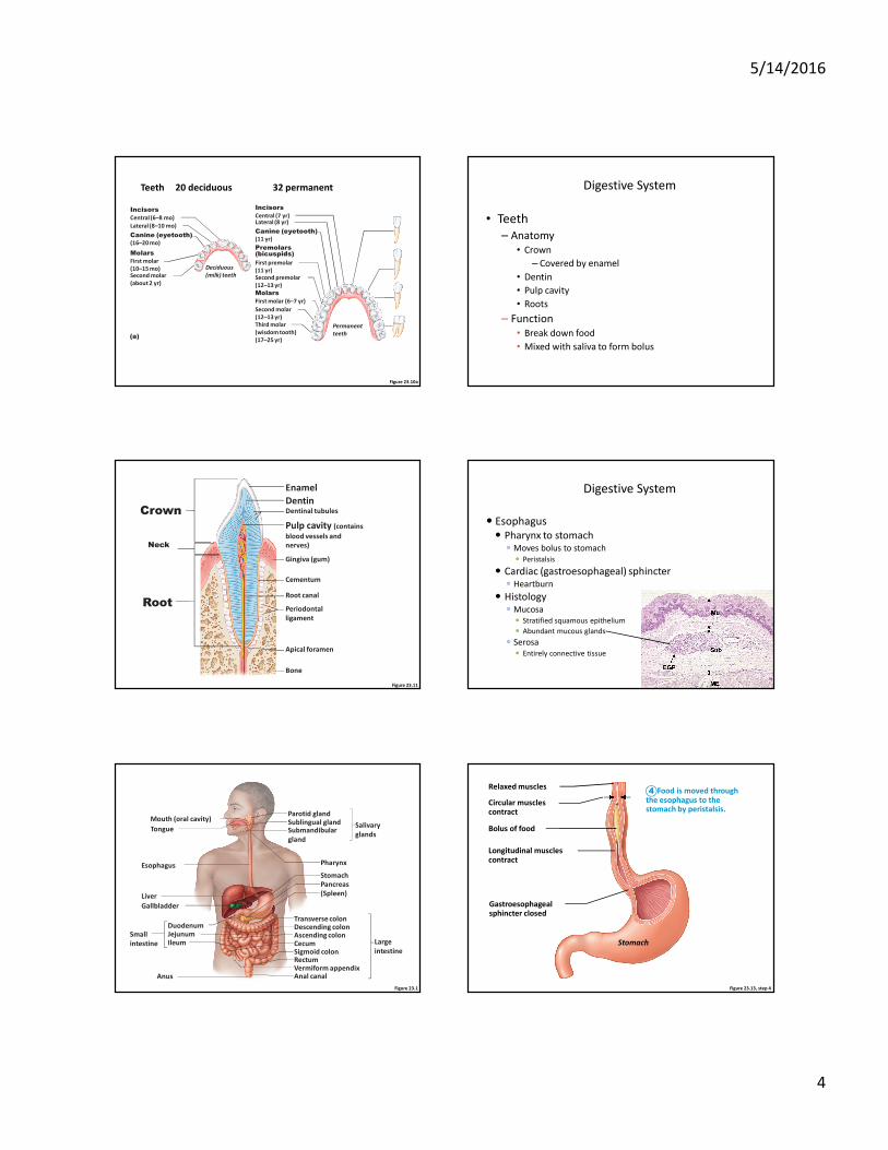
Digestive System
• Mouth
– Cheek, palate,
tongue
– Opens into
oropharynx
– Salivary glands
Figure 23.7a
Uvula
Soft palate
Palatoglossal arch
Palatine tonsil
Hard palate
Oral cavity
Tongue
Lingual tonsil
Oropharynx
Epiglottis
Hyoid bone
Laryngopharynx
Esophagus
Trachea
(a) Sagittal section of the oral cavity and pharynx
Figure 23.9
Teeth
Ducts of
sublingual
gland
Sublingual
gland
Submandibular
duct
Posterior belly
of digastric
muscle
Parotid duct
Masseter muscle
Body of
mandible (cut)
Parotid
gland
Tongue
Submandibular
gland
(a)
Frenulum
of tongue
Mylohyoid
muscle (cut)
Anterior belly of
digastric muscle Mucous
cells
(b)
Serous cells
forming
demilunes
Salivary glands secrete about 1250 ml of saliva per day! Digestive System
• Teeth
– Held in place by periodontal ligaments
– Gingiva covers bone
– 2 dentitions
• Deciduous
• Permanent
5/14/2016
4
Figure 23.10a
Incisors
Central (6–8 mo)
Incisors
Central (7 yr)
Canine (eyetooth)
(16–20 mo)
Canine (eyetooth)
(11 yr)
Premolars(bicuspids)
First premolar
(11 yr)
Molars
First molar
(10–15 mo)
Molars
First molar (6–7 yr)
Lateral (8–10 mo)Lateral (8 yr)
Second molar
(about 2 yr)
Second molar
(12–13 yr)
Third molar
(wisdom tooth)
(17–25 yr)(a)
Permanent
teeth
Deciduous
(milk) teeth Second premolar
(12–13 yr)
Teeth 20 deciduous 32 permanent Digestive System
• Teeth
– Anatomy
• Crown
– Covered by enamel
• Dentin
• Pulp cavity
• Roots
– Function
• Break down food
• Mixed with saliva to form bolus
Figure 23.11
Crown
Neck
Root
Enamel
DentinDentinal tubules
Pulp cavity (contains
blood vessels and
nerves)
Gingiva (gum)
Cementum
Root canal
Periodontal
ligament
Apical foramen
Bone
� Esophagus
� Pharynx to stomach� Moves bolus to stomach
� Peristalsis
� Cardiac (gastroesophageal) sphincter� Heartburn
� Histology� Mucosa
� Stratified squamous epithelium
� Abundant mucous glands
� Serosa
� Entirely connective tissue
Digestive System
Figure 23.1
Mouth (oral cavity)
Tongue
Esophagus
Liver
Gallbladder
Anus
DuodenumJejunumIleum
Small
intestine
Parotid glandSublingual glandSubmandibular
gland
Salivary
glands
Pharynx
Stomach
Pancreas
(Spleen)
Transverse colonDescending colonAscending colonCecumSigmoid colonRectumVermiform appendixAnal canal
Large
intestine
Figure 23.13, step 4
Relaxed muscles
Bolus of food
Stomach
Circular musclescontract
Longitudinal musclescontract
Gastroesophagealsphincter closed
Food is moved throughthe esophagus to thestomach by peristalsis.
4
5/14/2016
5
Figure 23.13, step 5
Relaxedmuscles
Gastroesophagealsphincter opens
The gastroesophagealsphincter opens, and foodenters the stomach.
5
Figure 23.12a
Mucosa(contains a stratifiedsquamous epithelium)
Submucosa (areolarconnective tissue)
Lumen
Muscularis externa
Adventitia (fibrousconnective tissue)
(a)
• Circular layer
• Longitudinal layer
Figure 23.12b
Mucosa
(contains a stratified
squamous epithelium)
(b)
Esophagus stomach junction
simple columnar
epithelium
Figure 23.14a
Cardia
Esophagus
Pyloric sphincter
(valve) at pylorus
Pyloric
canal
Pyloric
antrum
Rugae of
mucosa
Body
Lumen
Serosa
Fundus
Lesser
curvature
Greater
curvature
Muscularis
externa• Longitudinal layer• Circular layer• Oblique layer
(a)
Duodenum
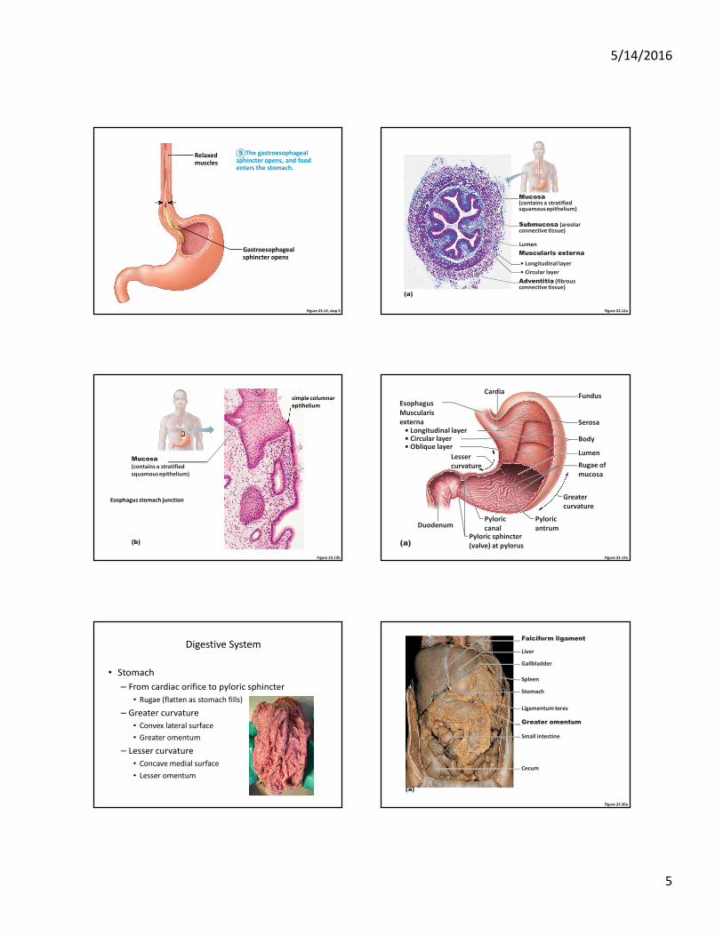
Digestive System
• Stomach
– From cardiac orifice to pyloric sphincter
• Rugae (flatten as stomach fills)
– Greater curvature
• Convex lateral surface
• Greater omentum
– Lesser curvature
• Concave medial surface
• Lesser omentum
Figure 23.30a
Falciform ligament
Liver
Gallbladder
Spleen
Stomach
Ligamentum teres
Greater omentum
Small intestine
Cecum
(a)
5/14/2016
6
Figure 23.30b
Liver
Lesser omentum
Gallbladder
Stomach
Duodenum
Transverse colon
Small intestine
Cecum
Urinary bladder
(b)
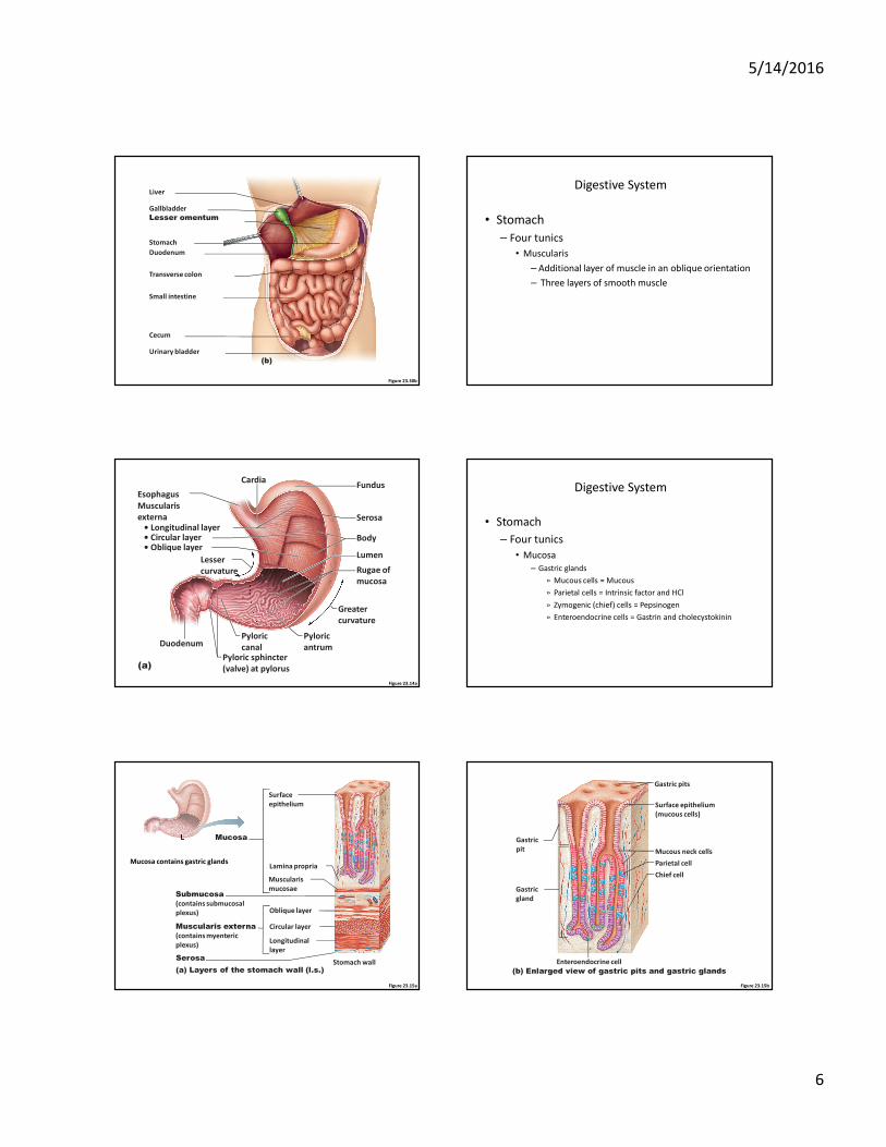
Digestive System
• Stomach
– Four tunics
• Muscularis
– Additional layer of muscle in an oblique orientation
– Three layers of smooth muscle
Figure 23.14a
Cardia
Esophagus
Pyloric sphincter
(valve) at pylorus
Pyloric
canal
Pyloric
antrum
Rugae of
mucosa
Body
Lumen
Serosa
Fundus
Lesser
curvature
Greater
curvature
Muscularis
externa• Longitudinal layer• Circular layer• Oblique layer
(a)
Duodenum
Digestive System
• Stomach
– Four tunics
• Mucosa
– Gastric glands
» Mucous cells = Mucous
» Parietal cells = Intrinsic factor and HCl
» Zymogenic (chief) cells = Pepsinogen
» Enteroendocrine cells = Gastrin and cholecystokinin
Figure 23.15a
Mucosa
Surface
epithelium
Lamina propria
Muscularis
mucosae
Oblique layer
Circular layer
Longitudinal
layerSerosa
(a) Layers of the stomach wall (l.s.)Stomach wall
Muscularis externa
(contains myenteric
plexus)
Submucosa
(contains submucosal
plexus)
Mucosa contains gastric glands
Figure 23.15b
(b) Enlarged view of gastric pits and gastric glands
Mucous neck cells
Parietal cell
Surface epithelium
(mucous cells)
Gastric pits
Chief cell
Enteroendocrine cell
Gastric
pit
Gastric
gland
5/14/2016
7
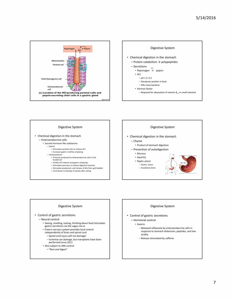
Figure 23.15c
(c) Location of the HCl-producing parietal cells and
pepsin-secreting chief cells in a gastric gland
Pepsinogen
Mitochondria
PepsinHCl
Chief (Zymogenic) cell
Enteroendocrine
cell
Parietal cell
• Chemical digestion in the stomach
– Protein catabolism → polypeptides
– Secretions
• Pepsinogen → pepsin
• HCl
– pH 1.5–3.5
– Denatures protein in food
– Kills many bacteria
• Intrinsic factor
– Required for absorption of vitamin B12 in small intestine
Digestive System
HCl
• Chemical digestion in the stomach
– Enteroendocrine cells
• Secrete hormone-like substances
– Gastrin
» Stimulates parietal cells to release HCl
» Increases gastric motility, emptying
– Cholecystokinin
» Primarily produced by enteroendocrine cells in the duodenum
» Inhibits HCl release and gastric emptying
» Stimulates pancreas to release digestive enzymes
» Stimulates production and release of bile from gall bladder
» Contributes to feelings of satiety after eating
Digestive System
• Chemical digestion in the stomach
– Chyme
• Product of stomach digestion
– Prevention of autodigestion
• Mucous
• Gastritis
• Peptic ulcers
– Gastric ulcers
– Duodenal ulcers
Digestive System
• Control of gastric secretions
– Neural control
• Seeing, smelling, tasting, thinking about food stimulates gastric secretions via the vagus nerve
• Enteric nervous system provides local control independently of brain and spinal cord
– Spinal cord injury will not damage!
– Ischemia can damage, but transplants have been performed since 2011
• Also subject to ANS control
– “Rest and digest”
Digestive System
• Control of gastric secretions
– Hormonal control
• Gastrin
– Released reflexively by enteroendocrine cells in
response to stomach distension, peptides, and low
acidity
– Release stimulated by caffeine
Digestive System
5/14/2016
8
� Control of gastric secretions
� Stimulatory and inhibitory events occur in three phases
1. Cephalic
2. Gastric
3. Intestinal
Digestive System
Figure 23.17
Presence of low
pH, partially digested
foods, fats, or
hypertonic solution
in duodenum when
stomach begins to
empty
Distension;
presence of
fatty, acidic,
partially
digested food
in the
duodenum
Brief
effect
Intestinal
(enteric)
gastrin
release
to blood
Entero-
gastric
reflex
Release of intestinal
hormones (secretin,
cholecystokinin, vasoactive
intestinal peptide)
Local
reflexes
Vagal
nuclei
in medulla
Pyloric
sphincter
Stimulate
Inhibit
1
1
2
Stomach
secretory
activity
Sight and thought
of food
Stomach
distension
activates
stretch
receptors
Stimulation of
taste and smell
receptors
Food chemicals
(especially peptides and
caffeine) and rising pH
activate chemoreceptors
Loss of
appetite,
depression
Emotional
upset
Lack of
stimulatory
impulses to
parasym-
pathetic
center
Cerebral
cortex
Cerebral cortex
Conditioned reflex
Vagovagal
reflexes
Local
reflexes
Medulla
G cells
Hypothalamus
and medulla
oblongata
Vagus
nerve
Vagus
nerve
Gastrin
release
to blood
Gastrin
secretion
declines
G cells
Overrides
parasym-
pathetic
controls
Sympathetic
nervous
system
activation
1
11
1
2
2
2
Stimulatory events Inhibitory events
Cephalic
phase
Gastric
phase
Intestinal
phase
Excessive
acidity
(pH <2)
in stomach
Distension
of duodenum;
presence of
fatty, acidic,
hypertonic
chyme, and/or
irritants in
the duodenum
� Control of gastric secretions
� Stimulatory and inhibitory events occur in three phases
1. Cephalic
� Hearing, seeing, smelling, tasting, thinking about food
� Vagus nerve stimulated
» Gastric secretion starts
Digestive System
• Control of gastric secretions
– Stimulatory and inhibitory events occur in three
phases
1.Cephalic
2.Gastric
• Arrival of food in stomach
– Stomach distension, peptides, low acidity →
gastrin released
» Relaxes pyloric sphincter
» Increases stomach motility
Digestive System
• Control of gastric secretions
– Stimulatory and inhibitory events occur in three
phases
1.Cephalic
2.Gastric
3.Intestinal
• Chyme reaches duodenum
– Intestinal distention → enterogastric reflex
– Release of secretin, CCK, VIP
» Inhibit stomach motility and delay emptying
Digestive System
Figure 23.17
Presence of low
pH, partially digested
foods, fats, or
hypertonic solution
in duodenum when
stomach begins to
empty
Distension;
presence of
fatty, acidic,
partially
digested food
in the
duodenum
Brief
effect
Intestinal
(enteric)
gastrin
release
to blood
Entero-
gastric
reflex
Release of intestinal
hormones (secretin,
cholecystokinin, vasoactive
intestinal peptide)
Local
reflexes
Vagal
nuclei
in medulla
Pyloric
sphincter
Stimulate
Inhibit
1
1
2
Stomach
secretory
activity
Sight and thought
of food
Stomach
distension
activates
stretch
receptors
Stimulation of
taste and smell
receptors
Food chemicals
(especially peptides and
caffeine) and rising pH
activate chemoreceptors
Loss of
appetite,
depression
Emotional
upset
Lack of
stimulatory
impulses to
parasym-
pathetic
center
Cerebral
cortex
Cerebral cortex
Conditioned reflex
Vagovagal
reflexes
Local
reflexes
Medulla
G cells
Hypothalamus
and medulla
oblongata
Vagus
nerve
Vagus
nerve
Gastrin
release
to blood
Gastrin
secretion
declines
G cells
Overrides
parasym-
pathetic
controls
Sympathetic
nervous
system
activation
1
11
1
2
2
2
Stimulatory events Inhibitory events
Cephalic
phase
Gastric
phase
Intestinal
phase
Excessive
acidity
(pH <2)
in stomach
Distension
of duodenum;
presence of
fatty, acidic,
hypertonic
chyme, and/or
irritants in
the duodenum
5/14/2016
9
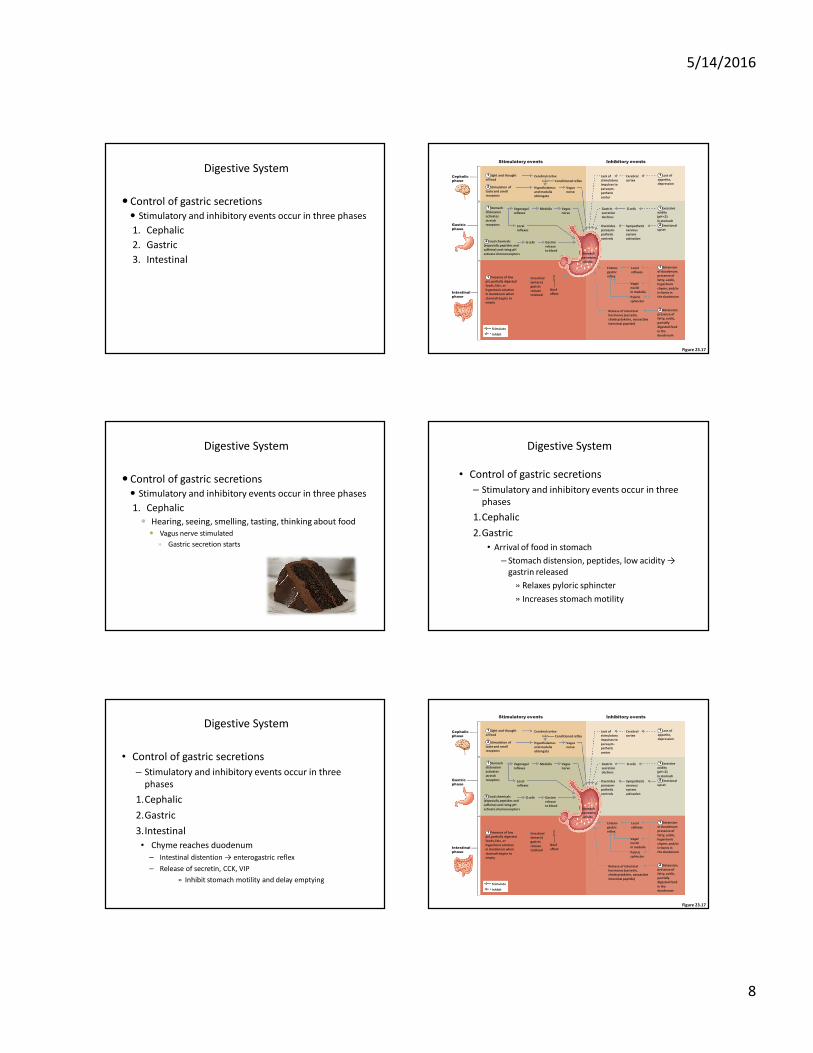
Figure 23.19
1 Propulsion: Peristaltic
waves move from the
fundus toward the
pylorus.
2 3Grinding: The most
vigorous peristalsis and
mixing action occur
close to the pylorus.
Retropulsion: The pyloric
end of the stomach acts as a
pump that delivers small
amounts of chyme into the
duodenum, simultaneously
forcing most of its contained
material backward into the
stomach.
Pyloric
valve
closed
Pyloric
valve
closed
Pyloric
valve
slightly
opened
Figure 23.20
Presence of fatty, hypertonic,
acidic chyme in duodenum
Duodenal entero-
endocrine cells
Chemoreceptors and
stretch receptors
Enterogastrones(secretin,
cholecystokinin,
vasoactive intestinal
peptide)
Duodenal
stimuli
decline
Via short
reflexes
Via long
reflexes
Enteric
neurons
Initial stimulus
Physiological response
Result
Contractile force and
rate of stomach
emptying decline
CNS centers
sympathetic
activity;
parasympathetic
activity
Stimulate
Inhibit
Secrete Target
Enterogastric Reflex
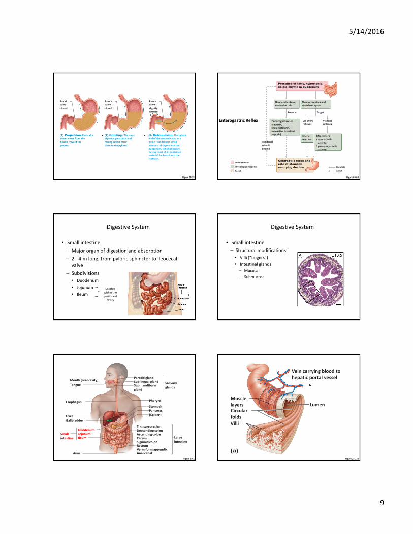
Digestive System
• Small intestine
– Major organ of digestion and absorption
– 2 - 4 m long; from pyloric sphincter to ileocecal
valve
– Subdivisions
• Duodenum
• Jejunum
• Ileum
Located
within the
peritoneal
cavity
Digestive System
• Small intestine
– Structural modifications
• Villi (“fingers”)
• Intestinal glands
– Mucosa
– Submucosa
Figure 23.1
Mouth (oral cavity)
Tongue
Esophagus
Liver
Gallbladder
Anus
DuodenumJejunumIleum
Small
intestine
Parotid glandSublingual glandSubmandibular
gland
Salivary
glands
Pharynx
Stomach
Pancreas
(Spleen)
Transverse colonDescending colonAscending colonCecumSigmoid colonRectumVermiform appendixAnal canal
Large
intestine
Figure 23.22a
Vein carrying blood to
hepatic portal vessel
Muscle
layersCircular
folds
Villi
(a)
Lumen
5/14/2016
10
Figure 23.22b
(b)
Absorptive cells
Lacteal
Intestinal crypt
Mucosa
associated
lymphoid tissue
Muscularis
mucosaeDuodenal gland Submucosa
Enteroendocrine
cellsVenule
Lymphatic vessel
Goblet cell
Blood
capillaries
Vilus
Microvilli
(brush border)
Figure 23.3b
(b)
Microvilli
Absorptive
cell
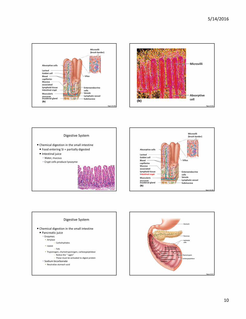
Digestive System
� Chemical digestion in the small intestine
� Food entering SI = partially digested
� Intestinal juice
� Water, mucous
� Crypt cells produce lysozyme
Figure 23.22b
(b)
Absorptive cells
Lacteal
Intestinal crypt
Mucosa
associated
lymphoid tissue
Muscularis
mucosaeDuodenal gland Submucosa
Enteroendocrine
cellsVenule
Lymphatic vessel
Goblet cell
Blood
capillaries
Villus
Microvilli
(brush border)
Digestive System
� Chemical digestion in the small intestine
� Pancreatic juice� Enzymes
� Amylase
» Carbohydrates
� Lipase
» Fats
� Trypsinogen, chymotrypsinogen, carboxypeptidase
» Notice the “-ogen”
» These must be activated to digest protein
� Sodium bicarbonate� Neutralize stomach acid
Figure 23.27
Stomach
Pancreas
Epithelial
cells
Trypsinogen
(inactive)
Chymotrypsinogen
(inactive)
Procarboxypeptidase
(inactive)
Trypsin
Chymotrypsin
Carboxypeptidase
Membrane-boundenteropeptidase
5/14/2016
11
Digestive System
• Chemical digestion in the small intestine
– Intestinal juice
• Alkaline, mucous rich watery secretion
• Lysosozymes – why are these defensive enzymes so important here?
– Brush border enzymes
• Enzymes for carbohydrates and proteins
– Pancreatic secretions
• Bicarbonate rich watery secretion
• Amylases, lipases, proteases, and nucleases
– Bile
• Bile salts emulsify lipids
Digestive System
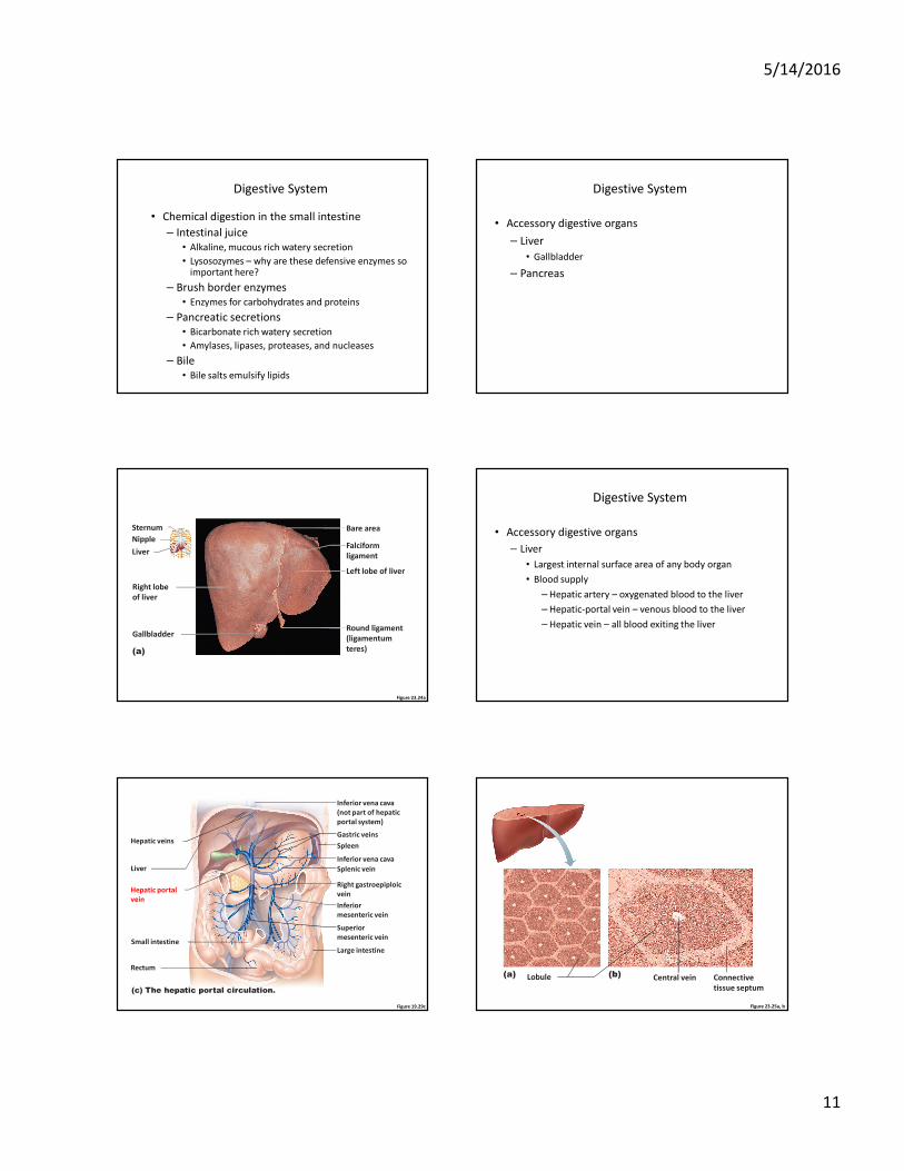
• Accessory digestive organs
– Liver
• Gallbladder
– Pancreas
Figure 23.24a
Sternum
Nipple
Liver
Right lobe
of liver
Gallbladder
(a)
Bare area
Falciform
ligament
Left lobe of liver
Round ligament
(ligamentum
teres)
Digestive System
• Accessory digestive organs
– Liver
• Largest internal surface area of any body organ
• Blood supply
– Hepatic artery – oxygenated blood to the liver
– Hepatic-portal vein – venous blood to the liver
– Hepatic vein – all blood exiting the liver
Figure 19.29c
(c) The hepatic portal circulation.
Hepatic veins
Liver
Spleen
Gastric veins
Inferior vena cava
Inferior vena cava
(not part of hepatic
portal system)
Splenic vein
Right gastroepiploic
vein
Inferior
mesenteric vein
Superior
mesenteric vein
Large intestine
Hepatic portal
vein
Small intestine
Rectum
Figure 23.25a, b
(a) (b)Lobule Central vein Connective
tissue septum
5/14/2016
12
Interlobular veins
(to hepatic vein) Central vein
Sinusoids
Portal triad
Plates of
hepatocytes
Portal vein
Fenestrated
lining (endothelial
cells) of sinusoids
Bile duct (receives
bile from bile
canaliculi)
Bile duct
Portal arteriole
Portal venuleHepatic
macrophages
in sinusoid walls
Bile canaliculi
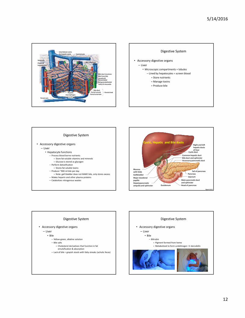
Digestive System
• Accessory digestive organs
– Liver
• Microscopic compartments = lobules
– Lined by hepatocytes = screen blood
» Store nutrients
» Manage toxins
» Produce bile
Digestive System
• Accessory digestive organs
– Liver
• Hepatocyte functions
– Process blood borne nutrients
» Store fat-soluble vitamins and minerals
» Glucose is stored as glycogen
– Perform detoxification
» Stores fat soluble toxins
– Produce ~900 ml bile per day
» Note: gall bladder does not MAKE bile, only stores excess
– Makes heparin and other plasma proteins
– Catabolizes nitrogenous wastes
Figure 23.21
Jejunum
Mucosa
with folds
Cystic duct
DuodenumHepatopancreatic
ampulla and sphincter
Gallbladder
Right and left
hepatic ducts
of liver
Bile duct and sphincter
Main pancreatic duct
and sphincter
Pancreas
Tail of pancreas
Head of pancreas
Common hepatic duct
Major duodenal
papilla
Accessory pancreatic duct
Cystic, Hepatic and Bile ducts
Digestive System
• Accessory digestive organs
– Liver
• Bile
– Yellow-green, alkaline solution
– Bile salts
» Cholesterol derivatives that function in fat
emulsification & absorption
– Lack of bile = grayish stools with fatty streaks (acholic feces)
Digestive System
• Accessory digestive organs
– Liver
• Bile
– Bilirubin
» Pigment formed from heme
» Metabolized to form urobilinogen → stercobilin
5/14/2016
13
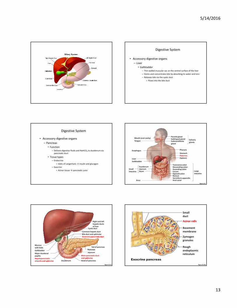
Digestive System
• Accessory digestive organs
– Liver
• Gallbladder
– Thin-walled muscular sac on the ventral surface of the liver
– Stores and concentrates bile by absorbing its water and ions
– Releases bile via the cystic duct
» Flows into the bile duct
Digestive System
• Accessory digestive organs
– Pancreas
• Function
– Delivers digestive fluids and NaHCO3 to duodenum via
pancreatic duct
• Tissue types
– Endocrine
» Islets of Langerhans → insulin and glucagon
– Exocrine
» Acinar tissue → pancreatic juice
Figure 23.1
Mouth (oral cavity)
Tongue
Esophagus
Liver
Gallbladder
Anus
DuodenumJejunumIleum
Small
intestine
Parotid glandSublingual glandSubmandibular
gland
Salivary
glands
Pharynx
Stomach
Pancreas
(Spleen)
Transverse colonDescending colonAscending colonCecumSigmoid colonRectumVermiform appendixAnal canal
Large
intestine
Figure 23.21
Jejunum
Mucosa
with folds
Cystic duct
DuodenumHepatopancreatic
ampulla and sphincter
Gallbladder
Right and left
hepatic ducts
of liver
Bile duct and sphincter
Main pancreatic duct
and sphincter
Pancreas
Tail of pancreas
Head of pancreas
Common hepatic duct
Major duodenal
papilla
Accessory pancreatic duct
Figure 23.26a
Small
duct
Acinar cells
Basement
membrane
Zymogen
granules
Rough
endoplasmic
reticulum
Exocrine pancreas
5/14/2016
14
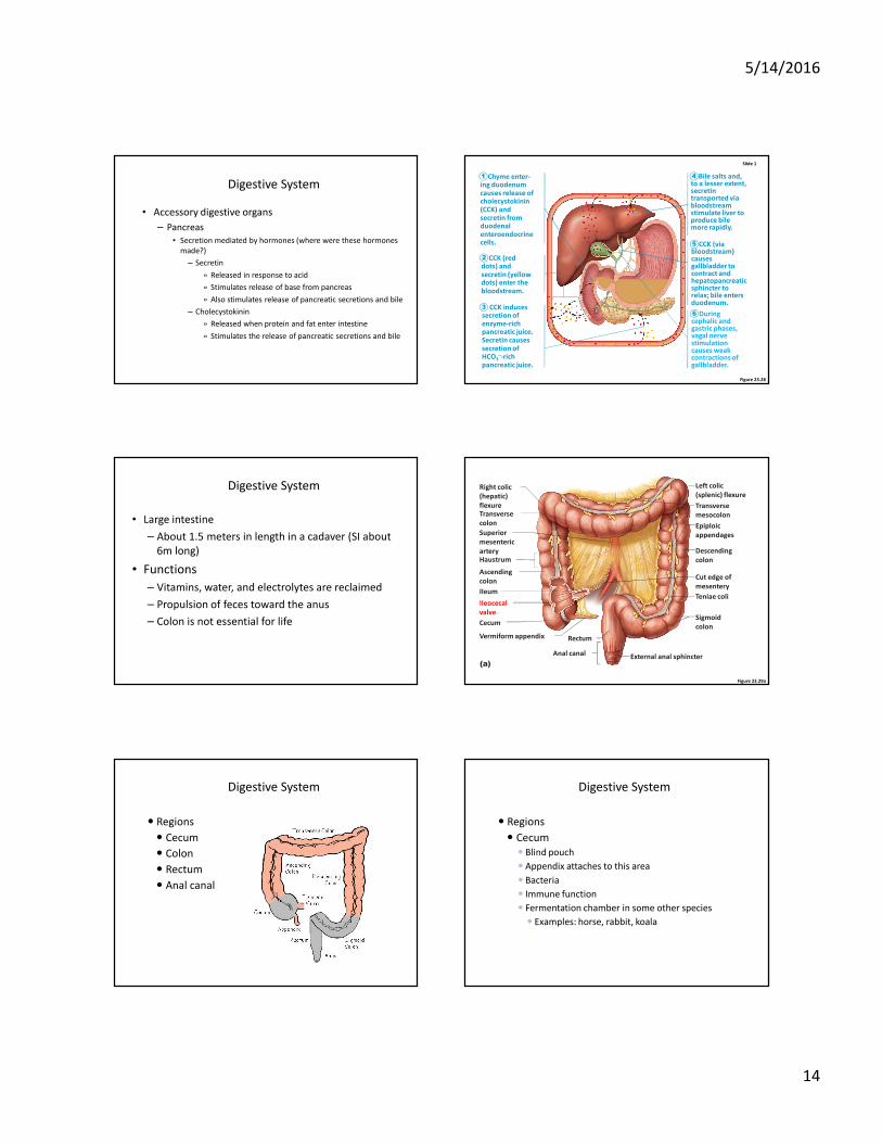
Digestive System
• Accessory digestive organs
– Pancreas
• Secretion mediated by hormones (where were these hormones
made?)
– Secretin
» Released in response to acid
» Stimulates release of base from pancreas
» Also stimulates release of pancreatic secretions and bile
– Cholecystokinin
» Released when protein and fat enter intestine
» Stimulates the release of pancreatic secretions and bile
Figure 23.28
Chyme enter-ing duodenum causes release of cholecystokinin (CCK) and secretin from duodenal enteroendocrine cells.
CCK (red dots) and secretin (yellow dots) enter the bloodstream.
CCK induces secretion of enzyme-rich pancreatic juice. Secretin causes secretion of HCO3
–-rich pancreatic juice.
Bile salts and, to a lesser extent, secretin transported via bloodstream stimulate liver to produce bile more rapidly.
CCK (via bloodstream) causes gallbladder to contract and hepatopancreatic sphincter to relax; bile enters duodenum.
During cephalic and gastric phases, vagal nerve stimulation causes weak contractions of gallbladder.
Slide 1
1
2
3
4
5
6
Digestive System
• Large intestine
– About 1.5 meters in length in a cadaver (SI about
6m long)
• Functions
– Vitamins, water, and electrolytes are reclaimed
– Propulsion of feces toward the anus
– Colon is not essential for life
Figure 23.29a
Left colic
(splenic) flexure
Transverse
mesocolon
Epiploic
appendages
Descending
colon
Teniae coli
Sigmoid
colon
Cut edge of
mesentery
External anal sphincter
Rectum
Anal canal
(a)
Right colic
(hepatic)
flexureTransverse
colon
Superior
mesenteric
arteryHaustrum
Ascending
colon
IIeum
IIeocecal
valve
Vermiform appendix
Cecum
Digestive System
� Regions
� Cecum
� Colon
� Rectum
� Anal canal
Digestive System
� Regions
� Cecum
� Blind pouch
� Appendix attaches to this area
� Bacteria
� Immune function
� Fermentation chamber in some other species
� Examples: horse, rabbit, koala
5/14/2016
15
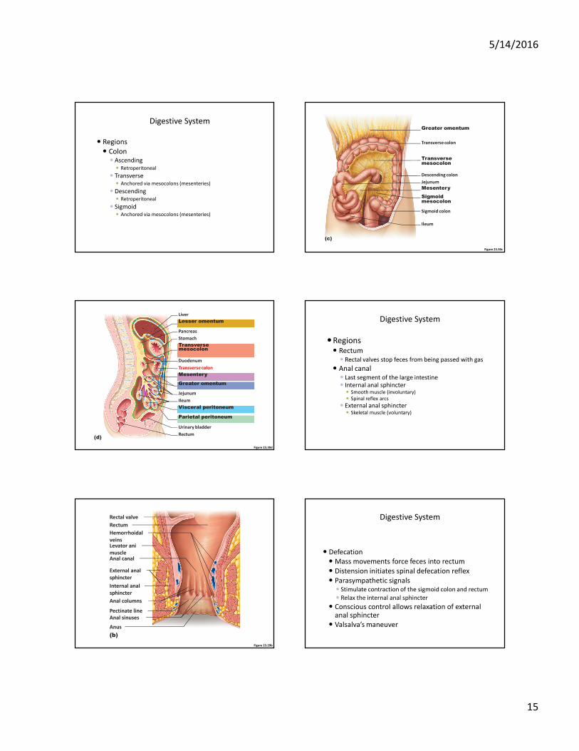
Digestive System
� Regions
� Colon� Ascending
� Retroperitoneal
� Transverse� Anchored via mesocolons (mesenteries)
� Descending� Retroperitoneal
� Sigmoid� Anchored via mesocolons (mesenteries)
Figure 23.30c
Transverse colon
Greater omentum
Descending colon
Jejunum
Mesentery
Transversemesocolon
Sigmoidmesocolon
Sigmoid colon
Ileum
(c)
Figure 23.30d
(d)
Pancreas
Liver
Lesser omentum
Stomach
Duodenum
Transversemesocolon
Greater omentum
Mesentery
Jejunum
Visceral peritoneum
Urinary bladder
Transverse colon
Ileum
Parietal peritoneum
Rectum
Digestive System
�Regions� Rectum
� Rectal valves stop feces from being passed with gas
� Anal canal� Last segment of the large intestine
� Internal anal sphincter� Smooth muscle (involuntary)
� Spinal reflex arcs
� External anal sphincter� Skeletal muscle (voluntary)
Figure 23.29b
(b)
Rectal valve
Rectum
Anal canal
Levator ani
muscle
Anus
Anal sinuses
Anal columns
Internal anal
sphincter
External anal
sphincter
Hemorrhoidal
veins
Pectinate line
Digestive System
� Defecation
� Mass movements force feces into rectum
� Distension initiates spinal defecation reflex
� Parasympathetic signals� Stimulate contraction of the sigmoid colon and rectum
� Relax the internal anal sphincter
� Conscious control allows relaxation of external anal sphincter
� Valsalva’s maneuver
5/14/2016
16
Figure 23.31
Impulses from
cerebral cortex
(conscious
control)
Voluntary motor
nerve to external
anal sphincter
External anal
sphincter
(skeletal muscle)
Internal anal sphincter
(smooth muscle)
Sensory
nerve fibers
Involuntary motor nerve
(parasympathetic division)
Stretch receptors in wall
Rectum
Sigmoid
colon
3
1
2
Distension, or stretch, of therectal walls due to movement of feces into the rectum stimulates stretch receptors there. The receptors transmit signals along afferent fibers to spinal cord neurons.
A spinal reflex is initiated in which parasympathetic motor (efferent) fibers stimulate contraction of the rectal walls and relaxation of the internal anal sphincter.
If it is convenient to defecate, voluntary motor
neurons are inhibited, allowing the external anal
sphincter to relax so that feces may pass.
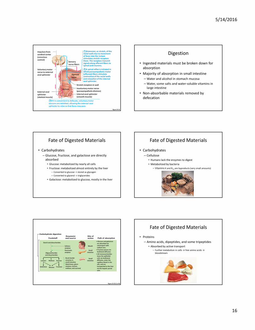
Digestion
• Ingested materials must be broken down for
absorption
• Majority of absorption in small intestine
– Water and alcohol in stomach mucosa
– Water, some salts and water-soluble vitamins in
large intestine
• Non-absorbable materials removed by
defecation
Fate of Digested Materials
• Carbohydrates
– Glucose, fructose, and galactose are directly
absorbed
• Glucose: metabolized by nearly all cells
• Fructose: metabolized almost entirely by the liver
– Converted to glucose -> stored as glycogen
– Converted to glycerol -> triglycerides
• Galactose: metabolized to glucose, mostly in the liver
Fate of Digested Materials
• Carbohydrates
– Cellulose
• Humans lack the enzymes to digest
• Metabolized by bacteria
– Vitamins K and B12 are byproducts (very small amounts)
Figure 23.32 (1 of 4)
Carbohydrate digestion
• Glucose and galactose
are absorbed via
cotransport with
sodium ions.
• Fructose passes via
facilitated diffusion.
• All monosaccharides
leave the epithelial
cells via facilitated
diffusion, enter the
capillary blood in the
villi, and are
transported to the liver
via the hepatic portal
vein.
Starch and disaccharides
Oligosaccharides
and disaccharides
Lactose Maltose Sucrose
Glucose Fructose
Salivary
amylase
Mouth
Pancreatic
amylase
Brush border
enzymes in
small intestine
(dextrinase, gluco-
amylase, lactase,
maltase, and sucrase)
Small
intestine
Small
intestine
Foodstuff
Galactose
Path of absorptionEnzyme(s)
and source
Site of
action
Fate of Digested Materials
• Proteins
– Amino acids, dipeptides, and some tripeptides
• Absorbed by active transport
– Further metabolism in cells → free amino acids →
bloodstream
5/14/2016
17
Figure 23.32 (2 of 4)
Protein digestion
• Amino acids are absorbed
by cotransport with
sodium ions.
• Some dipeptides and
tripeptides are absorbed
via cotransport with H+
and hydrolyzed to amino
acids within the cells.
+
• Amino acids leave the
epithelial cells by
facilitated diffusion, enter
the capillary blood in the
villi, and are transported
to the liver via the hepatic
portal vein.
Small
intestine
Small
intestine
Stomach
Foodstuff
Protein
Large polypeptides
Pepsin
(stomach glands)
in presence
of HCl
Small polypeptides,
small peptides
Pancreatic
enzymes
(trypsin, chymotrypsin,
carboxypeptidase)
Amino acids
(some dipeptides
and tripeptides)
Brush border
enzymes
(aminopeptidase,
carboxypeptidase,
and dipeptidase)
Path of absorptionEnzyme(s)
and source
Site of
action
Figure 23.33
Absorptive
epithelial
cell
Apical membrane (microvilli)
Amino
acid
carrier
Capillary
Lumen of
intestine
Pancreatic
proteases
Amino acids of protein fragments
Brush border enzymes
Na+
Na+
1 Proteins and protein fragments
are digested to amino acids by
pancreatic proteases (trypsin,
chymotrypsin, and carboxy-
peptidase), and by brush border
enzymes (carboxypeptidase,
aminopeptidase, and dipeptidase)
of mucosal cells.
2 The amino acids are then
absorbed by active transport into
the absorptive cells, and move to
their opposite side (transcytosis).
3 The amino acids leave the
villus epithelial cell by facilitated
diffusion and enter the capillary
via intercellular clefts.
Active transport
Passive transport
Fate of Digested Materials
• Lipids
Emuslified by bile salts and digested by lipase
into monoglycerides and FFAs
Micelles formed (lipid and bile salts) and move between microvilli
Lipids diffuse into intestinal epithelium
(bile salts later reabsorbed in ileum)
Fate of Digested Materials
• Lipids
– Within intestinal cells
Triglycerides are formed
Combined with proteins and cholesterol in the cell
Chylomicrons
Enter lymphatics through lacteal
Enter blood vascular system
Figure 23.34
Epithelial
cells of
small
intestine
Fat droplets
coated with
bile salts
Fat globule
Lacteal
Bile salts
Micelles made up of fatty
acids, monoglycerides,
and bile salts
1 Large fat globules are emulsified
(physically broken up into smaller fat
droplets) by bile salts in the duodenum.
2 Digestion of fat by the pancreatic
enzyme lipase yields free fatty acids and
monoglycerides. These then associate
with bile salts to form micelles which
“ferry” them to the intestinal mucosa.
3 Fatty acids and monoglycerides leave
micelles and diffuse into epithelial cells.
There they are recombined and packaged
with other lipoid substances and proteins
to form chylomicrons.
4 Chylomicrons are extruded from the
epithelial cells by exocytosis. The
chylomicrons enter lacteals. They are
carried away from the intestine by lymph.
Fate of Digested Materials
• Lipids
Plasma enzymes generate FFAs and glycerol
Pass thru capillary wall to serve tissues
The remaining protein-cholesterol combo returns to liver
Additional proteins added
HDL and LDL created (carriers for lipids)
5/14/2016
18
Figure 23.32 (3 of 4)
Fat digestion
Small
intestine
Small
intestine
Foodstuff
Unemulsified
fats
Emulsification by
the detergent
action of bile
salts ducted
in from the liver
Pancreatic
lipases
Monoglycerides
and fatty acids
Glycerol
and
fatty acids
Path of absorptionEnzyme(s)
and source
Site of
action
• Fatty acids and monoglycerides enter the intestinal cells via diffusion.
• Fatty acids and monoglycerides are recombined to form triglycerides and then combined with other lipids and proteins within the cells, and the resulting chylomicrons are extruded by exocytosis.
• The chylomicrons enter the lacteals of the villi and are transported to the systemic circulation via the lymph in the thoracic duct.
• Some short-chain fatty acids are absorbed, move into the capillary blood in the villi by diffusion, and are transported to the liver via the hepatic portal vein.
Fate of Digested Materials
• Water
– After digested nutrients removed, large volumes
of salt and water remain in LI
– Active Na+ uptake → passive Cl- and water uptake
• Undigested materials (cellulose) cause water to be
retained in LI
• Antibiotics may kill bacteria → digestion impaired
Disorders of the Digestive System
• Colon cancer
– Second most common cause of cancer death in
U.S. in men
• 98,000 new cases annually
• 48,000 deaths/year
– Diagnosis
• Colonoscopy
– Fiberoptic endoscope
– Polyps often occur before tumor
Disorders of the Digestive System
• Colon cancer
– Contributing factors
• Contact time with carcinogenic material in colon
– Diets high in animal material = slowed motility
• P53 (tumor suppressor gene) mutation
• Hereditary component
Disorders of the Digestive System
• Gallstones
– A.K.A. cholelithiasis
– Bile salts precipitate
• Block bile ducts
• Jaundice due to bilirubin
– Treatment
• Lithotripsy
• Medications
• Cholecystectomy
Disorders of the Digestive System
• Celiac disease
– Autoimmune disease
– Immune system destroys intestinal villi in
response to gluten
– No villi = no nutrient absorption


![Presentation2 - Linn–Benton Community Collegecf.linnbenton.edu/mathsci/bio/waitea/upload/... · Microsoft PowerPoint - Presentation2 [Compatibility Mode] Author: U0076978 Created](https://img.pdfslide.us/doc/110x75/5ec8bc059aa0e7580969d92f/presentation2-linnabenton-community-microsoft-powerpoint-presentation2-compatibility.jpg)


![Presentation1 - Linn–Benton Community Collegecf.linnbenton.edu/mathsci/bio/waitea/upload/Lecture_01_Neurons.pdfMicrosoft PowerPoint - Presentation1 [Compatibility Mode] Author: U0076978](https://img.pdfslide.us/doc/110x75/5f0eb6cd7e708231d44093df/presentation1-linnabenton-community-microsoft-powerpoint-presentation1-compatibility.jpg)