Embed Size (px)
Citation preview

`
The microstructure of muscle
Muscle Terminology
• myofiber (muscle fiber)- a single muscle cell
• sarcolemma- muscle cell membrane
• sarcoplasm- muscle cell cytoplasm
• myofibril- long contractile protein structure– actin and myosin
• sarcomere- the contractile unit between two z-lines
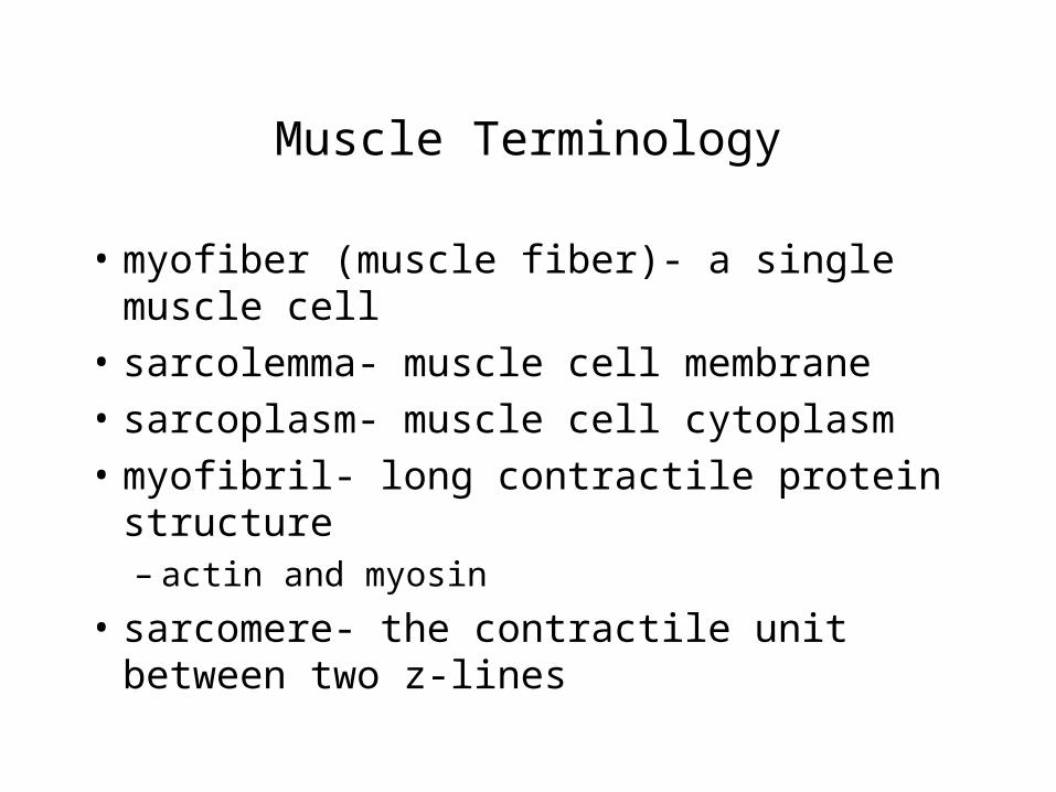
Connective tissue surrounding muscle
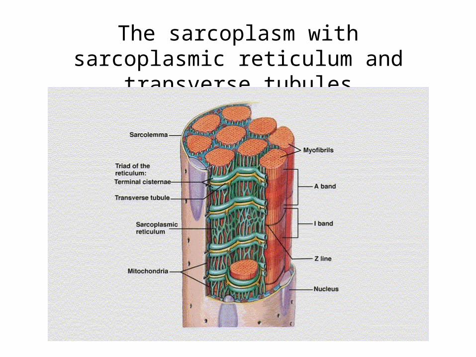
The sarcoplasm with sarcoplasmic reticulum and transverse tubules
More Terms
• sarcoplasmic reticulum- storage and release site of calcium
• transverse tubule- also involved in calcium flux

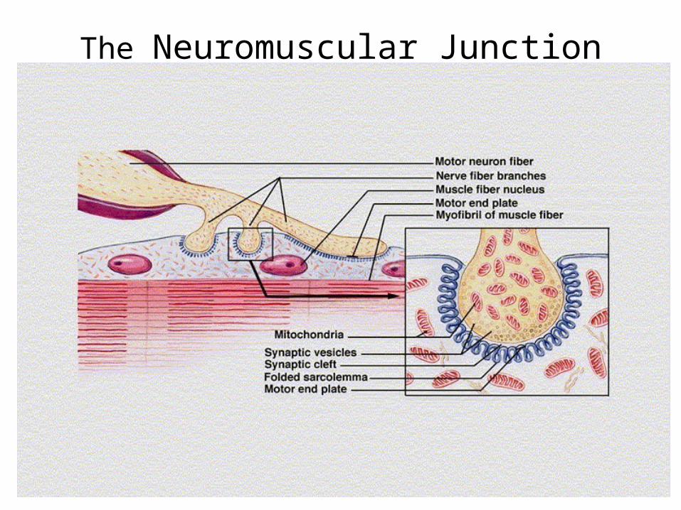
The Neuromuscular Junction
Neuromuscular Junction
1) Impulse travels down motor neuron
2) at end of neuron, acetylcholine released
3) Acetylcholine diffuses across synaptic cleft
4) acetylcholine binds to receptors on sarcolemma
causing permeabilty
5) sodium enters cell causing depolarization and muscle contraction
Muscular Contraction
• functions to produce force for locomotion
• force for breathing
• force for postural support
• heat production in cold (no force)
How do skeletal muscles contract?
• Sliding filament model of contraction
• the interaction of actin and myosin
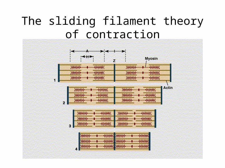
The sliding filament theory of contraction
Sliding Filament Animation
• -- sliding filament animation.htm
•http://intro.bio.umb.edu/111-112/112s99Lect/muscle/contract.html
How do the Actin and Myosin Interact?
• The myosin head binds to the actin filament in a weak state initially (or unbound)
• the signal to contract initiates a strong binding state– Binding of calcium to troponin regulates this
strong-weak state
The Contraction Itself
• during the strong binding the myosin pulls the actin past
• this effectively shortens or contracts the muscle
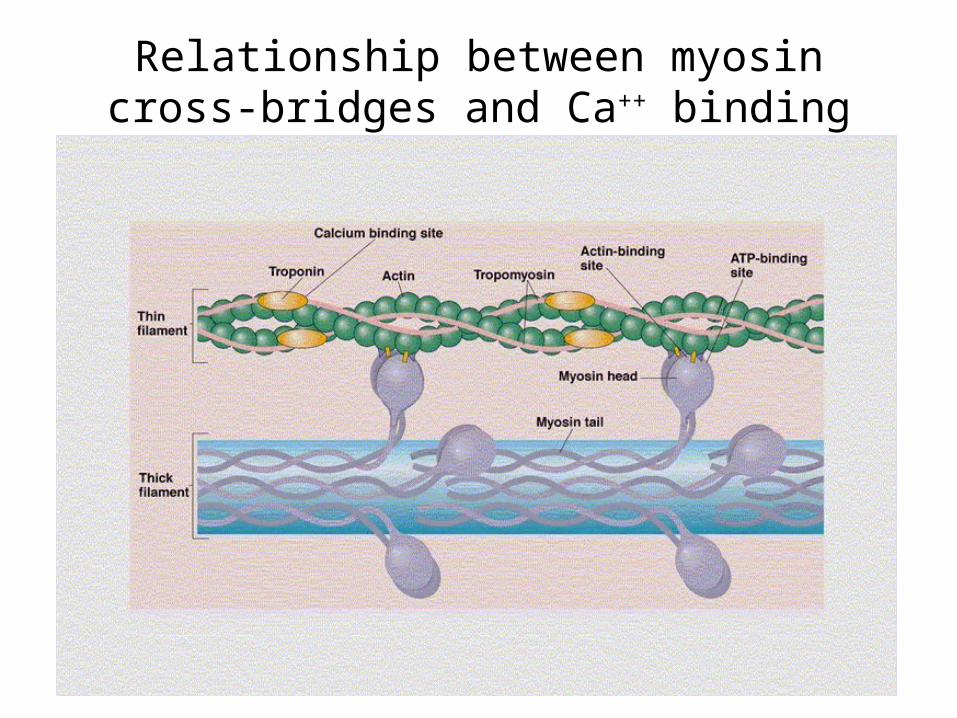
Relationship between myosin cross-bridges and Ca++ binding
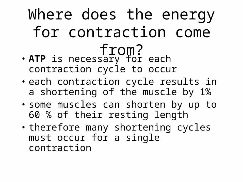
Where does the energy for contraction come from?
• ATP is necessary for each contraction cycle to occur
• each contraction cycle results in a shortening of the muscle by 1%
• some muscles can shorten by up to 60 % of their resting length
• therefore many shortening cycles must occur for a single contraction
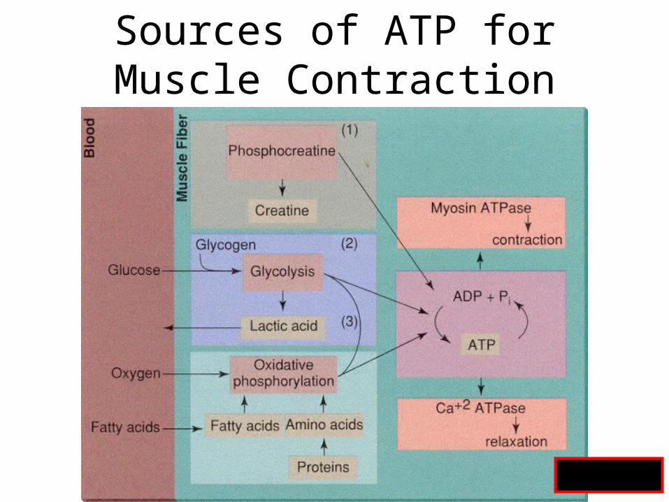
Sources of ATP for Muscle Contraction
Fig 8.7
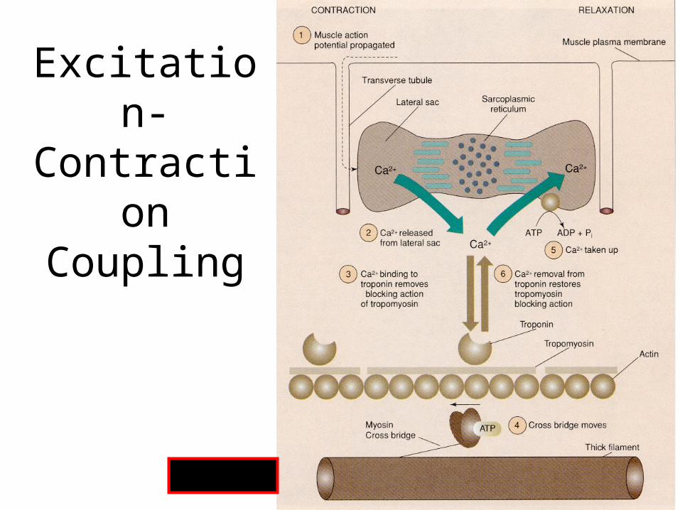
Excitation-Contraction Coupling
Fig 8.9
Crossbridge Animation
• Quicktime - Actin Myosin Crossbridge 3D Animation.htm
•http://www.sci.sdsu.edu/movies/actin_myosin.html
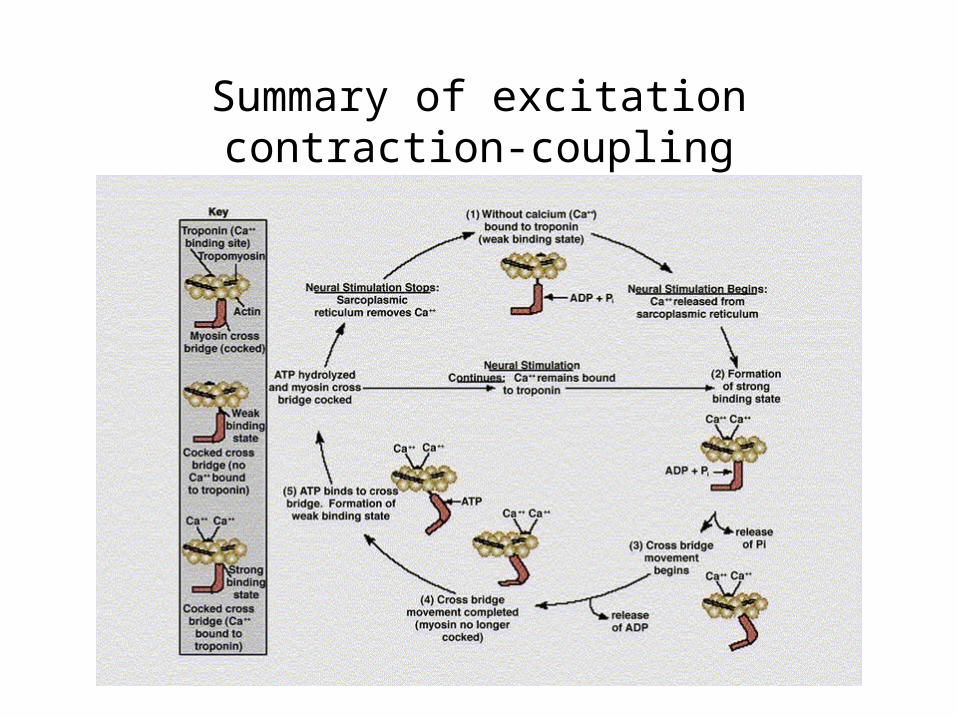
Summary of excitation contraction-coupling
Steps in Excitation - Contraction coupling
• at rest actin and myosin are weakly bound (or unbound)
• an excitation impulse from the a motor nerve causes an end-plate potential
• the potential depolarizes the muscle cell beginning at the sarcolemma
The Neuromuscular Junction
Excitation- Contraction cont’d
• depolarization travels down the T-tubules to the sarcoplasmic reticulum
• the impulse reaches the SR and calcium is released
• calcium binds to troponin and causes the strong binding state
Excitation- Contraction (one more)
• during strong binding, myosin head cocks
• this action moves actin filament along myosin
• Binding of ATP causes the weak binding (or release) again enabling another contraction

Summary of excitation contraction-coupling
Important Points
• depolarization causes release of calcium by SR
• calcium enables the strong binding state
• ATP provides energy for cocking of myosin head, BUT
• binding of ATP causes the weak binding state (or release) of actin and myosin
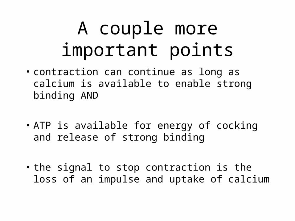
A couple more important points
• contraction can continue as long as calcium is available to enable strong binding AND
• ATP is available for energy of cocking and release of strong binding
• the signal to stop contraction is the loss of an impulse and uptake of calcium
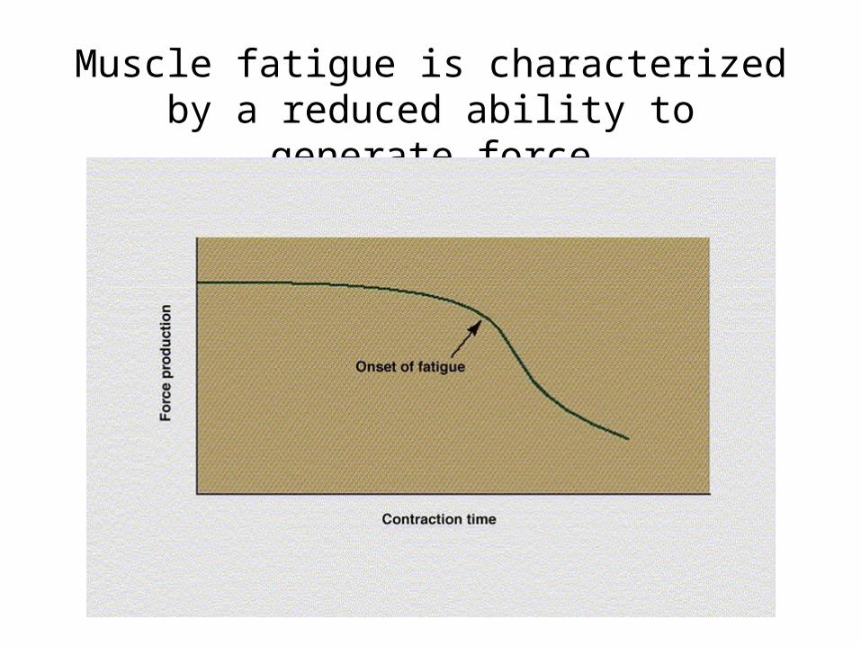
Muscle fatigue is characterized by a reduced ability to generate force
Properties of Muscle Fiber Types
• Biochemical properties– Oxidative capacity– Type of ATPase
• Contractile properties– Maximal force production– Speed of contraction– Muscle fiber efficiency
Individual Fiber TypesFast fibers• Type IIx fibers
– Fast-twitch fibers
– Fast-glycolytic fibers
• Type IIa fibers– Intermediate fibers
– Fast-oxidative glycolytic fibers
Slow fibers
• Type I fibers– Slow-twitch fibers– Slow-oxidative
fibers
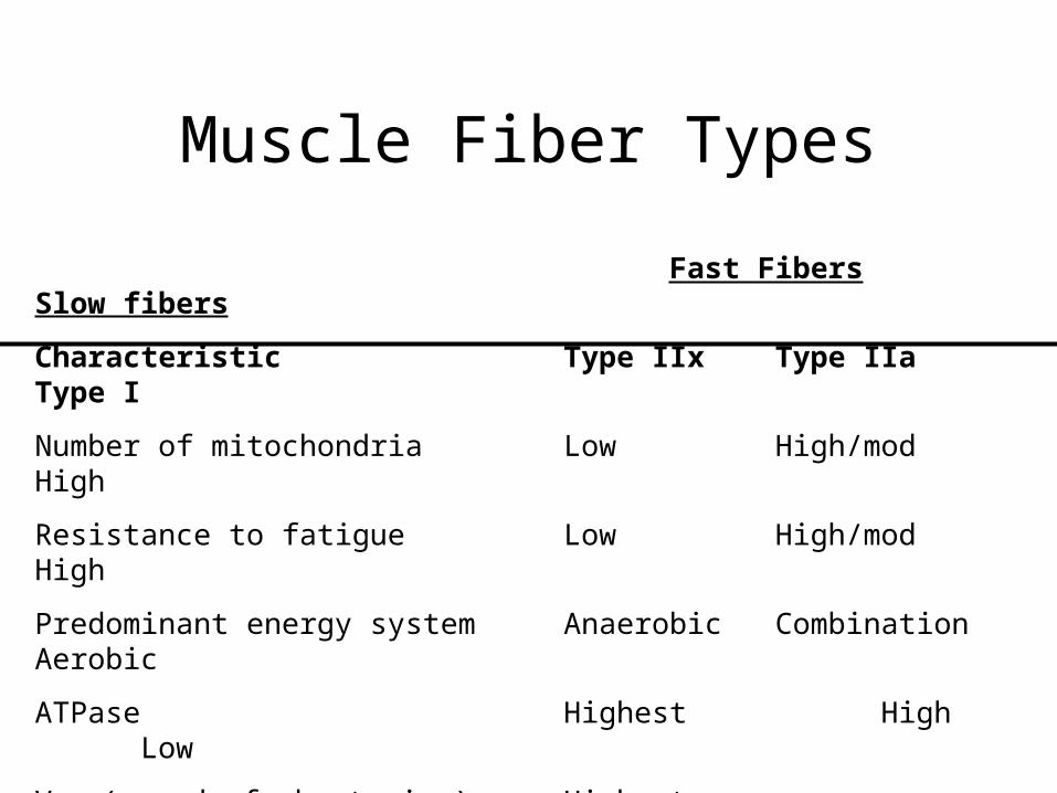
Muscle Fiber Types
Fast Fibers Slow fibers
Characteristic Type IIx Type IIa Type I
Number of mitochondria Low High/mod High
Resistance to fatigue Low High/mod High
Predominant energy system Anaerobic Combination Aerobic
ATPase Highest High Low
Vmax (speed of shortening) Highest Intermediate Low
Efficiency Low Moderate High
Specific tension High High Moderate
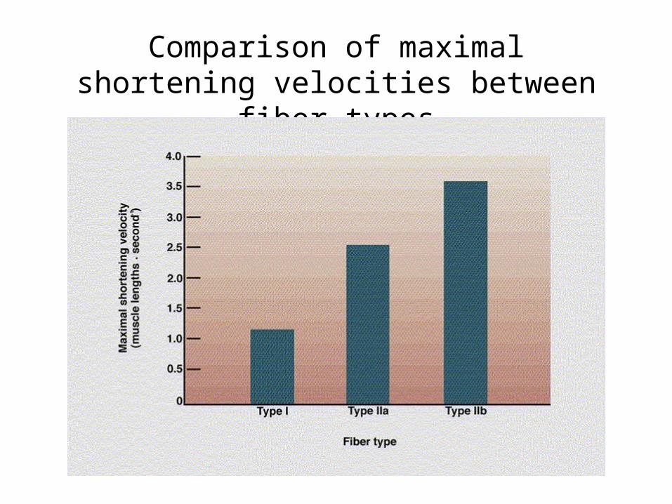
Comparison of maximal shortening velocities between fiber types
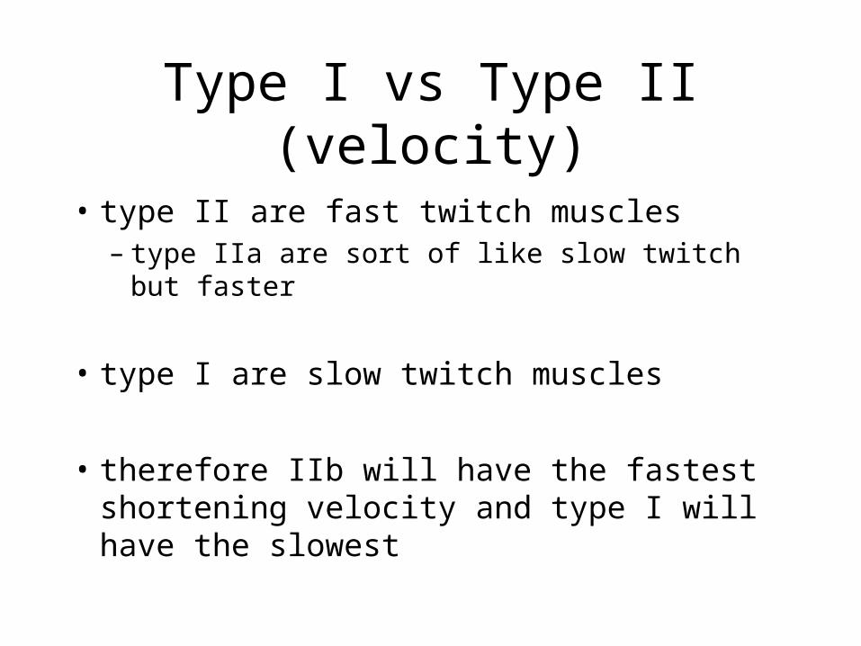
Type I vs Type II (velocity)
• type II are fast twitch muscles– type IIa are sort of like slow twitch but faster
• type I are slow twitch muscles
• therefore IIb will have the fastest shortening velocity and type I will have the slowest
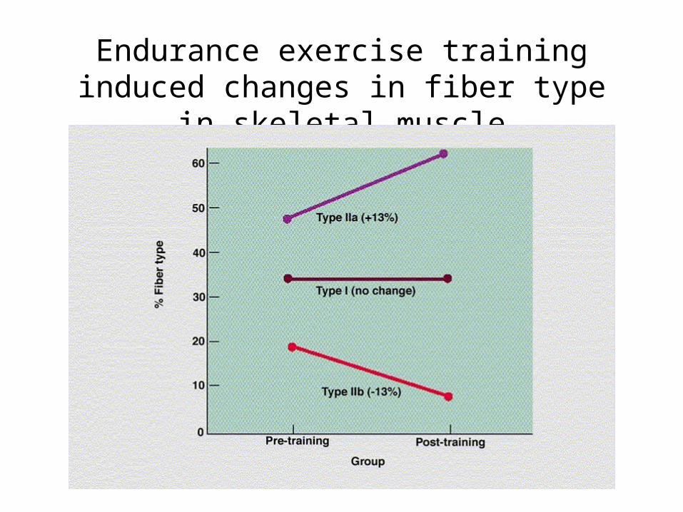
Endurance exercise training induced changes in fiber type in skeletal muscle
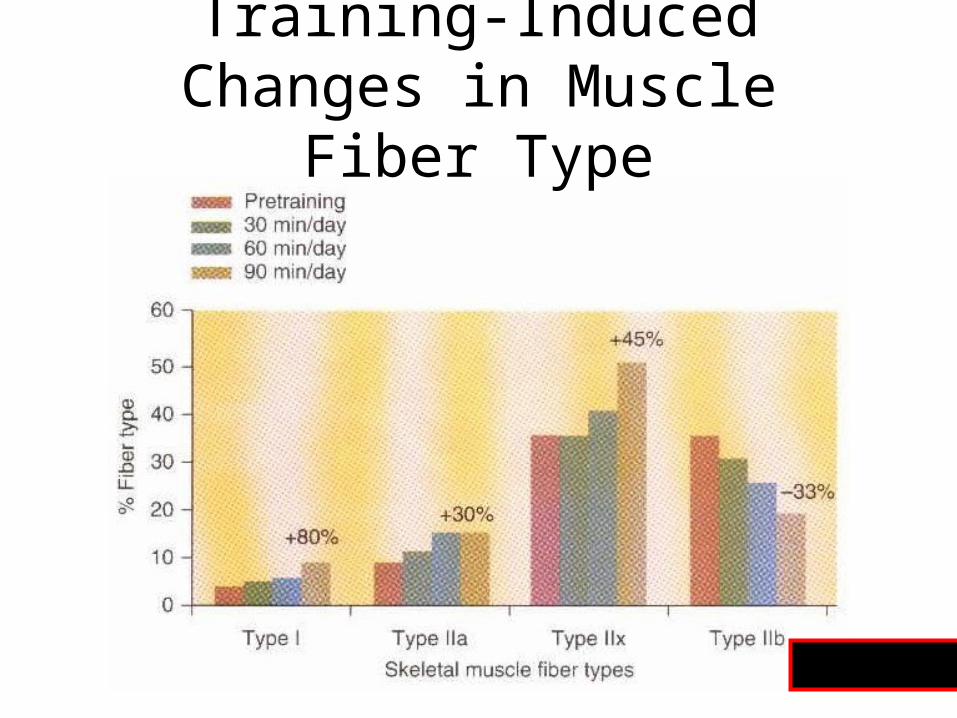
Training-Induced Changes in Muscle Fiber Type
Fig 8.13
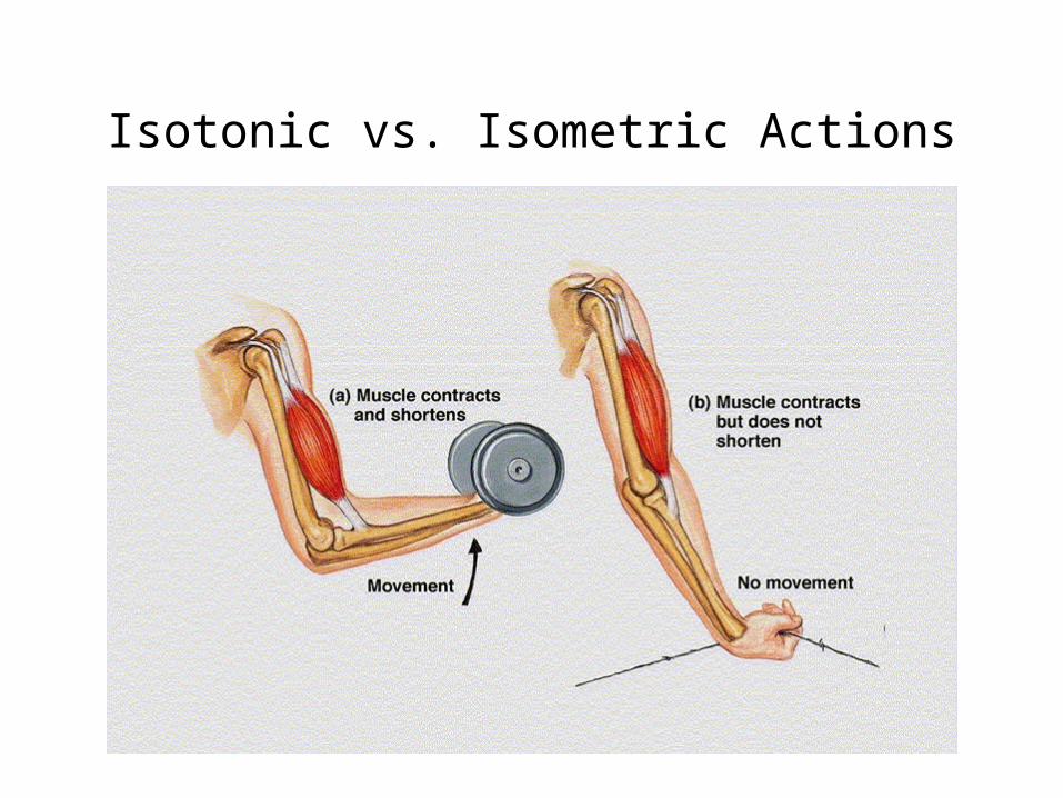
Isotonic vs. Isometric Actions
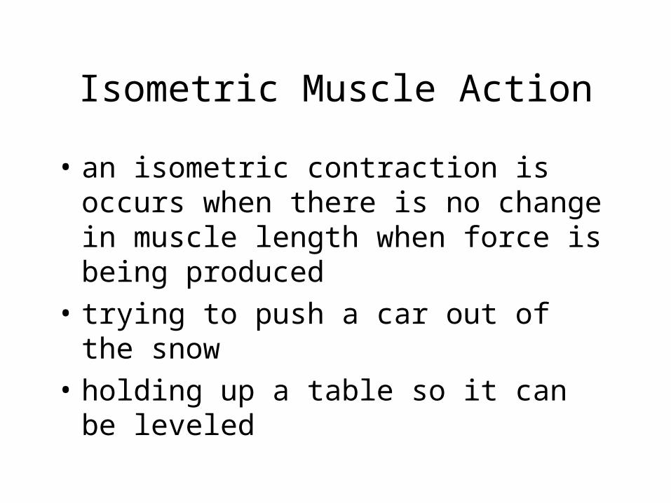
Isometric Muscle Action
• an isometric contraction is occurs when there is no change in muscle length when force is being produced
• trying to push a car out of the snow
• holding up a table so it can be leveled
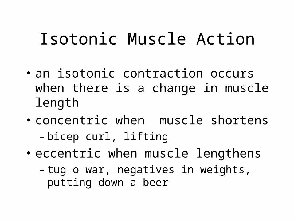
Isotonic Muscle Action
• an isotonic contraction occurs when there is a change in muscle length
• concentric when muscle shortens– bicep curl, lifting
• eccentric when muscle lengthens– tug o war, negatives in weights, putting down a
beer
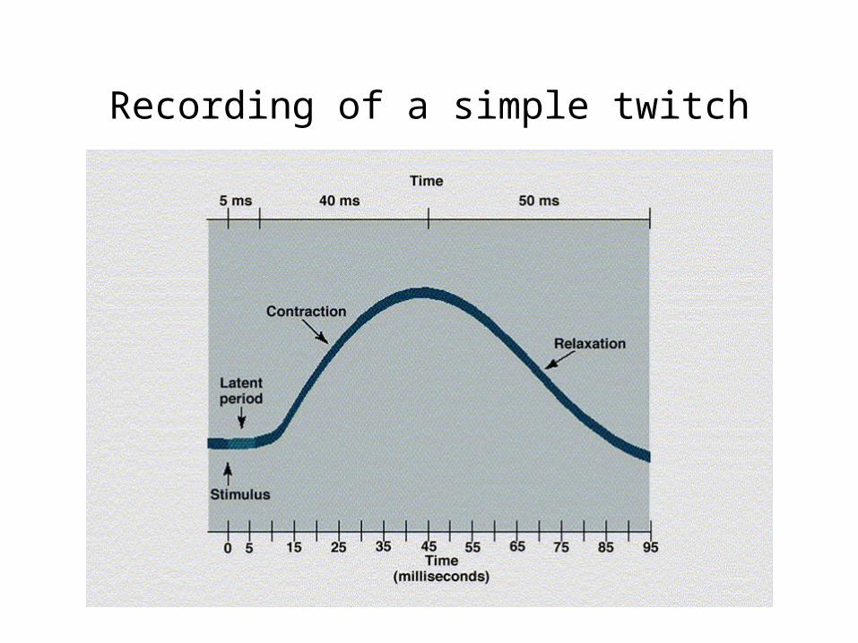
Recording of a simple twitch
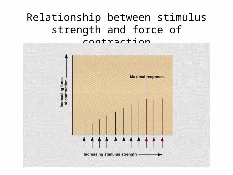
Relationship between stimulus strength and force of contraction
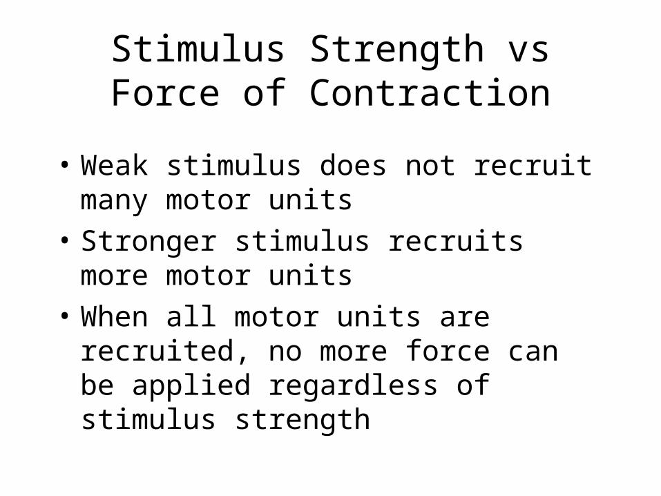
Stimulus Strength vs Force of Contraction
• Weak stimulus does not recruit many motor units
• Stronger stimulus recruits more motor units
• When all motor units are recruited, no more force can be applied regardless of stimulus strength
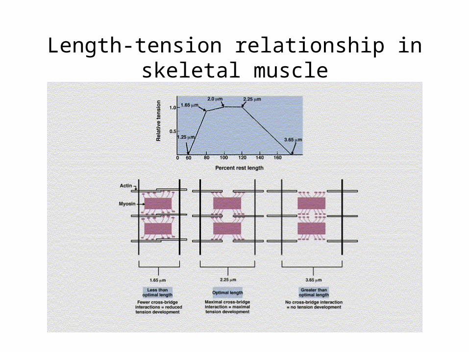
Length-tension relationship in skeletal muscle
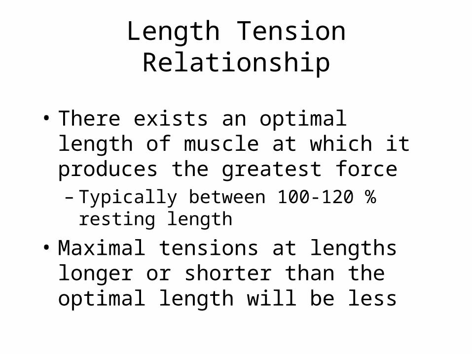
Length Tension Relationship
• There exists an optimal length of muscle at which it produces the greatest force– Typically between 100-120 % resting length
• Maximal tensions at lengths longer or shorter than the optimal length will be less
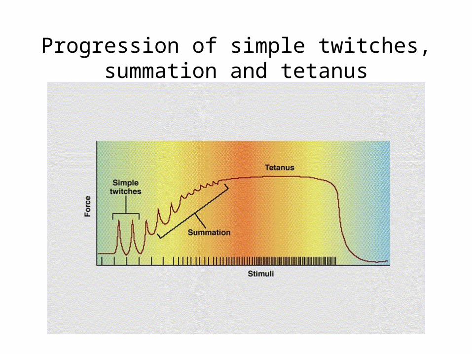
Progression of simple twitches, summation and tetanus
Tetanus
• If twitches become more frequent, greater force can be developed during summation than for a single twitch
• If twitches become to frequent, tetanus will develop and the muscle will not relax
• Typically results only from electrical stimulation
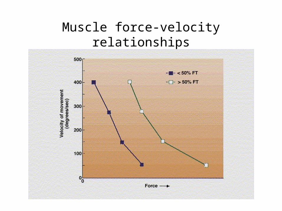
Muscle force-velocity relationships
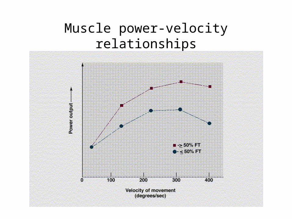
Muscle power-velocity relationships

The Golgi tendon organ
GTO
• Provides info to the CNS about tension development in the muscle
• Acts like a governor to prevent damaging tension from being generated
• Can be overridden to a certain extent by training– Supraphysiological strength in crisis
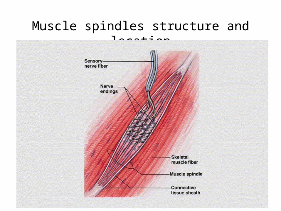
Muscle spindles structure and location
Spindle
• Provides info to the CNS about muscle length or stretch
• Excessive muscle stretch, especially during contraction is damaging
• Helps prevent damaging stretch during contraction