Embed Size (px)
Citation preview

iii
I N T R O D U C T I O N
IntroductionThis Heinemann Queensland Science Project–Biology: A Contextual ApproachStudent Activity Pack consists of a Student Activity Manual and eBiology,an interactive student CD.The pack provides strong support for Heinemann Queensland ScienceProject–Biology: A Contextual Approach and will assist students developtheir practical, research and information and communicationtechnology skills while enhancing their grasp of biological principlesand processes.
STUDENT ACTIVITY MANUALThe Student Activity Manual contains a comprehensive series ofactivities, experiments and investigations students can undertake to heighten their understanding of concepts. Activities will helpstudents develop a range of manipulative skills, including thoserequired by the syllabus. Students will also have opportunities tocollect and analyse both quantitative and qualitative data; use first-and second-hand data; and design their own investigations.
Major features of the manualinclude:
The key concepts and key ideas related to each activity are listed. Each activity includes a comprehensive list of materials required forthe activity. Any potential safety issues are noted in a box labelled‘Caution’. Key terms used in the activity are listed. Assessmentsuggestions are listed. Many activities involve the optional use ofinformation and communication technology and computersimulations.Activities are classified as simple, medium or complex.Simple activities:• Generally relate to one Key Concept and a limited number of Key
Ideas;• Contain all the information students need to complete the activity;
and• Tend to be relatively short in duration.Medium activities:• Generally relate to one or two Key Concepts and a number of
Key Ideas;• May require students to provide input into the method;• May require a longer time period; and• May require students to engage in higher-order thinking.Complex activities:• Generally relate to a number of Key Concepts and Key Ideas;• Will require students to develop aspects of the methodology;• May require students to undertake extra reading and research
before starting or during completion of the task;• May be extended activities requiring students to record results
over a longer time period;• Will require students to engage in higher-order thinking; and• Often form the basis for Extended Experimental Investigations.
these numbers indicateyou must record or answer something in your workbook
these numbers indicatesteps in practicalprocedures

Activities and assessmentAssessment suggestions are provided for each activity. These can beused for formative assessment or, when used under appropriateconditions, summative assessment. Complex activities may be suitableas Extended Experimental Investigations. Activities in Part One meetthe field work requirements of the syllabus. Presenting the results ofinvestigations could be an Extended Response task. If students areasked to write a scientific report of an activity, or answer questionsunder supervised conditions, the criteria for a Written Task are met.Many of the ‘Further Activities’ at the end of each activity are furtheropportunities for assessment tasks of all types.
HEINEMANN QUEENSLANDSCIENCE PROJECT-eBIOLOGY
Major features of eBiology include:• Interactive Tutorials, which model and simulate key biology
concepts. Many are directly integrated with the activities.• Worksheets to support the activities.• Data-logging worksheets, which provide an alternative method
involving sensors and computers.• A wide range of information and communication technology
support for the activities, including spreadsheets, presentation templates in PowerPoint and Word, webpage templates and more.
• Interactive glossary.• Exam and test self timer.
eBiology also includes a complete copy of the Student Activity Manualin electronic format, with hyperlinks to all worksheets, templates andtutorials. A direct link is also provided to the Heinemann QueenslandScience Project–Biology: A Contextual Approach Website, where internetsite URLs relevant to the activity are located, monitored and upgradedas necessary. Icons appear in the activities to indicate where these areavailable. Simply click on the icon to open the worksheet, template or tutorial.
Provides direct link to relevant websites.
Indicates a worksheet is available that either supports orsupplements the activity.
Indicates a spreadsheet is available as an alternative methodfor recording, graphing and processing data.
Indicates a PowerPoint template is available as an alternative method for presenting your results or findings or as an accompaniment to a class presentation.
Indicates a Webpage template is available as an alternative method to posters and other static presentations.
iv
I N T R O D U C T I O N
WS
SS
WP
PPT

Indicates a data-logging worksheet is available, which provides an alternative method involving sensors and computers.
Indicates an extra photograph or illustration relevant to the activity is available.
HEINEMANN QUEENSLANDSCIENCE PROJECT–BIOLOGY:ACONTEXTUAL APPROACH TEACHER’SRESOURCE AND ASSESSMENT DISKThe Teacher’s Resource and Assessment Disk supports both the textbookand Student Activity Manual and assists teachers in planning andimplementing a course of study and assessment programme.
Major features of the Teacher’sResource and Assessment Diskinclude:
• Detailed information relating to the implementation of each of the activities. This includes preparation notes, hints and answers to some of the questions within the activities.
• Sample work programme.• Units of work for both Years 11 and 12, showing how the
requirements of the syllabus could be met using the HeinemannQueensland Science Project–Biology: A Contextual Approach package.
• A sample assessment task for each of the assessment categories.• Suggested answers to all the review and further questions in the
text book.• A full copy of Heinemann Queensland Science Project–eBiology.
v
I N T R O D U C T I O N
DL
IM

ContentsIntroduction iii
Part 1: Living in an environmentActivity 1.1 What fruit is that?
CONSTRUCTING A DICHOTOMOUS KEY 2
Activity 1.2 Out of sightINVERTEBRATES IN ECOSYSTEMS 6
Activity 1.3 All in the familyINVESTIGATION OF TWO NATIVE PLANT GENERA 10
Activity 1.4 Plants in profileDISTRIBUTION OF PLANTS ALONG A TRANSECT 13
Activity 1.5 The weed censusSAMPLING PLANT POPULATIONS WITH QUADRATS 17
Activity 1.6 The underworld INVESTIGATING THE LEAF LITTER AND SOIL 23
Activity 1.7 The quiet provider ENERGY AND YOUR LAWN 32
Activity 1.8 A forest ecosystem AN INVESTIGATION OF INTERACTIONS 36
Activity 1.9 Up the creek! ADAPTATIONS AND INTERACTIONS 40
Activity 1.10 A little goes a long way BIOLOGICAL MAGNIFICATION 44
Activity 1.11 A numbers game FACTORS AFFECTING POPULATION SIZE 47
Activity 1.12 Ways and means ADAPTATIONS FOR SURVIVAL 50
Activity 1.13 Wattles—the fire weeds HEAT AND SEED GERMINATION 54
Activity 1.14 Changing over time ZONATION AND SUCCESSION 57
Part 2: Functioning organismsActivity 2.1 Cells–units of life
CELL STRUCTURE AND FUNCTION 63
Activity 2.2 That’s about the size and shape of itRELATIONSHIP BETWEEN SHAPE, SIZE AND DIFFUSION RATE 70
Activity 2.3 Moving moleculesOBSERVING DIFFUSION AND OSMOSIS 75
Activity 2.4 The facilitatorsENZYMES AND FACTORS AFFECTING ENZYME ACTIVITY 81
Activity 2.5 Useful yeastsFACTORS AFFECTING RESPIRATION 90
Activity 2.6 Green or variegated?RELATIONSHIP BETWEEN CHLOROPHYLL AND STARCH PRODUCTION 94
Activity 2.7 Using enzymesMAKING YOGHURT 99
Activity 2.8 Life’s necessitiesELEMENTS NEEDED FOR PLANT GROWTH 101
vi
C O N T E N T S

Activity 2.9 Food tubesCOMPARISON OF ANIMAL DIGESTIVE SYSTEMS 103
Activity 2.10 Respiring seeds and rodentsOXYGEN CONSUMPTION BY SEEDS AND ANIMALS 108
Activity 2.11 Leaves the losersPLANT LEAF STRUCTURE AND FUNCTIONING 113
Activity 2.12 An inspirational effortGAS EXCHANGE IN ANIMALS 118
Activity 2.13 An uplifting experiencePLANT STRUCTURE AND TRANSPORT 123
Activity 2.14 The heart of the matterHEART STRUCTURE AND FUNCTION 129
Activity 2.15 Red, white and … ?BLOOD COMPONENTS AND CIRCULATION 136
Activity 2.16 Fine-tuned filtersKIDNEY STRUCTURE AND FUNCTION 141
Activity 2.17 An evolutionary developmentWATER BALANCE IN ANIMALS 146
Activity 2.18 Mind over matterSTRUCTURE AND FUNCTION OF THE MAMMALIAN NERVOUS SYSTEM 150
Activity 2.19 Plant hormones and typical tropismsEFFECTS OF HORMONES AND TROPISMS ON PLANTS 155
Activity 2.20 Temperature tales—keeping warm staying coolINVESTIGATION OF TEMPERATURE REGULATION IN ANIMALS 162
Activity 2.21 A question of balanceHORMONAL REGULATION OF BLOOD GLUCOSE 169
Activity 2.22 TrainingEFFECTS OF EXERCISE ON BODY SYSTEMS 175
Activity 2.23 SurvivalHOMEOSTASIS IN EXTREME ENVIRONMENTS 180
Activity 2.24 Nature’s clonesASEXUAL REPRODUCTION IN PLANTS 184
Activity 2.25 It works for us!INTERNAL AND EXTERNAL FERTILISATION 188
Activity 2.26 Investigation: Going troppo!HUMAN INTERVENTION IN PLANT REPRODUCTION 190
Activity 2.27 Variations on a themeDIFFERENCES IN FLOWER STRUCTURE 194
Activity 2.28 Of mice and menEXAMINATION OF MAMMALIAN REPRODUCTIVE SYSTEMS 197
Activity 2.29 From seed to plantFACTORS AFFECTING SEED GERMINATION 199
Activity 2.30 Helping natureHUMAN INTERVENTION IN THE REPRODUCTION OF PLANTS AND ANIMALS 202
Activity 2.31 When things go wrongCONGENITAL MALFORMATIONS 205
Activity 2.32 A storm in a test tube?REPRODUCTIVE TECHNOLOGIES 212
Activity 2.33 Fellow travellersSTUDY OF ECTOPARASITES 222
vii
C O N T E N T S

viii
Activity 2.34 Life on the insideSTUDY OF ENDOPARASITES 225
Activity 2.35 Fit to drinkTHE TREATMENT OF DRINKING WATER 229
Activity 2.36 Defence forces in actionIMMUNITY AND THE IMMUNE SYSTEM 233
Activity 2.37 Blitzing bacteriaMETHODS OF PREVENTING BACTERIAL GROWTH 236
Activity 2.38 Under attackINVESTIGATION OF SOME PLANT INFECTIONS 241
Activity 2.39 Division for growthMITOSIS—SOMATIC CELL DIVISION 244
Activity 2.40 Division for gametesMEIOSIS—CELL DIVISION FOR REPRODUCTION 248
Part 3: Genetics and evolutionActivity 3.1 As easy as PCR
DNA REPLICATION 251
Activity 3.2 Making master moleculesDNA, RNA AND PROTEIN SYNTHESIS 255
Activity 3.3 Tigers and devilsA DISCUSSION OF CLONING 259
Activity 3.4 Genetic rouletteHUMAN VARIATION AND PEDIGREES 261
Activity 3.5 One, two or more genesPATTERNS OF GENETIC INHERITANCE 267
Activity 3.6 Growing up in the hillsENVIRONMENTAL INFLUENCES ON PHENOTYPE 274
Activity 3.7 Digging up the pastFOSSILS AND EVOLUTION 278
Activity 3.8 Similarities and differencesCONVERGENT AND DIVERGENT EVOLUTION 282
Activity 3.9 Lucy and her friendsHUMAN EVOLUTION 287
Activity 3.10 Adelie penguin populationsSIMULATING NATURAL SELECTION 291
Activity 3.11 Some people don’t like broad beansSELECTION IN SMALL POPULATIONS 293
Activity 3.12 Living in isolationTHE FOUNDER EFFECT AND GENETIC DRIFT 296
Activity 3.13 Diversity or bust!GENETIC DIVERSITY AND EXTINCTION 300
Activity 3.14 Fossils and the southern continentsEXPLAINING PATTERNS OF FOSSIL DISTRIBUTION 302
Activity 3.15 Exploring the pastCHANGES IN AUSTRALIA OVER GEOLOGICAL TIMES 306
Appendix 308
C O N T E N T S

2.1 Cells–units of lifeCELL STRUCTURE AND FUNCTION
IntroductionRobert Hooke was the first person to describe cells. In 1665, he observedthem for the first time by using a light microscope and wrote that hesaw ‘many little boxes’ (cellulae). Our knowledge and understandingof the structure of cells and the functions they perform have progressedsignificantly since then. Instruments for observing cell structure andtechniques for investigating the processes that go on inside a cell areconstantly being improved.
Scientists who study cells (cytologists) want to find out what cellsdo. This generally relates to the location of a cell in an organism, itsshape and its organelles (the structures that make up a cell). Livingthings are made up of cells or the chemical substances that cells make.Consequently, the cells of a living organism depend on, and produce, alarge range of chemicals that enable them to make new cell parts (togrow) and to relate to the rest of the organism of which they are a part.Therefore, the functions of a cell can be worked out by determining therange of organelles in a cell and the roles each of the organelles have inthe cell.
Pathologists diagnose a range of diseases by looking at changes inthe normal structure and function of cells and their organelles (seeBiology: A Contextual Approach, p. 177).
By looking at cells in a number of two-dimensional views, we canwork out the three-dimensional shape of cells. In most instances, cellsare stained to make them easier to see. To see inside a cell, a specialisedlight microscope, such as a phase-contrast microscope, can be used. Tosee the fine detail of cellular organelles, however, an electron micro-scope is necessary.
Purpose1 To observe, and record in diagrams, the shape of several different
plant and animal cells and to relate this to their function.
2 To observe and record the cell organelles that are visible under alight microscope and to relate this to the cell’s function.
3 To observe the effects of staining on cells.
4 To observe and record the structure of the cell organelles that arevisible under an electron microscope and to relate this to the cell’sfunction.
Plant cells
Procedure1 Take an onion scale and break it as shown in Figure 1. Quickly
cut or peel two pieces with sides 1 –1 cm long from the thin, transparent epidermal tissue. Mount one in a drop of water on a microscope slide and the other in a drop of toluidine blue oriodine/potassium iodide stain on another slide. If necessary,straighten the tissue using dissecting needles. Add a coverslip toeach preparation.
A
63
C E L L S — U N I T S O F L I F E
KEY CONCEPT 1
KEY IDEAS 1, 2, 3
• cell• eukaryote• organelle• stain
KEY TERMS
ER, WT
Assessment Suggestions
● Microscope
● Microscope slides and coverslips
● Scalpel, forceps and dissectingneedles
● Sharp, single-edged razor blade
● Foam or carrot tissue block forsection cutting (optional)
● Half Petri dish
● Fine paintbrush for section handling(optional)
● Paper tissues
● Iodine/potassium iodide stain ortoluidine blue stain
● Onion scale
● Moss or Elodea leaf
● Cabbage seedling (several per class)or celery petiole
● Cells and Their Organellesinteractive activity from HeinemannQueensland Science Project-eBiology:A Contextual Approach
● Websites listed on hi.com.au/biol
M A T E R I A L S
12
S I M P L E
A C T I V I T Y
14
Interactive

2 Examine the unstained tissue mounted under the microscope, firstusing low power, then high power. Adjust the iris diaphragm forthe greatest clarity at each magnification. Observe the regular shapeof the cells and the way they are arranged. Identify the cell wall andany organelles that are visible. Use Biology: A Contextual Approach,Figure 7.4 p. 147 as a guide.
Record your observations in a carefully drawn and labelled diagramof several cells. Show the magnification at which the observationswere made.
3 Examine the other slide of the same tissue mounted in toluidineblue or iodine/potassium iodide stain.
●1 Describe any further cell detail or cell structures that can now beobserved as a result of staining. Add these to your diagram.
●2 How many layers of the cells are there in the tissue sample?Suggest what the three-dimensional shape of these cells mightbe. Give your reasons.
4 Carefully detach a moss or Elodea leaf using fine forceps and mountit in a drop of water on a clean slide. Examine the slide first underlow power to observe the arrangement of cells and then under highpower to observe the structure and contents of an individual cell.Identify the cell organelles you can see.
Draw a leaf cell viewed under high power. Label all structures youcan see and state the magnification.
5 Look carefully at several cells under high power for a minute ortwo. Your observations may be more successful if you first warmthe slide gently over the microscope light for a minute.
●3 Is there any evidence that the cells you are looking at are alive?Explain the evidence you used to answer this question.
6 Cut several thin sections of the stem of a cabbage seedling (or celerypetiole) using the method demonstrated by your teacher. Select thetwo thinnest sections and mount one in a drop of water on aslide and add a coverslip. Mount the other section in a drop ofiodine/potassium iodide or toluidine blue on a second slide, andlet this slide stand for several minutes before adding a coverslip.
7 Examine the section mounted in water. You will observe a numberof different types of cells arranged in different ways. Now examinethe section mounted in iodine/potassium iodide or toluidine blue.If you use toluidine blue, cells that lack a thickened wall turnpurple and cells with thickened walls turn turquoise (blue–green).
●4 Have all cells stained the same colour? Is there any pattern to thestaining? A sketch diagram may help you to answer thisquestion.
●5 Explain why staining might be an advantage when examiningcells and tissues.
●6 a What structures within a cell make it possible to identify thosecells that are dividing?
b Would it be possible to make this identification without theuse of a stain? Explain.
64
F U N C T I O N I N G O R G A N I S M S
FIGURE 1Preparing onion epidermis.
thin layer of onionepidermis
Cut or tear small pieceand mount on slide.Do not let epidermisdry out.
S A F E T YBe careful when using sharpobjects, such as razor blades.
Chemicals must be used only forthe purpose specified in the
activity.
Dispose of chemicals and plantmaterial as instructed by your
teacher.

DiscussionDuring this part of the activity you observed some plant cells.Summarise your findings in a table using the headings:• cell type• cell shape• organelles• organelle function• cell functions
You may need to use your textbook or websites to find the functionof the organelles that you have seen.
Animal cells
Procedure1 Examine the prepared slide of cheek lining cells. Observe the shape
of the cells and look for the presence of any organelles.
Make a labelled drawing of a cheek cell under high power.
●7 What is the likely three-dimensional shape of these cells?Explain.
2 Examine the prepared slides of mammalian blood and frog bloodunder high power. The most common cells are red blood cells.
Make a labelled diagram of a red blood cell of each species as seenunder high power.
●8 a What are the major visible differences between mammalianand frog red blood cells?
b Suggest possible advantages and disadvantages to an animalof having red blood cells without nuclei.
3 Search the mammalian blood slide for white blood cells. There willbe very few of them in each field of view. Different types of whiteblood cells can be recognised by the different shapes of their nuclei.
Make a labelled diagram of two different types of white blood cellsseen under high power.
4 Examine the prepared slide of motor nerve cell bodies and fibrestaken from a spinal cord. Locate a cell body that can be clearly seen,and observe it under low and high power. A motor nerve cell orneuron consists of a cell body located in the spinal cord and a longaxon fibre, which extends out of the spinal cord and connects to amuscle.
Draw and label a motor nerve cell body.
●9 Using a reference book or websites to assist you, draw and labela complete motor nerve cell.
Hydra is a simple, sac-like animal whose body is made up of onlytwo cell layers and a small number of cell types.
5 Examine the prepared longitudinal section of Hydra and, underhigh power, identify several different types of cells.
Make clear drawings of two different types of cells, showing anyorganelles present.
●10 Suggest reasons why all cells on this slide do not show nuclei.
B
65
C E L L S — U N I T S O F L I F E
● Microscope
Prepared slides:
● Cheek cells (epithelial or liningcells)
● Mammalian blood
● Frog (or chicken) blood
● Motor nerve smear (nerve cells)
● Longitudinal section of Hydra(a variety of cells)
● Reference texts such as Biology:A Contextual Approach
● Websites listed on hi.com.au/biol
M A T E R I A L S
DiscussionDuring this activity you observed some animal cells. Summarise yourfindings in a table using the headings:• cell type• cell shape• organelles• organelle function• cell functions
You may need to use a text book to find the function of the organellesthat you have seen.
Discussion (for Parts A and B)Using a text book and the information from the tables produced inPart A and Part B, draw up a new table to list the organelles that canbe seen under a light microscope using the headings:• found only in plants• found only in animals• found in both plants and animals
Cell organellesThis part of the activity is based on second-hand data and the Cells andOrganelles interactive, and can be done as an out-of-class activity. It canbe completed using the PowerPoint template.
Electron microscopy (EM) and scanning electron microscopy (SEM) have made it possible to observe a great deal more in cells (seeBiology: – A Contextual Approach, pp. 175–7).
Use the electron microscope photographs on pages 66–69, the notesprovided in the captions, reference books and websites toproducea table using the headings:
• organelle• found in plants and animals• function• description• size in micrometres (1 micrometre [1 µm] = 0.001 mm)
C
66
F U N C T I O N I N G O R G A N I S M S
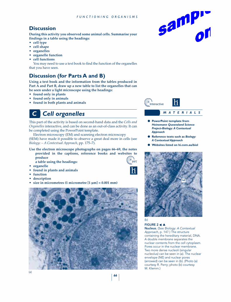
FIGURE 2Nucleus. (See Biology: A ContextualApproach, p. 147.) The structurecontaining the hereditary material, DNA.A double membrane separates thenuclear contents from the cell cytoplasm.Pores occur in the nuclear membrane.Two more dense nucleoli (singularnucleolus) can be seen in (a). The nuclearenvelope (NE) and nuclear pores(arrowed) can be seen in (b). (Photo (a)courtesy R. Perry; photo (b) courtesy M. Klemm.)
(a)
(b)
▲▲
NE
● PowerPoint template fromHeinemann Queensland ScienceProject-Biology: A ContextualApproach
● Reference texts such as Biology:A Contextual Approach
● Websites listed on hi.com.au/biol
M A T E R I A L S
PPT
Interactive
67
C E L L S — U N I T S O F L I F E
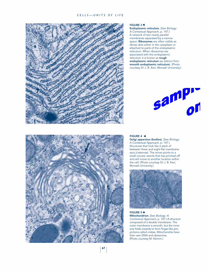
FIGURE 3Endoplasmic reticulum. (See Biology:A Contextual Approach, p. 147.) A network of two nearly parallelmembranes separated by a narrow space. Ribosomes are often visible asdense dots either in the cytoplasm orattached to parts of the endoplasmicreticulum. When ribosomes areassociated with the endoplasmicreticulum it is known as roughendoplasmic reticulum (as distinct fromsmooth endoplasmic reticulum). (Photocourtesy Dr J. B. Kerr, Monash University.)
▲
FIGURE 4Golgi apparatus (bodies). (See Biology:A Contextual Approach, p. 147.)Structures that look like a stack ofbetween three and eight flat membranesacs (cisternae). The arrow points to asmall circular vesicle that has pinched offand will move to another location withinthe cell. (Photo courtesy Dr J. B. Kerr,Monash University.)
FIGURE 5Mitochondrion. (See Biology: AContextual Approach, p. 147.) A structurecomposed of a double membrane. Theouter membrane is smooth, but the innerone folds inwards to form finger-like pro-jections called cristae. Mitochondria havetheir own DNA and ribosomes.(Photo courtesy M. Klemm.)
▲
▲

68
F U N C T I O N I N G O R G A N I S M S
cell A cell B
FIGURE 6Chloroplast. (See Biology: A ContextualApproach, p. 147.) Found in green parts of photosynthetic eukaryotes(plants) and in photosynthetic protists.The outer envelope encloses a complexseries of membrane stacks (grana). The chlorophyll is bound to proteinembedded in the membrane.Chloroplasts contain ribosomes and DNA (see Biology: A ContextualApproach, p. 147). (a) A completechloroplast (arrowed) in the cell of amangrove; (b) a close-up of part of achloroplast shows the grana (G) andstroma (S). (Both photos courtesy J. Carpenter, Monash University.)
(a)
(b)
▲ ▲
SS
GG
FIGURE 7Cell wall. (See Biology: A ContextualApproach, p. 147.) The rigid outerboundary of plant cells. The cell wall ismade up of layers of cellulosemicrofibrils. The cell membrane, which isnormally pressed against the cell wall, haspulled away from it (arrowed). (Photo courtesy G. Jaudzems.)
▲
69
C E L L S — U N I T S O F L I F E
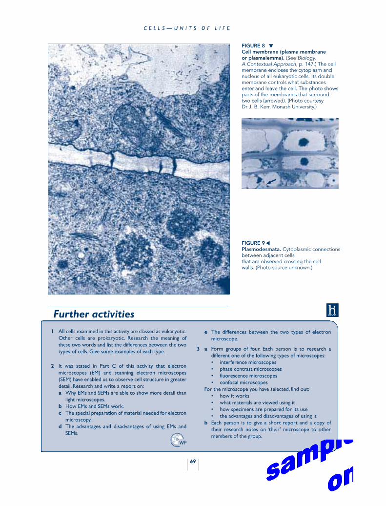
FIGURE 8Cell membrane (plasma membrane or plasmalemma). (See Biology:A Contextual Approach, p. 147.) The cellmembrane encloses the cytoplasm andnucleus of all eukaryotic cells. Its doublemembrane controls what substancesenter and leave the cell. The photo showsparts of the membranes that surroundtwo cells (arrowed). (Photo courtesy Dr J. B. Kerr, Monash University.)
FIGURE 9Plasmodesmata. Cytoplasmic connectionsbetween adjacent cells that are observed crossing the cell walls. (Photo source unknown.)
▲
1 All cells examined in this activity are classed as eukaryotic.Other cells are prokaryotic. Research the meaning ofthese two words and list the differences between the twotypes of cells. Give some examples of each type.
2 It was stated in Part C of this activity that electronmicroscopes (EM) and scanning electron microscopes(SEM) have enabled us to observe cell structure in greaterdetail. Research and write a report on:a Why EMs and SEMs are able to show more detail than
light microscopes.b How EMs and SEMs work.c The special preparation of material needed for electron
microscopy.d The advantages and disadvantages of using EMs and
SEMs.
Further activities
e The differences between the two types of electronmicroscope.
3 a Form groups of four. Each person is to research adifferent one of the following types of microscopes:• interference microscopes• phase contrast microscopes• fluorescence microscopes• confocal microscopes
For the microscope you have selected, find out:• how it works• what materials are viewed using it• how specimens are prepared for its use• the advantages and disadvantages of using it
b Each person is to give a short report and a copy oftheir research notes on ‘their’ microscope to othermembers of the group.
▲
WP
90
F U N C T I O N I N G O R G A N I S M S
2.5 Useful yeastsFACTORS AFFECTING RESPIRATION
IntroductionLong before yeasts were identified with the aid of a microscope, theireffects were well known. A yeast is a single-celled fungus that is capableof multiplying rapidly in the right conditions. There are a number ofdifferent yeast strains. For centuries, they have been used for makingalcohol (wine and beer) and bread. In both cases, the enzymes pro-duced by the yeast cells break sugars down into ethanol and carbondioxide. This process is known as cellular respiration. In wines andbeers, the carbon dioxide escapes and the alcohol remains in solution.In bread, the carbon dioxide causes the dough to rise and the alcohol isevaporated off during baking. (For details about aerobic and anaerobicrespiration, see Biology: A Contextual Approach, pp. 187–90.)
The relationship between sugars and carbon dioxide in respirationcan be demonstrated in an experiment such as the one that is outlinedbelow.
Some carbon atoms are radioactive. They are known as carbon-14 or14C atoms, and their presence can be detected with a radiation counter.
In an experiment, a mouse was fed water containing a small amountof glucose made with radioactive carbon. The mouse was placed in alarge, sealed jar containing air from which carbon dioxide had beenremoved. After a short time, a sample of the air inside the jar was takenand, using a radiation detector, some of the carbon dioxide was foundto be radioactive. The only radioactive carbon atoms present in the jarat the start of the experiment were in the glucose fed to the mouse. Atthe end of the experiment, they were found in the carbon dioxideexhaled by the mouse. This means that the radioactive carbon atomshad been transferred from glucose to carbon dioxide during the processof cellular respiration.
A mouse respires aerobically. However, in this activity, the linkbetween glucose and carbon dioxide in anaerobic respiration isassumed.
Purpose1 To identify the gas produced when yeast cells respire.
2 To investigate factors affecting the rate of respiration in yeast bymeasuring changes in gas output.
Identification of gas producedThis part of the activity may be set up as a demonstration for you. Sothat you can follow what has been done, an outline of the procedure isgiven. Observe the demonstration and answer Questions 1 and 2.
KEY CONCEPTS 1, 4, 5
KEY IDEAS 2, 4, 20
ER, EEI, WT
Assessment Suggestions
C O M P L E X
A C T I V I T Y
A
• cellular respiration• yeast• aerobic respiration• anaerobic respiration• enzyme
KEY TERMS
91
Procedure1 Put 25 mL of glucose solution in the 100 mL conical flask and add
the cube of compressed yeast. Disperse the yeast into the glucosesolution using the stirring rod.
2 Stand the flask in the 500 mL beaker and add warm water to makea water bath at 35–40°C. Do not add so much water that the flaskfloats. Alternatively, you could hold the flask in the water bath witha retort stand and clamp. Try to maintain the temperature duringthe experiment by removing cool water from, and adding warmwater to, the beaker.
3 Rinse the clean boiling tube with a little limewater and then addmore limewater to a depth of 3 cm. Stand the boiling tube in a racknear the water bath.
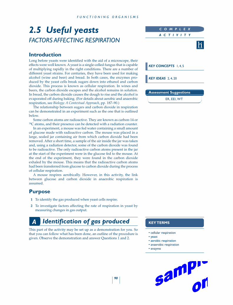
4 Connect up the stoppers and tubing as shown in Figure 1. Carefullyswirl the flask every 2–3 min for 15–20 min.
●1 Describe what you observed during the 15–20 min that theapparatus was set up.
●2 Explain how your observations allow you to:
a determine that a gas was released
b determine the identity of the gas.
Factors affecting respiration rate
ProcedureWork in groups of three. Your teacher will allocate a temperature foryour group’s water bath. The temperature ranges are:• low temperature (less than 5°C)• medium temperature (15–20°C)• high temperature (35–40°C)• very high temperature (greater than 60°C)Data for all temperatures will be pooled at the end of the activity so thatall students have a full range of data to process.
U S E F U L Y E A S T S
● 100 mL conical flask
● One-holed stopper with glass tubinginserted, to fit flask
● Boiling tube and test-tube rack
● Two-holed stopper to fit boilingtube, one hole with glass tubinginserted
● 500 mL beaker or similar containerfor water bath
● Thermometer (10–110°C)
● Plastic tube to connect conical flaskto boiling tube
● 100 mL measuring cylinder
● Glass stirring rod
● 1 cm cube of compressed yeast
● Limewater
● 20% glucose solution—class supply
● Retort stand and clamp
M A T E R I A L S
C A U T I O NBe careful when handling hot
materials.
clear plasticconnecting tube
water atrequired
temperature
yeast/glucosesuspension
Note: flask may need to beheld down in water bath
boiling tube
lime water
FIGURE 1Diagram of apparatus showing detail of tube connections.
B
(for part B)
● PET soft-drink bottle cut to size(see Figure 3), or
● 2 L beaker
● Three test tubes 20 mm × 150 mm(approximately)
● Three one-holed stoppers withdroppers inserted to fit the testtubes (see Figure 2)
● Glass stirring rod
● Thermometer
● 10 or 25 mL measuring cylinder
● Marking pen
● Timing device
● Metal nut or sinker to stick or tapeto bottom of test tubes
● 20% glucose solution—class supply
● Three 1 cm cubes of compressedyeast
● Spreadsheet from HeinemannQueensland Science Project-eBiology:A Contextual Approach (optional)
● Data-logging activity fromHeinemann Queensland ScienceProject-eBiology: A ContextualApproach (optional)
M A T E R I A L S
DL

92
F U N C T I O N I N G O R G A N I S M S
Copy Tables 1 and 2 into your workbook.
TABLE 1Yeast respiration rate at ____°C.
Time (min) Number of bubbles per min
Tube 1 Tube 2 Tube 3 Average
1
23
45
6
78
9
10
TABLE 2Yeast respiration rate at different temperatures.
Time Average gas bubbling rate at different temperatures(min) Low Medium High Very high
(temp. ____°C) (temp. ____°C) (temp. ____°C) (temp. ____°C)
12
3
45
67
8
9
10
one hole stopper
dropper(glass portion)
end of dropper(free of liquid
and not touchingyeast/glucose)
yeast andglucose
(in well-mixedsuspension)
FIGURE 2Test tube with dropper.
cut down PETsoft-drink bottle
stopper belowwater surface
yeast/glucosemixture
FIGURE 3Arrangement of test tubes in water bath.
1 Each member of the group should be responsible for one of thethree tubes. Label your test tube with your name and ‘1’, ‘2’ or ‘3’.To each tube, add 12 mL of 20% glucose solution and one cube ofyeast. Use the stirring rod to thoroughly disperse the yeast in theglucose solution.
2 Into each test tube insert a stopper with dropper attached. Makesure it is a firm fit (Figure 2). Note: the dropper must not have anyliquid in it and the bottom must be above the yeast/glucosesuspension.
3 Fill the PET bottle water bath and adjust the water temperature towithin your group’s allocated temperature range. Try to maintainthe water bath at this temperature throughout the activity byreplacing some of the water, if necessary, with ice water or hotwater.
4 Submerge the three stoppered test tubes in the water bath. Thetubes must be totally under water (Figure 3). If they tend to float,use masking tape to stick a metal ‘weight’ to the bottom of each
SS
SS
93
tube. Leave the tubes to stand in the water bath for 3 min, thencount the number of bubbles released per minute from each tubefor 10 min.
Record your data in Table 1.
Also record in Table 1 the data for the tubes observed by the othertwo members of your group. Calculate the average and enter it on theclass data sheet. When the class data are complete, copy the detailsinto Table 2.
5 Dispose of the solution and equipment as directed by your teacher.
Draw a graph of the data.
Discussion
●3 Describe the effect of temperature on the rate of yeast respirationin a glucose solution.
●4 What is the optimum temperature for the fermentation processinvestigated?
●5 Suggest possible reasons why the rate of respiration is affectedby temperature. Support your answer by quoting from the data,and referring to Biology: A Contextual Approach, Chapter 9 andreferences from other sources if necessary.
U S E F U L Y E A S T S
Here are some ideas for you to investigate.The technique you have used can be modified and used to
investigate the effect of a number of substances on yeastrespiration rate. Use the optimum temperature (which youfound in Part B of this activity) as the temperature at which you carry out the investigations. A thermostatically controlledlaboratory water bath would be useful. Make sure that thetubes are clearly identified in some way. Experimental control is important.
Some suggestions are:a Investigate the effect of solutions of different pH on
rates of fermentation. Make up buffer solutions or usebuffer tablets. Use a standard volume of glucosesolution and add a fixed volume of buffer solution. Fora control, replace the buffer solution with a similarvolume of distilled water.
Further activity
b Investigate the effect of alcohol on rates of fermen-tation. Use a standard volume of glucose solution and add a fixed volume of ethanol. For a control,replace the ethanol with a similar volume of distilledwater.
c Investigate the effects of carbohydrates other thanglucose on rates of fermentation. For example, as wellas glucose, choose from 20% solutions of fructose,mannose, galactose, sucrose, maltose and lactose.
Write a report outlining the purpose of your investigation,the procedure you used, the reason for any experimentalcontrols, the results you obtained and a discussion of them.Make suggestions for improving the experimental design and technique.
SS