Embed Size (px)
DESCRIPTION
INTRODUCTION. Thyroid Disease is the Most Common Endocrinopathy Observed in Children Incidences, Presentations , and Clinical Consequences Differ Markedly than in adults Failure to Diagnose and Treat Promptly may Lead to Irreversible Neurologic Damage. THYROID GLAND. - PowerPoint PPT Presentation
Citation preview
Thyroid Disease is the Most Common Endocrinopathy Observed in Children
Incidences, Presentations, and Clinical Consequences Differ Markedly than in adults
Failure to Diagnose and Treat Promptly may Lead to Irreversible Neurologic Damage
INTRODUCTION
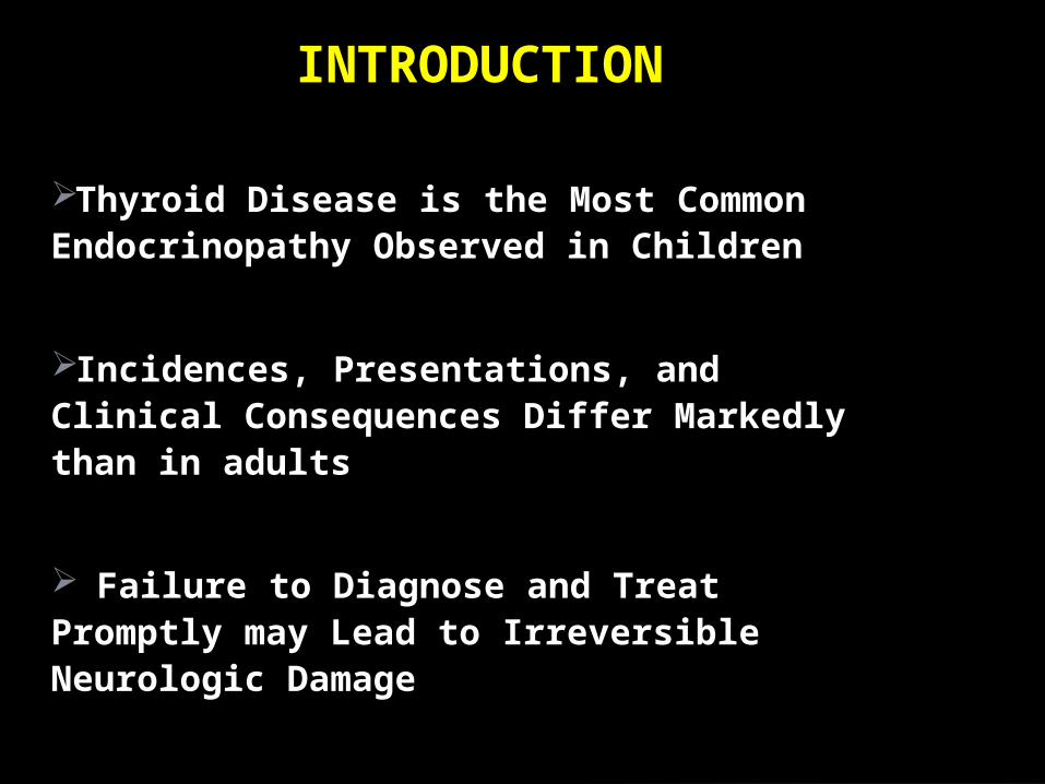
Derived from pharyngeal endoderm at 4/40
Migrate from base of the tongue to cover the 2&3 tracheal rings.
Blood supply from ext. carotid & subclavian and blood flow is twice as renal blood flow/g tissue.
Starts producing thyroxin at 14/40.
THYROID GLAND

Orignates from thyroid diverticulum and ultimobranchial bodies
Ontogeny influenced by several transcription factors (TTF, PAX8, HOX3)
Largely complete by 10-12 weeks
Gradual Maturation in Hypothalamic-Pituitary-Thyroid Axis
Thyroid Development

• TSH detectable by 12 wks
• Feedback mechanisms established by 20 wks
• T3 levels remain low
• Reverse T3 levels high
Fetal Thyroid Maturation:
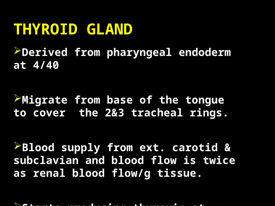
Maternal and fetal glands are independent
Little T4 transplacental transfer
TSH does not cross the placenta although it is
permeable to TRH, IgG, and thionamides
Fetal brain converts T4 to T3 efficiently
Effect of maternal hypothyroidism is most important in
first trimester
Placental and Fetal ThyroidMetabolism
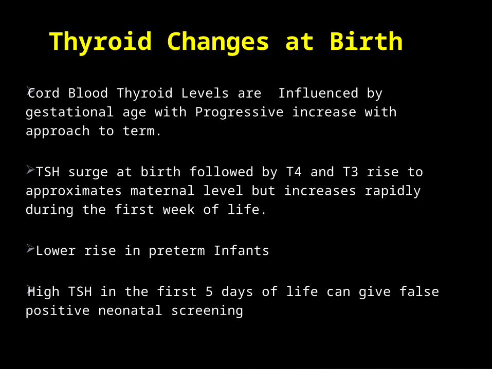
Cord Blood Thyroid Levels are Influenced by gestational age
with Progressive increase with approach to term.
TSH surge at birth followed by T4 and T3 rise to approximates
maternal level but increases rapidly during the first week of life.
Lower rise in preterm Infants
High TSH in the first 5 days of life can give false positive
neonatal screening
Thyroid Changes at Birth

Iodine & tyrosine form both T3 & T4 under TSH stimulation.
10% of T4 production is autonomous and is present in patients with central hypothyroidism.
Less than 1% of T4 & T3 is free in plasma.
T4 is deiodinated in the tissues to either T3 (active) or reverse T3 (inactive).
When released into circulation T4 binds to:Globulin TBG 75%Prealbumin TBPA 20%Albumin TBA 5%
THYROID HORMONES

Is a Glico-protein with Molecular Wt of 28000
Secreted by the anterior pituitary under influence of TRH
It stimulates iodine trapping,oxidation,organification, coupling and proteolysis of T4 & T3
It also has trophic effect on thyroid gland
TSH

T4 & T3 are feed-back regulators of TSH
TSH is stimulated by a-adrenergic agonists
TSH secretion is inhibited by:
DopamineBromocreptineSomatostatinCorticosteroids

Conversion of T4 to T3 is decreased by:Acute & chronic illnessesb-adrenergic receptor blockersStarvation & severe PEMCorticosteroidsPropylthiouracilHigh iodine intake (Wolff-Chaikoff effect
THYROXINE (T4)

Total T4 level is decreased in:Premature infantsHypopituitarismNephrotic syndromeLiver cirrhosisPEMProtein losing entropathy
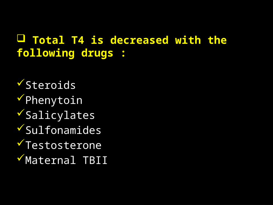
Total T4 is decreased with the following drugs :
SteroidsPhenytoinSalicylatesSulfonamidesTestosteroneMaternal TBII

Total T4 is increased with:Acute thyroiditisAcute hepatitisEstrogen therapyClofibrateiodidesPregnancyMaternal TSI
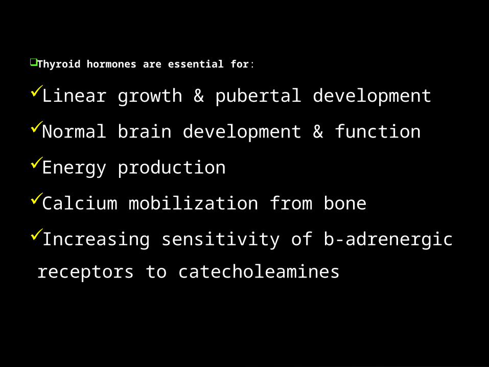
Thyroid hormones are essential for:
Linear growth & pubertal development
Normal brain development & function
Energy production
Calcium mobilization from bone
Increasing sensitivity of b-adrenergic receptors
to catecholeamines

•Congenital• Acquired
– Primary
Surgery
Radiation
Autoimmune
Iodine Deficiency
– Secondary
Surgery
Radiation
Infiltrative
Tumor
– Primary Thryoid Agenesis, Hypoplasia & mal-
descent
Dyshormonogenesis
Iodine Deficiency
– Secondary
Hypopituitarism Intake of goitrogens during
pregnancy
Idiopathic
Hypothyroidism
CONGENITAL HYPOTHYROIDISM

Incidence 1:4000
– Slightly higher in female infants
– Higher in Asian babies
– Lower in Black babies
Overt symptoms may not be present at birth
Profound effects on brain development, thus it is The most
common cause of preventable mental retardation in children
Reliable testing available (T4 and/or TSH)
No sequelae if treatment initiated by 4 wks– 10-15 mcg/kg/d
Epidemiology:

Relatively High Prevalence
Deleterious Consequence of Delayed
Diagnosis
Difficult ClinicalRecognition
Reliable Method of Screening
(sensitive & specific)
Safe, Effective Treatment available
Principles of NewbornScreening

Extensive testing for precise
etiology is generally not necessary (will not change immediate care plans)
May allow assessment of risk in future pregnancies
May allow early determination of transient vs permanent disease
Etiology of Congenital Hypothryoidism:
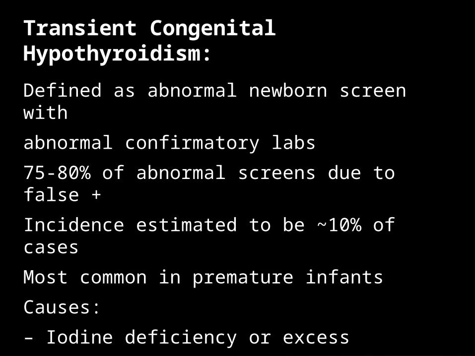
Defined as abnormal newborn screen with
abnormal confirmatory labs
75-80% of abnormal screens due to false +
Incidence estimated to be ~10% of cases
Most common in premature infants
Causes:
– Iodine deficiency or excess
– Maternal antithyroid medication
– Maternal TSH receptor blocking antibodies
Transient Congenital Hypothyroidism:

Incidence estimated at 1:180,000
Often history of treated Graves in mom
Mothers may have unrecognized hypothryoidism
Infant will not have goiter
Difficult to distinguish from thyroid dysgenesis May
have permanent neurocognitive deficit if present in utero
Resolves in 2-3 months as antibody clears
Maternal TSH receptor blocking antibodies:

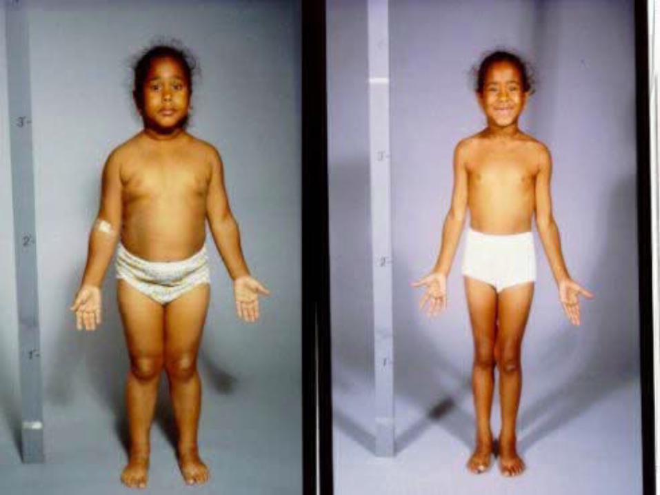
A condition of severely stunted physical
and mental growth due to untreated
congenital deficiency of thyroid hormones (
congenital hypothyroidism)
Cretinism is :


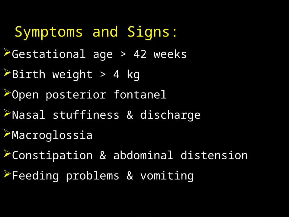
Gestational age > 42 weeks
Birth weight > 4 kg
Open posterior fontanel
Nasal stuffiness & discharge
Macroglossia
Constipation & abdominal distension
Feeding problems & vomiting
Symptoms and Signs:

Non pitting edema of lower limbs & feet
Coarse features
Umbilical hernia
Hoarseness of voice
Anemia
Decreased physical activity
Prolonged (>2/52) neonatal jaundice
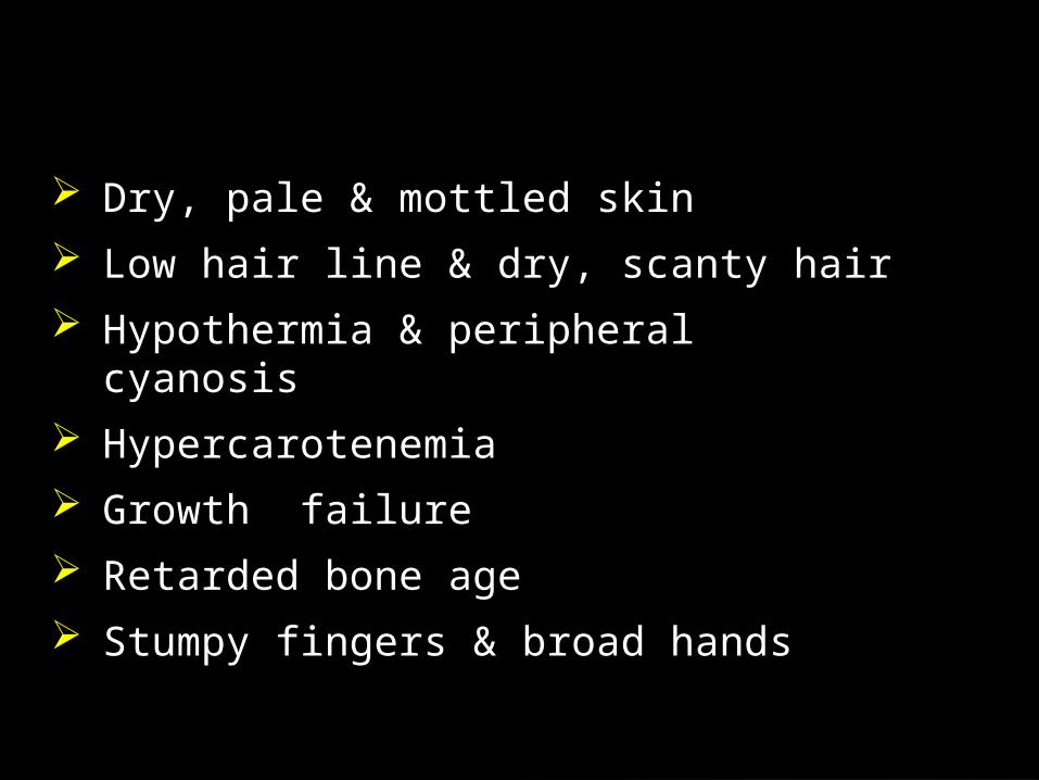
Dry, pale & mottled skin
Low hair line & dry, scanty hair
Hypothermia & peripheral cyanosis
Hypercarotenemia
Growth failure
Retarded bone age
Stumpy fingers & broad hands

Infantile proportions
Hip & knee flexion
Exaggerated lumbar lordosis
Delayed teeth eruption
Under developed mandible
Delayed closure of anterior fontanel
Skeletal abnormalities:
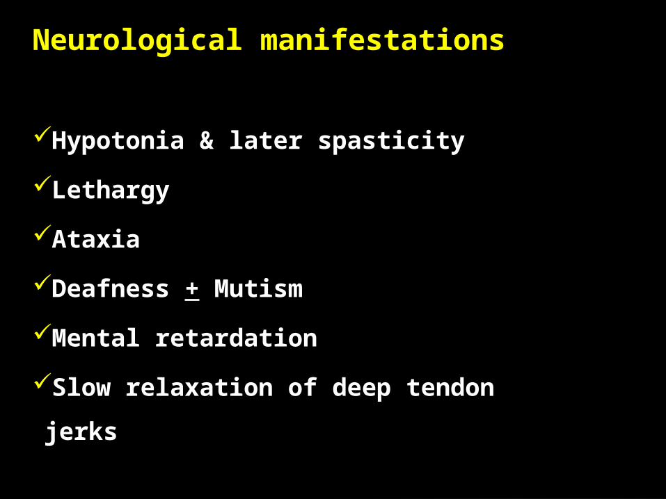
Hypotonia & later spasticity
Lethargy
Ataxia
Deafness + Mutism
Mental retardation
Slow relaxation of deep tendon jerks
Neurological manifestations
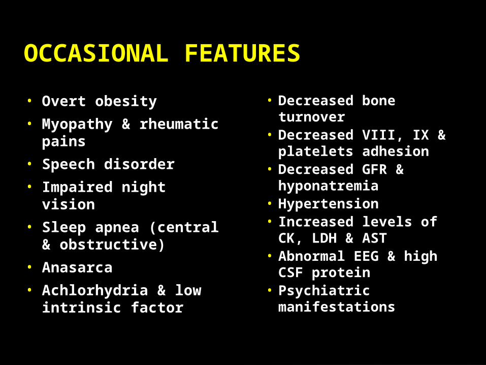
• Decreased bone turnover
• Decreased VIII, IX & platelets adhesion
• Decreased GFR & hyponatremia
• Hypertension• Increased levels of
CK, LDH & AST • Abnormal EEG &
high CSF protein• Psychiatric
manifestations
• Overt obesity
• Myopathy & rheumatic pains
• Speech disorder
• Impaired night vision
• Sleep apnea (central & obstructive)
• Anasarca
• Achlorhydria & low intrinsic factor
OCCASIONAL FEATURES


Autoimmune diseases (Diabetes Mellitus)
Cardiomyopathy & CHD
Galactorrhoea
Muscular dystrophy + pseudohypertrophy
(Kocher-Debre-Semelaigne)
ASSOCIATIONS
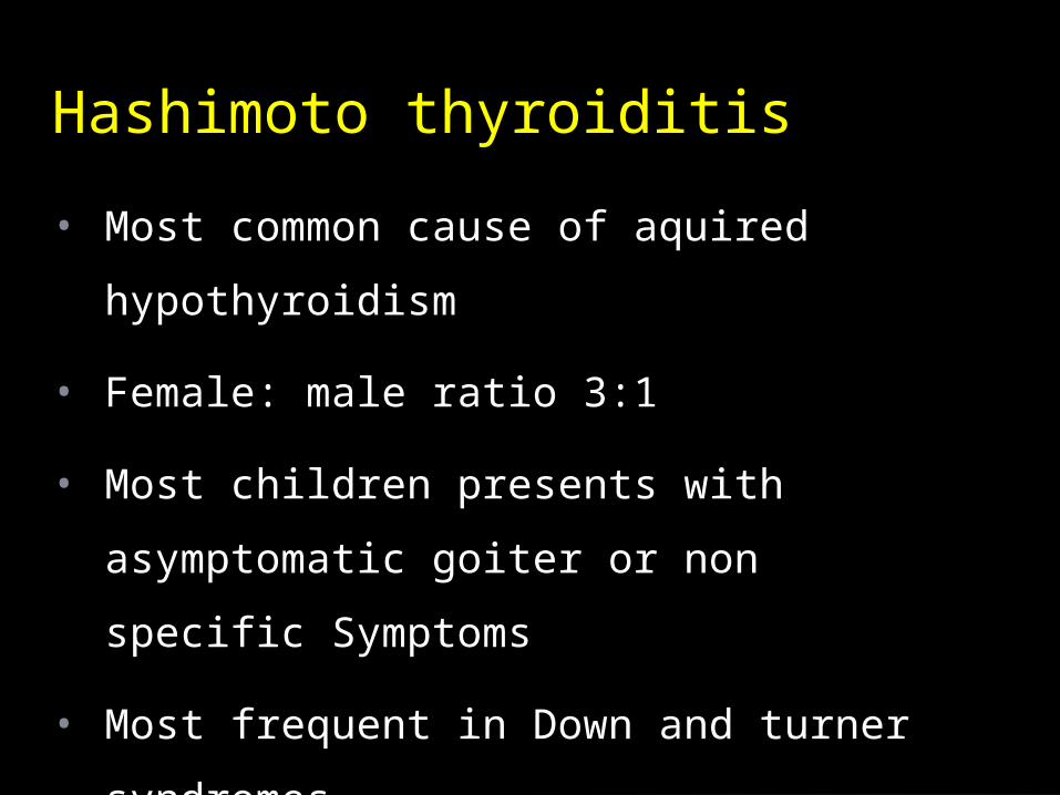
• Most common cause of aquired hypothyroidism
• Female: male ratio 3:1
• Most children presents with asymptomatic
goiter or non specific Symptoms
• Most frequent in Down and turner syndromes
Hashimoto thyroiditis
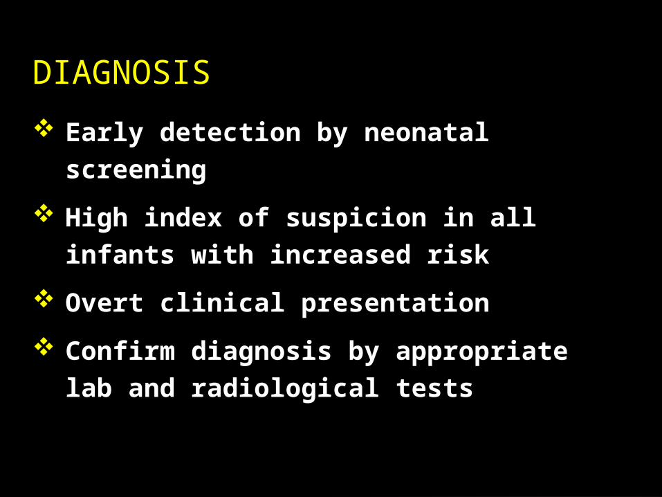
Early detection by neonatal screening
High index of suspicion in all infants with
increased risk
Overt clinical presentation
Confirm diagnosis by appropriate lab and
radiological tests
DIAGNOSIS
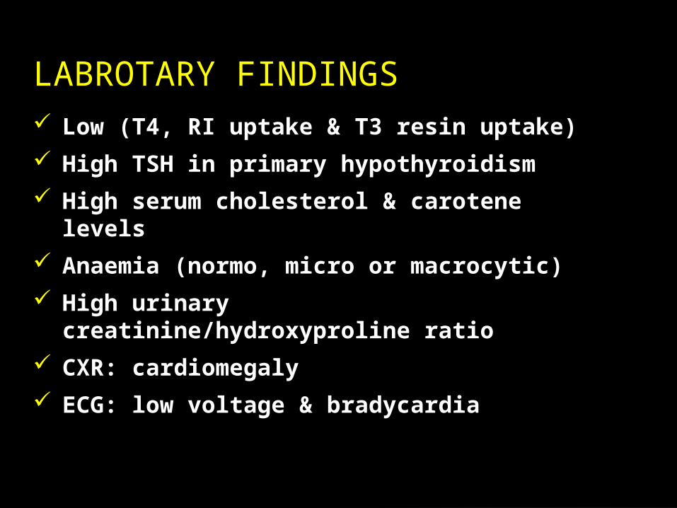
Low (T4, RI uptake & T3 resin uptake)
High TSH in primary hypothyroidism
High serum cholesterol & carotene levels
Anaemia (normo, micro or macrocytic)
High urinary creatinine/hydroxyproline ratio
CXR: cardiomegaly
ECG: low voltage & bradycardia
LABROTARY FINDINGS

X-ray films can show:Delayed bone age or epiphyseal dysgenesis
Anterior peaking of vertebraeCoxavara & coxa plana
Thyroid radio-isotope scan
Thyroid ultrasound
CT or MRI
IMAGING TESTS

Confirm all abnormal newborn screens with laboratory TSH and free T4
Borderline results may require repeat testing in 2-4 Wks
If repeat labs abnormal, begin thryoxine (25-37.5 mcg/day)
Goal is to start treatment within first month of life
Recheck q 2-3 months and adjust dose if Necessary
If no need to increase dose by 2 ½ -3 yrs, give 4 wk trial off of thyroxine
Treatment Guidelines
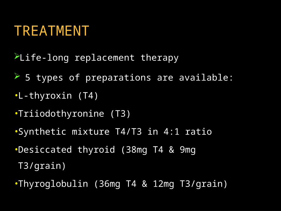
Life-long replacement therapy
5 types of preparations are available:
•L-thyroxin (T4)
•Triiodothyronine (T3)
• Synthetic mixture T4/T3 in 4:1 ratio
•Desiccated thyroid (38mg T4 & 9mg T3/grain)
•Thyroglobulin (36mg T4 & 12mg T3/grain)
TREATMENT
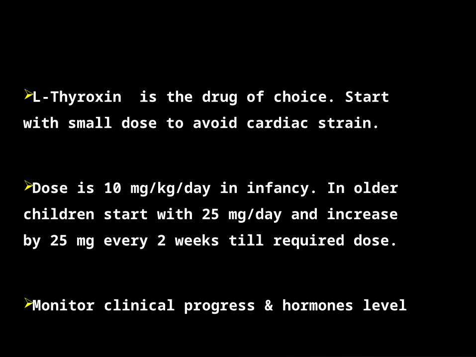
L-Thyroxin is the drug of choice. Start with small dose
to avoid cardiac strain.
Dose is 10 mg/kg/day in infancy. In older children start
with 25 mg/day and increase by 25 mg every 2 weeks till
required dose.
Monitor clinical progress & hormones level
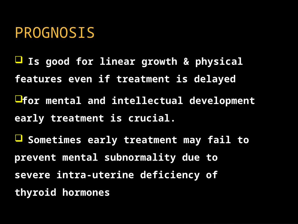
Is good for linear growth & physical features
even if treatment is delayed
for mental and intellectual development early
treatment is crucial.
Sometimes early treatment may fail to
prevent mental subnormality due to severe
intra-uterine deficiency of thyroid hormones
PROGNOSIS

• Acquired
– Inflammation
– Colloid
– Iodine Deficiency
– Goiterogen
– Infiltrative disease
– Toxic goiter
– Thyroglossal duct
cyst
– Adenoma
– Carcinoma
• Congenital
– Dyshormonogenesis
– Maternal Antibodies
• Blocking
• Stimulating
– Maternal Antithyroid drug
PTU, methimazole
– TSH receptor Activating
Mutation
– McCune Albright Syndrome
– Thyroid Tumor
Goiter: Differential Diagnosis
• Usually euthryoid
• Diffuse gland enlargement
• Rare in US (iodized salt provides adequate iodine
source)
• Rule out autoimmune thyroiditis
• Treament Doses in Children (6-12 months)
– Infants 100 mcg/d
– Children 200 mcg/day
– Adolescents 200-300 mcg/d
Endemic Goiter:

• Graves Disease (>95% of Cases)
– Relatively rare in children
– Incidence increases with puberty
– Female:Male (3-5:1)
• Neonatal Graves; Transplacental Antibodies
• Hashitoxicosis
• TSH receptor mutations (gain of function); McCune Albright syndrome
• Subacute Thyroiditis
• Exogenous thyroxine Exposure
Hyperthyroidism:
• Almost always transient
• Usually associated with maternal Graves:
– Transplacental passage of TSI
– Blocking and stimulating Abs may coexist
• Incidence ~1:50,000 infants
– 1-2% of moms with Graves disease
• Often presents in first week of life
– Emerges with clearance of maternal thionamide
Neonatal Hyperthyroidism

– PTU or Methimazole
– SSKI (If severe symptoms)
– Propranolol (If significant sympathetic
symptoms (HR>160)
• Treatment:

• Change in School Performance
• Insomnia
• Restlessness and Irritability
• Nocturia
• Bone age advancement
• Infants: Premature birth, Craniosynostosis, Poor
feeding, Failure to Thrive
Signs of Hyperthyroidism in Children

• Other classic signs:
– Weight Loss, Polyphagia, Tachycardia, Increased
Pulse
Pressure, Heat Intolerance, Diarrhea, Tremor

• Suppressed TSH
• Elevated T4, Free T4, T3 levels
• Positive Thyroid Stimulating Antibodies:
(May be helpful if exophthalmos absent)
– Thyroid Peroxidase
– Thyroglobulin
– Thyroid Stimulating Immunoglobulin
Grave’s Disease: Diagnosis

• Radioactive Iodine
– Preferred treatment in older children and adolescents
– Theoretical risk of radiation not established
– Possible increased risk of thryoid cancer (<5yrs)
• Thionamides (methimazole, PTU)
– Agranulocytosis, hepatitis, rash
– Poor long term remission rates
– Difficult to titrate dose, frequent monitoring
– Poor compliance in adolescents
• Surgical Thyroidectomy; Rarely indicated
Treatment of Graves’ Disease
• Diffuse enlargement of thryoid gland evident
usually during pubertal years
• Normal thyroid function tests
• Often family history
• May represent mild autoimmune thryoiditis; TPO Ab titer may be helpful to distinguish
• May be associated with nodular goiter as adults
• Therapy usually not necessary; May respond to thryoid suppression (controversial)
Colloid (Nontoxic) Goiter
• Low Prevalence in Children (0.2% <16 yrs)
• Higher Incidence of Malignancy (18-22%)
• Evaluation:
– Ultrasound can assist in detection but not helpful to distinguish benign from malignant nodules
– Uptake scan generally not helpful (Hot nodules can be malignant)
– Fine needle aspiration (90% accuracy)
– Excisional biopsy
• Majority are due to colloid cysts or follicular adenomas
Thryoid Nodules
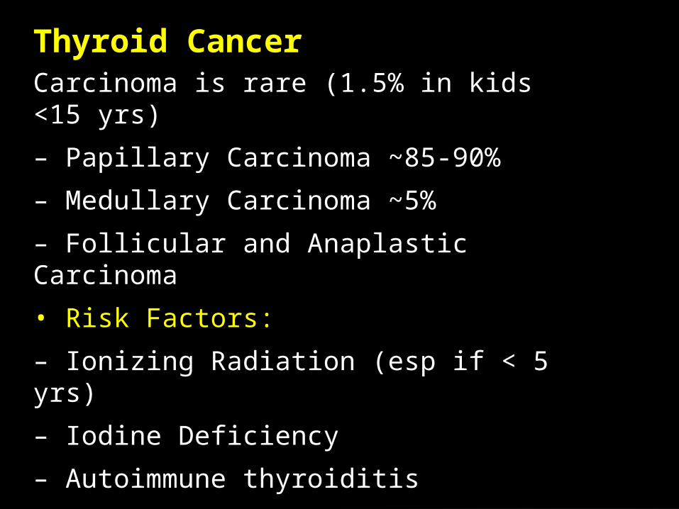
Carcinoma is rare (1.5% in kids <15 yrs)
– Papillary Carcinoma ~85-90%
– Medullary Carcinoma ~5%
– Follicular and Anaplastic Carcinoma
• Risk Factors:
– Ionizing Radiation (esp if < 5 yrs)
– Iodine Deficiency
– Autoimmune thyroiditis
– Prolonged TSH elevation
– Family history (MEN)
Thyroid Cancer