Embed Size (px)
Citation preview

Article
Intrinsic Variable Learning
for Brain-MachineInterface Control by Human Anterior IntraparietalCortexHighlights
d AIP neurons learn to modulate their activity to compensate
for errors in BMI tasks
d Changes in the neural activity reflect a cognitive re-
adaptation mechanism
d AIP fails to compensate for errors when novel neural activity
patterns are required
d Learning in AIP is constrained by the pre-existing neuronal
structure
Sakellaridi et al., 2019, Neuron 102, 694–705May 8, 2019 ª 2019 Elsevier Inc.https://doi.org/10.1016/j.neuron.2019.02.012
Authors
Sofia Sakellaridi,
Vassilios N. Christopoulos,
Tyson Aflalo, ..., Debra Ouellette,
Nader Pouratian, Richard A. Andersen
In Brief
Sakellaridi, Christopoulos, et al. studied
the learning mechanism in human AIP
using brain-machine interface
paradigms. They found that changes in
neural activity during learning reflect
cognitive re-adaptation mechanisms.
When cognitive strategies were not
adequate for compensation, AIP failed
to learn.

Neuron
Article
Intrinsic Variable Learning for Brain-MachineInterface Control by Human AnteriorIntraparietal CortexSofia Sakellaridi,1,2,5,6,* Vassilios N. Christopoulos,1,2,5 Tyson Aflalo,1,2 Kelsie W. Pejsa,1,2 Emily R. Rosario,3
Debra Ouellette,3 Nader Pouratian,4 and Richard A. Andersen1,21Division of Biology and Biological Engineering, California Institute of Technology, Pasadena, CA 91125, USA2Tianqiao and Chrissy Chen Brain-Machine Interface Center, Chen Institute for Neuroscience, California Institute of Technology, Pasadena,CA 91125, USA3Casa Colina Hospital and Centers for Healthcare, Pomona, CA 91767, USA4Department of Neurosurgery, Interdepartmental Program in Neuroscience, and Brain Research Institute, David Geffen School of Medicine
at UCLA, Los Angeles, CA 90095, USA5These authors contributed equally6Lead Contact
*Correspondence: [email protected]
https://doi.org/10.1016/j.neuron.2019.02.012
SUMMARY
Although animal studies provided significant insightsin understanding the neural basis of learning andadaptation, they often cannot dissociate betweendifferent learning mechanisms due to the lack of ver-bal communication. To overcome this limitation, weexamined the mechanisms of learning and its limitsin a human intracortical brain-machine interface(BMI) paradigm. A tetraplegic participant controlleda 2D computer cursor by modulating single-neuronactivity in the anterior intraparietal area (AIP). Byperturbing the neuron-to-movement mapping, theparticipant learned to modulate the activity of the re-corded neurons to solve the perturbations by adopt-ing a target re-aiming strategy. However, when nocognitive strategies were adequate to produce cor-rect responses, AIP failed to adapt to perturbations.These findings suggest that learning is constrainedby the pre-existing neuronal structure, although it ispossible that AIP needs more training time to learnto generate novel activity patterns when cognitivere-adaptation fails to solve the perturbations.
INTRODUCTION
The brain-machine interface (BMI) is an emerging technology
that translates brain activity into motor actions. Besides being
a powerful tool that enables people with paralysis to control
external devices with their thoughts, the BMI has also shed light
on the neural representation of cognitive functions, such as
learning and motor adaptation (Armenta Salas and Helms Tillery,
2016; Carmena, 2013; Chase et al., 2012; Ganguly and Car-
mena, 2010; Golub et al., 2018; Hwang et al., 2013; Koralek
694 Neuron 102, 694–705, May 8, 2019 ª 2019 Elsevier Inc.
et al., 2012; Sadtler et al., 2014; Wander et al., 2013). The main
advantage of BMIs for studying learning is that brain plasticity
associated with learning and adaptation can be explored within
a particular node in a network, without taking into account the
biomechanics of the limb or networks at lower levels, such as
the spinal cord (Carmena et al., 2003). Although recent BMI
studies have showed that behavioral performance improves
with practice, there is no strong consensus on the nature of
learning and its constraints in humans (Aflalo et al., 2015; Collin-
ger et al., 2013; Hochberg et al., 2006, 2012; Truccolo et al.,
2008; Wang et al., 2013). The two prevailing mechanisms that
have been considered to account for changes in neuronal activ-
ity associatedwith learning are the individual-neuronmechanism
and the intrinsic-variable mechanism. The first one posits that
entirely new patterns of neural activity can be learned by individ-
ual neurons or populations of neurons (Cerf et al., 2010; Chapin
et al., 1999; Fetz, 1969, 2007; Fetz and Baker, 1973; Fetz and Fi-
nocchio, 1971; Gage et al., 2005). In groundbreaking research,
Fetz and colleagues provided evidence that non-human pri-
mates (NHPs) could modulate single motor cortex neurons or
even pairs of neurons in opposite directions (Fetz, 1969, 2007;
Fetz and Baker, 1973). Moritz and colleagues further showed
that pairs of motor cortex neurons could arbitrarily change their
tuning to control stimulation of antagonist muscles in NHP limbs
in a limb movement task (Moritz et al., 2008). Although these
findings suggest that certain brain areas can generate novel
and arbitrary patterns of activity, an equally plausible interpreta-
tion is that animals generate only existing patterns of activity
within new contexts (Chase et al., 2010; Golub et al., 2018;
Hwang et al., 2013; Jarosiewicz et al., 2008). These patterns of
activity may be constrained by the pre-existing structure of the
network, and animals learn to manipulate an intrinsic variable
of the natural movement, such as target direction, to generate
activity patterns that comply with the new environmental de-
mands (Jarosiewicz et al., 2008; Paz et al., 2003; Paz and Vaadia,
2004; Wise et al., 1998). This mechanism prevents independent
adaptation of individual neurons, because the cognitive strategy

influences a global network of neurons that are sensitive to the
intrinsic variable. Therefore, it is still unclear whether changes
in neural activity that accompany learning reflect individual-
neuron or intrinsic-variable learning.
To dissociate between these two learning mechanisms, we
need to explore whether brain areas are capable of generating
activity patterns that cannot be associatedwith any possible nat-
ural movement. The lack of any introspective description of stra-
tegies in animal BMI models of learning make the dissociation
between the two mechanisms at best correlative and inferred.
For instance, given that animals cannot be verbally instructed
on which effector to use to perform intended movements (e.g.,
thinking of using only the right wrist) in a BMI task, it would not
be possible to rule out that animals learn to produce the correct
responses by using cognitive strategies to explore portions of
the intrinsic manifold that were not identified in the baseline por-
tions of the task (e.g., generating unexplored patterns by thinking
about wiggling the toes) rather than generating truly novel pat-
terns of activity. These limitations reveal the importance of using
human subjects, who can be instructed to follow specific rules
during BMI tasks and can report whether cognitive strategies
were adopted to produce the correct responses. In the current
study, a human participant with tetraplegia (C3-C4 complete
lesion; 6 years post-injury) was implanted with a microelectrode
array in the anterior intraparietal area (AIP) and learned to modu-
late the activity of a single neuron to control a 2D cursor in a goal-
decoding BMI center-out task with two peripheral stimuli. The
participant was instructed to attempt intendedmovements using
only the right wrist and to not switch effectors during the task.
She was also instructed to verbally report the direction of the in-
tended movement after each trial in addition to any other strate-
gies she employed to solve the task. Only the start and the end
position of the cursor were presented to the participant. In a
BMI-pro task, the neuron’s intrinsic coding for desired direction
of wrist movements was used to decode the desired goal. In a
BMI-anti task, the neuron was trained to switch its activity so
that it was active for desired movements opposite the direction
of the goal stimulus. Besides the verbal feedback from the
participant, we also looked at the behavior of neurons that
were not used for decoding but were found to be selective for
wrist movement and therefore were part of the pre-existing
structure that plans wrist movements. These neurons are
referred to as untrained neurons, as opposed to trained neurons
that are used for decoding and, therefore, directly contribute to
the BMI output. If the untrained neurons do not change their
turning in the BMI-anti task, then the individual neuron mecha-
nism predominates; if they flip their preferred directions, then
intrinsic variable learning predominates. In favor of the intrinsic
variable mechanism, we found that both trained and untrained
neurons changed their tuning in the BMI-anti task. This change
reflected an explicit strategic choice of the subject, as the partic-
ipant reported that she solved the task by attempting intended
wrist movements in the direction opposite to the stimulus (i.e.,
re-aiming strategy).
We further tested whether the learning mechanism in AIP
varies with the complexity of the BMI task. In a second BMI
perturbation experiment, the participant had to control the
cursor by simultaneously modulating the activity of two trained
neurons. Similar to the first experiment, the activity patterns of
the trained and untrained neurons, aswell as the verbal feedback
from the participant, provide evidence in favor of the intrinsic
variable learning mechanism. These findings suggest that the
intrinsic variable learning mechanism predominates in AIP
regardless of the task complexity. However, it could be argued
that these tasks favor the intrinsic variable learning over the indi-
vidual neuron learning, because there is always a cognitive solu-
tion that can produce the correct responses. We designed a BMI
perturbation task in which no cognitive strategy could produce
the neural activity patterns necessary to solve the task. In this
case, an individual neuron mechanism was required. The partic-
ipant could not produce the correct responses, indicating that
AIP neurons were not capable of generating novel and arbitrary
patterns of activity to solve the task. This suggests that either AIP
neuron can only engage pre-existing structures of the neural
network for short-term learning (about 1 h) or a much more
extensive period of training may be required if AIP neurons can
learn to generate entirely new neuronal activity patterns.
RESULTS
Volitional Control of Single Neurons in a Goal-DecodingBMI TaskWe designed a BMI center-out task in which a participant with
tetraplegia intended wrist movements toward one of two stimuli
presented at diametrically opposing locations (i.e., up-down;
Figure 1A). Each session consisted of three task blocks in the
following order: BMI-calibration; BMI-pro; and BMI-anti. BMI-
calibration trials started with a peripheral cue randomly pre-
sented for 0.8 s at one of the two potential locations on the
screen. Following the cue offset, a delay period of 1.5 s ensued.
After the go signal, the participant had 2 s to attempt a wrist
movement toward the direction of the stimulus location. The
only verbal instruction given prior to the beginning of each ses-
sion was ‘‘attempt a wrist movement to the stimulus location af-
ter the go signal.’’ The average firing rate activity of the recorded
neurons after the go signal in the BMI-calibration was used to
identify a single neuron that best decodes the stimulus loca-
tion—i.e., a neuron that fires more spikes when intended wrist
movements are made to one stimulus over the other. We refer
to this neuron as trained, because it was used for decoding
and therefore directly contributed to the BMI output.
The BMI-pro task had a similar sequence to the calibration,
with an extra feedback period following the go period. The stim-
ulus feedback location was decoded from the firing rate of the
trained neuron during the go period, according to the following
stimulus response rule. If the firing rate of the trained neuron
was higher for stimulus S1 than for stimulus S2 in the calibration,
the BMI-pro rule was that higher firing rates for stimulus S1 than
for S2would result in correct trials—i.e., the participant should in-
crease the activity of the neuron in response to S1 and suppress
the activity of the neuron in response to S2 (Figure 1B). We used
linear discriminant analysis (LDA) to identify the firing rate
threshold that divides the high and the low firing rates (see
STAR Methods section for more details). If the firing rate con-
formed to the stimulus response rule, the stimulus feedback
location was placed at the same location as the stimulus location
Neuron 102, 694–705, May 8, 2019 695

Figure 1. Task Event Sequences and Stimulus-Response Rules of the Brain-Machine Interface Learning Experiment with a Single Neuron
(A) A graphical representation of the brain-machine interface (BMI) learning paradigm with a single neuron.
(B) The stimulus response rules for the BMI-calibration (left), the BMI-pro task (middle), and BMI-anti task (right). Each panel illustrates the firing rate (mean ± SEM)
of a hypothetical trained neuron for successful trials in each of the three tasks.
in the cue target period and a ‘‘yes’’ message was displayed on
the screen, indicating a successful trial. Otherwise, the stimulus
feedback location was placed opposite to the cue stimulus and a
‘‘no’’ message was presented, indicating an unsuccessful trial.
The BMI-anti task enforced the opposite stimulus response
rule of lower firing rate for stimulus S1 than for S2. This rule forced
the trained neuron to flip its preferred stimulus associated with
higher firing rates, between the BMI-pro and the BMI-anti trials.
In the BMI-anti task, the stimulus feedback location was placed
opposite to the intended wrist direction for successful trials and
at the same location otherwise.
The participant performed 8 experimental sessions in total,
each on different days. For each session, the neuron that
best discriminated between the two stimuli was selected as
the ‘‘trained’’ neuron. We did not attempt to maintain the
same neuron across sessions, in part, because we found that
neurons recorded on a channel would change from day to
day. The activities of two single trained neurons recorded in
the 1st and 3rd sessions are illustrated in Figures 2A and 2B,
respectively. In the BMI-pro task, the firing rates of the trained
neurons were properly discriminated between the two stimuli in
both sessions (Figures 2A and 2B, left column), because the
participant intended movements in the direction of the dis-
played stimulus. However, in the BMI-anti task, the trained
neuron of the 1st session was not correctly discriminated, indi-
cating that the participant was not able to modulate the activity
of the trained neuron to conform to the new stimulus response
rule (Figure 2A, middle and right panels). According to the ver-
bal report, the participant was still mostly performing intended
wrist movements to the direction of the presented stimulus,
696 Neuron 102, 694–705, May 8, 2019
even though she was also exploring some new strategies,
such as intending faster wrist movements. During the 3rd
session, and particularly at the end of the 1st block of the
BMI-anti task, the participant found and reported that the
best strategy to solve the task was to intend wrist movements
in a direction opposite to the stimulus goal (anti-wrist move-
ments). At this time, the firing rate of the trained neuron was
able to properly discriminate by producing the opposite pattern
from the BMI-pro task (Figure 2B, right panel). To confirm that
the activity of the trained neuron was modulated by anti-wrist
movements, we looked at the activity of the untrained neu-
rons—i.e., neurons that do not participate in the BMI control
but are part of the pre-existing structure that plans the wrist
movements. If the trained neuron modulates its activity inde-
pendently from the pre-existing structure (i.e., individual-neuron
mechanism), the untrained neurons will not flip their preferred
stimulus, similar to the trained neuron. On the other hand, if
the participant learns a new cognitive strategy (i.e., attempting
anti-wrist movements), the untrained neurons will also flip their
preferred stimulus (i.e., intrinsic variable learning mechanism).
Figures 3A and 3B depict the firing rates of one trained and
two untrained neurons, respectively, from a BMI-pro (top row)
and BMI-anti (bottom row) task from a typical session (i.e., neu-
rons recorded from a single day). Consistent with intrinsic var-
iable learning, the trained and untrained neurons flipped their
preferred stimuli in the BMI-anti task, verifying that the partici-
pant aimed in the opposite direction to the stimulus. The
average performance in the BMI-pro task across all 8 sessions
was 0.89 ± 0.11 (mean ± SD). For the BMI-anti task, in the first 2
sessions and the first block of the 3rd session, the average

Figure 2. Firing Rate Distributions of Single Neurons in BMI-Pro and BMI-Anti Tasks
(A and B) The firing-rate distributions of two different single trained neurons for each of the two stimuli in the BMI-pro and BMI-anti task blocks in (A) session 1
and (B) session 3 for both correct and incorrect trials.
performance was 0.21 ± 0.16. After adopting the anti-wrist
movement strategy, the performance increased to 0.84 ±
0.09 (the last block of the 3rd session + the last 5 sessions).
Moreover, 91 untrained cells (out of a total of 281 recorded un-
trained cells across the last 5 sessions and the last block of the
3rd session) showed activity that discriminated between S1 and
S2 during the go period of the BMI-pro task. Eighty of those 91
cells (�88%) flipped their preferred stimulus between BMI-pro
and BMI-anti tasks during the go period (two-tailed t test p <
0.05; Figure 3C). The average activity of the untrained neurons,
whose activity flipped between the BMI-pro and BMI-anti task,
did not change between these two tasks (two-tailed t test p >
0.05 for all the untrained neurons that flipped their preferred
stimulus). The remaining 11 units (�12%) did not flip their
preferred stimulus.
Learning Mechanisms and Task ComplexityAlthough the single-neuron BMI experiment provides evidence in
favor of the intrinsic variable learning mechanism, it could be hy-
pothesized that individual neuron learning would be more likely
to be pursued for more cognitively complex tasks. In a second
experiment, we explored whether the preferred learning mecha-
nism varies with the task complexity. We designed a BMI-fsb
(fsb, feasible) task in which the cursor location was controlled
by the activity of two trained neurons at the same time. We refer
to this task as feasible, because it is still possible for the partic-
ipant to solve it by adopting a new cognitive strategy. Each
session consisted of three different task blocks in the following
order: BMI-calibration; BMI-pro; and BMI-fsb (Figure 4A). At
each stage, there were three epochs, cue target, delay, and
go, similar to the single-neuron BMI experiment. In the BMI-cali-
bration, the participant attempted wrist movements to eight radi-
ally arranged targets. Based on the tuning properties of two
trained neurons in calibration, we selected a pair of stimulus
locations (out of
�82
�= 28 possible pairs of stimuli) to be pre-
sented in the BMI-pro and the BMI-fsb tasks. The linear bound-
ary in the BMI-pro task was the one that best separated the two
firing-rate clusters, each formed by the activity of the two trained
neurons during the go period, for each stimulus in the BMI-cali-
bration. On the other hand, we selected the linear boundary for
the BMI-fsb task under two constraints: (1) it provides about
50% classification accuracy for both stimuli and (2) it best sepa-
rates the two clusters of another pair of targets. Therefore, the
participant could either alter the cognitive strategy (i.e., re-aim-
ing to directions away from the presented stimuli) or produce
new activity patterns through independent adaptation of the
trained neurons to match with the new stimulus response rule.
Both the BMI-pro and BMI-fsb tasks had the same sequence
as the BMI-calibration with an extra feedback period following
the go signal. A simplified, conceptual illustration of the stimulus
response rules in the two BMI tasks for the pair of stimuli (1, 5) is
shown in Figure 4B. The linear threshold (black continuous line)
in the BMI-pro task accurately separates the two firing-rate clus-
ters. On the other hand, the BMI-fsb threshold (black discontin-
uous line) divides the two clusters such that half of the trials lie
above and half below the threshold for both stimuli. However,
it accurately separates the clusters for intended wrist move-
ments toward the direction of targets 3 and 7 presented in the
BMI-calibration. Therefore, the participant could either attempt
wrist movements to the direction of 3 and 7 in response to stimuli
1 and 5, respectively, or she has to learn to generate activity
patterns that conform to the stimulus response rule through in-
dependent adaptation of the two trained neurons.
The participant performed 15 experimental sessions in total,
each on different days, with different pairs of stimuli and different
Neuron 102, 694–705, May 8, 2019 697

Figure 3. Trained and Untrained Neurons Flip Their Preferred Stimuli in the BMI-Anti Task(A) The temporal dynamics of the firing rates for a trained AIP neuron that directly contributed to the BMI output in a typical session.
(B) Similar to (A) but for two untrained AIP neurons.
(C) The percentage of untrained neurons that flip their activity between BMI-pro and BMI-anti tasks after the participant adopted the anti-wrist movement
strategy.
linear threshold boundaries that ranged in direction between 0�
(i.e., horizontal line) and 180�. The average direction of the
threshold in the BMI-pro task was 88.8� ± 59.2� (mean ± SD)
and in the BMI-fsb task was 86.2� ± 55.8� across all 15 trials.
The average angle between the BMI-pro and the BMI-fsb
threshold was 95� ± 28.4�. In the BMI-pro task, the participant
immediately produced the appropriate activity patterns for
both trained neurons, achieving an average performance of
0.88 ± 0.12 (mean ± SD) across all 15 sessions. Within a time-
scale of a session, she also learned to generate activity patterns
that comply with the stimulus response rule in the BMI-fsb task,
achieving 0.78 ± 0.21 decode performance across all 15 ses-
sions. A characteristic example is illustrated in Figure 5, in which
the pair of stimuli (1, 4) was selected based on the tuning prop-
erties of the two trained neurons in BMI-calibration (Figure 5A).
According to the stimulus response rule in the BMI-pro task,
the averaged activity of the two trained neurons should fall above
the linear threshold (black line) for stimulus 4 and below the
threshold for stimulus 1 for successful trials. The task perfor-
mance accuracy was over 95% in the BMI-pro task, in which in-
tended wrist movements were planned to the direction of the
presented stimuli (Figure 5B). In the BMI-fsb task, the linear
threshold was selected so that it substantially deteriorated the
performance for intended wrist movements to the presented
stimuli (black discontinuous trace; Figure 5A). However, the
participant learned to generate rule-complying activity patterns
(Figure 5C), achieving performance greater than 90%. One way
to modulate the neural activity of the trained neurons was to
aim in different directions. We examined the behavior of the
trained neurons during the BMI-calibration to determine whether
planning movements to any of the tested directions could
have produced activity patterns that comply with the stimulus
response rule in the BMI-fsb task. The activity for stimulus 1 in
the BMI-fsb task matched the activity for intended wrist move-
ment toward stimulus 3, and the activity for stimulus 4 matched
the activity for intended wrist movements to stimulus 7 (Fig-
698 Neuron 102, 694–705, May 8, 2019
ure 5D). This is consistent with the verbal feedback provided
by the participant: she reported that she was mostly aiming in
the direction of stimulus 3 (cyan) in response to stimulus 1 and
toward stimulus 7 (red) in response to stimulus 4 (Figure 6A).
These findings suggest that the BMI-fsb task was solved by
re-aiming intended wrist movements—a form of intrinsic vari-
able learning. To confirm this hypothesis, we compared the ac-
tivity of the two trained neurons with 20 untrained neurons,
whose activity was significantly discriminated between the two
stimuli, for each successful BMI-fsb trial. We used a nearest-
neighbor decoding algorithm to infer the stimulus that the
neuronal population encoded. The decoding algorithm selected
the stimulus associated with the ensemble activity in the BMI-
calibration that was closest to the ensemble activity of a given
BMI-fsb trial in terms of the Euclidean distance. Figure 6B de-
picts the eight-target decoding results from the BMI-fsb task
across all successful trials (correspondence matrix). The x and
y axes represent the stimulus decoded by the two trained and
untrained neurons, respectively. The correspondence matrix
therefore allows us to visualize the degree of correspondence
between the stimulus specified by the trained and untrained
units. The two bright spots for stimuli 3 and 7 indicate that
they were encoded concurrently for the trained and the un-
trained neurons in most of the trials. These two stimuli were
the same two best-matching stimuli as inferred by the activity
pattern (Figure 5D) and reported by the participant (Figure 6A).
A more detailed analysis of how the activity distribution of the
untrained neurons is modulated to counter the perturbation of
the stimulus response rule in this particular example is pre-
sented in Figure S1. Overall, in about 50% of the BMI-fsb trials
across all sessions, both trained and untrained neurons en-
coded the same matching target (49.6% ± 3.93%; mean ±
SEM; across all 15 sessions; Figure 6C, blue curve). Similarly,
in about 50% of the BMI calibration trials, both trained and un-
trained neurons encoded the same cued target (48.4% ± 4.95%
trials across all 15 sessions; Figure 6C, red curve).

Figure 4. Task Event Sequences and Stimulus-Response Rules of the Brain-Machine Interface Learning Experiment with Two Neurons
(A) A graphical representation of the BMI learning paradigm with two neurons.
(B) A simplified, conceptual illustration of the stimulus response rules for the BMI-pro and BMI-fsb trials for the pair of stimuli 1 and 5. Points correspond to the
hypothetical firing rates of the two trained neurons in response to four stimuli (color coded) in the BMI-calibration.
Learning Mechanisms in BMI Tasks with No FeasibleCognitive StrategiesSo far, we explored the learning mechanisms for BMI tasks in
which there was always a feasible intrinsic variable remapping
strategy to compensate for perturbations. An interesting ques-
tion is what would happen if no cognitive strategy was adequate
to solve the BMI tasks. To address this question, we designed a
BMI-nfsb task (nfsb, non-feasible) in which there was no strategy
that could produce rule-complying activity patterns.We selected
a linear threshold that performed poorly on classifying the firing-
rate clusters of the two trained neurons for at least one of the two
stimuli presented in the BMI-nfsb task. Ideally, we looked for
a linear boundary that results in chance performance (50%)
regardless of the intended movement direction. We used evolu-
tionary algorithms (EAs) to identify such a linear threshold (see
STAR Methods section for more details). EAs are adaptive heu-
ristic algorithms that mimic the process of natural selection (Ban-
dyopadhyay et al., 1995; de la Fraga and Coello Coello, 2011;
Goldberg, 1989). As such, they represent a principled exploita-
tion of a random search to solve complex optimization problems.
The results show that, in the timescale of a session (i.e., about
1 h), neurons could not generate activity patterns that conform
to the stimulus response rule in the BMI-nfsb task.
A characteristic example is depicted in Figure 7. Based on the
tuning properties of the two trained neurons from the BMI-cali-
bration (Figure 7A), we selected the pair of stimuli 2 and 7 in
the BMI-pro and BMI-nfsb tasks. This pair was selected because
there is a linear threshold (black solid line), named BMI-pro
threshold, which accurately separates the two firing-rate clus-
ters of the trained neurons—i.e., the firing rates are above this
linear threshold for intended wrist movements to the stimulus 7
and below it for intended movements to the stimulus 2. We
used this threshold in the BMI-pro task. On the other hand, the
EAs generated a linear threshold for the BMI-nfsb task (black
discontinuous line), in which the activity of the two trained neu-
rons should fall within the right and the left side of this boundary
in response to the stimuli 2 and 7, respectively. Figure 7B depicts
the percentage of the calibration trials that fall into the right (blue)
and the left (red) side of the BMI-nfsb threshold for intendedwrist
movements to all possible 8 targets. Notice that the threshold
poorly separates the firing-rate clusters of the two trained neu-
rons for intended wrist movements to the seven out of eight
possible targets—except for target 2, in which the activity of
the two trained neurons of all calibration trials falls into the right
side of the threshold. On the other hand, the highest percentage
(�65%) of trials that fall into the left side of the threshold corre-
sponds to intended movements toward target 1. Therefore,
when stimulus 7 is presented in the BMI-nfsb task, the best per-
formance that the participant could achieve is about 65% by
aiming toward the direction where target 1 was located in the
BMI-calibration. This suggests that there is no cognitive strategy
that can produce high performance accuracy in response to
stimulus 7 in the BMI-nfsb task. Consequently, an individual
neuron mechanism is required to compensate for changes on
the stimulus response rule. On the other hand, when stimulus 2
is presented, the best performance (100%) could be achieved
for intended wrist movements toward target 2, suggesting that
the participant should not modify her strategy.
We found that, in the BMI-pro task, the participant immedi-
ately produced appropriate activity patterns for the two trained
neurons to solve the task, achieving performance accuracy
over 90% (Figure 7C). On the other hand, the two neurons could
not learn to generate rule-complying activity patterns for trials in
which stimulus 7 was presented in the BMI-nfsb task, achieving
performance accuracy of about 50% (Figure 7D). The participant
reported that, although she was re-aiming to different directions
to compensate for the applied perturbation, none of them
seemed to offset the perturbation and improve her performance
when stimulus 7 was presented. Overall, we ran 6 sessions on
different days, in which there was always a feasible intrinsic var-
iable remapping strategy to acquire one stimulus (i.e., cognitively
solvable stimulus), but not the other (i.e., cognitively non-solv-
able stimulus), after perturbing the stimulus response rule (i.e.,
BMI-nfsb task). Note that both stimuli could be achieved with
Neuron 102, 694–705, May 8, 2019 699

Figure 5. Evidence of Intrinsic-Variable Learning in the BMI-fsb Task
(A–C) The firing rates for the two trained neurons in response to the stimuli 1 and 4 (colored coded) in (A) the 8-target BMI-calibration, (B) BMI-pro, and (C) BMI-fsb
tasks. The black solid and dashed lines represent the linear thresholds used for decoding in the BMI-pro and BMI-fsb tasks, respectively.
(D) The firing rates of the same two trained neurons in response to targets 3 (cyan) and 7 (red) in the BMI-calibration (the arrows indicate the relationship between
the stimulus and matched target locations). The two stimuli (cyan and red) correspond to the matched target locations.
high performance in the BMI-pro task by aiming directly to their
locations. We measured the performance as the number of trials
in which the activity of the two trained neurons fell on the correct
side of the boundary. The participant was able to produce the
appropriate activity pattern for the pair of trained neurons in
the BMI-pro and BMI-nfsb tasks for the cognitively solvable
stimulus, achieving performance of 94.03% ± 2.65% (mean ±
SEM) in the BMI-pro and 87.22% ± 3.0791% in the BMI-fsb
tasks across 6 sessions (no significant differences in the perfor-
mance between the two tasks for the cognitively solvable stim-
ulus; two-tailed t test p = 0.1324; Figure 7E). However, she could
not learn to produce rule-complying activity patterns for the
cognitively non-solvable stimulus in the BMI-nfsb task, and her
performance was significantly deteriorated from 88.90% ±
3.93% in the BMI-pro task to 50.0% ± 5.55% in the BMI-nfsb
task across 6 sessions (two-tailed t test p < 0.001; Figure 7E).
We interpret these findings as evidence that AIP neurons cannot
learn to produce novel and arbitrary patterns of activity to
comply with the stimulus response rule in the BMI-nfsb task.
These results suggest that either there is a constraint on learning
imposed by the existing neural structure of AIP ormore extensive
training is required to generate entirely novel neuronal activity
patterns.
DISCUSSION
GeneralAlthough a large body of evidence suggests that both humans
and animals are able to learn a variety of new tasks and adapt
to unpredictable changes of the environment, to our knowledge,
there have been no systematic studies of single-neuron learning
in humans. The prevailing hypotheses suggest that neurons
learn new tasks by either generating entirely new patterns of ac-
tivity, independently of the pre-existing neuronal structure (indi-
vidual neuron learning; Cerf et al., 2010; Chapin et al., 1999; Fetz,
1969, 2007; Fetz and Baker, 1973; Fetz and Finocchio, 1971;
Gage et al., 2005) or they combine existing patterns of activity
700 Neuron 102, 694–705, May 8, 2019
that are already part of the natural repertoire of the neural popu-
lation (intrinsic variable learning; Golub et al., 2018; Hwang et al.,
2013; Paz et al., 2003; Paz and Vaadia, 2004; Wise et al., 1998).
However, most of these studies have used laboratory animal
models, making it challenging (or even impossible) to distinguish
between these two mechanisms without verbal instruction and
feedback from the participants. To overcome this issue, we de-
signed BMI center-out intended wrist movement tasks, where a
human participant with tetraplegia controls a 2D computer
cursor by modulating the activity of AIP neurons. The participant
was instructed to use only the right wrist. Verbal feedback
reports were obtained on every trial about the strategy that
was followed to solve the BMI tasks. By perturbing the mapping
between the neural activity and the cursor location, we provide
evidence in favor of the intrinsic variable learning mechanism.
In particular, we found that the participant learned to volitionally
control neuronal activity in AIP by re-aiming in directions that
offset the perturbation. Importantly, when there were no cogni-
tive strategies that could solve the BMI tasks, AIP neurons could
not generate novel patterns of activity, independent of the pre-
existing neuronal structure. These findings suggest that the
pre-existing neuronal structure constrains learning, and this
may explain why learning a new task is easier when it is related
to skills that we already have.
From Animal- to Human-BMI: Human Participants Helpto Dissociate the Two Learning MechanismsAnimal BMI studies have significantly contributed in understand-
ing the mechanisms underlying learning and adaptation (Golub
et al., 2016). Their main advantage is that we can directly
compare neural activity between manual control and BMI con-
trol, enabling insights on the cortical reorganization during the
process of learning neuroprosthetic control (Carmena et al.,
2003; Chapin et al., 1999; Ganguly et al., 2011; Vyas et al.,
2018; Zacksenhouse et al., 2007). For instance, a growing
body of research indicates that, when switching from manual
control to neuroprosthetic control, the brain enters into a novel

Figure 6. Trained and Untrained Neurons
Encode the Same Matching Targets in the
BMI-fsb Task
(A) The proportion of trials for the eight targets that
the participant verbally reported to have attempted a
wrist movement in response to stimuli 1 and 4 during
the BMI-fsb task in the example session described in
Figure 5.
(B) Correspondence matrix for the BMI-fsb session
presented in Figure 5. It shows the proportion of
trials for the eight targets decoded by the trained
versus untrained neurons. The x axis represents the target decoded by the trained neurons and the y axis the target decoded by the untrained neurons in the
example session described in Figure 5.
(C) Probability distribution (blue trace) across 15 sessions of the difference between the targets decoded by the trained and untrained neurons in the BMI-fsb task
when the target decoded from the trained neurons was a matching target. The red trace describes the probability distribution from 15 sessions of the difference
between the targets decoded from the trained and untrained neurons in the 8-target BMI-calibration when the target decoded by the trained neurons was the
BMI-calibration target. The peak at zero corresponds to the case in which the trained and the untrained neurons encode the same matching target. Error bars
represent SEM, and red and blue lines show the spline interpolation to the data.
state, producing new representations in the untrained neurons
(Clancy et al., 2014; Ganguly et al., 2011; So et al., 2012). Animal
models also provide a valuable tool to gain additional insights
into the mechanism of neuroplasticity in learning during BMI by
disrupting neural activity using invasive techniques, such as op-
togenetics (Gulati et al., 2017). Similar to our work, previous BMI
studies in NHPs perturbed the neuron-to-movement map and
provided evidence that animals learn to volitionally control the
activity of the recorded neurons to adapt to perturbations (Golub
et al., 2018; Hwang et al., 2013; Jarosiewicz et al., 2008). Consis-
tent with our findings, Hwang et al. (2013) showed that NHPs can
alter the pattern of activity of a trained posterior parietal cortex
(PPC) neuron in a BMI task; however, the simultaneously re-
corded untrained neurons also changed their activity patterns
in a predictable manner consistent with the intrinsic variable
learning mechanism (Hwang et al., 2013). The activity of the un-
trained PPC neuronsmaintained consistent strength of tuning af-
ter the animals adapted to external perturbations, as we also
found in the untrained AIP neurons. Additionally, in a neural pop-
ulation M1 study, after dimensionality reduction, NHPs were
trained to make BMI movements within or out of the manifold
of intrinsic dimensions in M1 (Sadtler et al., 2014). Within the
manifold, perturbations could be solved by adopting new
cognitive strategies, whereas out-of-the-manifold perturbations
require the animals to generate novel neuronal activity patterns.
The results showed that the animals could solve the within the
manifold perturbations in a single day by re-associating existing
activity patterns with different intended movements (a form of
intrinsic variable learning; Golub et al., 2018). However, the
out-of-the-manifold perturbations could not be solved. These
findings are consistent with easier intrinsic variable (i.e., within
themanifold) rather than individual neuron (i.e., outside themani-
fold) learning mechanisms.
Although providing important insights, it could be argued that
NHP studies cannot conclusively determine the type of learning
mechanisms employed to overcome external perturbations. This
is because NHPs cannot be asked what strategy they follow to
solve a task and they cannot be instructed to constrain how
they solve the task. A constrained strategy is important to disso-
ciate between learning mechanisms, especially when recording
from cortical regions with high-dimensional mixed representa-
tions. Recent electrophysiological studies reported that neurons
in NHP prefrontal cortex, rodent PPC, and human AIP are tuned
to mixtures of task-related variables (Fusi et al., 2016; Raposo
et al., 2014; Rigotti et al., 2013; Zhang et al., 2017). Therefore,
a BMI-nfsb task does not necessarily require implicit generation
of novel activation profiles. Instead, it is possible that the partic-
ipant could solve the task by attempting movements using other
effectors or cognitive strategies (e.g., finger, arm, or foot move-
ments). Given that NHPs and rodents are non-verbal animals,
they can explore and select other intrinsic variables to solve
the BMI tasks. In our study, we constrained the participant to
attempt intended movements using only her right wrist. The
participant was allowed to explore different cognitive strategies
for improving her performance, but she had to always perform
the intended actions by using her right wrist. By verbally instruct-
ing the participant to use a constrained strategy, we restrict the
available interpretations to intrinsic variable or individual neuron
learning mechanisms.
Verbal reporting is also important to interpret the behavior
of the untrained neurons—i.e., neurons that do not directly
contribute to the BMI output but are part of the pre-existing
neuronal structure that plans the wrist movements. According
to our findings, both trained and untrained neurons fluctuate their
activity together and in a predictable way based on the cognitive
strategy used. Although this is a form of intrinsic variable
learning, it could be equivalently interpreted as individual neuron
learning that affects both the trained and the untrained neurons.
In other words, cortical plasticity may result in new patterns of
activity in both trained and untrained neurons, even when indi-
vidual neuron learning occurs. However, the participant reported
a target re-aiming strategy, indicating that the changes of the
neuronal activity in the trained and untrained neurons reflect
an intrinsic variable learning mechanism. Overall, verbally in-
structed strategies and self-reports are major advantages,
because they can better reveal how individuals learn new tasks
and compensate for perturbations. However, it could be argued
that the participant may not be capable of providing an accurate
description of her strategies, because she might be unaware of
changes in her mental strategy when trying to compensate for
the perturbation. Although this is possible, whatever changes
in her strategy may have occurred proved incapable of
Neuron 102, 694–705, May 8, 2019 701

Figure 7. AIP Fails to Generate Novel Patterns of Activity in the BMI-nfsb Task
(A) The firing rates for the 2 trained neurons in response to the eight stimuli (color and shape coded) in the 8-target BMI-calibration. The pair of stimuli 2 and 7 was
selected in the BMI-pro and BMI-nfsb tasks. The black solid and dashed lines represent the linear boundaries used for decoding in the BMI-pro and BMI-nfsb
tasks, respectively.
(B) Percentage of trials that fall into the left (red) and right (blue) side of the BMI-nfsb task threshold in response to the eight stimuli in the 8-target BMI-calibration.
(C) The firing rates for the two trained neurons in response to the stimuli 2 and 7 in the BMI-pro. To be successful, the activity of the two trained neurons should fall
below and above the BMI-pro threshold in response to stimuli 2 and 7, respectively.
(D) Similar to (C) but for the BMI-nfsb task. To be successful, the activity of the two trained neurons should fall on the right and left side of the BMI-nfsb threshold in
response to stimuli 2 and 7, respectively.
(E) Proportion of successful trials (i.e., performance) across 6 sessions. Successful trials were achieved when the activity of the trained neurons during the go
period fell on the correct side of the boundary. Participant achieved performance accuracy 94.03%±2.65% (mean ± SEM) in the BMI-pro and 87.22%±3.0791%
in the BMI-nfsd across 6 sessions (no significant difference in the performance; two-tailed t test; p = 0.1324) for the cognitively solvable stimulus—i.e., stimulus
that could be acquired by adopting a cognitive strategy. However, the performance was significantly deteriorated from 88.90% ± 3.93% in the BMI-pro to
50.0% ± 5.55% in the BMI-nfsb (two-tailed t test p < 0.001) across the 6 sessions for the cognitively non-solvable stimulus—i.e., stimulus that could not be
acquired by adopting any cognitive strategy. Instead, an individual neuron mechanism was required to generate novel patterns of activity and was not
accomplished during each daily session.
generating novel activity patterns that comply with the stimulus
response rule in the BMI-nfsb task. Further, from this perspec-
tive, an inability of the participants to explain how a solution is
achieved itself becomes an interesting finding, as may be the
case for individual neuron learning.
When Cognitive Strategies Are Not Enough to Solve theBMI TasksOne major limitation in most of the previous studies is that they
assessed the neural mechanisms of learning in different brain
areas by using BMI tasks that favor one hypothesis over the
other. For instance, it could be argued that perturbations that
can be solved by adopting new cognitive strategies, such as
re-aiming to different directions, favor the intrinsic variable
702 Neuron 102, 694–705, May 8, 2019
learning over the individual neuron learning mechanism. Hence,
it is likely that trained neurons could modulate their activity inde-
pendently from the pre-existing structure of the brain network
only when cognitive strategies fail to solve the BMI tasks. To
test this hypothesis, we designed a BMI task in which there
was no cognitive strategy that can produce neural activity pat-
terns that conform to the stimulus response rule. In this case,
an individual neuron mechanism is required. Because the partic-
ipant was instructed to use only the right wrist for intending
movements, successful outcomes would indicate that AIP neu-
rons are capable of generating arbitrary patterns of activity that
do not belong to the response set of the BMI movements. The
results show that AIP neurons could not generate novel and arbi-
trary patterns of neural activity, at least within a single session, to

solve the BMI perturbation task. This result suggests that either
the individual neuron mechanism is a slow learning process—
and therefore extensive training is required before neurons can
generate novel and arbitrary activity patterns—or AIP cannot
elicit arbitrary activity patterns.
Constraints on LearningAlthough our findings suggest that learning is constrained by the
neural structure of the implanted area, it does not imply that
intrinsic variable learning is the only mechanism for learning in
AIP or other brain areas. Instead, it could be hypothesized that
learning in BMI tasks, in which cognitive strategies can produce
rule-complying activity patterns, involves fast timescale mecha-
nisms that underlie adaptation (Salinas 2004). On the other hand,
when no cognitive strategies are adequate to solve the BMI
tasks, learning may engage mechanisms required for acquiring
new skills (Picard et al., 2013; Rioult-Pedotti et al., 2000), and
therefore, people may need an extensive period of training to
improve their performance (Sadtler et al., 2014). Additionally, it
is possible that the individual neuron learning mechanism exists
in other brain areas, such as the primary motor cortex (M1)
(Ganguly et al., 2011). For instance, neural recordings in M1
from NHPs during brain control showed that the effects were
subtle on untrained neuron activity, usually consisting of a reduc-
tion in gain rather than changes in tuning direction (Ganguly et al.,
2011). Noticeably, the gain reduction of the untrained neurons af-
ter learning brain control of a cursor seems to be at odds with our
findings—i.e., no significant gain modulation of the untrained
neurons after learning to adapt to perturbations in BMI tasks.
However, Ganguly et al. (2011) observed the down-modulation
of the untrained neurons when comparing manual control with
brain control. On the other hand, the comparison in our study
was between two BMI tasks—i.e., BMI-pro and BMI-anti—and
therefore it is possible that the gain of the untrained neurons
was already reduced in the BMI-pro task—a hypothesis that
could not be tested due to lack of manual control data from
the participant. Furthermore, Jarosiewicz et al. (2008) found
that, although the dominant response to a rotation of a subset
of the neural population was compensation based on re-aiming
(a form of intrinsic-variable learning that affects all of the neur-
ons in the population equally), re-weighting and re-mapping
compensation (forms of individual neuron learning in which the
tuning curves of a subset of neurons in the population are modu-
lated through a plastic mechanism) were also present in the neu-
ral responses (Chase et al., 2010; Jarosiewicz et al., 2008).
Although these findings favor some degree of short-term individ-
ual neuron learning, the lack of verbal feedback cannot conclu-
sively support one mechanism over the other for the reasons
that we explained above.
Where Does Intrinsic Variable Learning Occur?We showed that intrinsic variable learning is the predominant
strategy in AIP to compensate for perturbations in BMI tasks.
However, this does not necessarily mean that learning occurs
within AIP. In fact, it is likely that learning emerges in other brain
regions and then is transferred to AIP. An intrinsic variable
learning mechanism, such as re-aiming to directions that offset
the perturbation, should be implemented by brain areas that
remap the actual stimulus location to intended motor actions.
Most of the recorded AIP neurons in our study encode only the
selected motor action (see example units in Figures 3A and
3B)—i.e., activity increases only after the go signal. One candi-
date area that transforms sensory information (i.e., target loca-
tion) into motor actions (i.e., intended arm movements) is the
human homolog of the macaque parietal reach region (PRR). A
study in our lab showed that PRR neurons encode the task
rule before the appearance of the stimulus location (Gail and An-
dersen, 2006). By training NHPs to perform pro- and anti-reach
movements and presenting the task rule (i.e., pro- versus anti-
reaches) prior to stimulus onset, Gail and Andersen (2006) re-
ported that PRR neurons integrate cue location with the task
rule to encode the desired action plan. Therefore, one hypothesis
is that human PRR combines the stimulus location with the stim-
ulus response rule (i.e., intended movements to the stimulus
location versus re-aiming to different directions) to generate
the appropriate action plans. Then, this information is transferred
to AIP and other motor regions to specify the characteristics of
the action plans. This hypothesis is also supported by other
studies showing that PRR neurons encode both the potential
and the selected reach plans in contrast to other regions within
PPC, such as area 5d (Cui and Andersen, 2011) and the primary
motor cortex (Cisek and Kalaska, 2005), which encode only the
selected reach plan. Overall, it is likely that intrinsic variable
involving spatial remapping of reach targets occurs in PRR
before it is transferred to AIP. However, we do not have
recording data from human PRR, and therefore, further work is
required to validate this hypothesis.
ConclusionsThis study is the first systematic examination of single neuron
learning in a human BMI paradigm, which is an important
advance considering previous studies used laboratory animal
models for eventual application to humans. We recorded neural
activity from a tetraplegic human participant implanted with a
96-channel microelectrode array in AIP to test two potential
mechanisms of learning in BMI tasks. By perturbing themapping
between the neural activity and the cursor location, we found
that the participant learned to volitionally control single neuron
activity in AIP by using existing patterns of activity that are
already part of the natural repertoire of the neuronal populations.
These findings suggest that the ability to learn new BMI tasks is
limited by the pre-existing structure of the brain network. It re-
mains to be determined whether AIP and/or other brain areas
in humans can learn to generate novel and arbitrary patterns of
activity over longer timescales.
STAR+METHODS
Detailed methods are provided in the online version of this paper
and include the following:
d KEY RESOURCES TABLE
d CONTACT FOR REAGENT AND RESOURCE SHARING
d EXPERIMENTAL MODEL AND SUBJECT DETAILS
d METHOD DETAILS
B Behavioral setup
Neuron 102, 694–705, May 8, 2019 703

704
B Implant methodology and physiological recordings
B Data collection
B Task procedure
d QUANTIFICATION AND STATISTICAL ANALYSIS
B Linear discriminants in the BMI tasks with one neuron
B Linear discriminants in the BMI tasks with two neurons
d DATA AND SOFTWARE AVAILABILITY
d ADDITIONAL RESOURCES
SUPPLEMENTAL INFORMATION
Supplemental Information can be found with this article online at https://doi.
org/10.1016/j.neuron.2019.02.012.
ACKNOWLEDGMENTS
This work was supported by the National Institute of Health (5R01EY01554512),
the Tianqiao and Chrissy Chen Brain-Machine Interface Center at Caltech, the
Boswell Foundation, and the Swartz Foundation. The authors would also like
to thank subject N.S. for participating in the studies and Viktor Scherbatyuk
for technical assistance.
AUTHOR CONTRIBUTIONS
S.S., V.N.C., T.A., and R.A.A. designed the study. S.S., V.N.C., and T.A. devel-
oped the experimental tasks. S.S. and V.N.C. collected the data. S.S., V.N.C.,
and T.A. analyzed the results. S.S., V.N.C., T.A., and R.A.A. interpreted results
and wrote the paper. E.R.R. provided experimental facilities, administrative
assistance, and coordination with Casa Colina Hospital and Centers for
Healthcare. K.W.P. provided administrative assistance. D.O. provided onsite
assistance during experimental sessions. N.P. performed the surgery implant-
ing the recording arrays in subject N.S.
DECLARATION OF INTERESTS
The authors declare no competing interests.
Received: May 17, 2018
Revised: November 5, 2018
Accepted: February 6, 2019
Published: March 7, 2019
REFERENCES
Aflalo, T., Kellis, S., Klaes, C., Lee, B., Shi, Y., Pejsa, K., Shanfield, K., Hayes-
Jackson, S., Aisen, M., Heck, C., et al. (2015). Neurophysiology. Decoding mo-
tor imagery from the posterior parietal cortex of a tetraplegic human. Science
348, 906–910.
Armenta Salas, M., and Helms Tillery, S.I. (2016). Uniform and non-uniform
perturbations in brain-machine interface task elicit similar neural strategies.
Front. Syst. Neurosci. 10, 70.
Bandyopadhyay, S., Murthy, C.A., and Pal, S.K. (1995). Pattern classification
with genetic algorithms. Pattern Recognit. Lett. 16, 801–808.
Brainard, D.H. (1997). The Psychophysics Toolbox. Spat. Vis. 10, 433–436.
Carmena, J.M. (2013). Advances in neuroprosthetic learning and control. PLoS
Biol. 11, e1001561.
Carmena, J.M., Lebedev, M.A., Crist, R.E., O’Doherty, J.E., Santucci, D.M.,
Dimitrov, D.F., Patil, P.G., Henriquez, C.S., and Nicolelis, M.A. (2003).
Learning to control a brain-machine interface for reaching and grasping by
primates. PLoS Biol. 1, E42.
Cerf, M., Thiruvengadam, N., Mormann, F., Kraskov, A., Quiroga, R.Q., Koch,
C., and Fried, I. (2010). On-line, voluntary control of human temporal lobe
neurons. Nature 467, 1104–1108.
Neuron 102, 694–705, May 8, 2019
Chapin, J.K., Moxon, K.A., Markowitz, R.S., and Nicolelis, M.A. (1999). Real-
time control of a robot arm using simultaneously recorded neurons in themotor
cortex. Nat. Neurosci. 2, 664–670.
Chase, S.M., Schwartz, A.B., and Kass, R.E. (2010). Latent inputs improve es-
timates of neural encoding in motor cortex. J. Neurosci. 30, 13873–13882.
Chase, S.M., Kass, R.E., and Schwartz, A.B. (2012). Behavioral and neural cor-
relates of visuomotor adaptation observed through a brain-computer interface
in primary motor cortex. J. Neurophysiol. 108, 624–644.
Cisek, P., and Kalaska, J.F. (2005). Neural correlates of reaching decisions in
dorsal premotor cortex: specification of multiple direction choices and final se-
lection of action. Neuron 45, 801–814.
Clancy, K.B., Koralek, A.C., Costa, R.M., Feldman, D.E., and Carmena, J.M.
(2014). Volitional modulation of optically recorded calcium signals during neu-
roprosthetic learning. Nat. Neurosci. 17, 807–809.
Collinger, J.L., Wodlinger, B., Downey, J.E., Wang, W., Tyler-Kabara, E.C.,
Weber, D.J., McMorland, A.J., Velliste, M., Boninger, M.L., and Schwartz,
A.B. (2013). High-performance neuroprosthetic control by an individual with
tetraplegia. Lancet 381, 557–564.
Cui, H., and Andersen, R.A. (2011). Different representations of potential and
selected motor plans by distinct parietal areas. J. Neurosci. 31, 18130–18136.
de la Fraga, L.G., and Coello Coello, C.A. (2011). A review of applications of
evolutionary algorithms in pattern recognition. In Pattern Recognition,
Machine Intelligence and Biometrics, P.S.P. Wang, ed. (Springer).
Fetz, E.E. (1969). Operant conditioning of cortical unit activity. Science 163,
955–958.
Fetz, E.E. (2007). Volitional control of neural activity: implications for brain-
computer interfaces. J. Physiol. 579, 571–579.
Fetz, E.E., and Baker, M.A. (1973). Operantly conditioned patterns on precen-
tral unit activity and correlated responses in adjacent cells and contralateral
muscles. J. Neurophysiol. 36, 179–204.
Fetz, E.E., and Finocchio, D.V. (1971). Operant conditioning of specific pat-
terns of neural and muscular activity. Science 174, 431–435.
Fusi, S., Miller, E.K., and Rigotti, M. (2016). Why neurons mix: high dimension-
ality for higher cognition. Curr. Opin. Neurobiol. 37, 66–74.
Gage, G.J., Ludwig, K.A., Otto, K.J., Ionides, E.L., and Kipke, D.R. (2005).
Naive coadaptive cortical control. J. Neural Eng. 2, 52–63.
Gail, A., and Andersen, R.A. (2006). Neural dynamics in monkey parietal reach
region reflect context-specific sensorimotor transformations. J. Neurosci. 26,
9376–9384.
Ganguly, K., and Carmena, J.M. (2010). Neural correlates of skill acquisition
with a cortical brain-machine interface. J. Mot. Behav. 42, 355–360.
Ganguly, K., Dimitrov, D.F., Wallis, J.D., and Carmena, J.M. (2011). Reversible
large-scale modification of cortical networks during neuroprosthetic control.
Nat. Neurosci. 14, 662–667.
Goldberg, D.E. (1989). Genetic Algorithms in Search (Optimization, and
Machine Learning Addison-Wesley Professional).
Golub, M.D., Chase, S.M., Batista, A.P., and Yu, B.M. (2016). Brain-computer
interfaces for dissecting cognitive processes underlying sensorimotor control.
Curr. Opin. Neurobiol. 37, 53–58.
Golub, M.D., Sadtler, P.T., Oby, E.R., Quick, K.M., Ryu, S.I., Tyler-Kabara,
E.C., Batista, A.P., Chase, S.M., and Yu, B.M. (2018). Learning by neural reas-
sociation. Nat. Neurosci. 21, 607–616.
Gulati, T., Guo, L., Ramanathan, D.S., Bodepudi, A., and Ganguly, K. (2017).
Neural reactivations during sleep determine network credit assignment. Nat.
Neurosci. 20, 1277–1284.
Hochberg, L.R., Serruya, M.D., Friehs, G.M., Mukand, J.A., Saleh, M., Caplan,
A.H., Branner, A., Chen, D., Penn, R.D., and Donoghue, J.P. (2006). Neuronal
ensemble control of prosthetic devices by a human with tetraplegia. Nature
442, 164–171.
Hochberg, L.R., Bacher, D., Jarosiewicz, B., Masse, N.Y., Simeral, J.D., Vogel,
J., Haddadin, S., Liu, J., Cash, S.S., van der Smagt, P., and Donoghue, J.P.

(2012). Reach and grasp by people with tetraplegia using a neurally controlled
robotic arm. Nature 485, 372–375.
Hwang, E.J., Bailey, P.M., and Andersen, R.A. (2013). Volitional control of
neural activity relies on the natural motor repertoire. Curr. Biol. 23, 353–361.
Jarosiewicz, B., Chase, S.M., Fraser, G.W., Velliste, M., Kass, R.E., and
Schwartz, A.B. (2008). Functional network reorganization during learning in
a brain-computer interface paradigm. Proc. Natl. Acad. Sci. USA 105,
19486–19491.
Koralek, A.C., Jin, X., Long, J.D., 2nd, Costa, R.M., and Carmena, J.M. (2012).
Corticostriatal plasticity is necessary for learning intentional neuroprosthetic
skills. Nature 483, 331–335.
Moritz, C.T., Perlmutter, S.I., and Fetz, E.E. (2008). Direct control of paralysed
muscles by cortical neurons. Nature 456, 639–642.
Paz, R., and Vaadia, E. (2004). Learning-induced improvement in encoding
and decoding of specificmovement directions by neurons in the primarymotor
cortex. PLoS Biol. 2, E45.
Paz, R., Boraud, T., Natan, C., Bergman, H., and Vaadia, E. (2003). Preparatory
activity in motor cortex reflects learning of local visuomotor skills. Nat.
Neurosci. 6, 882–890.
Picard, N.,Matsuzaka, Y., and Strick, P.L. (2013). Extended practice of amotor
skill is associated with reduced metabolic activity in M1. Nat. Neurosci. 16,
1340–1347.
Raposo, D., Kaufman, M.T., and Churchland, A.K. (2014). A category-free
neural population supports evolving demands during decision-making. Nat.
Neurosci. 17, 1784–1792.
Rigotti, M., Barak, O., Warden, M.R., Wang, X.J., Daw, N.D., Miller, E.K., and
Fusi, S. (2013). The importance of mixed selectivity in complex cognitive tasks.
Nature 497, 585–590.
Rioult-Pedotti, M.-S., Friedman, D., and Donoghue, J.P. (2000). Learning-
induced LTP in neocortex. Science 290, 533–536.
Sadtler, P.T., Quick, K.M., Golub, M.D., Chase, S.M., Ryu, S.I., Tyler-Kabara,
E.C., Yu, B.M., and Batista, A.P. (2014). Neural constraints on learning. Nature
512, 423–426.
Salinas, E. (2004). Fast remapping of sensory stimuli onto motor actions on the
basis of contextual modulation. J. Neurosci. 24, 1113–1118.
So, K., Ganguly, K., Jimenez, J., Gastpar, M.C., and Carmena, J.M. (2012).
Redundant information encoding in primary motor cortex during natural and
prosthetic motor control. J. Comput. Neurosci. 32, 555–561.
Truccolo, W., Friehs, G.M., Donoghue, J.P., and Hochberg, L.R. (2008).
Primary motor cortex tuning to intended movement kinematics in humans
with tetraplegia. J. Neurosci. 28, 1163–1178.
Vyas, S., Even-Chen, N., Stavisky, S.D., Ryu, S.I., Nuyujukian, P., and Shenoy,
K.V. (2018). Neural population dynamics underlying motor learning transfer.
Neuron 97, 1177–1186.e3.
Wander, J.D., Blakely, T., Miller, K.J., Weaver, K.E., Johnson, L.A., Olson, J.D.,
Fetz, E.E., Rao, R.P., and Ojemann, J.G. (2013). Distributed cortical adaptation
during learning of a brain-computer interface task. Proc. Natl. Acad. Sci. USA
110, 10818–10823.
Wang,W., Collinger, J.L., Degenhart, A.D., Tyler-Kabara, E.C., Schwartz, A.B.,
Moran, D.W., Weber, D.J., Wodlinger, B., Vinjamuri, R.K., Ashmore, R.C., et al.
(2013). An electrocorticographic brain interface in an individual with
tetraplegia. PLoS ONE 8, e55344.
Wise, S.P., Moody, S.L., Blomstrom, K.J., and Mitz, A.R. (1998). Changes in
motor cortical activity during visuomotor adaptation. Exp. Brain Res. 121,
285–299.
Zacksenhouse, M., Lebedev, M.A., Carmena, J.M., O’Doherty, J.E.,
Henriquez, C., and Nicolelis, M.A. (2007). Cortical modulations increase in
early sessions with brain-machine interface. PLoS ONE 2, e619.
Zhang, C.Y., Aflalo, T., Revechkis, B., Rosario, E.R., Ouellette, D., Pouratian,
N., and Andersen, R.A. (2017). Partially mixed selectivity in human posterior
parietal association cortex. Neuron 95, 697–708.e4.
Neuron 102, 694–705, May 8, 2019 705

STAR+METHODS
KEY RESOURCES TABLE
REAGENT or RESOURCE SOURCE IDENTIFIER
Software and Algorithms
MATLAB R2016a Mathworks RRID:SCR_001622; https://www.mathworks.com/
Psychophysics toolbox PTB3 N/A RRID:SCR_002881; http://psychtoolbox.org/
Other
Neuroport Recording System, Utah array implant Blackrock Microsystems https://blackrockmicro.com/
CONTACT FOR REAGENT AND RESOURCE SHARING
Further information and requests for resources should be directed to andwill be fulfilled by the Lead Contact, Sofia Sakellaridi (sofia@
caltech.edu).
EXPERIMENTAL MODEL AND SUBJECT DETAILS
The participant of this study was N.S., a 60 years old female tetraplegic patient, who has a complete spinal cord injury at cervical level
C3-C4 (implanted 6 years post injury) with paralysis of all limbs. All procedures were approved by the California Institute of Technol-
ogy, University of California Los Angeles, and Casa Colina Center for Rehabilitation Internal Review Boards. Informed consent was
obtained fromNS after the nature of the study and the possible risks were explained. Study sessions were performed at most 3 days/
week at Casa Colina Center for Rehabilitation.
METHOD DETAILS
Behavioral setupAll the tasks were performed with N.S. seated in her motorized wheelchair in front of a 27-inch LCDmonitor in a lit room. The monitor
was positioned so that the screen occupied approximately 40 degrees of visual angle. Stimulus presentation was controlled using the
psychophysics toolbox for MATLAB (Mathworks) (Brainard, 1997).
Implant methodology and physiological recordingsSubject N.S. was implanted with two 96-channel Neuroport arrays. One array was implanted on the surface of the Superior Parietal
Lobule (putative human BA5) and one at the junction of the intraparietal sulcus with the postcentral sulcus (putative human AIP), two
areas that are specialized in reaching and grasping movements, respectively. The implantation locations were determined based on
preoperative fMRI (Aflalo et al., 2015). NS performed an imagined hand reaching and grasping task during a functional magnetic reso-
nance imaging (fMRI) scan. A Siemens Trio 3T scanner at the University of California, Los Angeles Semel Institute for Neuroscience &
Human Behavior Imaging Center was used for scanning. Anatomical sequences were acquired using a T1-weighted MP-RAGE
sequence with the following parameters (TR = 1590 ms; TE = 2.7ms; fov = 176 3 256 3 256 mm; 1 mm isotropic voxels). The pa-
rameters for the functional scans were as follows: T2*-weighted single-shot echo-planar acquisition sequence (TR = 2000 ms; slice
thickness = 3 mm; in-plane resolution = 3x3mm; TE = 30ms; flip angle = 80; fov = 192 3 192mm; matrix size = 64 x64; 33 slices
(no-gap) oriented 20 degrees relative to ACPC line). Surface reconstruction of the cortex was performed using the Freesurfer soft-
ware (http://surfer.nmr.mgh.harvard.edu/). NS was asked to imagine or attempt different reaching and grasping movements with
visual and attentional controls in order to localize limb and hand areas in SPL and AIP. The BOLD responses from the reaching
and the grasping tasks were used to determine the implantation sites for the surgery. The surgery was performed at the UCLA
Medical Center on August 26, 2014. A single craniotomy wasmade over SPL and AIP of the dominant hemisphere and the two arrays
were implanted in the hand/limb regions of SPL and AIP. The AIP array was placed at Talairach coordinate [-36 lateral, 48 posterior,
53 superior]. More information about the implantation method can be found in a previous study from our lab (Aflalo et al., 2015).
Although a different subject participated, the same procedure was followed to implant and record the neural data.
Data collectionThe data were collected 2 years after implanting the recording arrays, over a period of about 1 year (including pauses) in 2-3 study ses-
sions per week. Each session lasted about 1h. Neural activity was amplified, digitized, and recorded at 30 kHz from each recording
e1 Neuron 102, 694–705.e1–e3, May 8, 2019
electrode using the Neuroport neural signal processor (NSP). Unsorted action potential threshold crossings of �4.5 times the root-
mean-square of the high-pass filtered (250Hz) full bandwidth signal was used to register action potentials for on-line analysis. Clearly
isolatable single-units were sorted using a window discriminator approach when possible. For offline analysis single and multiunit ac-
tivitywassortedusingmixtureofGaussianmodelingof thewaveformsdetectedby thresholdcrossings (for further information regarding
the data collection see (Aflalo et al., 2015)). Due to a lack of single units in SPL, only data from AIP was analyzed in the current study.
Task procedureIn the single-neuron experiment, each session consisted of three primary task blocks: BMI-calibration, BMI-pro, BMI-anti. The first
4 sessions were performed in separate blocks, in which one block of BMI-pro trials preceded two blocks of BMI-anti trials. In the last
4 sessions, blocks of BMI-pro trials were randomly interleaved with blocks of BMI-anti trials. NS ran 20 calibration trials (10 per stim-
ulus), 48.88 ± 2.8 (MEAN ± SEM) BMI-pro trials and 61.25 ± 10.92 BMI-anti trials per session. In the two-neuron experiments, each
session consisted of three primary blocks in the following order: BMI-calibration, BMI-pro and BMI-fsb (for a pair of feasible stimuli) or
BMI-nfsb (for one feasible and one non-feasible stimulus). NS ran 48 calibration trials (6 trials per stimulus), 32.67 ± 1.18 BMI-pro
trials, 97.33 ± 5.89 BMI-fsb trials and 72 BMI-nfsb trials per session. In between blocks, NS rested for about 1 min. Prior to exper-
imental trials, NS was instructed to control the 2D location of the cursor on the screen using only her right wrist and no other body
parts. In particular, the only instruction given to NS was ‘‘attempt a wrist movement with your right hand after the go-signal.’’ We also
instructed NS after each trial to verbally report the direction of the intended movement based on points on a clock (e.g., intended
movement at 12 o’clock).
QUANTIFICATION AND STATISTICAL ANALYSIS
All analyses were performed using MATLAB 2016a.
Linear discriminants in the BMI tasks with one neuronIn the single-neuron experiment, we used linear discriminant analysis (LDA) in the BMI-calibration to identify the linear threshold that
divides the high and low firing rates under the assumption of a uniform prior distribution. In particular, the classifier took as input a
vector comprised of the number of spikes occurring after the go-signal for the trained neuron. The following assumptions were made
for the classification models: (a) the prior probability across the classes was uniform, (b) the conditional probability distribution of
each feature on any given class was normal, (c) only the mean firing rates differ for each class (the covariance of the normal distri-
butionswere the same for each class), and (d) the firing rates of each input are independent (covariance of the normal distribution was
normal). We evaluated the classification accuracy offline using the calibration data based on leave-one out cross-validation. The
linear threshold from the LDA classification was used both in the BMI-pro and the BMI-anti tasks, with the difference that the stimuli
associated with high versus low firing rate was flipped in the BMI-anti task. Classification accuracy for the on-line decoding in the
BMI-pro and BMI-anti tasks was based on subject’s performance during the testing session.
Linear discriminants in the BMI tasks with two neuronsSimilarly, in the BMI-pro task with two trained neurons, the linear boundary that divides the two-firing rate clusters was found using
LDA under the assumption of a uniform prior and independent distribution. The linear boundary for the BMI-pro task was the one that
best separated the two-firing rate clusters acquired during the BMI-calibration– i.e., ideally 100% classification accuracy for both
stimuli. For the BMI-fsb task, we selected a linear threshold based on the classification accuracy in the BMI-calibration of each
possible pair of stimuli, except the one used in the BMI-pro task. In particular, we computed all 27 linear thresholds for each pair
of stimuli in the BMI-calibration that was not presented in the BMI-pro task and selected one under the following constraints: i) it
poorly classifies the two firing-rate clusters for the presented pair of stimuli in the BMI-pro task and ii) it best separates the firing-
rate clusters formed by intended wrist movements to another pair of stimuli.
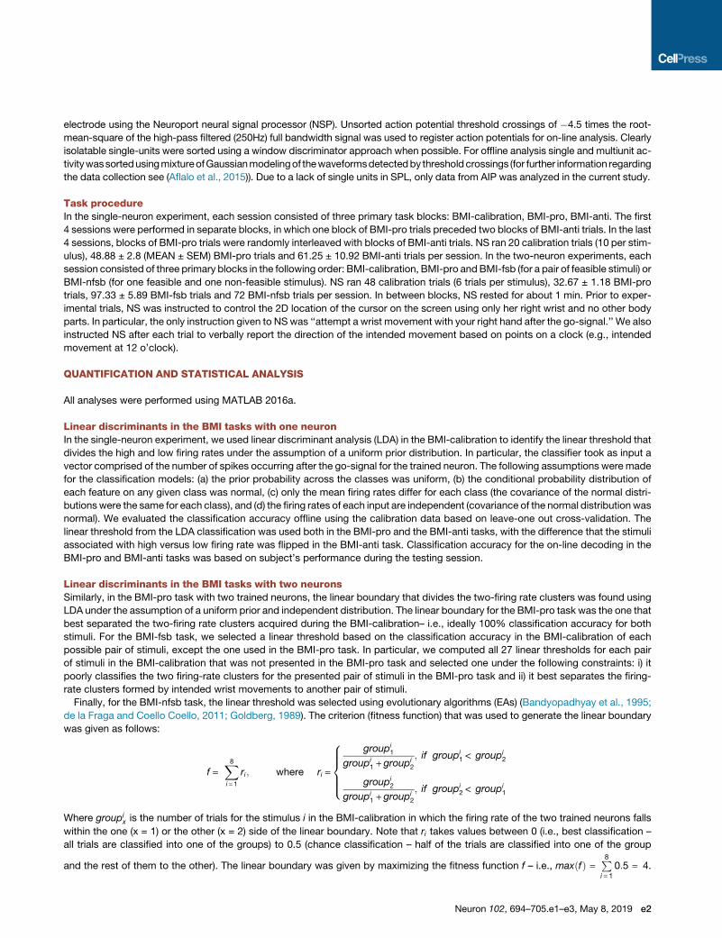
Finally, for the BMI-nfsb task, the linear threshold was selected using evolutionary algorithms (EAs) (Bandyopadhyay et al., 1995;
de la Fraga and Coello Coello, 2011; Goldberg, 1989). The criterion (fitness function) that was used to generate the linear boundary
was given as follows:
f =X8
i = 1
ri; where ri =
8>>><>>>:
groupi1
groupi1 +groupi
2
; if groupi1 < groupi
2
groupi2
groupi1 +groupi
2
; if groupi2 < groupi
1
Where groupix is the number of trials for the stimulus i in the BMI-calibration in which the firing rate of the two trained neurons falls
within the one (x = 1) or the other (x = 2) side of the linear boundary. Note that ri takes values between 0 (i.e., best classification –
all trials are classified into one of the groups) to 0.5 (chance classification – half of the trials are classified into one of the group
and the rest of them to the other). The linear boundary was given by maximizing the fitness function f – i.e., maxðfÞ = P8i = 1
0:5 = 4.
Neuron 102, 694–705.e1–e3, May 8, 2019 e2

Each chromosome in the EA population consists of 4 genes, which correspond to the 2-dimensional points ðx1; y1Þ and ðx2; y2Þ thatdefine the linear threshold. The parameters of the EA algorithm were as follows: Population = 100 (i.e., number of chromosomes),
Generations = 100 (i.e., total number of iterations), probability of crossover = 0.8, probability of mutations = 0.2.
DATA AND SOFTWARE AVAILABILITY
Data and MATLAB analysis scripts available upon request from Sofia Sakellaridi ([email protected]).
ADDITIONAL RESOURCES
This study was conducted as part of NIH clinical trial NCT01958086.
e3 Neuron 102, 694–705.e1–e3, May 8, 2019