Embed Size (px)
Citation preview
A Review of the Literature on the Determination of Brain Death
Acknowledgements The Planning Committee for the Forum on Severe Brain Injury to Neurological Determination of Death (April 9-11, 2003) commissioned this paper, a working draft, as a background information piece for Forum participants. This review of literature considers issues surrounding existing clinical practices in Canada. This paper represents a detailed summary of more recent medical literature on brain death and a summary description of the evolution of the brain death concept from the 1950s until the early 1990s. It has been prepared by Leonard B. Baron, BSc(Eng), MBA, MD, DABA, FRCPC(P) (See final tab, workshop binder). The views in the paper do not reflect the official policy of the Canadian Council for Donation and Transplantation and are not intended for publication in their current format. A bibliography of references is available from the Canadian Association of Donation and Transplantation by writing to [email protected]. Extension of Thanks from the Author: Dr. Baron notes, “This paper represents the cumulative input of a number of individuals. I am deeply indebted for the assistance of many during this project. Many thanks to Ms. Maggie Shane for performing a detailed search of the medical literature to initiate this project, to Ms. Vanessa Boyko and Mr. Gregory Workun for obtaining the relevant review articles, and to Ms. Kim Young from CCDT for spearheading this endeavour. The insight and constructive criticisms supplied by Sam Shemie, MD, and Christopher (Chip) Doig, MD, were invaluable in completing this task, as were the suggestions provided by G. Fred MacDonald, MD, in reviewing the first draft of the primary document. Finally, sincere thanks to Ms. Kathryn Burke for asking me to be involved in this project, for always being available as a sounding board during the execution of this undertaking and for formatting the final documents.”
i
A Review of the Literature on the Determination of Brain Death
Contents Executive Summary ii Introduction 1 Evolutionary History of the Brain Death Concept 2 The Brain Death Concept 3 Brain Death Criterion Explained 4
The Circulation Formulation 4 The Whole-Brain Criterion 4 The Higher Brain Formulation 5 The Brainstem Formulation 6
Pathophysiology of Brain Death 7 Clinical Diagnosis of Brain Death 9
Table I: Clinical Criteria for Brain Death in Adults and Children 10 Table II: Examples of Observation Periods in Hours before Testing Ventilated Patients
11
International Brain Death Criteria 12 Clinical Inconsistencies in Diagnosing and Managing Brain-Injured Patients
13
Certification of Brain Death 14 Timing in Declaration of Brain Death 15 Hypothermia 16 Drug Intoxication 17 Reflex Movements in Brain Death 18 Pediatric Brain Death versus Adult Brain Death 18 Fetal Salvage in Maternal Brain Death 19
Supplementary Diagnostic Testing for Determination of Brain Death 20 Cerebral Angiography 20 Radionuclide Imaging Techniques 21 Transcranial Doppler Ultrasonography (TCD) 21 Neurophysiological Testing 22
Electroencephalography 22 Evoked Potential Monitoring 23
Investigational Methodologies 23 Heart Rate Variability (HRV) Analysis 23 Ocular Micro-tremor 24 Evolving Radiological Technologies 24
Conclusion 25 Appendix I: Brain Death Formulation 28 Appendix II: Brain Death Criteria by Country 29 Appendix III: Confirmatory/Supplementary Tests 32 Appendix IV: International Comparison of Key Features on Brain Death Determination
35
References 36
ii
A Review of the Literature on the Determination of Brain Death
Executive Summary The introduction of new technologies designed for the critical care of severely injured patients in the 1950s allowed physicians to intervene in a manner that had never been possible prior to that era. It was not long before anecdotal reports of previously unseen clinical conditions began to appear in the medical literature. In 1959, Mollaret and Goulon published the first seminal work on brain death where they coined the term “le come dépassé”. Although largely ignored by the North American medical community, this work later became the foundation for brain death determination as we know it today. In 1968, the Ad Hoc Committee of the Harvard Medical School developed the first North American clinical criteria for the determination of brain death. Later, the Uniform Determination of Death Act (UDDA) gave statutory recognition to the concept of brain death and equated this concept with traditional cardiorespiratory death, which is still the most common formulation for death applied in the world. Notably, the UDDA did not address the clinical methodology for determination of brain death. Rather, the UDDA specified that brain death should be declared in accordance with accepted medical standards. It would be expected that these standards might evolve over time. Both the Ad Hoc Committee criteria and the UDDA were based upon a whole-brain formulation of brain death. This formulation supplemented the traditional definition of death represented by the circulation formulation. Other formulations of brain death were also proposed, including the higher brain formulation and the brainstem formulation, with the latter becoming the fundamental principal underlying what is now known as the U.K. code for determination of brain death. These formulations are summarized in Appendix I. The whole-brain formulation that was adopted in the United States is not dramatically different from that of the U.K. code that was proposed by the Conference of Medical Royal Colleges and Their Faculties and later championed by Pallis and Harley. From the clinical perspective, they are largely similar. The U.K. code is explicit in advising clinicians that, once a patient is identified as being in non-responsive coma of known etiology, two preconditions must be met. These are (1) that the patient is apneic, requiring ventilation, and (2) that the brain injury is irremediable. Failure to meet either of these criteria is sufficient to abandon determination of brain death. The clinical criteria for determination of brain death from several major review articles are summarized in Appendix II. The clinical examinations used in the United States, as described by Wijdicks, and those of the United Kingdom, as described by Pallis and Harley, are essentially identical. Lack of cortical activity and damage to the reticular activating stem are substantiated by deep unresponsive coma, and brainstem injury is determined by the absence of cranial nerve reflexes. An apnea test is performed to ensure that spontaneous respiration is absent, although thresholds for apnea vary between guidelines. Determination of brain death varies from nation to nation and sometimes from jurisdiction to jurisdiction. In Canada, clinical guidelines for brain death determination have been developed and promulgated by national organizations such as the Canadian Neurocritical Care Group. From a statutory perspective in Canada, however, health
iii
issues fall under the purview of provincial and territorial governments, which have not addressed the issue of brain death to any degree. Wijdicks recently published a review of international standards for the determination of brain death. Part of his work has been summarized in Appendix III. Once again, as is the case with the United States and the United Kingdom, there is significant similarity in the clinical evaluation for brain death among responding countries. Despite this commonality, numerous questions and issues remain unresolved. There are dramatic variations in the qualifications and numbers of physicians required to evaluate a patient for brain death. From one to four physicians are required in various guidelines, despite lack of evidence for having more than one physician involved in determination of brain death. Extensive clinical experience in evaluating patients for brain death is probably more important than the academic designations of those performing these evaluations. Nonetheless, several guidelines specifically recommend that specialists in the neurosciences (e.g., neurology and neurosurgery) and intensive care medicine should be recruited for these assessments. This review attempted to identify the historic basis for some of the parameters that are quoted in guidelines and that are applied by various organizations and jurisdictions. The search was unable to identify evidenced-based references for a number of often-quoted parameters. Some of the issues that remain incompletely addressed include
• the processes to be followed when the patient is exposed to drugs, either through self-medication or as provided by physicians during the course of clinical care
• the waiting period from the time of brain injury to the first evaluation for determination of brain death
• the interval waiting time between first and subsequent examinations for brain death
• relevant waiting times for examination when comparing structural brain-injured patients (e.g., traumatic hematoma) to those suffering from ischemic-hypoxic brain injury
• age limits for which adult brain death criteria are applicable. • variable brain death criteria for children, neonates and premature infants • a minimum threshold temperature for determination of brain death • the PaCO2 threshold for apnea determination, especially when the patient is
known to have suffered from premorbid pulmonary disease. • the application of supplementary tests for the determination of brain death in the
presence of confounding medical conditions such as physical injury not allowing completed clinical examination, hypothermia, drug intoxication and metabolic disturbances
• the most appropriate supplementary test when clinical examination cannot be entirely completed due to the nature of the injuries
• variable application of supplementary testing based upon patient age • the definition of legal time of death considering that a minimum of two clinical
examinations is required for determination of brain death This literature review also examined newer supplementary diagnostic tests for brain death. The more commonly available and rapidly evolving tests that may become useful in the determination of brain death are summarized in Appendix III. The “gold standard”
iv
four-vessel cerebral angiogram is now recognized to have some significant limitations, and radionuclide angiography with Tc-99m HMPAO has become more widely accepted in the last decade. This technology allows both dynamic and static imaging of the brain with a lipid-soluble radiopharmaceutical capable of traversing the blood-brain barrier. Both the anterior and posterior cerebral circulations can be evaluated with this technology. Traditionally accepted electroencephalography suffers from several serious shortcomings, which are summarized in Appendix III. Despite these shortcomings, electroencephalography continues to play a pivotal role in brain death determination and is a requirement in many international guidelines for brain death. Multiple evoked potential monitoring may provide a more useful approach to evaluating brainstem function on a global basis. Full validation of this latter technique is still pending. The need to optimally preserve organs for transplantation has resulted in the development of newer monitoring techniques intended to minimize the time from actual brain death to organ retrieval. With this purpose in mind, two interesting new technologies were identified during this literature review. Heart rate variability analysis and ocular micro-tremor monitoring are in the early stages of evaluation. Both techniques involve methods that are not costly, yet are readily adaptable for the bedside. The former technique can be seamlessly incorporated into existing monitoring capabilities, and the latter requires only modest technical expertise. Donor organ dysfunction is thought to be related to autonomic storm, which occurs at the point of death, and hemodynamic instability, which arises following brain death. Timely declaration of brain death could serve to enhance the quality of organs retrieved for transplantation. Assuming that heart rate variability analysis and ocular micro-tremor monitoring are proven to document the evolution from brain injury to total brain death, organ explantation could be executed in a more timely fashion where appropriate. Ultimately, a timely detection of brain death might address issues of scarce resource allocation in the intensive care environment.
1
A Review of the Literature on the Determination of Brain Death
Introduction A review of the recent medical literature might give one the erroneous impression that the debate about classical death and brain death is a recent one centring on a discussion about the criteria that define each of these phenomena. James Bernat has been engaged in this deliberation for some time and summarizes many of the salient historic and ethical points in a chapter of his recently published book Ethical Issues in Neurology.1 Although the concept of brain death is relatively recent, having arisen following the introduction of technologies that allowed the ventilation of seriously ill patients, dialogue about death dates into antiquity. The ancient Greeks considered the heart to be “the seat of life; the first organ to live and the last to die”.1 In the 12th century, Rabbi Moses Maimonides was first to suggest that the brain was primary in the demise of the human organism, arguing in favour of this theory by illustrating that a decapitated individual would invariably die. This was in contrast to Hebrew law of the time, which considered breathing to be central to the preservation of life. Fears of burying the living have been long held. As far back as the 17th and 18th centuries, this fear provided the impetus for innovation such as the provision of bells within caskets to warn observers that the deceased had returned to the realm of the living. Medical practitioners of the era sought clear-cut criterion for the determination of death in order to assure the common man that a false positive diagnosis was not about to occur. Today, the concept of brain death, defined as the irreversible absence of brain function, is widely accepted both in the medical community and in most international societies. Legal provisions defining brain death are common, particularly in more technologically advanced nations.2 In the United States, the Uniform Determination of Death Act3 forms the legal foundation upon which the concept of brain death is gauged. This statute characterizes the deceased and death in the following quotation:
An individual who has sustained either: 1) irreversible cessation of circulatory and respiratory functions, or 2) irreversible cessation of all functions of the entire brain, including the brainstem, is dead. A determination of death must be made in accordance with accepted medical standards.
It is noteworthy that this statute does not provide specific standards for the determination of death and/or brain death. Some observers suggest that this terminology is deliberately open-ended to accommodate the natural evolution of medical standards as science learns more about the pathophysiology of death and develops newer modalities of treatment and diagnosis. Recent developments in the medical literature with respect to brain death cannot be presented without some background regarding the origin of the concept of brain death and the natural history of the brain death concept since its inception.
2
Evolutionary History of the Brain Death Concept With the introduction of the mechanical ventilator and advanced resuscitative practices into clinical medicine, the first cases of primary brain injury in the presence of persistent cardiac function became apparent in the 1950s. The first extensive description of this phenomenon came from France in 1959, where Mollaret and Goulon coined the phrase “le coma depassé”, meaning “a state beyond coma”.4 These patients showed some signs of life, with apparently normal cardiac activity and peripheral tissue perfusion, persistent urinary output and preservation of metabolic activities. On the other hand, these patients were in deep coma, unresponsive to the most intense stimuli, and displayed signs of absent brainstem function, including absent cranial nerve reflexes and apnea. It was apparent that many vital physiologic functions could be maintained long after the brain had apparently ceased to function. These patients were also observed to have absent electroencephalographic activity, cessation of cerebral blood flow and, on autopsy, evidence of brain necrosis. The initial focus of discussions on brain death was meant to address the issue of continued medical intervention in the presence of hopelessly irreversible brain injury. However, as organ transplantation technologies were being pioneered at nearly the same time, an increased demand for donor organs inextricably linked the issues of brain death with those of organ procurement and transplantation. In 1968, the Ad Hoc Committee of the Harvard Medical School to Examine the Definition of Brain Death5 undertook to define irreversible coma and brain death. The Committee defined brain death as “unresponsiveness and lack of receptivity, the absence of movement and breathing, the absence of brain-stem reflexes and coma whose cause had been identified”. In the 1970s, the importance of irreversible loss of brainstem function was identified.6 In 1976, the Conference of Medical Royal Colleges and Their Faculties in the United Kingdom published a statement on the diagnosis of brain death.7 Clinical diagnostic testing for brain death became more refined with the application of this guideline. In 1981, the U.S. President’s Commission for the Study of Ethical Problems in Medicine and Biomedical and Behavioral Research published guidelines regarding brain death.8 These guidelines recommended the use of supplementary diagnostic tests to augment the clinical examination in the diagnosis of brain death. The Commission also recommended that patients who had suffered ischemic hypoxic brain injury should be observed for no fewer than 24 hours prior to declaration of brain death. Additional refinements in the diagnosis and management of brain injury and brain death have been identified since that time, and the American Academy of Neurology finally published an evidence-based review on brain death in 1995.9 This guideline is particularly noteworthy for the affirmation that the diagnosis of brain death is clinical in nature and for the clarification that it provides regarding the use of supplementary tests in the management of brain death in the presence of clinical confounding factors. Ultimately, in 1995, the U.K. code took the position that brainstem death was equivalent to brain death. Whereas supplementary testing such as electroencephalography and cerebral blood flow studies had been recommended earlier, the U.K. code did not require any supplementary testing for determination of brain death where a complete clinical examination could be performed. Notably, this guideline required that an etiology
3
for brain death must be first established and that conditions that mimic brain death must be excluded prior to clinical evaluation for brain death. The Brain Death Concept Discussions about death and dying frequently become mired in philosophical and ethical debate. For many members of the public, death is perceived as an “all-or-nothing” event, but from a medical perspective, death is generally viewed as a physiologic process. Despite this broadly accepted view, medical literature may also contribute to confusion. For example, Bernat’s contention that there are only two fundamental states of being for any organism, living or dead, tends to depict death as an event rather than as a physiologic process.1 Pallis and Harley are not as rigid on this matter, although they are definitive in stating that “there are points of no return” in the physical process of dying.10 Like Maimonides, they use the example of the decapitated individual, whose heart continues to function for a short time following decapitation. To Pallis and Harley, brain death is synonymous with “physiologic decapitation”; the cause of decapitation may be different but the outcome is no less grim. This debate about whether death is a process or an event contributes to confusion amongst the lay public. Complicating these issues, legal processes related to an individual’s death are triggered by specific events following death.11 Beyond the issue of long-term ventilator support for the apparently brain-dead patient, the need for clear-cut definitions and criteria for brain death was also driven, in part, by advances in organ transplantation. Timely procurement of viable organs prior to significant deterioration is a key factor in successful organ transplantation. In Ethical Issues in Neurology, Bernat describes a three-step process addressing philosophical and ethical issues regarding death and dying. He first encourages the reader to consider a definition of death.1 He argues that death is largely a social and moral concept based upon philosophically held tenets and beliefs. In defining death, Bernat asserts that our implicit internalized beliefs and concepts about death and dying become explicitly stated. In a similar vein, some authors believe that lay literature confuses the debate concerning death because different segments of society choose to define death in differing ways while seldom explicitly expressing their definition of death to the fullest extent. Bernat suggests that one must therefore develop a rational and coherent criterion for death. He has been a proponent of the whole-brain formulation of death, which has dominated debate about brain death in the United States. This criterion became the foundation for discussions held by the ad hoc Harvard committee and the President’s Commission. Since then, the whole-brain death concept has been explicitly acknowledged in the Uniform Determination of Death Act, which has been recognized by a majority of the states within the United States. Bernat’s final step charges the medical community to develop and validate bedside clinical and diagnostic supplementary tests that are compatible with the accepted criteria for brain death. This article will address some of the controversial medical issues pertaining to the formulation of brain death criteria and processes related to the declaration of brain death.
4
Brain Death Criterion Explained During its deliberations, the President’s Commission considered three possible criteria for death: a non-brain criterion that some refer to as the “circulation formulation”, a whole-brain criterion that had been proposed by the ad hoc Harvard committee, and a higher brain formulation that was popularized by Veatch12 in the mid 1970s. Since that time, a fourth formulation known as the brainstem formulation was conceptualized and popularized by Pallis and Harley. This formulation represents the underlying principle behind brain death determination in the United Kingdom. Appendix I provides a comparison of the four currently recognized formulations. The Circulation Formulation The non-brain criterion or circulation formulation reflects what might be considered the traditional signs of death represented by a cessation of tissue perfusion detected by the absence of peripheral pulses in the absence of spontaneous respiration. This formulation is consistent with the manner in which most deaths are documented today. Some theologians argue that death is solely manifested by absence of circulation and that brain death is a construct that is inherently untenable. Although the concept of brain death is accepted by the vast majority of the lay public, this small group of individuals rejects all criteria for brain death that have been developed. In the context of current technological capabilities and in support of organ transplantation efforts, a non-brain criterion for brain death suffers from some serious drawbacks. It is distinctly possible to support life for extended periods of time.13 Such cases impose significant ethical and legal challenges for medical caregivers as well as stress for the families of patients being supported by medical technology. There is also a significant economic burden associated with sustaining patients in these situations. Moreover, individuals sustained on artificial support systems may experience cardiorespiratory events that could lead to the deterioration of organ systems that might otherwise be appropriate for organ transplantation. Not surprisingly, the President’s Commission7 looked beyond a non-brain criterion for death because of some of these considerations. The Whole-Brain Criterion The whole-brain criterion has been central to much of what is written regarding brain death in the United States. The fundamental tenet of this criterion is based upon a “cessation of the integrative functioning of the organism as a whole”. Today’s whole-brain death criterion characterizes this loss of integrative function as absence of cerebral functioning represented by non-responsiveness to supramaximal stimulation plus evidence of brainstem death manifested as absence of brain stem reflexes and documented apnea in response to properly performed apnea testing. Arguing in favour of the whole-brain criterion, the President’s Commission wrote,
This view gives the brain primacy not merely as the sponsor of consciousness (since even unconscious persons may be alive), but also as the complex organizer and regulator of bodily functions. Only the brain can direct the entire organism.
Bernat et al attempted to provide an explicit definition for death in the context of the whole-brain model.14 They defined death as the “permanent cessation of functioning of the organism as a whole”. This reference to “the organism as a whole” has unfortunately created some confusion in the medical literature. Bernat attempts to clarify the intent of
5
this clause by explaining that the authors intended to “refer not to the whole organism (the sum of the parts of the organism) but rather to the set of functions of integration, control and behavior that provide the unity of the organism and are greater than the sum of the organism’s parts”.1 Persistence of some neuroregulatory function may be observed long after the clinical criteria for brain death are fully met.15 For example, clinical laboratory evidence of antidiuretic hormone activity may be seen in brain-dead patients. For some observers, the persistence of this activity suggests that the whole-brain death criterion is not being fulfilled. Bernat counters that this activity may represent retained functionality of isolated nests of brain cells while the remainder of the brain (cerebral cortex, diencephalon, brainstem and cerebellum) may have been totally destroyed by the pathologic process. Wijdicks16 further elucidates the physiology of hypothalamic-pituitary functioning by illustrating that pituitary blood flow arises from extracranial sources and that preservation of neurohypophyseal functioning is not inconsistent with the whole-brain formulation of brain death. Bernat also does not appear to have difficulty resolving whole-brain death in the context of persistent neurohumoral activity.1 His argument centres upon the fact that the integrative unity of the organism has been disrupted once the remainder of the brain has been pathophysiologically destroyed. Once this integrative unity no longer exists, the organism meets all requirements to be declared dead if all other clinical indicators of brain death are met. In determining the death of a human organism Bernat et al identified two general sets of tests that could be used to confirm the presence of brain death: (1) traditional signs of cardiorespiratory death manifested by prolonged absence of vital signs, which would irrevocably lead to the demise of the brain, and (2) a set of neurologic tests and procedures based upon a clear identification of irreversible coma of known etiology that documented the loss of brain function observed as absence of brainstem reflex activity and apnea persisting for an appropriate period of time. This latter set of manifestations is expressed in the whole-brain death criterion that is widely published in today’s medical literature. The Higher Brain Formulation The higher brain formulation for death was pioneered by Veatch and further refined by others such as Stuart Youngner, Michael Green and Daniel Wikler. This formulation suggests that death involves the disintegration of the nature of the organism. In the case of humans, it entails the dissolution of personhood, which may be identified as the consciousness and cognition that are fundamental to the life experience. In pathophysiologic terms, this formulation of death physically equates death to destruction of the cerebral cortex. There are significant practical problems associated with this formulation. Using the higher brain formulation, two clinical conditions, anencephaly and persistent vegetative states, would satisfy the criteria for death. The higher brain criterion is a radical departure from traditional thinking for many people. The case of Karen Ann Quinlan underscores some of the concerns with a higher brain criterion. Quinlan remained in a persistent vegetative state requiring chronic ventilatory support following a serious brain injury. Her family made the decision to remove her from her ventilator so that she might die with dignity, but she continued to breathe unassisted after doing so. During the 10 years that Quinlan survived without ventilatory support, she fulfilled the criteria for higher brain death. The adoption of a higher brain criterion of
6
death would have rendered Quinlan legally dead even though her brainstem function was undeniably intact. Given the choice between a whole-brain and a higher brain criterion for brain death, the President’s Commission settled on a whole-brain criterion for a number of reasons. From a practical perspective, the Commission identified some significant shortcomings in defining death using a higher brain criterion because the brain structures and physiologic processes critical to preservation of consciousness and cognition were incompletely understood. Moreover, the Commission held that the loss of function within critical brain structures could be reasonably ascertained with current medical technologies to meet the requirements of statutory regulations. The Brainstem Formulation In 1976 and 1979, two memoranda were issued by the Conference of Medical Royal Colleges and Their Faculties in the United Kingdom. The first of these memoranda stated that “permanent functional death of the brainstem constitutes brain death”. Irreversible loss of brainstem function is determined in clinical terms and is identified in the context of irremediable structural brain damage, but only if reversible causes of brainstem dysfunction are excluded. Pallis and Harley refer to this declaration of brain death as the U.K. code. Proponents of this formulation conceptually have little problem with the whole-brain criterion. The clinical determinants of brain death in the whole-brain formulation are fundamentally similar to those employed by supporters of the brainstem formulation. This formulation is explained in detail in the writings of Pallis and Harley. The reticular formation, integral to “generating the capacity for consciousness”, is a key anatomic structure within the brainstem. The brainstem also harbours nuclei that sustain respiratory mechanisms and many of the centres that control and maintain blood pressure. Furthermore, the brainstem acts as a central clearinghouse through which sensory input (excluding vision and olfaction) passes and all motor output originating in the cortex is transmitted peripherally. In summary, the brainstem is accepted as the site where all integrative capacities for consciousness and involuntary integrative physiologic functioning reside. This United Kingdom criterion for brain death has come under criticism in several BBC documentaries examining the issue of brain death. Dissenting American neurologists have argued that patients have survived life-threatening events in which the U.K. code for brainstem death had been fully met. Pallis and Harley reject these statements based upon two central arguments: (1) that some of the patients did not meet the essential preconditions for evaluation of brain death in that they did not have evidence of irremediable brain damage and (2) that a small number of patients were not examined in a timely fashion after having been exposed to hypoxic ischemic brain injury and, thus, were given insufficient time for spontaneous recovery of neurologic function. To further his arguments, Pallis and Harley summarize clinical reports and studies that account for 1,862 patients who were brain dead by U.K. brainstem death criteria. They concluded that continued ventilatory support invariably led to cardiac death by asystole in all cases. In rejecting the brainstem formulation, Bernat contends that this criterion suffers from a significant conceptual flaw related to a condition referred to as “super locked-in
7
syndrome”.1 In this condition, cerebral cortical activity is retained in the presence of totally absent brainstem function. An actual case of this syndrome was not described in any of the articles identified during this review and appears to be a theoretical concept more than a practical concern. While anecdotal cases describing this syndrome are sometimes described, it is unclear why this syndrome has not been reported in the literature. In the presence of super locked-in syndrome, diagnosing brain death might also be inconsistent with the interpretation of currently accepted supplementary tests for detection of brain death. It is presumed that cortical EEG activity and cerebral blood flow could be retained in these patients while all other requirements for the declaration of brain death would be met. At the moment, it is unclear if any clinician would be willing to declare brain death if these findings were encountered on supplementary testing. Bernat concedes that the criteria for brain death may ultimately move in the direction of accepting a brainstem formulation. This shift in criteria might be facilitated by the development of new medical technologies capable of examining isolated brainstem activities. It is distinctly possible that these new technologies, used singularly or in unison, may become reflective of the functional integrative unity of the organism as a whole. Pathophysiology of Brain Death Although the primary mechanisms of brain injury in closed head injury and intracranial hemorrhage differ, the secondary pathophysiology characterized by massive increase in intracranial pressure (ICP) is similar in both conditions.17 In the early evolution of closed head injury or intracranial hemorrhage, there is displacement of cerebrospinal fluid from the cranial vault into the spinal subarachnoid space, offering some attenuation of ICP. Persistent increases in ICP result in an intracranial shift of brain structures with downward displacement of the diencephalon and brainstem. At the extreme, when intracranial compliance becomes limited, ICP rises dramatically with minimal change in cerebral blood flow or edema. This increase in ICP is accompanied by a significant reduction in cerebral perfusion pressure (CPP) and cerebral blood flow. As ICP increases, transtentorial uncal herniation may ensue. Coning at the foramen magnum may further contribute to brainstem damage. Because the rate of increase in ICP is highly variable, the evolutionary time course of coma and brain death is equally unpredictable. Case reports in the literature also describe the persistence of some cerebral perfusion in the face of extreme increases in intracranial pressure in patients who have sustained severe disruptive cranial vault trauma or those who have undergone surgical decompression with large craniotomy.18 A similar phenomenon may also be observed in the neonate whose fontanelles remain open. Compression of the pituitary stalk against the edge of the diaphragma sellae has also been described.19 This is most likely to be seen with centrally located brain injury and is less common in diffuse cerebral pathophysiologic processes such as hypoxic-ischemic brain injury and encephalitis. Disruption of pituitary functioning most commonly involves the posterior pituitary gland, while the anterior pituitary gland may be largely spared from damage. While some authors have used this preservation of pituitary functioning as an argument against the whole-brain death criterion, Wijdicks is puzzled by this argument, considering the vascular anatomy of the hypothalamic-pituitary axis.16
8
Reperfusion injury is a pathophysiologic process associated with hypoxic-ischemic brain injury. A complex detailed biochemical description of reperfusion injury is provided elsewhere.20 The authors describe reperfusion damage as “aggravation of tissue injury which is triggered by mechanisms set in motion be reperfusion, i.e., by the re-supply of oxygen and glucose”. At the time of primary ischemic injury, there is a loss of bio-energetic functioning of the brain, likely due to mitochondrial dysfunction. Reperfusion damage occurs following restoration of oxygen delivery to the brain. The likely mechanism for late neuronal damage appears to involve the accumulation of calcium within neurons once perfusion has been re-established, but the exact mechanisms involved in these processes remain to be identified. The reviewed literature did not provide insight into the time course of reperfusion injury. It is possible that the time evolution of brain injury may be temporally related to other factors, such as the rapidity and degree of diminution of oxygen delivery to the brain and the restoration of cerebral blood flow once reperfusion is established. Ultimately, following brain death, patients often display signs of diminished sympathetic activity manifested by systemic hypotension, vasodilatation and diminished myocardial contractility. To maintain end-organ perfusion. it is frequently necessary to provide inotropic or vasopressor support and to restore intravascular fluid volume status at this stage. Clinical investigators have sought to clarify the pathophysiology of brain death in an effort to identify potential new modalities of therapy in brain-injured patients. Therapeutic interventions based upon a better understanding of the pathophysiology of brain death could also contribute to optimal preservation of organs destined for transplant following declaration of brain death. This research, however, may have unintentionally contributed to the debate about the nature of the current brain death criteria as reflected by the Uniform Determination of Death Act. The early manifestations of evolving brain death may be an expression of increased circulating catecholamines.21 Animal models in which rapid inflation of an epidural balloon catheter induces brain death reveal that histologic myocardial changes in these animals is remarkably similar to that seen in catecholamine-stressed humans.22 Powner et al studied circulating catecholamine concentrations in patients in whom evolving brain death was anticipated.23 Measured catecholamine concentrations were no different before and after documented brain death. Furthermore, catecholamine concentrations of injured patients who did not progress to brain death were similar to those of similarly injured patients who did evolve to full brain death. Those patients who did not progress to brain death did not display any evidence of myocardial injury or clinical hypotension. Powner et al concluded that a more significant injury to the myocardium was likely to be caused by an abrupt increase in circulating catecholamines in those patients progressing to brain death. Other articles have documented extensive changes in myocardial contractility in patients who fulfil brain death criteria.24 Echocardiographic studies revealed myocardial dysfunction in 42% of brain-dead patients studied by Dujardin et al. Although the pathophysiologic mechanism leading to myocardial dysfunction has yet to be fully elucidated, it would appear to be related to surges in catecholamine concentrations at the time of brain death. It is commonly held that whole-brain death should ultimately result in disrupted neurohumoral autoregulation, identified as panhypopituitarism. Clinical studies have revealed that this clinical scenario is variably present.25 Changes in circulating hormones
9
have been documented in a number of brain death animal studies, but the findings in these studies are inconsistent with those of human studies. Schrader et al evaluated the presence of neurohumoral autoregulation using an insulin-induced hypoglycemic test in two patients identified as brain dead using the Harvard criteria.26 In one case an immediate growth hormone response was clearly identified, indicating that pituitary functioning was preserved in spite of the person having met brain death criteria. Posterior pituitary functioning, especially antidiuretic hormone secretion, has also been studied. If the hypothalamus and neurohypophysis were totally non-functional, then the brain-dead patient should develop a clinical picture of central diabetes insipidus. Grenvik et al reported that, in a study of brain-dead patients meeting brain death criteria, only 8.5% of patients showed clinical manifestations of central diabetes insipidus.27 Hohenegger et al assayed antidiuretic hormone in 11 patients meeting brain death criteria and in whom clinical manifestations of diabetes insipidus were identified. Surprisingly, they found either normal or increased antidiuretic hormone in all patients.28 The preservation of some neurohumoral regulatory activities raises concerns for several reasons. As described above, clinical studies illustrate definitively that some preservation of hormonal regulation is seen in many patients who would be considered brain dead using generally accepted brain death criteria. These same studies further support the assertion that functionality of regulatory mechanisms is preserved as compared to simple uncontrolled activity and, finally, that some integrative role of the brain in systemic autoregulation may also be preserved. The Uniform Determination of Death Act, which was based on a whole-brain death concept, requires the “irreversible cessation of all functions of the brain, including the brainstem”. Quoted clinical studies provide evidence that the conditions required by the Uniform Determination of Death Act may not be fully met in many of our traditionally designated brain-dead patients. A better understanding of the pathophysiologic processes related to brain death may aid in the resolution of this apparent conflict. Clinical Diagnosis of Brain Death Comparative tables of brain death guidelines are provided in Appendix II. The Canadian Neurocritical Care Group guidelines29 are compared with those published by Wijdicks,30 Pallis and Harley, and the Australia and New Zealand Intensive Care Society.31 The most recent major review article on brain death is that provided by Wijdicks in the New England Journal of Medicine. The American clinical criteria for brain death, as published by Wijdicks, are outlined in Table I. Several points in the article by Wijdicks are particularly noteworthy. Unlike the U.K. code, there is no explicit mention of preconditions for the clinical determination of brain death. These preconditions might be perceived by some as rather self-evident, but there are case reports and anecdotal accounts of brain death declaration where these preconditions were clearly not met.32 Like Pallis and Harley, Wijdicks addresses the issue of remediable coma, stating that clinical examination for brain death must not proceed when irremediable coma cannot be confirmed.
10
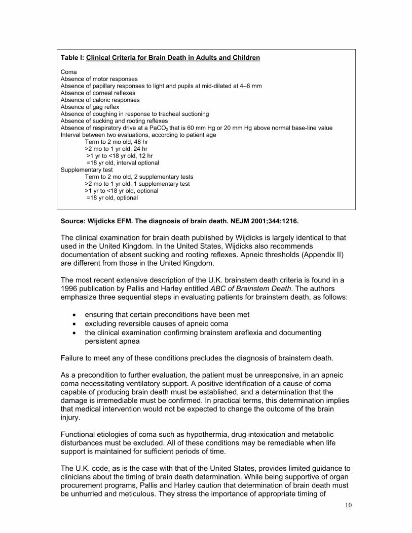
Table I: Clinical Criteria for Brain Death in Adults and Children
Coma Absence of motor responses Absence of papillary responses to light and pupils at mid-dilated at 4–6 mm Absence of corneal reflexes Absence of caloric responses Absence of gag reflex Absence of coughing in response to tracheal suctioning Absence of sucking and rooting reflexes Absence of respiratory drive at a PaCO2 that is 60 mm Hg or 20 mm Hg above normal base-line value Interval between two evaluations, according to patient age Term to 2 mo old, 48 hr >2 mo to 1 yr old, 24 hr >1 yr to <18 yr old, 12 hr =18 yr old, interval optional Supplementary test Term to 2 mo old, 2 supplementary tests >2 mo to 1 yr old, 1 supplementary test >1 yr to <18 yr old, optional
=18 yr old, optional
Source: Wijdicks EFM. The diagnosis of brain death. NEJM 2001;344:1216. The clinical examination for brain death published by Wijdicks is largely identical to that used in the United Kingdom. In the United States, Wijdicks also recommends documentation of absent sucking and rooting reflexes. Apneic thresholds (Appendix II) are different from those in the United Kingdom. The most recent extensive description of the U.K. brainstem death criteria is found in a 1996 publication by Pallis and Harley entitled ABC of Brainstem Death. The authors emphasize three sequential steps in evaluating patients for brainstem death, as follows:
• ensuring that certain preconditions have been met • excluding reversible causes of apneic coma • the clinical examination confirming brainstem areflexia and documenting
persistent apnea Failure to meet any of these conditions precludes the diagnosis of brainstem death. As a precondition to further evaluation, the patient must be unresponsive, in an apneic coma necessitating ventilatory support. A positive identification of a cause of coma capable of producing brain death must be established, and a determination that the damage is irremediable must be confirmed. In practical terms, this determination implies that medical intervention would not be expected to change the outcome of the brain injury. Functional etiologies of coma such as hypothermia, drug intoxication and metabolic disturbances must be excluded. All of these conditions may be remediable when life support is maintained for sufficient periods of time. The U.K. code, as is the case with that of the United States, provides limited guidance to clinicians about the timing of brain death determination. While being supportive of organ procurement programs, Pallis and Harley caution that determination of brain death must be unhurried and meticulous. They stress the importance of appropriate timing of
11
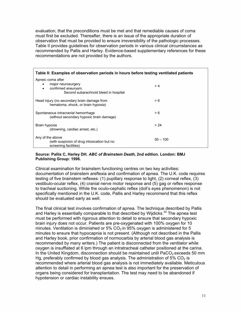
evaluation, that the preconditions must be met and that remediable causes of coma must first be excluded. Thereafter, there is an issue of the appropriate duration of observation that must be provided to ensure irreversibility of the pathologic processes. Table II provides guidelines for observation periods in various clinical circumstances as recommended by Pallis and Harley. Evidence-based supplementary references for these recommendations are not provided by the authors.
Table II: Examples of observation periods in hours before testing ventilated patients Apneic coma after
• major neurosurgery • confirmed aneurysm.
Second subarachnoid bleed in hospital > 4
Head injury (no secondary brain damage from hematoma, shock, or brain hypoxia)
> 6
Spontaneous intracranial hemorrhage (without secondary hypoxic brain damage)
> 6
Brain hypoxia (drowning, cardiac arrest, etc.)
> 24
Any of the above
(with suspicion of drug intoxication but no screening facilities)
50 – 100
Source: Pallis C, Harley DH. ABC of Brainstem Death, 2nd edition. London: BMJ Publishing Group: 1996. Clinical examination for brainstem functioning centres on two key activities: documentation of brainstem areflexia and confirmation of apnea. The U.K. code requires testing of five brainstem reflexes: (1) pupillary response to light, (2) corneal reflex, (3) vestibulo-ocular reflex, (4) cranial nerve motor response and (5) gag or reflex response to tracheal suctioning. While the oculo-cephalic reflex (doll’s eyes phenomenon) is not specifically mentioned in the U.K. code, Pallis and Harley recommend that this reflex should be evaluated early as well. The final clinical test involves confirmation of apnea. The technique described by Pallis and Harley is essentially comparable to that described by Wijdicks.30 The apnea test must be performed with rigorous attention to detail to ensure that secondary hypoxic brain injury does not occur. Patients are pre-oxygenated with 100% oxygen for 10 minutes. Ventilation is diminished or 5% CO2 in 95% oxygen is administered for 5 minutes to ensure that hypocapnia is not present. (Although not described in the Pallis and Harley book, prior confirmation of normocarbia by arterial blood gas analysis is recommended by many writers.) The patient is disconnected from the ventilator while oxygen is insufflated at 6 lpm through an intratracheal catheter positioned at the carina. In the United Kingdom, disconnection should be maintained until PaCO2 exceeds 50 mm Hg, preferably confirmed by blood gas analysis. The administration of 5% CO2 is recommended where arterial blood gas analysis is not immediately available. Meticulous attention to detail in performing an apnea test is also important for the preservation of organs being considered for transplantation. The test may need to be abandoned if hypotension or cardiac instability ensues.
12
The U.K. code requires retesting to ensure that observer error has not occurred. Pallis and Harley argue that there is a second, more important reason for retesting—i.e., that the period of observation exceeds the time required for ischemia to produce irreversible neuronal damage by a factor of several hundredfold. Lazar et al recently published a brief review of the Canadian perspective of brain death.33 While the bulk of this paper discusses philosophical, ethical and legal issues, it also includes a guideline for brain death determination. This guideline echoes that of the more detailed Canadian Neurocritical Care Group guidelines published in 1999. It requires determination of brain death by physicians with experience in brain death criteria and, in the event of organ donation, recommends that two physicians, involved neither in the patient’s care nor in proposed transplant procedures, be engaged in brain death determination. In this document, hypothermia is defined as a threshold temperature of 32.2OC and the observation period prior to assessment for brain death is said to vary with the etiology of coma, although specifics are not provided. Finally, the apnea test requires arterial blood gas determination of a rise in PaCO2 to at least 60 mm Hg with a pH of less than 7.28. The Canadian Neurocritical Care Group guidelines provide a more detailed discussion in relation to pediatric brain death and situations where clinical conditions do not allow complete examination of brainstem reflexes or where complicating medical factors such as hypothermia are identified.29 For the pediatric population, it is recommended that adult guidelines are applicable beyond 52 weeks post-conceptual age. In contrast to other authors, this group does not agree that clinical criteria alone are adequate for the declaration of brain death in children. Age-based recommendations for supplementary testing are provided. These are detailed in Appendix II. Despite the fact that guidelines frequently advocate supplementary testing in the determination of brain death in children, the literature is devoid of evidence-based support for this recommendation. International Brain Death Criteria Although this literature review identified articles pertaining to brain death criteria from individual countries and regions linked by geography, a major review article on international brain death criteria was published by Wijdicks in January 2002.2 This study was the first of its kind, using a distillation of the literature and a review of legal statutes as well as data acquired through contact with experts in brain death worldwide. In total, responses were documented from 80 nations, representing all continents. Having been recently completed, this article is the most detailed compilation of its nature. For the purpose of this review it can be assumed to be a reasonable approximation of the current status on brain death worldwide, recognizing that organizations within many nations continue to work on the development and evolution of brain death criteria. Of the responding countries, 70 of 80 have a guideline for the determination of brain death. The number of physicians required to provide a definitive brain death diagnosis ranges from one in 44% of responding countries to a maximum of four in Turkey. Turkey is unique in that it has passed an organ procurement law with strict requirements for brain death determination provided by four different medical and surgical specialties and confirmation using a combination of diagnostic tests. Notably, only 41 guidelines require an apnea test using arterial blood gas determined target PaCO2 values while 20 of 71 guidelines accept apnea following a 10-minute disconnection from the ventilator as a criteria for brain death. Supplementary testing is more commonly recommended in Europe and Asia than elsewhere.
13
Within the United States, 44 states and the District of Columbia have accepted the Uniform Determination of Death Act as the criterion for brain death. Because individual state statutes may also address brain death, there are some significant incongruities amongst the American states. For example, some states have statutes that specify the specialties allowed to test for brain death, while others, such as New York and New Jersey, have adopted statutes that allow the accommodation of religious, cultural and philosophical differences in respect of death. The clinical evaluation for brain death is strikingly similar across European nations. Requirements for supplementary testing, however, are variable. Eleven of 25 guidelines have mandatory supplementary diagnostic testing. Half of the European countries require more than one physician in evaluating for brain death. In Australia and New Zealand, guidelines have been prepared by the Australia and New Zealand Intensive Care Society (ANZICS).31 Updated guidelines are expected to be released shortly by ANZICS. The 1995 guidelines were in keeping with national statutes and regulations of the time. They applied solely to adults and children of more than 2 months chronological age. The guidelines did not address the issue of brain death in newborns less than this age. As in the United States, the ANZICS guidelines require “irreversible cessation of all function of the brain”. In spite of this statement, the guidelines advise that the terms whole-brain death and brainstem death should not be used. Details of the ANZICS guidelines are summarized in Appendix II. The ANZICS guidelines mandate the demonstration of absent intracranial blood flow either by 4-vessel angiography or by an equivalent radionuclide study in all cases where the conditions of clinical diagnosis cannot be completely met. A 6-hour interval of observation prior to the second declaration regarding brain death is also required prior to performing supplementary diagnostic testing in the ANZICS guidelines. Japan is unique and the most specific amongst nations with established guidelines for brain death.34 Wijdicks describes Japan’s requirements as follows:
CT scan should detect “irreparable lesion”; the cause of cardiac arrest should be known when it has caused brain death; the ciliospinal reflex should be performed; the apnea test should be performed after loss of seven specified brainstem reflexes and after isoelectric EEG; brain death determination is allowed only if intact tympanic membranes exist; and children < 6 years old are excluded.
Appendix III provides an edited summary of brain death criteria for those Western nations that provided data for the 2002 article by Wijdicks on the worldwide criteria for brain death.2 Clinical Inconsistencies in Diagnosing and Managing Brain-Injured Patients A literature review on brain death invariably identifies a number of confounding clinical scenarios that may significantly affect a clinician’s ability to diagnose brain death in a timely fashion. While the following is not exhaustive, some specific areas of concern that have received attention in the literature recently are presented. Comments are prefaced, where applicable, with reference to the Canadian Neurocritical Care Group guidelines, which broadly reflect current practices in the declaration of brain death in Canada.
14
Certification of Brain Death
Brain death must be determined clinically by an experienced physician and in accord with the accepted medical standards. … Because of the major consequences of the diagnosis of brain death, consultation with other physicians experienced in the relevant clinical examinations and diagnostic procedures is advisable.29
It is generally accepted that considerable clinical experience in the determination of brain death is desirable when evaluating the patient. Inasmuch as a precise clinical examination forms the foundation for brain death determination, it would appear to be desirable to have a neurologist or neurosurgeon involved in the assessment of all such patients. In practice, this may not always be feasible. Consequently, other medical personnel may be involved in the determination of brain death. Moreover, medical specialists outside the neurosciences may acquire considerable experience in evaluating for brain death, and their skills could feasibly exceed those of neuroscience specialists in this area. Wijdicks36 and Pallis and Harley10 both acknowledge that any physician with sufficient clinical experience may perform an examination for determination of brain death. To minimize perceptions of conflict of interest, guidelines may exclude those involved in transplants following organ procurement in evaluating patients for brain death. Requirements for determination of brain death vary across jurisdictions with respect to the degree of training required to perform such an evaluation and the number of physicians involved in the determination. Some of these requirements have been cited in articles and books.35 In his international review of brain death, Wijdicks documents some of the variability in requirements for the declaration of brain death. In the United States, state statutes may specify specific specialties and the number of examiners required for the determination of brain death.2 For example, in Virginia, a specialist in neurosciences is required. Although a single physician performing two examinations is more commonly required, some statutes mandate independent evaluations by two different physicians. There are no data to support the suggestion that having a second different physician involved in the evaluation for brain death reduces the risk of errors in clinical evaluation. Despite this lack of evidence, Florida requires two physicians, one of whom is the primary treating physician and the other a board-eligible or board-certified specialist in neurology, neurosurgery, internal medicine, pediatrics, surgery or anesthesiology. In two states, Georgia and Alaska, a registered nurse may be delegated authority to declare brain death, although a physician must subsequently certify brain death within 24 hours. Virginia confers limited authority to registered nurses in the certification of brain death. Requirements for declaration of brain death also vary internationally. In Turkey, four specialties must be involved in the determination of brain death: cardiology, neurology, neurosurgery, and anesthesiology. In Israel, two physicians are required, but, in contrast to the state of Florida, neither can be the treating physician. In India, a panel of physicians including the treating physician, a physician representing the treating hospital, an independent specialist of no particular designation, and a neurologist or neurosurgeon is convened to determine brain death. The burden of proof for diagnosis resides with the neurosciences specialist. Where organ procurement is proceeding, the ANZICS guidelines designate that two physicians be involved in certification; if organ donation has been excluded, the recommendation is that two physicians participate in
15
certification of brain death, although this is not required.31 Specialty standards are not defined. In the United Kingdom, two physicians with skills in a related specialty are recommended by code. One of these must be a consultant while the second may be either a consultant or a senior registrar. Although certain specialty consultants are preferred, there is no specified specialty requirement for determination of brain death. None of the guidelines and articles that address the level of expertise and number of physicians involved in brain death determination provide insight into the logic behind the establishment of these requirements. It might be surmised that availability of specific medical specialties may be a primary driver in the designation of specific specialties to this task. Furthermore, a specialist physician would be expected to have the requisite skills to do a thorough and complete examination consistent with national and regional requirements. Some authors have suggested that it may be desirable to institute a certification program for determination of brain death similar to those provided for advanced resuscitative programs.36 Timing in Declaration of Brain Death The Canadian Neurocritical Care Group guidelines are silent on the timing of the initial examination for determination of brain death. They do address the issue of a second evaluation in stating,
Re-evaluation is essential to ensure that the non-functioning state of the brain is persistent and to reduce the possibility of error. Depending on the etiology, the interval between such examinations may be as short as 2 hours or as long as 24 hours; observation for 24 hours is usually recommended to confirm brain death due to anoxic-ischemic insults (e.g. post-cardiac arrest).29
There are two key timing issues in respect to the declaration of brain death. The first relates to the timing of the first examination for brain death relative to the primary injury and the second to the time interval between successive examinations. Pallis and Harley describe the first time as the point where the preconditions for diagnosis of brain death have been met. The second precondition for brain death states that the cause of coma must be a disorder known to lead to brain death that is irremediable. In some clinical circumstances, compliance with this requirement may be most vexing. Hypoxic ischemic brain injury following cardiac resuscitation is a common example of this latter problem (see below - Hypothermia). The ANZICS guidelines are noteworthy for recommending that no fewer than four hours of documented coma should precede the first examination for brain death. The preconditions, as described by Pallis and Harley are also required by the ANZICS guidelines. No other guidelines reviewed mentioned a specific waiting period prior to initial examination. A second worrisome problem relates to the interval time between examinations where two or more examinations are required. Wijdicks provides a table of age-related interval times in his recently published guideline (Appendix II).30 Although interval times for re-examination are frequently provided in guidelines, no evidence-based support for these selected times could be identified.
16
Hypothermia The following prerequisites are recommended: i) core temperature should be at least 32.2OC, preferably >36.5OC, to allow an adequate rate of rise of PaCO2. (Great caution must be exercised in patients with subnormal body temperatures…) …. Some conditions may mimic brain death, e.g. hypothermia.29
Hypothermia may be seen in brain-injured patients for a number of different reasons, and temperature regulation is frequently impaired in brainstem injured patients. As temperature decreases, the cerebral metabolic demand for oxygen diminishes in a non-linear fashion. This combination of factors has a significant bearing on the management of the brain-injured patient and on the utility of supplementary testing for brain death. Most brain death guidelines identified provide the examiner with a minimum threshold temperature below which the diagnosis of brain death should not be established. Although there is a striking degree of variability in these guidelines regarding threshold temperature, an evidence-based rationale for selecting a given threshold is never provided. Guidelines are also inconsistent in their reference to threshold temperatures from a technical perspective. Although Pallis and Harley make a definitive distinction that core temperature must be measured, they provide no guidance with regard to the selection of an optimal site for core temperature determination.10 Many other guidelines make general reference to temperature without specifically acknowledging core temperature. The Pallis and Harley guidelines refer to rectal threshold temperature greater than 35OC, even though it is well documented that rectal temperature may be inconsistent with core temperature. Pallis and Harley also cite variable temperature thresholds for pediatrics, for which they advocate a core temperature greater than 36.1OC, and for adult organ donors, for which they recommend a core temperature greater than 35OC. No validation for this variability in recommended thresholds is provided. In guidelines for adult patients, the literature review found that acceptable threshold temperatures range from 32.2OC to 36.5OC. Validation for stated thresholds is absent in all articles. Although there is a poor correlation between level of consciousness and core temperature,37 neurologic function generally diminishes as hypothermia becomes more pronounced. Hyporeflexia is frequently seen at temperatures less than 32OC, and areflexia may ensue when core temperatures less than 28OC are seen.38 A recently published article studied the neurologic outcome following the application of mild hypothermia, defined as a urinary bladder temperature between 32OC and 34OC, in the management of patients who had been resuscitated following cardiac arrest.39 Hypothermic temperatures were maintained for 24 hours from the initiation of cooling. The study group concluded that neurologic mortality rates and the rate of neurologic recovery were superior in the hypothermia-treated patients. Increased enthusiasm for this mode of therapy may contribute to delays in determining brain death in the presence of hypoxic-ischemic brain injury, as many guidelines require minimum temperature thresholds of 35OC for determination of brain death.
17
Drug Intoxication Drug intoxication (particularly barbiturates, sedatives, and hypnotics) … must be excluded. Some conditions may mimic brain death, e.g., hypothermia, drug intoxication, the use of neuromuscular blocking and anticholinergic agents and shock. These should be excluded or reversed before applying clinical criteria.29
Drug intoxication is one of the three most common remediable causes of coma that must be excluded prior to diagnosis of brain death. This situation may arise in the context of acute drug intoxication (e.g., alcohol and psychoactive agents) or overdose (commonly, antidepressants and psychotropes) or it may be related to the administration of a multitude of therapeutic drugs including muscle relaxants, sedatives, narcotic analgesics, anesthetics and anticonvulsants. Traumatic brain injury in combination with drug intoxication is a relatively common clinical occurrence. Drug intoxication is the most common cause of coma of rapid onset where diagnosis of brain death is delayed. Although all brain death guidelines require the exclusion of drug-induced coma, specific details about how this is to be accomplished are usually minimal. The guidelines more typically advise the clinician to take into account the pharmacologic half-life of the agent if drug identity can be reliably established. Screening, although useful, may not identify a number of drugs that have been either ingested or administered. It is also recognized that plasma concentrations of a drug may not reflect brain concentrations of that agent. The uptake and distribution of drugs are well described in the anesthesiology literature.40 Lipid-soluble agents are rapidly delivered to high-perfusion end-organs such as the brain. The pharmacologic action of these agents is subsequently limited by redistribution of the agent from the brain to other tissues with lower rates of perfusion. If cerebral perfusion suddenly ceases subsequent to the administration of a lipophilic agent, redistribution of the drug will not occur. Consequently, it is not surprising that discordance between plasma and brain concentrations of drugs has been identified during autopsy of some brain dead patients. Wijdicks recommends that, where drug intoxication is identified and plasma concentration cannot be quantified, physicians should allow at least four agent half-lives to pass before determination of clinical brain death.38 This rule should be applied only where metabolism and excretion of a known agent are not otherwise altered by disease states or organ disturbances. With a high index of suspicion that drug intoxication may be present but the agent is unidentified, it is recommended that the patient be observed for 48 hours before evaluation for brain death. Supplementary diagnostic testing is also recommended in this instance. Pallis and Harley are in agreement when drug intoxication is likely but agent identity is unknown, recommending that 48 to 72 hours elapse prior to determination of brain death.10
Beyond these generalizations, no other recommendations about the management of brain death were identified in this literature search. However, this search did not investigate either the management of specific drug overdose or the issue of pharmacologic treatment of the brain-injured patient.
18
Reflex Movements in Brain Death There should be no spontaneous or elicited movements (dyskinesias, decorticate, or decerebrate posturing or epileptic seizures arising from the brain). However, various spinal reflexes may persist in brain death.29
Spinal reflex motor activity has been reported in up to 75% of patients progressing to brain death.41 This motor activity may range from finger jerking and muscle stretch reflexes to more complex “spinal automatisms” such as the Lazarus sign, which consists of “flexion of the arms at the elbow, abduction of the shoulders, lifting of the arms, dystonic posturing of the hands, and crossing of the hands”. Although the Harvard criteria invalidated brain death in the presence of persistent spinal reflex motor activity, the presence of reflex activity no longer precludes the diagnosis of brain death. Nonetheless, family members and physicians not involved in the declaration of brain death may find these movements disturbing. In the worst case scenario, spinal automatisms may present false hope that the patient may be regaining consciousness. Experienced clinicians may also find spinal automatisms disturbing, potentially creating doubts in ascertaining the diagnosis of brain death. Some physicians have recommended supraorbital ridge pressure as a preferred method of attempting to elicit a spontaneous response to supramaximal stimulation, since peripheral stimulation may facilitate local limb spinal reflex activity. Case reports have also documented low-frequency gasping during CPAP and disconnection from the ventilator during apnea testing, which some authors view as reflex motor activities.42,43 These gasps may mimic spontaneous respiration and generate doubts that the apneic criteria for brain death are being met. At autopsy, patients displaying this phenomenon had evidence of cortical and brainstem injury with bilateral uncal and tonsilar herniation but without medullary respiratory centre necrosis. Reflex motor activity may delay decision-making with respect to organ procurement, which may lead to deterioration of organs and increased cost of terminal patient care. Uncertainty about the nature of spontaneous reflex motor activity may warrant additional supplementary testing prior to acceptance of a diagnosis of brain death. Pediatric Brain Death versus Adult Brain Death
In children with a conceptional age of 52 weeks or older (more than 2 months post-term) the adult clinical criteria can be applied. Clinical criteria alone are not sufficient in the determination of brain death in infants under this age. … It is recommended that: (a) for term newborns (greater than 38 weeks gestation) and young infants, aged 7 days to 2 months, that the clinical examination and a radionuclide brain flow study be done, (b) for those 2 months to 1 year, two examinations and EEGs separated by at least 24 hours was suggested; a repeat examination and EEG would not be necessary if a concomitant radionuclide angiographic study failed to visualize cerebral arteries, and (c) in those over 1 year of age, an observation period of at least 12 hours is recommended. However, in those comatose due to hypoxic-ischemic encephalopathy, at least 24 hours of observation is suggested. The validity of the application of clinical
19
criteria t preterm infants is still uncertain. Further guidelines are needed.29
Ashwal has provided a summary on brain death in children in a chapter published in the book Brain Death.44 The most recently identified major journal review of brain death issues in pediatrics was published by Farrell in 1993.45 It is widely accepted that adult brain death criteria may be applied in children, although the age limits for using adult brain death criteria are inconsistent amongst guidelines. Farrell et al state that adult guidelines may be used in infants > 7 days of age and that special consideration should be given to preterm newborns. In terms of clinical examination for brain death, these recommendations are consistent with the recommendations provided by Ashwal, although the latter recommends additional supplementary testing in patients ranging in age from 7 days to 1 year based upon the work of the American Academy of Pediatrics Task Force, whose guidelines46 were published in 1987. These recommendations are not entirely consistent with other guidelines that were identified during this review. Wijdicks provides guidelines for all newborns, infants and children, with modifications in testing interval and requirements for supplementary testing varying with age. Where mentioned, traditional adult brain death criteria are applicable when the infant exceeds 8 weeks chronological age or, in some instances, 52 weeks post-conceptual age. This latter guide is useful in addressing brain death determination in the case of premature infants. Volpe provides a detailed discussion regarding brain death determination in the newborn that is beyond the scope of this paper.47 Farrell et al indicate that the clinical history, physical examination and an apnea test are sufficient in diagnosing pediatric brain death. Many guidelines recommend additional supplementary testing for determination of brain death in children. Some, like the Wijdicks and Ashwal recommendations, propose supplementary testing graded by chronological age of the child. In summary, the age limits for the application of adult brain death guidelines in children and the requirements for supplementary testing vary among guidelines. The interval times for observation and testing are also inconsistent. In 1993, Farrell et al wrote that “none of these intervals are based on hard data”. This literature review found that there is little, if any, evidence to support many of the age limits and time intervals quoted in the literature. Although guidelines frequently modify observation and testing time intervals based upon patient age, many writers remain unconvinced that this is inherently logical or supported by the literature. Anencephalic infants are a special subgroup frequently addressed in separate discussions concerning brain death.48 There are fundamentally two diverging schools of opinion on the declaration of brain death and organ procurement from these infants. Proponents for organ donation argue that these infants never meet the requirements for life as commonly recognized (referring to higher brain criterion for brain death) and that a greater societal need will be met if organs from these infants are used to sustain others. Those opposed cite the fact that these infants are few in number and that organ donation rates are extremely low in any event. Furthermore, decisions about brain death and organ donation create additional tensions and anxieties for families who are already being subjected to inordinate stress. There does not appear to be an easy solution or definitive answer to this difficult subject.
20
Fetal Salvage in Maternal Brain Death The Canadian Neurocritical Care Group guidelines do not make reference to fetal salvage following maternal brain injury. Several case reports in the medical literature address the issue of maternal preservation following catastrophic intracranial events in an effort to preserve a fetus for subsequent delivery. In some cases, the fetus was “previable”49 and in others the decision to provide maternal support involved a “periviable” fetus.50 In the latter instance, the decision to provide support on behalf of the fetus involves a careful consideration of risk–benefit, as medical interventions may also expose the fetus to significant risk. Medical management of these cases may range from relatively simple to extremely complex, involving cardiopulmonary support, maintenance of normothermia, nutritional support and maintenance of endocrine status. Aggressive tocolysis may also be required in some cases. These issues are extensively discussed in the available literature.51 Issues of maternal brain death with the possibility of fetal salvage are distinct from discussions regarding the declaration of brain death per se. The decision to provide maternal support for the preservation of a fetus raises significant ethical, moral and legal issues that are unique to that clinical problem and are beyond the scope of this review. A detailed account of these specific issues has been published elsewhere.52,53 Supplementary Diagnostic Testing for Determination of Brain Death Wijdick’s international survey on brain death documented the fact that supplementary testing is more often required in Asia and in Europe than elsewhere.2 Inaccessibility to technologically sophisticated techniques may have contributed to the lack of this requirement in a number of the responding nations. In North America, supplementary tests are recommended in pediatric brain death and when confounding clinical conditions are identified. A recent review of radiological techniques applicable to confirmation of brain death has been published.54 The utility of some of these techniques is diminished by evidence of persistent blood flow that may be artifactual. The causes of this phenomenon were summarized in an article by Flowers.55 Venous sinus opacification is a frequent finding in up to 57% of cases and is not considered to be a significant finding precluding the diagnosis of cerebral circulatory arrest. In a select number of complicated cases, cerebral blood flow may be marginally preserved in the presence of definitive clinical diagnosis of brain death. These cases may require additional supplementary testing using secondary methodologies. Cerebral Angiography Cerebral angiography remains the gold-standard supplementary test for brain death in the clinical literature. This review did not identify any new major articles related to this specific technology. A 4-vessel angiogram is the preferred technique for this purpose. Both the anterior and posterior cerebral circulation must be delineated, either with aortic arch injection or direct injection of the internal carotid arteries and the posterior circulation. Angiography is considered confirmatory for brain death when cerebral circulation is absent. Circulatory arrest represents inadequacy of cerebral perfusion pressure (CPP) caused by a massive increase in intracranial pressure. Transtentorial pressure coning may also contribute to diminution of cerebral blood flow through direct compression of arterial inflow. External
21
carotid circulation should be evident, and filling of the superior sinus may be present although delayed. Some authors have recommended that intra-aortic or intravenous injection techniques are preferable to selective arteriography because excessive injection pressure may contribute to a false negative study with artifactual evidence of intracranial blood flow. The use of controlled pressure injectors has resolved the issue of artifactual cerebral blood flow due to excessive injection pressure. This supplementary technique is not without risk, however. Angiography requires technical expertise and is performed in the radiology department, necessitating transport of a highly unstable patient from the treatment locale. Complications related to the arterial puncture and passage of a catheter have been described. Radiocontrast can produce an idiosyncratic reaction in both the patient and the recipient of a donated organ. End-organ damage, such as renal dysfunction, may occur following arteriography. Radionuclide Imaging Techniques Radionuclide angiography for brain death confirmation has been widely accepted for a number of years. Most of this work was published prior to 1992 and involved earlier generations of radiopharmaceuticals. Earlier radiopharmaceutical agents were not reliable indicators for brainstem and posterior fossa blood flow. Newer radiopharmaceuticals, especially Tc-99m hexamethylpropylene-amine oxime (Tc-99m HMPAO), have been studied extensively in the last decade.56 Tc-99m HMPAO is lipid soluble, crossing the blood-brain barrier with ease. Uptake into brain tissue and retention of this agent within perfused brain tissue provides an excellent opportunity to perform multiple dynamic and static examinations in multiple projections. Moreover, uptake of Tc-99m HMPAO into other tissue beds allows for confirmation that the injection was intravascular and may provide a gauge of regional tissue perfusion. Artifactual intracerebral blood flow is excluded, as a pressure-related injection phenomenon is irrelevant with this technique. The use of Tc-99m HMPAO can be enhanced with single photon emission computed tomography (SPECT) where available. This modification offers enhanced detail related to regional blood flow and offers information about posterior fossa and brainstem blood flow. Tc-99m HMPAO is significantly more costly than other radiopharmaceuticals.57 It has been suggested, however, that using Tc-99m HMPAO may still be more cost efficient if poor techniques with less costly mainstay radiopharmaceuticals must be repeated. Traditional gamma cameras used for this technique are immobile, necessitating patient transfer for study, but newer technologies are portable, allowing for studies to be performed at the bedside where available. Transcranial Doppler Ultrasonography (TCD) Although this non-invasive technology requires substantial clinical expertise for proper application, it offers a useful approach for the determination of cerebral circulatory arrest. With temporal and transorbital insonation techniques, brain-dead patients display either absent or reversed diastolic flow or small systolic spikes that are classic. Several papers support the utility of TCD. In a recent report from Ducrocq, TCD was found to be highly sensitive in determining absent cerebral perfusion. The authors studied 130 adults and children over 2 years of age.18 All patients had met the clinical
22
criteria for brain death in France and had undergone diagnostic confirmation by EEG or cerebral angiography prior to TCD evaluation. It is reported that 10% of patients cannot be insonated by a temporal approach. In the Ducrocq study, transorbital insonation was performed when a temporal approach was unsuccessful. Despite this modification in technique, 5 patients could not be adequately studied. The posterior circulation was not uniformly assessed by TCD during this study, with evaluation of this anatomic region having been completed during the latter part of the study only. An important case of a single false negative study was described by the authors. This patient had undergone extensive surgical decompressive craniotomy. Both cerebral angiography and TCD revealed persistent bilateral blood flow despite clinical examination wholly consistent with brain death. The authors postulated that the surgical decompression allowed intracranial pressure to be attenuated such that some cerebral perfusion was retained. A second patient who had EEG evidence of seizure activity was also documented to have a unilateral positive flow signal with TCD but died before either the EEG or the TCD could be repeated. Because of the non-invasiveness and portability of this technique, it may be useful in documenting the evolution of brain death. Specificity and sensitivity of TCD have been documented to be as high as 100% and 91% respectively. Neurophysiologic Testing Electroencephalography (EEG) Electroencephalography is readily available in most tertiary medical centres worldwide and has long been used as a supplementary test for brain death. Worldwide, EEG continues to be the most commonly applied supplementary test for brain death. It can be performed at the bedside, albeit with significant limitations. The most recent data on required EEG is found in Pallis and Harley’s ABC of Brainstem Death, which indicates that EEG confirmation in brain death determination was mandatory in a number of European countries in the mid-1990s.10
The EEG is known to be representative of electrical cortical activity. Proponents of the brainstem death criterion argue that the EEG is incapable of documenting whole-brain death, as deep structures such as the thalamus have been shown to retain electrical activity in the presence of an isoelectric cortical EEG.
Specific technical requirements for performance of an EEG must be met when evaluation for electrocerebral silence is being sought, including minimum numbers of electrodes, appropriate positioning of electrodes and appropriate adjustment of system impedance, gain and filtering. The trace should document no baseline changes to intense somatic and audiovisual stimuli. The high sensitivity requirement for EEG recording may result in detection of electric interference from many of the devices that are commonplace in the ICU setting. The EEG is also significantly affected by hypothermia, drug administration and metabolic disturbances, thus diminishing its clinical utility.
23
Evoked Potential Monitoring (EVP) It has been suggested that evoked potential monitoring, including somatosensory, visual and brainstem auditory evoked responses, may be more useful in evaluating brainstem function. More recently, multimodal evoked potential monitoring has been recommended for determination of brain death.58 As is the case with EEG, EVP monitoring requires considerable technical expertise for both the performance of the test and the interpretation of results. Wagner examined median nerve somatosensory EVPs in 181 patients in deep coma and brain death, 35 of whom progressed from deep coma to full brain death during the course of study.59 He specifically analysed the P14 potential amplitude and latency. The anatomic site for this potential has not been fully elucidated, but it was noted that the P14 potential was lost in 100% of patients who progressed to brain death when the Fz (midfrontal scalp) to Pgz (median nasopharyngeal) electrode was analysed. The author cautions that diseases that disrupt the lemniscal pathway, specifically isolated brainstem death and high cervical transverse spinal cord transaction and focal bilateral lemniscal lesions, must be excluded for this test to be 100% supplementary for brain death. The use of multimodal evoked potentials (MEPs) in the diagnosis of brain death was described by de Tourtchaninoff et al.59 Single evoked potentials have been criticized because the brainstem nuclei being evaluated may not reflect the functional integrity of the entire brainstem. A combination of median nerve somatosensory evoked potentials, brainstem auditory evoked potentials and flash visual evoked potentials were used by the authors in the evaluation of brain death. The authors advocate this approach especially in patients where confounding clinical factors such as hypothermia, drugs or metabolic disturbances are identified, since evoked potentials are not significantly affected by these conditions. Unlike EEGs, EVPs are not affected by the environmental factors that can make EEG confirmation of brain death technically difficult. Investigational Methodologies Heart Rate Variability (HRV) Analysis The early course of brain death is accompanied by signs of increased sympathetic autonomic activity characterized by hypertension and tachycardia associated with increased myocardial contractility and cardiac output. Some researchers describe autonomic storm, which is manifested by an abrupt increase in heart rate and systemic hypertension occurring at the time of brain death. It is thought that autonomic influences on heart rate diminish following brain death and that resting heart rate frequently diminishes at this point. Systemic arterial pressure may also decrease, although reductions in heart rate and systemic pressure may not move completely in tandem. In an interest to document and diagnose brain death as efficiently as possible and to preserve end-organ function for anticipated transplantation, research has also focused upon methods to detect imminent brain death. Rapenne used time domain and spectral analysis to assess autonomic activity in a set of patients progressing from deep coma to brain death.60 Evidence of autonomic storm at the time of brain death was followed by absent sympathetic and parasympathetic tone. HRV analysis appeared to be a sensitive method to detect brain death, but its sensitivity (not determined in this study) may be lacking in conditions where autonomic function may also be impaired, such as diabetes mellitus.
24
A second small study by Freitas reports 100% sensitivity and specificity in using HRV to detect evolving brain death in trauma patients.61 Compared to age-matched deep coma patients and a normal age-matched cohort, the brain-dead patients displayed statistically significant reduction in HRV parameters. The authors did not provide data to account for pre-existing autonomic disease in their study group. These technologies can be easily incorporated into existing ICU monitoring capabilities. At this point they remain under investigation and incompletely validated. Ocular Micro-tremor Ocular micro-tremor (OMT) is a high-frequency, low-amplitude tremor of the eye that has been described for over 50 years. Found even at rest, micro-tremor represents continual activity originating from the oculomotor nuclei of the brainstem. OMT has recently been under investigation in the diagnosis and management of various coma states.62 This study, the only of its kind that was identified, examined the utility of using OMT as a diagnostic tool to identify brainstem death in a series of patients evaluated using the U.K. code. The equipment used in determining OMT is portable and relatively inexpensive. No OMT activity could be detected in 28 of 32 patients studied who met all of the criteria for brain death. Two additional patients in whom a response to the apnea test was detected while all brainstem reflex activity was absent had retained OMT activity. Both patients subsequently failed the apnea test, at which point OMT activity was also undetectable. A third patient underwent surgical decompression of a hematoma following which some brainstem reflex activity was apparent. OMT activity was also evident at this point. The patient went on to lose all reflexes some time later, at which point OMT activity also vanished. The last case involved a patient who met all criteria for brain death and had a documented isoelectric EEG. CT demonstrated focal left frontal lobe enhancement of uncertain etiology. Persistent OMT activity was detected. Treatment was continued because the etiology of the coma remained uncertain. At 72 hours, OMT activity was notably absent, and the patient died several hours later. A post-mortem diagnosis of bacteria cerebritis and thrombosis of intracranial vessels was subsequently established. Evolving Radiological Technologies In reviewing imaging techniques and brain death, Monsein briefly discusses newer technologies such as computed tomography (CT), magnetic resonance imaging (MRI) and positron emission tomography (PET). While many case reports and small studies using these technologies are described in the literature, these newer radiological techniques remain incompletely validated. CT confers the advantage of providing evidence of underlying intracranial pathology. Studies performed with and without radiocontrast are performed first to demonstrate intracranial anatomy and then to confirm the presence of cerebral blood flow within the circle of Willis. Xenon computed tomographic determination of cerebral blood flow has also been described in the literature. Brain death evaluation can also be achieved using blood-flow-sensitive MRI techniques. As with CT, MRI provides an opportunity to evaluate the nature of intracranial pathology.
25
Phosphorus and proton magnetic resonance spectroscopy have also been used in the evaluation of brain death. The use of PET with fluorodeoxyglucose in the determination of brain death has been reported since the early 1990s. Medlock reported a case of pediatric brain death meeting all required criteria in which non-quantitative PET imaging revealed metabolic activity within the cerebral cortex and basal ganglia in the presence of an isoelectric EEG.63 Ventilation was discontinued 72 hours later, and post-mortem examination revealed extensive liquefaction of brain matter. The authors concluded that metabolic activity probably related to persistent metabolic activity within inflammatory cells rather than cerebral tissue. While enthusiasm for the application of PET persists, the role of PET in brain death determination is yet to be fully elucidated. Access to these more sophisticated diagnostic techniques necessitates transfer of patients to sites remote from treatment centres. Furthermore, access to these technologies may be limited by availability in some geographic areas. Conclusion The literature on determination of brain death describes two primary parallel models: (1) that of the U.K. code based upon the brainstem formulation for brain death and (2) that based upon the whole-brain death model as advocated in the United States. Upon examination, the similarities between these two models of determination appear more striking than the differences (Appendix II). In his booklet ABC of Brainstem Death, Pallis and Harley maintain that clinical examination for brain death must proceed only after a definitive cause for deep unresponsive coma has been established.10 The U.K. code first demands evidence of “irremediable structural brain damage”. This condition would be satisfied when, in the expert opinion of the consultants, knowledge of the underlying pathologic condition would inevitably lead to death despite appropriate maximal medical therapy and an appropriate length of time having passed in which one might reasonably expect recovery of some function to have occurred. Thereafter, it must also be determined that there are no reversible causes of coma such as hypothermia, drug intoxication or metabolic disturbances that would invalidate the clinical examination for brain death. Evidence-based answers to issues such as appropriate duration of observation are critically lacking in the literature, particularly when the question relates to confounding factors such as drug intoxication or hypoxic-ischemic brain injury. Wijdicks similarly addresses these concerns in his book Brain Death.16 He lists four prerequisites to the determination of brain death:
1. definitive acute catastrophic event involving both hemispheres or brainstem and irreversibility
2. exclusion of complicating medical conditions that may confound clinical assessment, particularly severe electrolyte, acid-base, or endocrine disturbances
3. core temperature ≥ 32oC 4. no documented evidence of drug intoxication, poisoning, or
neuromuscular blocking agents
Despite these clearly expressed directives, case reports examined during this review suggest that supplementary evidence of brain death is often sought before the
26
preconditions required by the U.K. code or the prerequisites expressed by Wijdicks are met. The 1997 review article by Van Norman is prefaced by three disturbing case accounts (one not independently verified) in which clinical criteria for the determination of brain death were not met.32 Two of these cases proceeded to organ procurement in spite of clinical evidence of persistent brainstem functioning. The clinical examinations for brain death described by Pallis and Harley and Wijdicks are largely similar. The only significant variation is that of the apneic PaCO2 threshold requirement for adults (Appendix II). In their booklet, Pallis and Harley also provide a checklist for the diagnosis of brainstem death. At first glance, this checklist appears to be superfluous in the determination of brain death, especially if the clinicians involved in the determination of brain death have extensive experience with the process. In practical terms, this superfluity may not be the case. It is unclear whether requirements for appropriate documentation and the use of documents such as those suggested by Pallis and Harley might prevent catastrophic medical decisions. Nonetheless, documentation of this nature might serve to reinforce the requirements for brain death at the time of declaration, especially when the clinical examination for brain death is being performed by people with lesser degrees of clinical experience with the process. A key strength of the U.K. code requirements is the deliberate attempt to focus on the clinical history and examination in the determination of brain death rather than to seek supplementary validation of the diagnosis through the application of radiological and neurophysiologic testing. In the United States and in some other European countries, there appears to be a significant reliance upon supplementary diagnostic testing for brain death (particularly electroencephalography in Europe), which does not appear to be the case in the United Kingdom. This phenomenon of relying on technology to confirm the diagnosis of brain death continues in the face of documents such as the American Academy of Neurology 1995 guidelines9 for brain death, which definitively caution,
Brain death is a clinical diagnosis. … A supplementary test is not mandatory but is desirable in patients in whom specific components of clinical testing cannot be reliably performed or evaluated. It should be emphasized that any of the suggested supplementary tests may produce similar results in patients with catastrophic brain damage who do not (yet) fulfill the clinical criteria of brain death.
Each of the available supplementary diagnostic tests has significant shortcomings and limitations. Some of the available tests are extremely complex and technically sophisticated. Access to ultra-sophisticated technologies is inconsistent across various regions of Canada. Furthermore, reliability and reproducibility of results have not been completely reported in the medical literature. Prudence would appear to dictate that, in Canada, the medical community place their trust in sound clinical evaluation for determination of brain death where appropriate. A role for supplementary testing in the determination of brain death must remain, especially where the ability to perform a clinical examination is restricted by the injuries sustained by the patient or whenever clinical confounding conditions are identified. It would appear that the decision to incorporate a specific formulation for brain death in legislation may be of more academic than practical concern. Bernat1 continues to
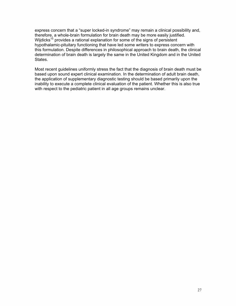
27
express concern that a “super locked-in syndrome” may remain a clinical possibility and, therefore, a whole-brain formulation for brain death may be more easily justified. Wijdicks15 provides a rational explanation for some of the signs of persistent hypothalamic-pituitary functioning that have led some writers to express concern with this formulation. Despite differences in philosophical approach to brain death, the clinical determination of brain death is largely the same in the United Kingdom and in the United States. Most recent guidelines uniformly stress the fact that the diagnosis of brain death must be based upon sound expert clinical examination. In the determination of adult brain death, the application of supplementary diagnostic testing should be based primarily upon the inability to execute a complete clinical evaluation of the patient. Whether this is also true with respect to the pediatric patient in all age groups remains unclear.
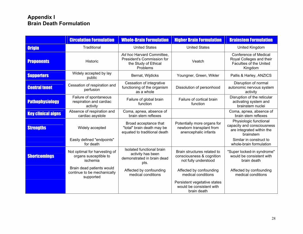
Appendix I Brain Death Formulation
28
Circulation Formulation Whole-Brain Formulation Higher Brain Formulation Brainstem Formulation
Origin Traditional United States United States United Kingdom
Proponents Historic Ad hoc Harvard Committee, President's Commission for
the Study of Ethical Problems
Veatch Conference of Medical
Royal Colleges and their Faculties of the United
Kingdom
Supporters Widely accepted by lay
public Bernat, Wijdicks Youngner, Green, Wikler Pallis & Harley, ANZICS
Central tenet Cessation of respiration and
perfusion Cessation of integrative
functioning of the organism as a whole
Dissolution of personhood Disruption of normal
autonomic nervous system activity
Pathophysiology Failure of spontaneous respiration and cardiac
activity Failure of global brain
function Failure of cortical brain
function Disruption of the reticular
activating system and brainstem nuclei
Key clinical aigns Absence of respiration and
cardiac asystole Coma, apnea, absence of
brain stem reflexes Coma, apnea, absence of brain stem reflexes
Strengths Widely accepted Broad acceptance that
"total" brain death may be equated to traditional death
Potentially more organs for newborn transplant from
anencephalic infants
Physiologic functional capacity and consciousness
are integrated within the brainstem
Easily defined "endpoints" for death Similar in construct to
whole-brain formulation
Shortcomings Not optimal for harvesting of
organs susceptible to ischemia
Isolated functional brain activity has been
demonstrated in brain dead pts.
Brain structures related to consciousness & cognition
not fully understood
"Super locked-in syndrome" would be consistent with
brain death
Brain dead patients would continue to be mechanically
supported
Affected by confounding medical conditions
Affected by confounding medical conditions
Affected by confounding medical conditions
Persistent vegetative states would be consistent with
brain death
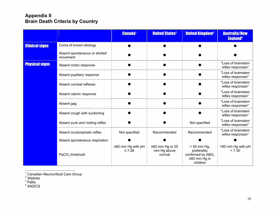
Appendix II Brain Death Criteria by Country
29
Canada1 United States2 United Kingdom3 Australia/New
Zealand4
Coma of known etiology Clinical signs
Absent spontaneous or elicited movement
Absent motor response "Loss of brainstem reflex responses"
Absent pupillary response "Loss of brainstem reflex responses"
Absent corneal reflexes "Loss of brainstem reflex responses"
Absent caloric response "Loss of brainstem reflex responses"
Absent gag "Loss of brainstem reflex responses"
Absent cough with suctioning "Loss of brainstem reflex responses"
Absent suck and rooting reflex Not specified "Loss of brainstem reflex responses"
Absent oculocephalic reflex Not specified Recommended Recommended "Loss of brainstem reflex responses"
Absent spontaneous respiration
Physical signs
PaCO2 threshold
≥60 mm Hg with pH ≤ 7.28
≥60 mm Hg or 20 mm Hg above
normal
> 50 mm Hg, preferably
confirmed by ABG, ≥60 mm Hg in
children
>60 mm Hg with pH < 7.30
1 Canadian Neurocritical Care Group 2 Wijdicks 3 Pallis 4 ANZICS
Appendix II Brain Death Criteria by Country
30
Canada1 United States2 United Kingdom3 Australia/New Zealand4
Time interval between evaluations (hrs.) Conditions
Term - 2 months old Not specified 48 hours Do not diagnose under 7 days
Guideline not applicable
2 months - 1 year old Not specified 24 hours Use caution under 5
yrs. 2 hrs. minimum
1 year - 18 years old Not specified 12 hours Use caution under 5
yrs. 3 hrs. minimum
Over 18 years Not specified Optional
Time to satisfy preconditions & to
exclude confounding factors
4 hrs. minimum
Confirmatory Test
Term - 2 months old Clinical exam &
radionuclide confirmation
2 tests required "Not required for diagnosis" Not specified
2 months - 1 year old
2 examinations with EEGs; 2nd exam & EEG not needed if
confirmed with radionuclide study
1 test required "Not required for diagnosis" Not specified
1 year - 18 years old
Optional. Observation period of at least 12 hrs. or greater than 24 hrs. for hypoxic ischemic
brain injury
Optional "Not required for diagnosis" Not specified
Over 18 years Optional. Optional "Not required for diagnosis" Not specified
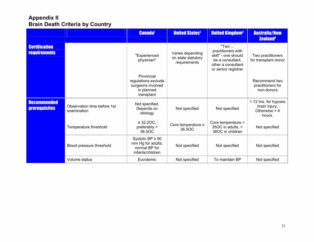
Appendix II Brain Death Criteria by Country
31
Canada1 United States2 United Kingdom3 Australia/New Zealand4
"Experienced physician"
Varies depending on state statutory
requirements
"Two ... practitioners with skill" - one should be a consultant,
other a consultant or senior registrar
Two practitioners for transplant donor
Certification requirements
Provincial regulations exclude surgeons involved
in planned transplant
Recommend two practitioners for
non-donors
Observation time before 1st examination
Not specified. Depends on
etiology Not specified Not specified
> 12 hrs. for hypoxic brain injury.
Otherwise > 4 hours.
Temperature threshold ≥ 32.2OC,
preferably > 36.5OC
Core temperature ≥ 36.5OC
Core temperature > 35OC in adults, > 36OC in children
Not specified
Blood pressure threshold
Systolic BP ≥ 90 mm Hg for adults,
normal BP for infants/children
Not specified Not specified Not specified
Recommended prerequisites
Volume status Euvolemic Not specified To maintain BP Not specified
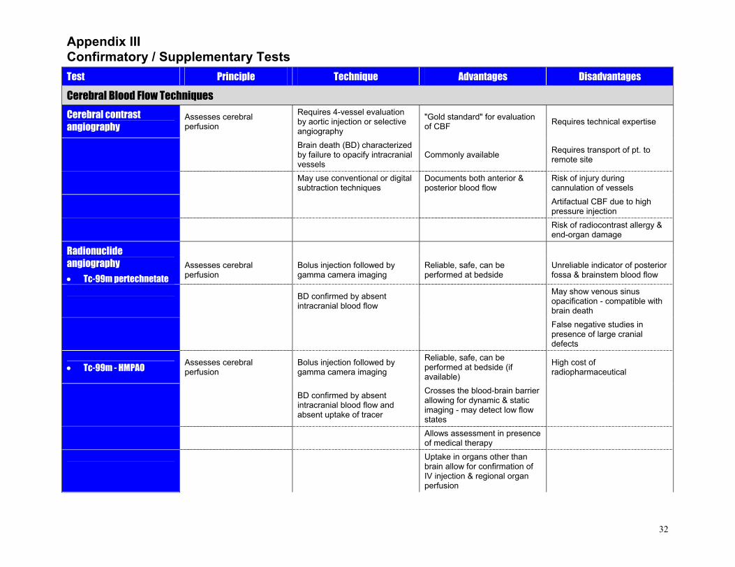
Appendix III Confirmatory / Supplementary Tests
32
Test Principle Technique Advantages Disadvantages
Cerebral Blood Flow Techniques
Cerebral contrast angiography
Assesses cerebral perfusion
Requires 4-vessel evaluation by aortic injection or selective angiography
"Gold standard" for evaluation of CBF Requires technical expertise
Brain death (BD) characterized by failure to opacify intracranial vessels
Commonly available Requires transport of pt. to remote site
May use conventional or digital subtraction techniques
Documents both anterior & posterior blood flow
Risk of injury during cannulation of vessels
Artifactual CBF due to high pressure injection
Risk of radiocontrast allergy & end-organ damage
Radionuclide angiography • Tc-99m pertechnetate
Assesses cerebral perfusion
Bolus injection followed by gamma camera imaging
Reliable, safe, can be performed at bedside
Unreliable indicator of posterior fossa & brainstem blood flow
BD confirmed by absent intracranial blood flow
May show venous sinus opacification - compatible with brain death
False negative studies in presence of large cranial defects
• Tc-99m - HMPAO Assesses cerebral perfusion
Bolus injection followed by gamma camera imaging
Reliable, safe, can be performed at bedside (if available)
High cost of radiopharmaceutical
BD confirmed by absent intracranial blood flow and absent uptake of tracer
Crosses the blood-brain barrier allowing for dynamic & static imaging - may detect low flow states
Allows assessment in presence of medical therapy
Uptake in organs other than brain allow for confirmation of IV injection & regional organ perfusion
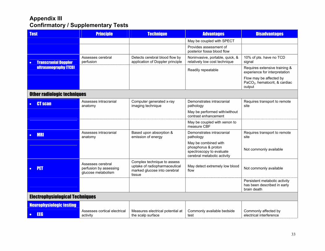
Appendix III Confirmatory / Supplementary Tests
33
Test Principle Technique Advantages Disadvantages
May be coupled with SPECT Provides assessment of
posterior fossa blood flow
Assesses cerebral perfusion
Detects cerebral blood flow by application of Doppler principle
Noninvasive, portable, quick, & relatively low cost technique
10% of pts. have no TCD signal • Transcranial Doppler
ultrasonography (TCD) Readily repeatable Requires extensive training & experience for interpretation
Flow may be affected by PaCO2, hematocrit, & cardiac output
Other radiologic techniques
• CT scan Assesses intracranial anatomy
Computer generated x-ray imaging technique
Demonstrates intracranial pathology
Requires transport to remote site
May be performed with/without
contrast enhancement
May be coupled with xenon to
measure CBF
• MRI Assesses intracranial anatomy
Based upon absorption & emission of energy
Demonstrates intracranial pathology
Requires transport to remote site
May be combined with phosphorus & proton spectroscopy to evaluate cerebral metabolic activity
Not commonly available
• PET Assesses cerebral perfusion by assessing glucose metabolism
Complex technique to assess uptake of radiopharmaceutical marked glucose into cerebral tissue
May detect extremely low blood flow Not commonly available
Persistent metabolic activity has been described in early brain death
Electrophysiological Techniques
Neurophysiologic testing
• EEG Assesses cortical electrical activity
Measures electrical potential at the scalp surface
Commonly available bedside test
Commonly affected by electrical interference
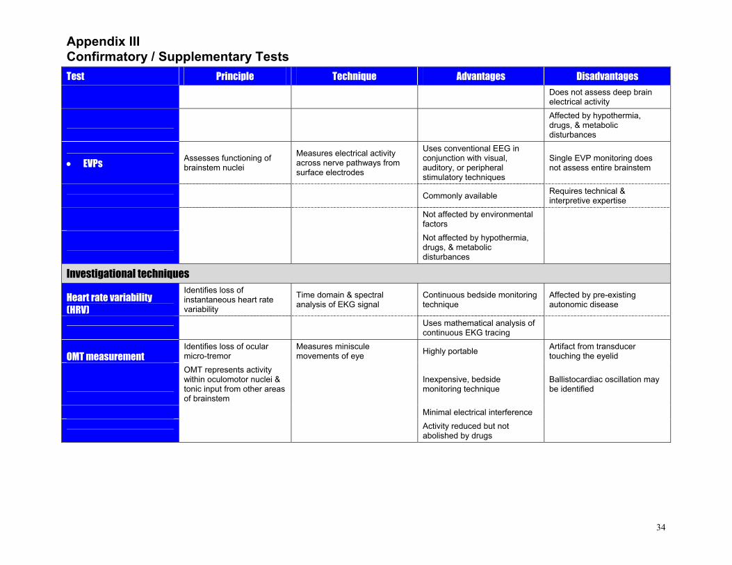
Appendix III Confirmatory / Supplementary Tests
34
Test Principle Technique Advantages Disadvantages
Does not assess deep brain electrical activity
Affected by hypothermia, drugs, & metabolic disturbances
• EVPs Assesses functioning of brainstem nuclei
Measures electrical activity across nerve pathways from surface electrodes
Uses conventional EEG in conjunction with visual, auditory, or peripheral stimulatory techniques
Single EVP monitoring does not assess entire brainstem
Commonly available Requires technical & interpretive expertise
Not affected by environmental factors
Not affected by hypothermia, drugs, & metabolic disturbances
Investigational techniques
Heart rate variability (HRV)
Identifies loss of instantaneous heart rate variability
Time domain & spectral analysis of EKG signal
Continuous bedside monitoring technique
Affected by pre-existing autonomic disease
Uses mathematical analysis of continuous EKG tracing
OMT measurement Identifies loss of ocular micro-tremor
Measures miniscule movements of eye Highly portable Artifact from transducer
touching the eyelid
OMT represents activity within oculomotor nuclei & tonic input from other areas of brainstem
Inexpensive, bedside monitoring technique
Ballistocardiac oscillation may be identified
Minimal electrical interference
Activity reduced but not abolished by drugs
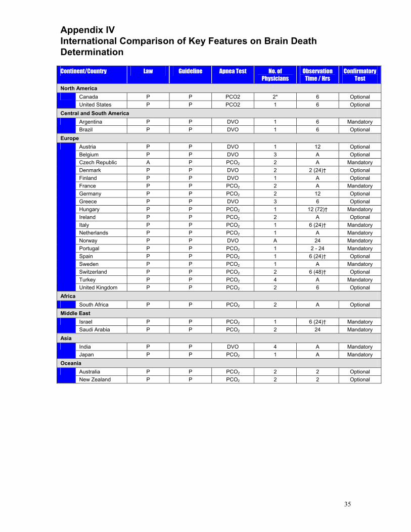
Appendix IV International Comparison of Key Features on Brain Death Determination
35
Continent/Country Law Guideline Apnea Test No. of
Physicians Observation
Time / Hrs Confirmatory
Test
North America Canada P P PCO2 2* 6 Optional United States P P PCO2 1 6 Optional
Central and South America Argentina P P DVO 1 6 Mandatory Brazil P P DVO 1 6 Optional
Europe Austria P P DVO 1 12 Optional Belgium P P DVO 3 A Optional Czech Republic A P PCO2 2 A Mandatory Denmark P P DVO 2 2 (24)† Optional Finland P P DVO 1 A Optional France P P PCO2 2 A Mandatory Germany P P PCO2 2 12 Optional Greece P P DVO 3 6 Optional Hungary P P PCO2 1 12 (72)† Mandatory Ireland P P PCO2 2 A Optional Italy P P PCO2 1 6 (24)† Mandatory Netherlands P P PCO2 1 A Mandatory Norway P P DVO A 24 Mandatory Portugal P P PCO2 1 2 - 24 Mandatory Spain P P PCO2 1 6 (24)† Optional Sweden P P PCO2 1 A Mandatory Switzerland P P PCO2 2 6 (48)† Optional Turkey P P PCO2 4 A Mandatory
United Kingdom P P PCO2 2 6 Optional Africa
South Africa P P PCO2 2 A Optional Middle East
Israel P P PCO2 1 6 (24)† Mandatory Saudi Arabia P P PCO2 2 24 Mandatory
Asia India P P DVO 4 A Mandatory Japan P P PCO2 1 A Mandatory
Oceania Australia P P PCO2 2 2 Optional New Zealand P P PCO2 2 2 Optional
36
References 1 Bernat JL. Ethical issues in neurology, 2nd edition. Boston: Butterworth Heinemann; 2002:243-81.
2 Wijdicks EFM. Brain death worldwide—Accepted fact but no global consensus in diagnostic criteria. Neurology 2002;58:20-5.
3 Uniform Determination of Death Act, 12 Uniform Laws Annotated 589 (West 1993 and West Supp. 1997), 706 U.S.C.
4 Mollaret P, Goulon M. Le coma dépassé. Rev Neurol (Paris) 1959;101:3-15.
5 A definition of irreversible coma: Report of the Ad Hoc Committee of the Harvard Medical School to Examine the Definition of Brain Death. JAMA 1968;205:337-40.
6 Mohandas A, Chou SN. Brain death—A clinical and pathological study. J Neurosurg 1971;35:211-8.
7 Diagnosis of brain death: Statement issued by the honorary secretary of the Conference of Medical Royal Colleges and Their Faculties in the United Kingdom on 11 October 1976. BMJ 1976;2:1187-8.
8 The President’s Commission for the Study of Ethical Problems in Medicine and Biomedical and Behavioral Research. Defining death. JAMA 1981;246:2184-6.
9 The Quality Standards Subcommittee of the American Academy of Neurology. Practice parameters for determining brain death in adults (summary statement). Neurology 1995;45:1012-24.
10 Pallis C, Harley DH. ABC of brain stem death, 2nd edition; London: BMJ Publishing Group; 1996. 11 Capron AM. Brain death—well settled yet still unresolved. NEJM 2001;344:1244-6.
12 Veatch RM. The whole-brain-oriented concept of death: an outmoded philosophical formulation. J Thanatol 1975;3:13-30.
13 Kantor JE, Hoskins IA. Brain death in pregnant women. Journal of Clinical Ethics 1993;4: 308-14.
14 Bernat JL, Culver CM, Gert B. On the definition and criterion of death. Annals of Internal Medicine 1981;94:389-94.
15 Powner DJ, Hendrich A, Lagler RG, Ng RH, Madden RL. Hormonal changes in brain dead patients. Crit Care Med 1990;18:702-8.
16 Wijdicks EFM. Brain death. Philadelphia: Lippincott, Williams & Wilkins. 2000; 29-43
17 Principles and practice of anesthesiology, Volume 2, 2nd edition. ST. Louis: Mosby. 1998; 1607-68.
37
18 Ducrocq X, Braun M, Debouverie M, Junges C, Hummer M, Vespignani H. Brain death and transcranial Doppler: Experience in 130 cases of brain dead patients. J Neurol Sciences 1998;160:41-6.
19 Wijdicks 2000: 45-60.
20 Choi DW, Dacey RG, Hsu CY, Powers WJ. Cerebrovascular disease—Momentum at the end of the second millennium. American Heart Association Monograph Series. Armonk: Futura Publishing Company. 2001; 251-67.
21 Powner DJ, Darby JM. Current considerations in the issue of brain death. Neurosurg 1999;45:1222-6.
22 Chen EP, Bittner HB, Kendall SW, Van Trigt P. Hormonal and hemodynamic changes in a validated animal model of brain death. Crit Care Med 1996;24:1352-59.
23 Powner DJ, Hendrich A, Hyhuis A. Strate R. Changes in serum catecholamine levels in patients who are brain dead. J Heart Lung Transplant 1992;11:1046-53.
24 Dujardin KS, McCully RB, Wijdicks EFM, Tazelaar HD, Seward JB, McGregor CG, Olson LJ. Myocardial dysfunction associated with brain death: clinical, echocardiographic, and pathologic features. J Heart Lung Transplant 2001;20:350-7.
25 Gramm HJ, Meinhold H, Bickel U, Zimmerman J, von Hammerstein B, Keller F, Deinhardt R, Voigt K. Acute endocrine failure after brain death? Transplantation 1992;54:851-7.
26 Schrader H, Krogness K, Aakvaag A, Sorland O, Purvis K. Changes of pituitary hormones in brain death. Acta Neurochr (Wien) 1980;52:239-48.
27 Grenvik A, Powner DJ, Snyder JV. Cessation of therapy in terminal illness and brain death. Crit Care Med 1978;6:284-91.
28 Hohenegger M, Vermes M, Mauritz W, Redl G, Sporn P, Eiselsberg P. Serum vasopressin (AVP) levels in polyuric brain-dead organ donors. Eur Arch Psychiatry Neurol Sci 1990;239:267-9.
29 Canadian Neurocritical Care Group. Guidelines for the diagnosis of brain death. Can J Neurol Sci 1999;26:64-6.
30 Wijdicks EFM. The diagnosis of brain death. NEJM 2001;344:1216.
31 Pearson IY. Australia and New Zealand Intensive Care Society Statement and Guidelines on Brain Death and Model Policy on Organ Donation. Anesthesia and Intensive Care 1995;23:104-8.
32 Van Norman G. A matter of life and death: What every anesthesiologist should know about the medical, legal, and ethical aspects of declaring brain death. Anesthesiology 1999;91:275-87.
33 Lazar NM, Shemie S, Webster GC, Dickens BM. Bioethics for clinicians: 24. Brain death. CMAJ 2001;164:833-6.
34 Watts J. Brain-death guidelines revised in Japan. Lancet 1999;354:1011.
38
35 Haupt WF, Rudolf J. European brain death codes: a comparison of national guidelines. J Neurol 1999;246:432-37. 36 Wijdicks 2000: 61-90. 37 Wijdicks 2000: 115-34.
38 Danzl DR, Pozos RS. Accidental hypothermia. NEJM 1994;331:1756-60. 39 The Hypothermia after Cardiac Arrest Study Group. Mild therapeutic hypothermia to improve the neurologic outcome after cardiac arrest. NEJM 2002;346:549-56.
40 Principles and practice of anesthesiology 1998: 1130-4.
41 Saposnik G, Bueri JA, Mauriňo J, Saizar R, Garetto NS. Spontaneous and reflex movements in brain death. Neurology 2000;54:221-4.
42 Turnbull J, Rutledge F. Spontaneous respiratory movement in a patient with clinical brain death. Neurology 1985;35:1260.
43 Saposnik G, Mauriňo J, Bueri JA. Movements in brain death. European Journal of Neurology 2001;8:209-13.
44 Ashwal, S as cited in Wijdicks 2000: 91-114. 45 Farrell MM, Levin DL. Brain death in the pediatric patient: Historical, sociological, medical, religious, cultural, legal and ethical considerations. Crit Care Med 1993;21:1951-65.
46 American Academy of Pediatrics Task Force on Brain Death in Children. Guidelines for the determination of brain death in children. Pediatrics 1987;80:298-300. 47 Volpe JJ. Commentary: Brain death determination in the newborn. Pediatrics 1987;80:293-7.
48 Girvin J, Capron AM. Debat III: Resolved: Brain death criteria must be revised so that society can readily benefit from families who offer their anencephalic infants as organ donors. J Heart Lung Transplant 1993:12:S369-78. 49 Bernstein IM, Watson M, Simmons GM, Catalano PM, Davis G, Collins R. Maternal brain death and prolonged fetal survival. Obstet Gynecol 1989;74:434-7.
50 Catanzarite VA, Willms DC, Holdy KE, Gardner SE, Ludwig DM, Cousins LM. Brain death during pregnancy: tocolytic therapy and aggressive maternal support on behalf of the fetus. Am J of Perinat 1997;14:431-44. 51 Kantor JE, Hoskins IA. Brain death in pregnant women. J Clinical Ethics 1993;4:308-14.
52 Chervenak FA, McCullough LB. Clinical management of brain death during pregnancy. J Clinical Ethics 1993;4:349-50.
53 Field DR, Gates EA, Creasy RK, Jonsen AR, Laos RK. Maternal brain death during pregnancy: medical and ethical issues. JAMA 1988;260:816-22.
54 Monsein LH. The imaging of brain death. Anaesth Intens Care 1995;23:44-50.

39
55 Flowers WM, Patel BR. Persistence of cerebral blood flow after brain death. South Med Jour 2000;93:364-70.
56 Schlake HP, Böttger IG, Grotemeyer KH, Husstedt IW, Brandau W, Schober O. Determination of cerebral perfusion by means of planar brain scitigraphy and 99mTc-HMPAO in brain death, persistent vegetative state and severe coma. Int Care Med 1992;18:76-81.
57 Spieth ME, Ansari AM, Kawada TK, Kimura RL, Siegel ME. Direct Comparison of TC-99m DTPA and TC-99m HMPAO for evaluating brain death. Clin Nuc Med 1994;19:867-72.
58 de Tourtchaninoff M, Hantson P, Mahieu P, Guérit JM. Brain death diagnosis in misleading conditions. Q J Med 1999;92:407-14.
59 Wagner W. Scalp, earlobe, and nasopharyngeal recordings of the median nerve somatosensory evoked P14 potential in coma and brain death. Brain 1996;119:1507-21.
60 Rapenne T, Moreau D, Lenfant F, Boggio V, Cottin Y, Freysz M. Could heart rate variability analysis become an early predictor of imminent brain death? A pilot study. Anesth Analg 2000;92:329-36.
61 Fretias JF, Azevedo AJ, Teixeira J, Carvalho MJ, Costa O, Falcão de Freitas A. Heart rate variability as an assessment of brain death. Transplantation Proceedings 2000;32:2584-5.
62 Bolger C, Bojanic S, Philips J, Sheahan N, Coakley D, Malone J. Ocular microtremor in brain stem death. Neurosurgery 1999;44:1201-6.
63 Medlock MD, Hanigan WC, Cruse RP. Dissociation of cerebral blood flow, glucose metabolism, and electrical activity in pediatric brain death. J Neurosurg 1993;79:752-5.