Embed Size (px)
Citation preview

Intrinsic Radiation Resistance of Primary Clonogenic Blasts from Children withNewly Diagnosed B-Cell Precursor Acute Lymphoblastic LeukemiaFatih M. Uckun,**Il Waclaw Jaszcz,' Mridula Chandan-Langlie,* Kevin G. Waddick,*Kazimiera Gaji-Peczalska,O and Chang W. Song**Tumor Immunology Laboratory, Sections of Cancer and Leukemia Biology and Radiation Biology, Department of TherapeuticRadiology-Radiation Oncology, tDivision of Hematology/Oncology/Bone Marrow Transplantation Program, Department of Pediatrics,§Department of Laboratory Medicine/Pathology, University of Minnesota Health Sciences Center, Minneapolis, Minnesota 55455; andIThe Children's Cancer Study Group, Arcadia, California 91066
Abstract
The radiation sensitivity of primary clonogenic blasts from 44children with newly diagnosed B-cell precursor acute lympho-blastic leukemia (ALL) was analyzed using leukemic progeni-tor cell (LPC) colony assays. The derived values for SF2 (sur-viving fraction at 200 cGy) and a (initial slope of radiationsurvival curves constructed according to the linear quadraticmodel) indicated a marked interpatient heterogeneity in intrin-sic radiation sensitivity of LPC populations. The SF2 valuesranged from 0.01 to 1.00 (median = 0.430; mean±SE= 0.47±0.04), and the a values ranged from 0.000 to 3.272Gyf' (median = 0.280 Gyf-; mean±SE = 0.430±0.093 Gy-1).When CD19' CD34' versus CD19' CD34- immunopheno-types were compared, a trend toward higher SF2 and lower avalues were observed in LPCfrom CD34+ patients, consistentwith greater radiation resistance. When patients were dividedinto three approximately equal groups based on increasing lev-els of CD34 expression, a clear ordering effect was observedindicating that increased CD34expression levels are associatedwith significantly higher radiation resistance at the level of B-lineage LPC. The highest CD34expression group (. 75%posi-tivity) had 1.4-fold higher SF2 (P = 0.05) and twofold lower avalues (P = 0.06) than the lowest group (< 30% positivity).Furthermore, the CD34 positivity of radiation resistant (a< 0.2 and SF2 . 0.5) B-cell precursor ALL cases was greaterthan the CD34 positivity of radiation sensitive (a > 0.2 and/orSF2 < 0.5) cases (56±9% versus 34±9%, P = 0.09). Whereasonly 6 of 16 (38%) of radiation sensitive cases were CD34+, 11of 15 (73%) of radiation resistant cases expressed CD34 (P= 0.04). Our results offer new insights into the inherent and/or acquired radiation resistance of primary clonogenic blastsfrom B-cell precursor ALL patients. (J. Clin. Invest. 1993.91:1044-1051.) Key words: immunophenotype - leukemia * ra-diation resistance * clonogenic cells * leukemic progenitor cell
Introduction
High risk acute lymphoblastic leukemia (ALL)' patients typi-cally suffer a poor outcome after conventional drug treatment
Address correspondence to Fatih M. Uckun, M.D., Ph.D., Box 356UMHC,Section of Cancer and Leukemia Biology, University of Min-nesota, 420 Delaware St., Minneapolis, MN55455.
Received for publication 12 August 1992 and in revised form 13October 1992.
1. Abbreviations used in this paper: ALL, acute lymphoblastic leuke-mia; BMT, bone marrow transplantation; L-BCGF, low molecular
protocols (1). Within the last decade, however, total body irra-diation (TBI) and high dose chemotherapy have been appliedbefore bone marrow transplantation (BMT) in aggressive regi-mens to enhance these patients' prospects for disease-free sur-vival (2-9). Although improvements in long-term, disease-freesurvival have been reported, recurrence of leukemia within thefirst 6 moafter TBI and BMTcontinues to be a major obstacleto a more successful outcome after BMTfor high risk ALL, andonly < 20% of high risk ALL patients become long-term, dis-ease-free survivors after autologous or allogeneic BMT(2-13).The inability of TBI to adequately destroy leukemia cells andprevent residual disease may involve several possibilities. Clon-ogenic blasts (i.e., leukemic progenitor cells [LPC] may (a)possess an intrinsic resistance to radiation; (b) contain a largefraction of noncycling dormant blasts; (c) effectively repairsublethal radiation injury; (d) be capable of rapid self-renewaland repopulation; and/or (e) survive in quantities sufficient tocause relapse as a result of a high leukemia burden at remission.
Until we learn more about the effects of TBI on LPC, theresults of radiation based pre-BMT conditioning regimens arenot likely to dramatically improve. A thorough radiobiologicanalysis of primary clonogenic blasts from ALL patients is arequisite step toward greater understanding of clinical radia-tion resistance. The present study initiated this task by evaluat-ing the radiation sensitivity of primary clonogenic blasts from44 newly diagnosed B-cell precursor ALL patients. Two com-puter-based models of cell survival were used (14). The multi-target model of survival curve analysis (14-16) provides SF2values as parameters to describe intrinsic radiation sensitivity.The surviving fraction at 200 cGy (SF2) value is the survivingfraction of clonogenic blasts after exposure to 200 cGy. Themajor parameter for intrinsic radiation sensitivity in the linearquadratic model (14-16) is the a value, representing the initialslope of the linear component ofthe continuously bending radi-ation dose survival curve. Clonogenic blasts are consideredsensitive to radiation based on high a values and low SF2values.
Weinitially recorded a marked interpatient variation in thevalues obtained from survival curves generated by LPC. Clono-genic blasts from different B-cell precursor ALL patients variedsubstantially in their sensitivity to radiation. Weexamined pos-sible relationships between LPC radiation responses and thestandard diagnostic features of these patients, including age,sex, white blood cell count( WBC)at diagnosis, immunopheno-type, and cell cycle distribution of bone marrow blasts. Ourobservations indicated that LPCare intrinsically more resistantto radiation when a high percentage of blasts are in the Go,
weight B-cell growth factor; LPC, leukemic progenitor cells; PKC, pro-tein kinase C; SF2, surviving fraction at 200 cGy; WBC, white blood
cell count.
1044 Uckun, Jaszcz, Chandan-Langlie, Waddick, Gajl-Peczalska, and Song
J. Clin. Invest.© The American Society for Clinical Investigation, Inc.0021-9738/93/03/1044/08 $2.00Volume 91, March 1993, 1044-1051
phases of the cell cycle or a low percentage of blasts are in the Sphase. Further results provided evidence that radiation resis-tance may be associated with a high level of CD34 antigenexpression. To our knowledge, this report represents the firstradiobiologic analysis comparing radiation responses of pri-mary LPC in relation to diagnostic features that distinguishB-cell precursor ALL patients.
Methods
Patient selection and patient material. Fresh leukemic bone marrowsamples from 44 newly diagnosed B-cell precursor ALL patients wereused to study the radiobiologic features of primary clonogenic blastpopulations. Diagnosis of B-cell precursor ALL was based on the mor-phologic, cytochemical, and immunophenotypic features of marrowblasts. There were 17 females and 27 males with a median age of 4 yr(range: 0.1-17 yr; mean±SE = 5.5±0.7 yr; n = 44). 37 of 44 patients(84%) were < 10 yr of age. The initial WBCranged from 4.0 X I09 to999.0 x 109/liter (median = 74.0 x 10⁹/liter; mean±SE= 138.1±33.6 x 10⁹/liter; n = 34). The cell cycle distribution of leuke-mic blasts was evaluated by DNAflow cytometry in a total of 18 pa-tients. The percentage of Go, I phase cells ranged from 72 to 95% (me-dian-85%; mean±SE = 85±1%). The percentage of S phase cellsranged from 4 to 23% (median = 13%; mean±SE = 12±1%) and theproliferation index (PI = %S+ %G2M)ranged from 5 to 28% (median= 15%; mean±SE = 15± 1%).
The immunophenotypic features of leukemic blasts were deter-mined by immunofluorescence staining and flow cytometry using abroad panel of mAb reactive with lymphoid differentiation antigens.None of 44 patients expressed the pan-T cell antigens CD2, CD5, andCD7or the mature B-cell marker surface immunoglobulin. In contrast,leukemic blasts from each of the 44 patients expressed high levels ofCD19 and/or CD24 B-lineage differentiation antigens. 89±1% of leu-kemic blasts from 44 of 44 (100%) expressed CD19, 83±3% of leuke-mic blasts from 34 of 40 patients (85%) expressed CD24, and 57±4% ofleukemic blasts from 10 of 28 patients (36%) expressed CD40antigen.The lymphoid precursor cell antigen CD1Owas expressed on 82±3% ofleukemic blasts from 33 of 44 (75%) patients. The progenitor cell asso-ciated antigen CD34was expressed on 76±5% of leukemic blasts from17 of 31 patients (55%). Thus, leukemic blasts displayed a commoncomposite immunophenotype (CD2-CD5 -CD7 -CD10'/-CD19+CD24+CD34+/-CD40+1-sIg-) consistent with B-cell precursor ALL.Fresh bone marrow aspirates were procured by conventional methodsas part of clinical treatment protocols. Mononuclear cells highlyenriched for leukemic blasts (i.e., containing > 90% blasts) were iso-lated from fresh bone marrow aspirates by density gradient separationon Ficoll-Hypaque gradients. All patient bone marrow samples wereused following the guidelines of the University of Minnesota Commit-tee on the Use of Human Subjects in Research for secondary use ofpathologic or surgical tissue.
Immunophenotyping. The surface antigen profiles of freshly iso-lated B-cell precursor ALL blasts were analyzed by indirect immunoflu-orescence and flow cytometry using a panel of monoclonal antibodies(mAb) that define human leukocyte differentiation antigens as previ-ously described (17-20). Specifically, we used the mAbs 13-B3/35.1(anti-CD2), T 101 / 10.2 (anti-CD5), G3.7 (anti-CD7), BA-3 (anti-CD1O), B43 (anti-CDl9), BA-l (anti-CD24), MY1O(anti-CD34),and G28-5 (anti-CD40). Samples were considered positive if> 30%ofcells bound the antibody used.
Analysis of cell cycle kinetics by quantitative DNAflow cytometry.DNAcontent and cell cycle distribution of leukemic marrow blastswere evaluated by flow cytometric DNAanalysis of fixed cells stainedwith propidium iodide (Sigma Chemical Co., St. Louis, MO) aftertreatment with RNAse (21, 22). Flow cytometric data of at least20,000 cells from each sample were acquired in linear mode using aflow cytometer (FACScan®; Becton Dickinson, Mountain View, CA).Data were analyzed using polynomial and broadened rectangle models
of DNA-ploidy-cell cycle and software (DNA Cell-Cycle Analysis Soft-ware; Becton Dickinson).
Irradiation of cells. Primary B-cell precursor ALL blasts, 1 X 10⁵cells/ml in alpha-MEM supplemented with 5% FCS, were irradiatedwith 50-800 cGy gamma rays in a single exposure (100 cGy/min,370C) using a 137CS irradiator (model Mark I; J. L. Shephard and Asso-ciates, Glendale, CA), as previously described (22-25). After irradia-tion, 1 X 10⁵ ALL blasts per sample were assayed in duplicate forLPC-derived B-cell precursor ALL blast colony formation, as describedin the next paragraph. The surviving fractions of clonogenic blasts weredetermined using the formula: Percent survival = 100 X (mean num-ber of colonies in irradiated samples)/(mean number of colonies inunirradiated control samples). The radiation survival curves were con-structed using computer programs for the analysis of cell survival dataaccording to the single-hit multitarget and linear quadratic models, asdetailed under Data analysis hereinafter. Data were expressed as themean±SE values for SF2 as well as the a (initial slope reflecting thesteepness of the linear component of cell killing) value of the linearquadratic model.
Colony assays for primary clonogenic blasts from B-cell precursorALL patients. A small fraction of leukemic B-cell precursors from ALLpatients proliferate in response to low molecular weight B-cell growthfactor (L-BCGF; Cellular Products, Buffalo, NY) and form compactblast colonies (20, 25, 26, 27). Day 7 ALL blast colonies are verycompact and have a small size containing 20-50 lymphoblasts (20,27). Therefore, colony numbers up to 3,000 per Petri dish can easily beenumerated on a grid using an inverted phase microscope of high opti-cal resolution (20, 27). In the present study, fresh ALL marrow blasts(1 X I0 5 blasts/ ml, duplicate I -ml samples) were irradiated and imme-diately plated in 35-mm Petri dishes for a 7-d culture at 370C in ahumidified 5%CO2atmosphere in alpha-MEM. Medium was supple-mented with 0.9% methylcellulose, 50 uM 2-mercaptoethanol, 15%human plasma + 15% fetal bovine serum (or 30% calf bovine serum),and 10% (vol/vol) L-BCGF (20, 25-27). On day 7, blast coloniescontaining > 20 cells were counted using an inverted phase microscopewith high optical resolution. After the enumeration of colonies in eachpetri dish, colony blasts were subjected to morphological and immuno-logical analyses, as described (20, 25-30). In all experiments, culturedishes were examined immediately and 24 h after plating to exclude thepossibility of clumping unrelated to proliferation, which might lead towrong conclusions regarding the radiation sensitivity of clonogenicblasts.
Data analysis. The radiation survival curves were constructed us-ing three computer programs for the MacIntosh, ENTER, DSUM,andFIT (kindly provided by Dr. N. Albright, Department of RadiationOncology, University of California, San Francisco, CA), for the analy-sis of cell survival data according to the singe-hit multitarget and linearquadratic models of cell survival ( 14, 22). The data in this study wereexpressed as SF2 value representing the radiation sensitivity parameterof the singe-hit multitarget model of cell survival, as well as the a valuerepresenting the radiation sensitivity parameter of the linear quadraticmodel of cell survival ( 14, 22). Weused standard statistical methods,including Student's t tests, to evaluate the in vitro radiation survivaldata and to compare the radiobiologic features of primary clonogenicblasts from different patients, as previously described (22, 25). Chisquare analyses were used to assess the degree of association betweenclinical/laboratory parameters and radiation resistance/sensitivity ofLPC. All computations were performed using an updated statistics pro-gram for the MacIntosh (StatWorks; Cricket Software, Philadel-phia, PA).
Results
Radiobiologicfeatures ofprimary clonogenic blastsfrom newlydiagnosed B-lineage ALL patients. Primary bone marrowblasts from 44 patients were exposed to ionizing radiation, cul-tured in the presence of L-BCGF, and assayed for LPC-derived
Radiation Resistance in B-Cell Precursor Acute Lymphoblastic Leukemia 1045

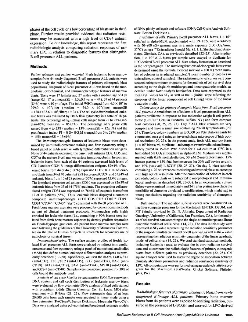
Figure 1. Composite ra-1 s . diation survival curve
of primary clonogenic0 ~ ~ Iblasts from newly diag-
nosed B-cell precursorALL patients. Survival
o 10.1 of primary clonogenicblasts from 44 newlydiagnosed B-cell precur-sor ALL patients is
.....B-CeP ... ALL (N=4) .. shown as a function of10-2 2 4 6 8 1 the radiation dose. Cells
2adiat4on 6ose 8G 10 were irradiated, assayedRadiation Dose (Gy) for blast colony forma-
tion in vitro, and thecomposite radiation survival curve was generated using a computerprogram according to the linear quadratic model of cell survival asdescribed in Methods. The calculated standard errors are also indi-cated. Each independent experiment was performed in duplicate.
blast colony formation. The median and mean numbers ofblast colonies in unirradiated control cultures were 1,097 colo-nies/ I05 cells (1.1% plating efficiency) and 1,668±217 colo-nies/ 105 cells (1.7±0.2% plating efficiency), respectively.
Radiation survival curves of pimary clonogenic blasts (i.e.,LPC) were constructed for each of, the 44 newly diagnosed
B-cell precursor ALL patients using computer programs for thesingle-hit multitarget, as well as the linear quadratic models ofcell survival. The computer-determined values for the radio-biologic parameters indicated a marked interpatient heteroge-neity in intrinsic radiation sensitivity of LPCpopulations. TheSF2 values ranged from 0.01 to 1.00 (median = 0.430;mean±SE = 0.47±0.04), and the a values ranged from0.000 to 3.272 Gy-' (median = 0.280 Gy-'; mean±SE= 0.430±0.093 Gy-'). The SF2, and a values of the compositeradiation survival curve were 0.42 and 0.337 Gy', respec-tively (Fig. 1). LPC from 17 of 44 patients (39%) had SF2values 2 0.50 and a values < 0.2 Gy', consistent with amarked radiation resistance at the level of LPCusing the multi-target and linear quadratic models of cell survival. The meanSF2 value for this subgroup of 17 patients was 0.75+0.04,which is significantly higher than the mean SF2 value of0.29±0.03 for the remaining 27 radiation sensitive patients (P< 0.0001). The mean a values were 0.063±0.016 Gy'- for theradiation resistant group (n = 17) and 0.660±0.133 Gy-' forthe radiation sensitive group (n = 27) (P < 0.0001).
Correlation between radiobiologic features of primary B-lineage LPC and patients' characteristics. Patients were di-vided into groups according to sex, age, WBCat diagnosis, cellcycle distribution of leukemic blasts, and immunophenotype(Table I). Patients with a high percentage of blasts in Go/,
Table I. Statistical Correlations
n SF2 P a (Gy-') P
All patients 44 0.47±0.04 0.430±0.093M 27 0.46±0.05 NS 0.434±0.099 NSF 17 0.48±0.07 0.423±0.185
Age<4 yr 19 0.46±0.05 0.356±0.098.4 yr 25 0.47±0.07 NS 0.486±0.146 NS
WBC< I00,000/1 20 0.49±0.06 0.327±0.064
I 00,000/,Ol 14 0.39±0.08 NS 0.616±0.240 0.2Plating efficiency (%)
< 1 22 0.48±0.06 0.409±0.117.1 22 0.46±0.06 NS 0.451±0.147 NS
%Go/,<85 8 0.41±0.08 0.653±0.381.85 10 0.73±0.07 0.006 0.112±0.061 0.1
%S<13 9 0.73±0.08 0.108±0.068. 13 9 0.45±0.08 0.017 0.598±0.340 0.1
CD19+CD10+ 33 0.46±0.05 0.480±0.121CDl9+CD10- 11 0.52±0.08 NS 0.108±0.068 NS
CD19+CD34+ 17 0.60±0.06 0.261±0.116CDl9+CD34- 14 0.46±0.06 0.09 0.300±0.058 0.2
CDl9+CD40+ 10 0.56±0.09 0.352±0.194CD19+CD40- 18 0.54±0.05 NS 0.252±0.050 NSCD34 expression (%)
<30 14 0.46±0.06 0.300±0.05830-74 8 0.57±0.09 0.388±0.241.75 9 0.63±0.06 0.05 0.148±0.048 0.05
The SF2 and a values (mean±SE) of B-lineage LPC from newly diagnosed B-cell precursor ALL patients were compared using two sample, two-sided Student's t tests.
1046 Uckun, Jaszcz, Chandan-Langlie, Waddick, Gajl-Peczalska, and Song

phases of the cell cycle or a low percentage of blasts in S phasehad higher SF2 and smaller a values, consistent with a higherintrinsic radiation resistance at the level of LPC (Table I, Fig.2). Whereas only 25%of cases with Go/, < 85%had SF2 values2 0.5, consistent with radiation resistance according to singlehit multitarget model of cell survival, 80% of cases with Go,,. 85% had SF2 values 2 0.5 (Fig. 2). Similarly, 90% of caseswith Go,, 2 85% versus 37.5% of cases with Go,, < 85% had avalues < 0.2, consistent with radiation resistance according tolinear quadratic model of cell survival (Fig. 2). Furthermore,univariate analysis using Go, I and S phase percentages as con-tinuous covariates established a significant association betweenGo,1, as well as S phase percentages and SF2 (%Go0I versus SF2:coefficient of correlation = 0.36, P = 0.1; %Sphase versus SF2:coefficient of correlation = 0.29, P = 0.1), as well as a values(%Go,, lversus a: coefficient of correlation = 0.29, P = 0. 1; %Sphase versus a: coefficient of correlation = 0.26, P = 0.1). Incontrast, patient age, WBC, or in vitro plating efficiency ascontinuous covariates did not correlate with SF2 or a values (Pvalues all > 0.3). Similarly, when patients were divided intogroups, patient sex, age, WBCat diagnosis, or in vitro platingefficiency did not have a significant impact on the radiationsensitivity of LPC (Table I).
In our initial analyses ofimmunophenotype-radiation sensi-tivity associations, we used antigen expression on 2 30%blastsas an arbitrary criterion for positivity. According to this conven-tional classification, 17 of 31 patients (55%) were CDl9+CD34+,and 14 of 31 patients (45%) were CD19'CD34-. WhenCDl9'-CD34' versus CD19'CD34- patients were com-pared, a trend towards higher SF2 and lower a values was ob-served in LPC from CD34+ patients, consistent with greaterradiation resistance (Table I). By comparison, a trend towardslower SF2 and higher a values was observed for CDl 9+CDI0+patients and CD40 expression was not associated with radia-tion resistance or sensitivity of LPC from CDl9 + B-lineageALL patients (Table I). There were not enough CD19- orCD24- patients to examine the potential influence of lack orpresence of CDl9 or CD24 on LPC radiation sensitivity. Tofurther investigate the influence of CD34 expression on radia-tion sensitivity at the level of LPC, patients were divided intothree approximately equal groups based on increasing levels ofCD34 expression (Table I). A clear ordering effect was ob-served, indicating that increased CD34 expression levels are
associated with significantly higher radiation resistance at thelevel of B-lineage LPC. The highest CD34 expression group(2 75%positivity) had 1.4-fold higher SF2 (P = 0.05) and two-fold lower a values (P = 0.05) than the lowest CD34expressiongroup (< 30% positivity). As shown in Fig. 3, increased CD34expression levels were associated with a higher radiation resis-tant fraction. Whereas only 35.7% of CD34- cases ( < 30%posi-tivity) had LPCwith SF2 20.5,62.5% of patients with interme-diate (< 75%, 2 30%) CD34 positivity, and 66.6% of patientswith high (2 75%) CD34positivity had LPCwith SF2 20.5 (X2= 2.10, P = 0.15). Similarly, only 35.7% of CD34 cases had a< 0.2, whereas 62.5% of cases with intermediate CD34positiv-ity and 77.8% of cases with high CD34 positivity had a < 0.2(x2 = 3.88, P <0.05).
Presenting features of B-cell precursor ALL patients withradiation-resistant LPC. LPC from 17 patients had SF2 values> 0.50 and a values < 0.2 Gy-'. The radiation resistant groupwas composed of slightly younger children than the radiation-sensitive group. Of 17 radiation-resistant cases, 16 (94%) werechildren < 10 yr of age. By comparison, 21 of 27 (78%) radia-tion sensitive cases were < 10 yr of age. The mean age was4.5±0.8 yr for the radiation resistant group and 6.2±0.9 yr forthe radiation-sensitive group (P = 0.2). A trend towards lowerWBCvalues was noted among radiation-resistant cases (TableII). Whereas only 25% of radiation resistant cases had WBC> 100,000, some 50% of radiation-sensitive cases had WBC> 100,000. The mean WBCvalues were 91±39 X 109/liter forradiation-resistant cases and 170±46 X 109/liter for radiation-sensitive cases (P = 0.2). The sex distribution of patients withradiation-resistant versus radiation-sensitive LPCwere not sig-nificantly different (Table II). Notably, a greater fraction ofradiation-resistant cases had high Go,1 phase percentages(2 85%) (80% versus 25%, P = 0.02) and low S phase percent-ages (< 13%) (70% versus 25%, P = 0.06) than radiation sensi-tive cases (Table II), in accordance with the association be-tween high GO/, phase percentage or low S phase percentageand radiation resistance shown in Table I and Fig. 3. 70% of
CD34<30%
64.3%
21%14.3%
CD34 <30%
C - w
3 -35.7 .
i28.6%2 21.4%. 1
-I
Go,, l 85%
:::::I41...... ........ .... .....ID ....62.5%.
0... .. ..... .... -.
>.. -
6AF--'-'f a 5
Z 50.0%
co
co0
05F 0 75
SF2
G. 85% en 75% >CD.
0~~~~~~~~~~~~~~. . I
2 3i5S---- 2-
. .
Go 85%E 8-
90/3%.
2 0 0 2 0 3 6( a
es (Gy-1)
CD34
.33.4%
i.. .,. .:
4- >30%0~~~~~~co -
50.0% 0 20) 2
C12.5% m
_ - Z
275%
C'' D 2 5 a 50
Sr2
44.4% 42--21
i. 22.2% 2
_ ,_ i,,_ .,
75%:CD34- 30%
62.5%i
!25 .0
D0 2 a 6 C. 8 C(
a (Gyrl)
Figure 2. Radiation sensitivity of primary clonogenic blasts fromnewly diagnosed B-cell precursor ALL patients according to the Go/,percentage. The distribution of SF2 and a values is shown for patientswith Go/, 85% or Go,/ < 85%.
Figure 3. Radiation sensitivity of primary clonogenic blasts fromnewly diagnosed B-cell precursor ALL patients according to CD34antigen expression. The distributions of SF2 and a values fromCD34' and CD34- patients are compared.
Radiation Resistance in B-Cell Precursor Acute Lymphoblastic Leukemia 1047
~3'
0 7 5 z "

Table II. Presenting Features of B-Cell Precursor ALL Patients According to Radiation Sensitivity of the LPC
Radiation resistant LPC Radiation sensitive LPCn = 17 n = 27 Pvalue
Do valueSF2 valuea ValueSex
MaleFemale
Age<10 yr.10 yrMean±SE yr
WBC<100,000/id. 100,000/z1Mean±SE (X 03h/d)
CD1OPositive casesNegative casesMean %positivity
CD19Positive casesNegative casesMean %positivity
CD24Positive casesNegative casesMean %positivity
CD34Positive casesNegative casesMean %positivity
CD40Positive casesNegative casesMean %positivity
Go/, phase<85%.85%
S phase<13%>13%
S + G2Mphase<16>16
353.9±65.50.75±0.04
0.063±0.016
11/17 (65%)6/17 (35%)
16/17 (94%)1/17 (6%)
4.5±0.8
9/12 (75%)3/12 (25%)
91±39
13/17 (76%)4/17 (24%)
65±9
17/17 (100%)0/17 (0%)
90±2
15/17 (88%)2/17 (12%)
70±7
11/15 (73%)4/15 (27%)
56±9
6/14 (43%)8/14 (57%)
31±6
2/10 (20%)8/10 (80%)
7/10 (70%)3/10 (30%)
9/11 (82%)2/11 (18%)
216.2±32.80.29±0.03
0.660±0.133
0.04<0.001<0.001
16/27 (59%)11/27 (41%)
21/27 (78%)6/27 (22%)
6.2±0.9
11/22 (50%)11/22 (50%)
170±46
20/27 (74%)7/27 (26%)
63±7
27/27 (100%)0/27 (0%)
88±2
21/24 (88%)3/24 (12%)
76±5
6/16 (38%)10/16 (63%)
34±9
0.04 (x2 = 4.0)
0.09
4/14 (29%)10/14 (71%)
21±7
6/8 (75%)2/8 (25%)
2/8 (25%)6/8 (75%)
3/10 (30%)7/10 (70%)
0.02 (X2 = 5.4)
0.06 (X2 = 3.6)
0.02 (X2 = 5.7)
Radiation resistance was defined as SF2 2 0.50 and a < 0.2. Continuous covariates were compared using two-sample, two-sided Student's t tests.Chi-square analyses were used to assess the degree of association between clinical/laboratory parameters and radiation resistance/sensitivity ofLPC. P values are given only for significant or nearly significant (P < 0.1) differences/associations.
radiation-sensitive cases but only 18% of radiation-resistant sensitive groups were noted relative to expression of CD10,cases had a high proliferative index (%S + G2M. 16%) (P CDl 9, CD24, or CD40 antigens.= 0.02) (Table II). Importantly, a greater fraction of radiation-resistant cases were CD34+ (73% versus 38%, P = 0.04) and the Discussionmean CD34 positivity of radiation-resistant B-cell precursorALL cases was greater than the mean CD34positivity of radia- Until recently, very little was known regarding the radiobio-tion-sensitive cases (56±9% versus 34±9%, P = 0.09) (Table logic features of primary leukemic blasts from ALL patients.II). No differences between radiation-resistant and radiation- This paucity of knowledge was caused by historic difficulties in
1048 Uckun, Jaszcz, Chandan-Langlie, Waddick, Gajl-Peczalska, and Song

cloning freshly isolated primary ALL blasts in vitro. Within thepast 6 yr, we have refined in vitro colony assay systems toculture primary blasts from T-lineage ALL ( 10, 18, 28, 29), aswell as B-lineage ALL ( 1 1, 20, 27, 30, 31 ) patients. Subsequentstudies using these assay systems provided strong evidence thatin vitro clonogenic ALL blasts, referred to as LPC, likely repre-sent counterparts of in vivo clonogenic ALL blasts ( 10, 1 1, 22,28). In the present study, we used the B-lineage ALL LPCcolony assay system to elucidate the radiobiologic features ofprimary clonogenic blasts from 44 newly diagnosed B-cell pre-cursor ALL patients.
This report extends our earlier work on the radiobiologicfeatures of primary leukemic blasts from ALL patients (22, 25,28), amplifies our knowledge of the radiobiologic features ofhuman tumor cells (32-46), and provides novel insights intopossible associations between cell cycle kinetics, immunophe-notype, and radiation sensitivity. The a value, reflecting theinitial slope of radiation survival curves constructed accordingto the linear quadratic model of cell survival, is one predictor ofthe sensitivity of human tumors to clinical radiation (44). Thereported a values for human tumor cells range from 0.2 to 0.6Gy'- ( 16, 32, 5 1 ). The mean a value for B-lineage LPCin thepresent study is 0.430±0.093 Gy-'. Notably, 17 of the 44(39%) newly diagnosed B-cell precursor ALL patients had SF2values 2 0.50, which are equivalent to the reported SF2 valuesfor the least radiation-responsive tumors in clinical radiationtherapy ( 16, 42). In conjunction with a values < 0.2 Gy-', ourresults are consistent with marked intrinsic radiation resistanceat the level of clonogenic blasts using the multitarget and linearquadratic models of cell survival. Thus, primary clonogenicblasts from some B-cell precursor ALL patients are clearlyamong the most radiation resistant human tumor cells re-ported to date. Early and frequent relapses experienced by B-cell precursor ALL patients within the first 6 moafter 800 cGysingle dose or 1,375 cGy hyperfractionated TBI and BMTwithonly 10-15% disease-free survival at 2 yr after BMTare inaccordance with this conclusion (2, 6, 11, 13). Taken together,these preclinical and clinical observations in B-cell precursorALL emphasize the need for therapeutic innovation and recom-mend a reevaluation of the role of TBI in BMT.
The observed radiobiologic heterogeneity among B-cellprecursor ALL patients encouraged us to assess the radiobio-logic features of primary B-lineage LPC in relation to the morefrequently measured diagnostic parameters of age, sex, WBCatdiagnosis, cell cycle distribution, proliferation index of leuke-mic blasts, and immunophenotype. Among these diagnosticparameters, only cell cycle distribution and immunopheno-type showed a significant correlation with the intrinsic radia-tion sensitivity of B-lineage LPC. A trend towards lower SF2and higher a values was observed for CDl 9+CDI0+ patients,consistent with greater radiation sensitivity. By comparison, anopposite relationship existed between CD34expression and ra-diation sensitivity of LPC. LPC from CD19+CD34+patientsappeared to be more radiation resistant than LPC fromCD19 + CD34- patients, as reflected by higher SF2 and lower avalues. When patients were divided into three approximatelyequal groups based on increasing levels of CD34 expression, aclear ordering effect was observed indicating that increasedCD34 expression levels are associated with significantly higherradiation resistance at the level of B-lineage LPC. The highestCD34expression group (2 75% positivity) had 1.4-fold higherSF2 (P = 0.05) and 2.0-fold lower a values (P = 0.05) than the
lowest group (< 30% positivity). Importantly, a greater frac-tion of radiation resistant cases were CD34+ (73% versus 38%,P = 0.04) and the mean CD34 positivity of radiation resistantB-cell precursor ALL cases was greater than the mean CD34positivity of radiation sensitive cases (56±9% versus 34±9%, P= 0.09). Taken together, these results prompt the hypothesisthat high level CD34 expression is associated with radiationresistance in B-cell precursor ALL. CD34 expression has alsobeen associated with resistance to chemotherapy in myeloidmalignancies (52, 53) and may be related to poor treatmentoutcome in B-cell precursor ALL (based on unpublished ob-servations in 1992 by F. M. Uckun, H. Sather, and D. Ham-mond of the Children's Cancer Study Group).
HumanCD34 is a 1l0-kD lymphohematopoietic progeni-tor cell associated surface sialomucin antigen that is expressedon normal as well as leukemic progenitor cell populationscorresponding to the earliest stages of differentiation (54-58).The gene for CD34 has been mapped to band 1q32 of the longarm of chromosome 1 (59, 60). CD34 cDNApredicts a 373-amino acid polypeptide that is a type I integral membrane pro-tein and has no sequence homology to any known protein(58). Notably, > 30% of the predicted amino acids in theNH2-terminal domain of this antigen are serine or threonineresidues and CD34 antigen is a substrate for protein kinase C(PKC) (58, 61 ). CD34antigen can be phosphorylated by PKCin CD34' leukemic B-cell precursors (58). Fackler et al. haverecently shown that multiple serine kinases including glycogensynthase kinase and casein kinase II can phosphorylate CD34antigen (62). Greaves et al. proposed that the currently unde-fined function of CD34is likely to be modulated by signals thatstimulate the activation of PKC (58). Intriguingly, more re-cent studies by Weichselbaum and colleagues demonstratedthat ionizing radiation activates PKC in irradiated myeloidcells, including KG-i cells, which are strongly CD34+ (63).Similarly, Uckun et al. reported that in normal and leukemicB-cell precursors, ionizing radiation activates multiple serinekinases including PKC (64). It is therefore likely that CD34becomes phosphorylated in irradiated cells. The relationshipbetween CD34 positivity and radiation resistance, as presentlyreported, suggests a possible role for phosphorylated CD34 inintrinsic radiation sensitivity.
Several investigators have noted cell cycle dependent varia-tions in radiation sensitivity of mammalian cells, with the high-est levels of sensitivity usually appearing in the G2- and M-phases of the cell cycle (65-69). By comparison, quiescent cellsand cells in early and mid-GI phase are relatively radiationresistant (65-69). Kimler and Anderson reported that 9L ratbrain tumor cells are most resistant to radiation while in G1phase (70). Weichselbaum explained that the repair of radia-tion-induced molecular damage may be facilitated if DNArep-lication is delayed by holding cells in G1 (71 ). Potmesil andGoldfeder reported that nonproliferating G. confined cellsfrom mouse mammaryadenocarcinoma cell lines DBAHandMT2 persist in irradiated tumors of mice and a transition ofthese nonproliferating cells to the proliferating pool takes placeat the start of tumor recurrence (67). In contrast, Wallen et al.reported that quiescent cells from the murine mammarycarci-noma cell lines 66 and 67 are significantly more sensitive thanproliferating cells from the same cell lines (72). Similarly, Ma-doc-Jones reported that rat sarcoma cells are most sensitive toionizing radiation during the GI phase of the cell cycle (73).These reports regarding the radiation sensitivity of quiescent
Radiation Resistance in B-Cell Precursor Acute Lymphoblastic Leukemia 1049

G1 cells indicate that the most sensitive and resistant phases ofthe cell cycle may be different for each cell type. Alternatively,not only the exact time of radiation exposure in relation to cellcycle, but also the length of a given cell cycle phase duringwhich the radiation exposure occurred may determine the bio-logical outcome of radiation exposure. In the present study, ahigh Go,, percentage or low S phase percentage in leukemicblast populations from newly diagnosed B-cell precursor ALLpatients was associated with radiation resistance at the level ofLPC. The observed relationship between cell cycle kinetic fea-tures and radiation sensitivity recommends agents that canstimulate S phase entry of quiescent ALL blast populations,such as mitogenic cytokines as potentially useful adjuncts tocurrent TBI/BMT regimens for high risk B-cell precur-sor ALL.
Notably, high WBCat diagnosis was not associated withradiation resistance at the level of LPC. Therefore, the pub-lished ability of this factor to predict relapse after TBI andBMTin B-cell precursor ALL (2-9) cannot be explained by thelevel of radiation resistance.
In summary, we have used in vitro colony assays to studyand compare the radiobiologic features of B-lineage LPC fromnewly diagnosed B-cell precursor ALL patients. Our findingsdemonstrate a marked interpatient variation in the radiationsensitivity of LPC. Wepostulate that clonogenic blasts with aninherent and/or acquired resistance to radiation contribute tothe high relapse rate after BMTfor B-cell precursor ALL. Dif-ferences in radiation sensitivity may partially explain the in-consistent responses of B-cell precursor ALL patients to TBIand BMT. The insights acquired from this study should pro-mote sequential comparative analyses of novel radiosensitizingagents, which may ultimately provide more effective condi-tioning regimens.
Acknowledaments
This work was supported in part by research grants, including U.S.Public Health Service grants R29 CA-42 1 1 1, ROI CA-42633, and Chil-drens Cancer Study Group (CCSG) Chairman's Grant CA-1 3539-19from the National Cancer Institute, Department of Human HealthServices (DHHS), by Minnesota Medical Foundation, Children'sCancer Research Fund, and a special gift from Louis Kitsis and theBank of Palm Springs. F. M. Uckun is a Scholar of the LeukemiaSociety of America. This is publication number 80 from the TumorImmunology Laboratory, University of Minnesota.
References
1. Champlin, R., and R. P. Gale. 1989. Acute lymphoblastic leukemia: recentadvances in biology and therapy. Blood. 73:2051-2066.
2. Kersey, J. H., D. Weisdorf, M. E. Nesbit, T. W. LeBien, W. G. Woods, P. B.McGlave, T. Kim, D. A. Vallera, A. I. Goldman, B. Bostrom, et al. 1987. Compar-ison of autologous and allogeneic bone marrow transplantation for treatment ofhigh-risk refractory acute lymphoblastic leukemia. N. Engl. J. Med. 317:461-467.
3. Barrett, A. J., M. M. Horowitz, R. P. Gale, J. C. Biggs, B. M. Camitta, K. A.Dicke, E. Gluckman, R. A. Good, R. H. Herzig, M. B. Lee, et al. 1989. Marrowtransplantation for acute lymphoblastic leukemia: factors affecting relapse andsurvival. Blood. 74:862-871.
4. Doney, K., C. D. Buckner, K. J. Kopecky, J. E. Sanders, F. R. Appelbaum,R. Clift, K. Sullivan, R. Witherspoon, R. Storb, and E. D. Thomas. 1987. Marrowtransplantation for patients with acute lymphoblastic leukemia in first marrowremission. Bone Marrow Transplant. 2:355-363.
5. Dicke, K. A., and G. F. Spitzer. 1986. Clinical studies of autografting inacute lymphoblastic leukemia. Clin. Haematol. 15:85-103.
6. Ramsay, N., T. LeBien, M. Nesbit, P. McGlave, D. Weisdorf, P. Kenyon,D. Hurd, A. Goldman, T. Kim, and J. Kersey. 1985. Autologous bone marrow
transplantation for patients with acute lymphoblastic leukemia in second or sub-
sequent remission: results of bone marrow treated with monoclonal antibodiesBA- 1, BA-2, and BA-3 plus complement. Blood. 66:508-513.
7. Coccia, P. F., S. E. Strandjord, P. I. Warkentin, N. V. Cheung, E. M.Gordon, L. J. Novak, D. C. Shina, and R. H. Herzig. 1982. High dose cytosinearabinoside and fractionated total body irradiation: an improved preparativeregimen for bone marrow transplantation of children with acute lymphoblasticleukemia in remission. Blood. 71:888-893.
8. Santos, G., and H. Kaizer. 1982. Bone marrow transplantation in acuteleukemia. Semin. Hematol. 19:227-239.
9. O'Reilly, R. J. 1983. Allogeneic bone marrow transplantation: currentstatus and future directions. Blood. 62:941-964.
10. Uckun, F. M., J. H. Kersey, D. A. Vallera, J. A. Ledbetter, D. Weisdorf,D. E. Myers, R. Haake, and N. K. C. Ramsay. 1990. Autologous bone marrowtransplantation in high risk remission T-lineage acute lymphoblastic leukemiausing immunotoxins plus 4-hydroperoxycyclophosphamide for marrow purging.Blood. 76:1723-1733.
11. Uckun, F. M., J. H. Kersey, R. Haake, D. Weisdorf, and N. K. C. Ramsay.1992. Autologous bone marrow transplantation in high-risk remission #-lineageacute lymphoblastic leukemia using a cocktail of three monoclonal antibodies(BA-I /CD24, BA-2/CD9, and BA-3/CD10) plus complement and 4-hydroper-oxycyclophosphamide for ex vivo bone marrow purging. Blood. 79:1094-1104.
12. Chao, N. J., S. J. Forman, G. M. Schmidt, D. S. Snyder, M. D. Amylon,P. N. Konrad, A. P. Nademanee, M. R. O'Donnell, P. M. Parker, A. S. Stein, et al.1991. Allogeneic bone marrow transplantation for high-risk acute lymphoblasticleukemia during first complete remission. Blood. 78:1923-1927.
13. Uckun, F. M., J. H. Kersey, R. Haake, D. Weisdorf, M. E. Nesbit, andN. K. C. Ramsay. 1992. Pretransplant leukemic progenitor cell burden as a pre-dictor of relapse after autologous bone marrow transplantation for high risk re-mission acute lymphoblastic leukemia. Blood. 80:1499 (Abstr.)
14. Albright, N. 1987. Computer programs for the analysis of cellular survivaldata. Radiat. Res. 112:331-340.
15. Hall, E. J. 1972. Cell survival curves. In Radiobiology for the Radiobiolo-gists. Harper & Row, Philadelphia. 31-62.
16. Steel, G. G., T. J. McMillan, and J. H. Peacock. 1989. The radiobiology ofhuman cells and tissues. In vitro radiosensitivity. The picture has changed in the1980s. Int. J. Radiat. Biol. 56:525-537.
17. Uckun, F. M., A. Muraguchi, J. A. Ledbetter, T. Kishimoto, R. T.O'Brien, J. S. Roloff, K. Gajl-Peczalska, A. Provisor, and B. Koller. 1989. Bi-phenotypic leukemic lymphocyte precursors in CD2+CD19+ acute lymphoblas-tic leukemia and their putative normal counterparts in human fetal hematopoi-etic tissues. Blood. 73:1000-1015.
18. Uckun, F. M., D. E. Myers, J. A. Ledbetter, S. E. Swaim, K. J. Gajl-Pec-zalska, and D. A. Vallera. 1988. Use of colony assays and leukemic anti-T-cellimmunotoxins to elucidate the immunobiological features of leukemic progeni-tor cells in T-lineage acute lymphoblastic leukemia. J. Immunol. 140:2103-21 1 1.
19. Uckun, F. M., D. E. Myers, W. Jaszcz, S. Haissig, K. Gajl-Peczalska, andJ. A. Ledbetter. 1990. Temporal association of CD40 antigen expression withdiscrete stages of human B-cell ontogeny and the efficacy of anti-CD40 immuno-toxins against clonogenic B-lineage acute lymphoblastic leukemia as well as B-lin-eage non-Hodgkin's lymphoma cells. Blood. 76:2449-2456.
20. Uckun, F. M., and J. A. Ledbetter. 1988. Immunobiologic differencesbetween normal and leukemic human B-cell precursors. Proc. Natl. Acad. Sci.USA. 85:8603-8607.
21. Crissman, H. A., Z. Darzynkiewicz, R. A. Tobey, and J. A. Steinkamp.1985. Correlated measurements of DNA, RNAand protein in individual cells byflow cytometry. Science (Wash. DC). 228:1321-1324.
22. Uckun, F. M., N. K. C. Ramsay, K. G. Waddick, W. Jaszcz, M. Chandan-Langlie, V. Obuz, R. Haake, K. Gajl-Peczalska, J. H. Kersey, and C. W. Song.1991. In vitro and in vivo radiation resistance associated with CD3 surface anti-gen expression in T-lineage acute lymphoblastic leukemia. Blood. 78:2945-2955.
23. Uckun, F. M., S. Gillis, L. Souza, and C. W. Song. 1989. Effects of recom-binant human growth factors on radiation survival of bone marrow progenitorcells. Int. J. Radiat. Oncol. Biol. Phys. 16:415-435.
24. Uckun, F. M., J. B. Mitchell, V. Obuz, H. J. Chae, K. Waddick, N.Friedman, L. Oubaha, W. S. Min, and C. W. Song. 1991. Radiation sensitivity ofhuman B-lineage lymphoid precursor cells. Int. J. Radiat. Oncol. Biol. Phys.21:1553-1560.
25. Uckun, F. M., and C. W. Song. 1988. Radiobiological features of leukemicprogenitor cells in acute lymphoblastic leukemia. Cancer Res. 48:5788-5795.
26. Uckun, F. M., and N. A. Heerema. 1990. Use of leukemic progenitor cellassays for a more detailed analysis of the cytogenetic changes occurring duringclonal evolution in acute lymphoblastic leukemia. Leuk. & Lymphoma. 2:1-16.
27. Uckun, F. M., A. S. Fauci, C. W. Song, S. R. Mehta, N. A. Heerema, K. J.Gajl-Peczalska, and J. L. Ambrus. 1987. B-cell growth factor receptor expressionand B cell growth factor response of leukemic B-cell precursors and B lineagelymphoid progenitor cells. Blood. 70:1020-1034.
28. Uckun, F. M., C. W. Song, M. Nesbit, J. H. Kersey, and N. K. C. Ramsay.1992. Immunophenotype predicts radiation resistance in T-lineage acute lympho-
1050 Uckun, Jaszcz, Chandan-Langlie, Waddick, Gajl-Peczalska, and Song

blastic leukemia and T-lineage non-Hodgkin's lymphoma. Int. J. Radial. Oncol.Biol. Phys. 24:705-712.
29. Uckun, F. M., K. J. Gajl-Peczalska, D. E. Myers, N. K. C. Ramsay, J. H.Kersey, M. Colvin, and D. A. Vallera. 1987. Marrow purging in autologous bonemarrow transplantation for T-lineage acute lymphoblastic leukemia: efficacy ofex vivo treatment with immunotoxins and 4-hydroperoxycyclophosphamideagainst fresh leukemic marrow progenitor cells. Blood. 69:361-366.
30. Uckun, F. M., K. J. Gajl-Peczalska, J. H. Kersey, L. L. Houston, and D. A.Vallera. 1986. Use of a novel colony assay to evaluate the cytotoxicity of animmunotoxin containing pokeweed antiviral protein against blast progenitorcells freshly obtained from patients with commonB-lineage acute lymphoblasticleukemia. J. Exp. Med. 163:347-368.
31. Uckun, F. M., J. H. Kersey, K. J. Gajl-Peczalska, N. A. Heerema, A. J.Provisor, D. Haag, G. Gilchrist, C. W. Song, D. C. Arthur, J. Roloff, et al. 1987.Heterogeneity of cultured leukemic lymphoid progenitor cells from B-cell precur-sor acute lymphoblastic leukemia patients. J. Clin. Invest. 80:639-646.
32. Weichselbaum, R. R., and J. B. Little. 1983. X-ray sensitivity and repair inhuman tumour cells. In The Biological Basis of Radiotherapy. W. Steel, M.Adams, and J. Peckham, editors. Elsevier Science Publishers B. V., Amsterdam.113-121.
33. Weichselbaum, R. R., J. Rotmensch, S. Ahmed-Swan, and M. A. Beckett.1989. Radiobiological characterization of 53 human tumor cell lines. Int. J. Ra-diat. Biol. 56:553-560.
34. Peckham, M. J. 1983. The Biological Basis of Radiotherapy G. G. Steele etal., editors. Elsevier Science Publishers, 1-50.
35. Weichselbaum, R. R., W. Dahlberg, and J. B. Little. 1985. Inherentlyradioresistant cells exist in some human tumors. Proc. Natl. Acad. Sci. USA.82:4732-4735.
36. Weichselbaum, R. R. 1986. Radioresistant and repair proficient cells maydetermine radiocurability in human tumors. Int. J. Radiat. Oncol. 12:637-639.
37. Weichselbaum, R. R., J. S. Greenberger, A. Karpas, A. Schmidt, and J. B.Little. 1981. Radiosensitivity of human hemapoietic cell lines of distinguishablebiochemical and physiological stages of differentiation. Radiology. 139:485-487.
38. Weichselbaum, R. R., J. S. Greenberger, A. Schmidt, A. Karpas, W. C.Moloney, and J. B. Little. 1981. In vitro radiosensitivity of human leukemia celllines. Radiology. 139:485-487.
39. Steel, G. G., and V. D. Courtenay. 1983. The radiobiology of humantumour cells. In The Biological Basis of Radiotherapy. W. Steel, M. Adams, andJ. Peckham, editors. Elsevier Science Publishers B. V., Amsterdam. 123-137.
40. Kimler, B. F., C. H. Park, D. Yakar, and R. M. Mies. 1985. Radiationresponse of human normal and leukemia hemopoietic cells assayed by in vitrocolony formation. Int. J. Radiat. Oncol. Biol. Phys. 11:809-816.
41. Rofstad, E. K., A. Wahl, and T. Brustad. 1987. Radiation sensitivity invitro of cells isolated from human tumor surgical specimens. Cancer Res. 47:106-110.
42. Fertil, B., and E. P. Malaise. 1985. Intrinsic radiosensitivity of human celllines is correlated with radioresponsiveness of human tumors: analysis of 101published curves. Int. J. Radiat. Oncol. Biol. Phys. 11:1699-1707.
43. Fertil, B., and E. P. Malaise. 1981. Inherent cellular radiosensitivity as abasic concept for human tumor radiotherapy. Int. J. Radial. Oncol. Biol. Phys.7:621-629.
44. Malaise, E. P., B. Fertil, P. J. Deschavanne, N. Chavaudra, and W. A.Brock. 1987. Initial slope of radiation survival curves is characteristic of the originof primary and established cultures of human tumor cells and fibroblasts. Radiat.Res. 11 1:319-333.
45. Fitzgerald, T. J., J. McKenna, K. Kase, C. Daugherty, L. Rothstein, andJ. S. Greenberger. 1986. Effect of X-irradiation dose rate on the clonogenic sur-vival of human and experimental animal hemotopoietic tumor cell lines: evi-dence for heterogeneity. Int. J. Radial. Oncol. Biol. Phys. 12:69-73.
46. Prasad, K. N. 1984. Factors affecting radiosensitivity. In Handbook ofRadiobiology. K. N. Prasad, editor. CRCPress, Inc., Boca Raton, FL. 247-255.
47. Uckun, F. M., and C. W. Song. 1989. Radiobiological features of humanpluripotent bone marrow progenitor cells (CFU-GEMM). Int. J. Radial. Oncol.Biol. Phys. 17:1021-1025.
48. Song, C. W., T. H. Kim, F. M. Khan, J. H. Kersey, and S. H. Levitt. 1981.Radiobiological basis of total body irradiation with different dose rate and frac-tionation: Repair capacity of hemopoietic cells. Int. J. Radial. Oncol. Biol. Phys.7: 1695-1701.
49. UNSCEAR. 1988. Sources, Effects and Risks of Ionizing Radiation.United Nations Scientific Committee on the Effects of Atomic Radiation, Reportto the General Assembly (with Annexes). United Nations Sales PublicationE.88.IX.7. United Nations, NY.
50. Fitzgerald, T. J., M. McKenna, L. Rothstein, C. Daugherty, K. Kase, andJ. S. Greenberger. 1986. Radiosensitivity of human bone marrow granulocyte-
macrophage progenitor cells and stromal colony forming cells: effect of dose rate.Radiat. Res. 107:205-215.
51. Deschavanne, P. J., and E. P. Malaise. 1989. The relevance of a/, ratiosdetermined in vitro for human cell lines to the understanding of in vivo values.Int. J. Radiat. Biol. 56:539-542.
52. Geller, R. B., M. Zahurak, C. A. Hurwitz, P. J. Burke, S. Piantadosi, andC. I. Civin. 1990. Prognostic importance of immunophenotyping in adults withacute myelocytic leukemia: the significance of the stem-cell glycoprotein CD34(MY10). Br. J. Haematol. 76:340-347.
53. Guinot, M., G. F. Sanz, A. Sempere, M. J. Arilla, I. Arque, F. Gomis, andM. A. Sanz. 1991. Prognostic value of CD34expression in de novo acute myelo-blastic leukemia. Br. J. Haematol. 79:533-534.
54. Uckun, F. M. 1990. Regulation of human B-cell ontogeny (review).Blood. 76:1908-1923.
55. Vaughan, W. P., C. I. Civin, D. D. Weisenburger, J. E. Karp, M. L.Graham, W. G. Sanger, H. L. Grierson, S. S. Joshi, and P. J. Burke. 1988. Acuteleukemia expressing the normal human hematopoietic stem cell membrane gly-coprotein CD34 (MY10). Leukemia (Basingstoke). 2:661-666.
56. Azuma, E., M. Umemoto, M. Kubo, Y. Ohta, S. L. Zhang, Y. Komada,M. Ito, and M. Sakurai. 1991. CD34antigen expression in children with Philadel-phia chromosome-positive acute lymphoblastic leukemia. Cancer (Phila.).67:1565-1569.
57. Greaves, M. F., J. Brown, H. V. Molgaard, N. K. Spurr, D. Robertson, D.Delia, and D. R. Sutherland. 1992. Molecular features of CD34: Hemopoieticprogenitor cell-associated molecule. Leukemia (Basingstoke). 6:31-36.
58. Simmons, D. L., A. B. Satterthwaite, D. G. Tenen, and B. Seed. 1992.Molecular cloning of a cDNAencoding CD34, a sialomucin of human hemato-poietic stein cells. J. Immunol. 148:267-271.
59. Tenen, D. G., A. B. Satterthwaite, R. Borson, D. Simmons, R. L. Eddy,and T. B. Shows. 1990. Chromosome I localization of the gene for CD34, asurface antigen of human stem cells. Cytogenet. Cell Genet. 53:55-57.
60. Howell, S. M., H. V. Molgaard, M. F. Greaves, and N. K. Spurr. 1991.Localisation of the gene coding for the haemopoietic stem cell antigen CD34 tochromosome 1q32. Hum. Genet. 87:625-627.
61. Fackler, M. J., C. I. Civin, D. R. Sutherland, M. A. Baker, and W. S. May.1990. Activated protein kinase C directly phosphorylates the CD34 antigen onhematopoietic cells. J. Biol. Chem. 265:11056-1106 1.
62. Fackler, M. J., P. T. Tauzon, J. A. Traugh, B. F. Khatra, B. T. Smith, andW. S. May. 1991. CD34 antigen appears to be phosphorylated in KGI cells bymultiple serine protein kinases. Blood. 76:91 a.
63. Brach, M. A., R. Hass, M. L. Sherman, H. Gunji, R. Weichselbaum, andD. Kufe. 1991. Ionizing radiation induces expression and binding activity of thenuclear factor KB. J. Clin. Invest. 88:691-695.
64. Uckun, F. M., L. Tuel-Ahlgren, G. Schieven, C. H. Park, I. Dibirdik, R.Smith, D. E. Myers, and C. W. Song. 1992. Tyrosine-phosphorylation is a man-datory proximal step in radiation-induced activation of protein kinase C signalingpathway in human B-lymphocyte precursors. Proc. Natl. Acad. Sci. USA. Inpress.
65. Potmesil, M., and A. Goldfeder. 1980. Cell kinetics of irradiated experi-mental tumors: cell transition from the non-proliferating to the proliferatingpool. Cell Tissue Kinet. 13:563-570.
66. Mendonca, M. S., A. Rodriguez, and E. L. Alpen. 1989. Quiescence in 9Lcells and correlation with radiosensitivity and PLD repair. Radiat. Res. 1 7:433-447.
67. Potmesil, M., D. Ludwig, and A. Goldfeder. 1975. Cell kinetics of irra-diated experimental tumors: relationship between the proliferating and the non-proliferating pool. Cell Tissue Kinet. 8:369-385.
68. Barendsen, G. W., H. Roelse, A. F. Hermens, H. T. Madhuizen, H. A. vanPeperzeel, and D. H. Rutgers. 1973. Clonogenic capacity of proliferating andnonproliferating cells of a transplantable rat rhabdomyosarcoma in relation to itsradiosensitivity. J. Nat!. Cancer Inst. (Bethesda). 51:1521-1526.
69. Little, J. B. 1969. Repair of sub-lethal and potentially lethal radiationdamage in plateau phase cultures of human cells. Nature (Lond.). 224:804-806.
70. Kimler, B. F., and S. D. Henderson. 1992. Cyclic responses of cultured 9Lcells to radiation. Radiat. Res. 91:155-168.
71. Weichselbaum, R. 1986. Radioresistant and repair proficient cells maydetermine radiocurability in human tumors. Int. J. Radiat. Oncol. Biol. Phys.12:637-639.
72. Wallen, A., D. N. Ridinger, and Dethlefsen. 1985. Heterogeneity of x-raycytotoxicity in proliferating and quiescent murine mammary carcinoma cells.Cancer Res. 45:3064-3069.
73. Madoc-Jones, H. 1964. Variations in radiosensitivity of a mammalian cellline with phase of the growth cycle. Nature (Lond.). 8:983-984.
Radiation Resistance in B-Cell Precursor Acute Lymphoblastic Leukemia 1051