Embed Size (px)
Citation preview

Intraosseous lipoma is a very rare lesion that constitutes no more than 0.1% of all bone tumors.1) It may undergo varying degrees of degenerative changes and manifests with areas of fat necrosis, cystic changes, and calcification. Based on histological findings, an intraosseous lipoma can be classified by Milgram classification system as fol-lows: stage I, a lesion composed of mature fat cells without
calcification; stage II, a predominantly fatty lesion with necrosis, focal calcification, or ossification; and stage III, a fat-containing lesion with multiple necroses, extensive calcification, and cystic degeneration.2) Diagnosis of in-traosseous lipoma with simple X-ray may not be easy. Pain is the major symptom reported by patients, but more than 30% of the cases are found incidentally on imaging stud-ies performed for other reasons.3-5) Intraosseous lipomas occur mainly in the lower limbs, but they can occur any-where in the skeleton.6) For this reason, the lesions are dif-ficult to diagnose.
Contrary to previous studies centered on radiologi-cal findings and degree of histological degeneration,2,6,7) this study focuses on radiographic and clinical character-
Intraosseous Lipoma: 18 Years of Experience at a Single Institution
Hyung Suk Kang, MD, Taehun Kim, MD, Sunju Oh, MD*, Sekyoung Park, MD†, So Hak Chung, MD
Departments of Orthopedic Surgery, *Pathology, and †Radiology, Kosin University Gospel Hospital, Busan, Korea
Background: Intraosseous lipoma is a very rare lesion that constitutes no more than 0.1% of all bone tumors. We analyzed 21 cases of intraosseous lipoma at a single institution for clinical and radiographic characteristics.Methods: A retrospective study was performed on 21 pathologically confirmed intraosseous lipomas treated in our hospital from 2000 to 2017. Simple X-ray and magnetic resonance imaging findings and medical records were reviewed. Patients’ age, sex, and clinical symptoms were investigated. From the radiographic images, the site of the lesion, calcification, bony expansion, and stage of the lesion were evaluated. Correlations between the degree of involution and clinical symptoms were analyzed.Results: The mean age of patients was 50 years (range, 20 to 67 years), and there were 13 males and eight females. The mean lesion size was 6.1 cm (range, 2.5 to 13.6 cm). The most common anatomical site of the lesion was the femur (seven cases), and three cases occurred in flat bones such as the ilium and scapula. Visual analogue scale score for pain was 3 to 6 in 15 patients. There were no complaints of functional limitation. There was no correlation between the degree of degeneration and clinical symp-toms (p = 1.000). Curettage was performed as a surgical treatment in 20 patients, and bone graft was performed using a bone chip. Excision was performed in one patient. Pain was resolved in seven of 11 patients with a complaint of preoperative pain; intermit-tent pain remained in four cases. There was no local recurrence or malignant change during the follow-up. Conclusions: There was no correlation between the degree of degeneration and clinical symptoms. Pain was the most common clinical symptom, but it was rarely accompanied by functional limitation. However, it is important to distinguish it from other pain-inducing disorders. The incidence of intraosseous lipomas is low, and detection based on various imaging findings can be difficult. Clear understanding of the radiographic findings and symptoms of intraosseous lipoma is helpful for diagnosis and differentiation.Keywords: Bone neoplasms, Intraosseous, Lipoma
Original Article Clinics in Orthopedic Surgery 2018;10:234-239 • https://doi.org/10.4055/cios.2018.10.2.234
Copyright © 2018 by The Korean Orthopaedic AssociationThis is an Open Access article distributed under the terms of the Creative Commons Attribution Non-Commercial License (http://creativecommons.org/licenses/by-nc/4.0)
which permits unrestricted non-commercial use, distribution, and reproduction in any medium, provided the original work is properly cited.Clinics in Orthopedic Surgery • pISSN 2005-291X eISSN 2005-4408
Received January 12, 2018; Accepted March 13, 2018Correspondence to: So Hak Chung, MDDepartment of Orthopedic Surgery, Kosin University Gospel Hospital, 262 Gamcheon-ro, Seo-gu, Busan 49267, KoreaTel: +82-51-990-6467, Fax: +82-51-243-0181E-mail: [email protected]

235
Kang et al. Intraosseous Lipoma: 18 Years of ExperienceClinics in Orthopedic Surgery • Vol. 10, No. 2, 2018 • www.ecios.org
istics of intraosseous lipoma. We reviewed intraosseous lipomas treated at our institution to elucidate the clinical and radiological characteristics and investigated the cor-relations between the stage of the lesion and clinical symp-toms. In addition, postoperative results were analyzed.
METHODS
The study was approved by the Institutional Review Board of Kosin University Gospel Hospital (IRB No. 2016-11-032-001). The study included a total of 21 patients who were pathologically confirmed as having an intraosseous lipoma and treated at Kosin University Gospel Hospital from January 2000 to December 2017. The mean follow-up period was 17 months (range, 12 to 38 months). The
diagnosis was made by radiographic and histological evaluation. The study was conducted retrospectively by reviewing plain radiographs, magnetic resonance imaging (MRI) scans, and medical records. Patients’ age, sex, and clinical symptoms were investigated. From radiographic images (simple X-ray and MRI), the site of the lesion, pat-tern, calcification, bony expansion, and stage of the lesion were analyzed. MRI included T1-weighted, T2-weighted, and fat-suppressed images in all cases. Subjective symp-toms reported by the patient and objective signs observed by the physician in physical examination were analyzed. Visual analogue scale (VAS) score was evaluated preop-eratively and during the follow-up in patients with a com-plaint of pain.
We analyzed the correlations between the stage of
Table 1. Clinical Characteristics of Intraosseous Lipoma
No. Sex Age (yr) Bone Site Symptom Symptom
duration (mo)Follow-up
(mo)Milgram stage
(histologic)Accompanying
disease
1 Male 20 Scapula Body Mass, pain - 28 I
2 Male 65 Humerus Prox E None 6 24 I
3 Male 54 Calcaneus Body Pain 3 12 I
4 Male 48 Tibia Prox E Pain 6 12 I Discoid meniscus
5 Male 34 Fibula Dist E Pain 4 12 I
6 Female 58 Humerus Prox E Pain 12 12 I Rotator cuff tear
7 Female 57 Humerus Prox E Pain 2 12 I
8 Male 63 Tibia Prox E Pain 6 18 II
9 Female 64 Tibia D Pain 12 12 III
10 Female 37 Femur Prox E None - 16 III
11 Male 53 Femur Prox E None - 16 III
12 Male 50 Tibia Prox E Pain 6 12 III
13 Male 49 Tibia Prox E Pain 6 12 III
14 Female 63 Femur D Pain 4 12 III
15 Male 43 Ilium - None - 18 III
16 Female 45 Femur Dist E Pain 2 17 III Discoid meniscus
17 Female 36 Humerus Prox E Pain 6 12 III
18 Male 67 Femur Dist E Pain 60 24 III Knee osteoarthritis
19 Female 44 Ilium - None - 24 III
20 Male 51 Femur Prox E None - 18 III
21 Male 58 Femur D Pain 6 38 III
Prox E: proximal epiphysis, Dist E: distal epiphysis, D: diaphysis.
236
Kang et al. Intraosseous Lipoma: 18 Years of ExperienceClinics in Orthopedic Surgery • Vol. 10, No. 2, 2018 • www.ecios.org
the lesion and clinical symptoms. For this, the patients were divided according to the Milgram classification sys-tem into two groups, stage I group and stage III group. Treatment results were also analyzed.
Statistical analysis was performed using IBM SPSS ver. 24.0 (IBM Corp., Armonk, NY, USA). Fisher exact test was used to compare clinical symptoms between the two groups. We considered a p-value < 0.05 to indicate statisti-cal significance.
RESULTS
The mean age of the patients was 50 years (range, 20 to 67 years), and the study included 13 males and eight females. The mean size of the lesions was 6.1 cm (range, 2.5 to 13.6 cm). The most common anatomical site of the lesion was the femur in seven cases, and three cases occurred in flat bones such as the ilium and scapula (Table 1). The size of the lesion defined as the longest diameter of the le-sion ranged from 2.5 cm to 13.6 cm; in 15 cases, the size was smaller than 6 cm. All lesions exhibited a geographic bone destruction pattern; 13 cases with a sclerotic rim, four cases without a sclerotic rim, and four cases with unclear margin. Bony expansion was observed in seven
Table 2. Radiographic Characteristics of Intraosseous Lipoma
Radiographic finding No. of patients
Bone destruction pattern
Geographic (I) 21
Well-defined border with sclerotic rim (Ia) 13
Well-defined border without sclerotic rim (Ib) 4
Ill-defined border (Ic) 4
Moth eaten (II) 0
Infiltrative (III) 0
Bony expansion
Negative 14
Positive 7
Cortical disruption
Negative 20
Positive 1
Endosteal erosion
Negative 21
Positive 0
C D
BA
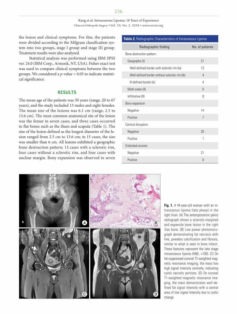
Fig. 1. A 44-year-old woman with an in-traosseous lipoma (late phase) in the right ilium. (A) The anteroposterior pelvic radiograph shows a sclerotic-margined and expansile bone lesion in the right iliac bone. (B) Low power photomicro-graph demonstrating fat necrosis with fine, powdery calcification and fibrosis, similar to what is seen in bone infarct. These features represent the late stage intraosseous lipoma (H&E, ×100). (C) On fat-suppressed coronal T2-weighted mag-netic resonance imaging, the mass has high signal intensity centrally, indicating cystic necrotic portions. (D) On coronal T1-weighted magnetic resonance ima-ging, the mass demonstrates well-de-fined fat signal intensity with a central area of low signal intensity due to cystic change.

237
Kang et al. Intraosseous Lipoma: 18 Years of ExperienceClinics in Orthopedic Surgery • Vol. 10, No. 2, 2018 • www.ecios.org
cases. There was one case in which cortical disruption was observed. Endosteal erosion was not observed (Table 2). Regarding the stage of the lesion according to the Milgram classification system, stage III was most common, which was found in 13 cases and accompanied by cystic forma-tion and extensive calcification (Fig. 1). Stage II was noted in one case and stage I, in seven cases (Fig. 2). In 15 of 21 cases, the VAS score for pain was between 3 and 6. There were four cases with other causes of pain, such as osteoar-thritis, rotator cuff tear, and discoid meniscus. Therefore, pain was caused by intraosseous lipoma in 11 cases. A pal-pable mass was present in one case. There was no range of motion limitation in all cases. In the stage I group, four of seven cases had pain; in the stage III group, six of 13 cases had pain. There was no correlation between the stage of the lesion and clinical symptoms (p = 1.000). There were no local recurrences or malignant changes during the follow-up. Curettage was performed as a surgical treat-ment in 20 cases, and bone graft was performed using a bone chip. Excision was performed in one case. Pain was resolved in seven of 11 cases with a complaint of preopera-tive pain. In four cases, intermittent pain remained.
DISCUSSION
Intraosseous lipoma is a very rare lesion that constitutes no more than 0.1% of all bone tumors.1) No sex differences in the incidence have been reported.2,8) In our study, 13 patients were males and eight were females. The age range of patients at presentation is wide, from youth to elderly, with high incidence in the fourth, fifth, and sixth decades of life.2,8-12) In our study, the mean age of the patients was 50 years (range, 20 to 67 years). The fifth decade was most common, in seven of 21 patients.
The calcaneus and long tubular bones are the com-mon anatomical sites of the lesion.2,6,10) When located in long bones, the lesion tends to be found in the metaphy-sis.2,6,10) But, intraosseous lipomas can be found essentially anywhere in the skeleton. Cases in the spine, pelvic bone, skull, and rib have been reported.2,6,10) In our study, as in a previous study, long tubular bones were common anatomical locations. The most common anatomical site of the lesion was the femur in seven cases, followed by the tibia in five cases and the humerus in four cases. Flat bones such as the ilium and scapula were the anatomical locations in three cases. Compared with previous studies,
Fig. 2. A 20-year-old man with an intra-osseous lipoma (early phase) in the body of scapula. (A) The outlet view shoul der radiograph demonstrates an expan sile bone lesion at the body of the right scapula. (B) Photomicrograph of the lesion consisting of mature adipocytes, similar to fatty marrow. However, there is no evidence of bone marrow elements (H&E, ×100). (C) On fat-suppressed coronal T2-weighted magnetic resonance imaging, the mass shows signal dropout with fat suppression. (D) On coronal T1-weighted magnetic resonance imaging, the corresponding lesion is isointense to subcutaneous fat.
A B
C D

238
Kang et al. Intraosseous Lipoma: 18 Years of ExperienceClinics in Orthopedic Surgery • Vol. 10, No. 2, 2018 • www.ecios.org
there was no notable difference in terms of sex distribu-tion, age, and anatomical locations. However, there were differences among studies2,3,6,7) probably due to differences in diagnostic methods (histological/radiological confirma-tion) and the development of imaging studies which may have resulted in the decrease in histological confirmation.
The pathogenesis of intramedullary lipoma has not been clarified. Several hypotheses have been proposed in-cluding secondary bone reaction after trauma, healing re-action of osteonecrosis, and primary benign tumors.2,13-15) It has been postulated that a secondary change in the tumor is due to an increase in pressure in the bone caused by proliferation of adipocytes.2,13) Milgram proposed a three-stage classification system based on the histologi-cal appearance of intraosseous lipomas: stage I, a lesion composed of mature fat cells without calcification; stage II, a predominantly fat lesion with necrosis and focal calci-fication or ossifications; and stage III, a fat-containing le-sion with multiple necroses, wide calcification, and cystic degeneration.2) The distribution of Milgram stages vary across studies.2,3,7) In our study, stage III was most com-mon, seen in 13 cases, stage II was noted in one case and stage I, in seven cases.
Pain is the leading symptom in most cases, but more than 30% of bone lipomas are found incidentally on imaging studies performed for other reasons. Some authors have reported up to 70% of patients with intraos-seous lipomas presented with pain,6,16) while other authors described most patients as asymptomatic.2,12) In this study, 15 of 21 cases complained of pain. Excluding osteoarthri-tis, rotator cuff tear, and discoid meniscus as the causes of pain, 11 patients complained of pain related to the lipoma. A palpable mass was present in one case in the scapula body; the lesion had an expansile pattern. No functional limitations were observed in all cases; therefore, it is neces-sary to distinguish the lesion from other sources of pain when there is functional limitation.
This study was based on the hypothesis that the degree of degenerative changes is related to increased intraosseous pressure. We investigated the relationship between the degree of degeneration and clinical symptoms (pain) with an assumption that severe degenerated changes would manifest with severe clinical symptoms. However, there was no correlation between the degree of degenera-
tion and clinical symptoms (p = 1.000). Considering that stage I cases with no clinical symptoms may be excluded from studies based on surgical histological examinations, more studies on radiologically confirmed cases are needed for better understanding of intraosseous lipoma.
Radiological follow-up with conservative treatment is recommended, except for rare cases with risk of patho-logical fractures or malignant transformation.2,17) Goto et al.12) suggested surgical indications of intraosseous lipoma as follows: (1) painful tumor, (2) occurrence of pathologi-cal fracture, (3) necessity for histological diagnosis, and (4) need to decrease the risk of malignant transformation.12) We performed curettage and packing with bone chips for surgical treatment in 20 cases. Excision was performed in one case. The indication for surgical treatment was symptomatic tumor (pain, mass) in 16 cases and necessity for histological diagnosis in five cases. Pain was resolved in seven of 11 cases with a complaint of preoperative pain. In cases presenting with clinical symptoms, surgical treatment (curettage, excision) is considered necessary; however, it is important to differentiate it from other pain-inducing disorders.
The limitations of this study include evaluation of only patients from a single institution. In addition, due to the nature of the lesion, the number of cases was not suf-ficient, and the follow-up period was short.
Intraosseous lipoma undergoes various degenera-tive changes according to the stage of the lesion. It occurs commonly in long bones of the lower limb and often in flat bones. There was no noticeable correlation between the degree of degeneration and clinical symptoms. Pain was the most common clinical symptom, but it was rarely accompanied by functional limitation. However, it is important to differentiate it from other sources of pain. The incidence of intraosseous lipomas is low, and various imaging findings make clinical diagnosis difficult. Clear understanding of the radiographic findings and symptoms will be useful for diagnosis and differentiation.
CONFLICT OF INTEREST
No potential conflict of interest relevant to this article was reported.
REFERENCES
1. Murphey MD, Carroll JF, Flemming DJ, Pope TL, Gan-non FH, Kransdorf MJ. From the archives of the AFIP:
benign musculoskeletal lipomatous lesions. Radiographics. 2004;24(5):1433-66.

239
Kang et al. Intraosseous Lipoma: 18 Years of ExperienceClinics in Orthopedic Surgery • Vol. 10, No. 2, 2018 • www.ecios.org
2. Milgram JW. Intraosseous lipomas: a clinicopathologic study of 66 cases. Clin Orthop Relat Res. 1988;(231):277-302.
3. Shin DS, Kwak ES, Choi JH. Intraosseous lipoma. J Korean Orthop Assoc. 2003;38(5):526-30.
4. Pappas AJ, Haffner KE, Mendicino SS. An intraosseous lipoma of the calcaneus: a case report. J Foot Ankle Surg. 2014;53(5):638-42.
5. Weinfeld GD, Yu GV, Good JJ. Intraosseous lipoma of the calcaneus: a review and report of four cases. J Foot Ankle Surg. 2002;41(6):398-411.
6. Campbell RS, Grainger AJ, Mangham DC, Beggs I, Teh J, Davies AM. Intraosseous lipoma: report of 35 new cases and a review of the literature. Skeletal Radiol. 2003;32(4):209-22.
7. Kim JW, Kim SJ, Kim GE, Ki SY, Lee SJ, Park JG. Radiologic findings of intraosseous lipoma of long bones. J Korean Soc Radiol. 2016;75(4):313-21.
8. Hart JA. Intraosseous lipoma. J Bone Joint Surg Br. 1973;55(3):624-32.
9. Bano S, Yadav SN, Chaudhary V, Jain VK. Radiological evaluation of bilateral intraosseous calcaneal lipoma in vari-ous stages of involution. Eur J Radiol Extra. 2011;78(1):e57-9.
10. Radl R, Leithner A, Machacek F, et al. Intraosseous li-poma: retrospective analysis of 29 patients. Int Orthop. 2004;28(6):374-8.
11. Muthuphei MN. Intra-osseous lipoma of the calcaneus. S Afr Med J. 1996;86(12):1554-5.
12. Goto T, Kojima T, Iijima T, et al. Intraosseous lipoma: a clinical study of 12 patients. J Orthop Sci. 2002;7(2):274-80.
13. Chow LT, Lee KC. Intraosseous lipoma: a clinicopathologic study of nine cases. Am J Surg Pathol. 1992;16(4):401-10.
14. Schatz SG, Dipaola JD, D'Agostino A, Hanna R, Quinn SF. Intraosseous lipoma of the calcaneus. J Foot Surg. 1992;31(4):381-4.
15. Greenspan A, Raiszadeh K, Riley GM, Matthews D. Intraosseous lipoma of the calcaneus. Foot Ankle Int. 1997;18(1):53-6.
16. Levin MF, Vellet AD, Munk PL, McLean CA. Intraosseous lipoma of the distal femur: MRI appearance. Skeletal Radiol. 1996;25(1):82-4.
17. Jebson PJ, Schock EJ, Biermann JS. Intraosseous lipoma of the proximal radius with extraosseous extension and a secondary posterior interosseous nerve palsy. Am J Orthop (Belle Mead NJ). 2002;31(7):413-6.