Embed Size (px)
Citation preview
Intraoperative Electrochemotherapy of Colorectal Liver Metastases
IBRAHIM EDHEMOVIC, MD, MSc,1 ERIK BRECELJ, MD, PhD,1 GORANA GASLJEVIC, MD,1
MAJA MAROLT MUSIC, MD, PhD,1 VOJKA GORJUP, MD, PhD,2 BARBARA MALI, PhD,3 TOMAZ JARM, PhD,3
BOR KOS, PhD,3 DENIS PAVLIHA, PhD,3 BILJANA GRCAR KUZMANOV, MD, PhD,1 MAJA CEMAZAR, PhD,1
MARKO SNOJ, MD, PhD,1 DAMIJAN MIKLAVCIC, PhD,3 ELDAR M. GADZIJEV, MD, PhD,1**AND GREGOR SERSA, PhD
1*1Institute of Oncology Ljubljana, Ljubljana, Slovenia
2University Medical Center Ljubljana, Ljubljana, Slovenia3Department of Biomedical Engineering, Faculty of Electrical Engineering, University of Ljubljana, Ljubljana, Slovenia
Background and Objectives: Electrochemotherapy is effective in treatment of various cutaneous tumors and could be translated into treatment ofdeep‐seated tumors. With this aim a prospective pilot study was conducted to evaluate feasibility, safety, and efficacy of intraoperativeelectrochemotherapy in the treatment of colorectal liver metastases.Methods: Electrochemotherapy with bleomycin was performed during open surgery, by insertion of long needle electrodes into and around thetumor according to the individualized pretreatment plan.Results:A 29metastases in 16 patients were treated in 16 electrochemotherapy sessions. No immediate (intraoperative) and/or postoperative seriousadverse events related to electrochemotherapy were observed. Radiological evaluation of all the treated metastases showed 85% complete responsesand 15% partial responses. In a group of seven patients that underwent a second operation at 6–12 weeks after the first one, during whichelectrochemotherapy was performed, the histology of resected metastases treated by electrochemotherapy showed less viable tissue (P¼ 0.001)compared to non‐treated ones.Conclusions: Electrochemotherapy of colorectal liver metastases proved to be feasible, safe, and efficient treatment modality, providing its specificplace in difficult to treat metastases, located in the vicinity of major hepatic vessels, not amenable to surgery or radiofrequency ablation.J. Surg. Oncol. 2014;110:320–327. � 2014 Wiley Periodicals, Inc.
KEY WORDS: electroporation; bleomycin; liver neoplasms; safety; treatment effectiveness; ablation
INTRODUCTION
The best management of patients with resectable colorectal livermetastases is surgical; however, many patients are presented withunresectable metastases, due to their size, location, and/or inadequateremnant liver volume. In such unresectable cases, several alternativelocal approaches are used, among which the most frequent isradiofrequency ablation [1]; however, its efficacy is reduced in thevicinity of major vessels due to heat sink effect [2]. In such specialcases and also in other unresectable cases, new electroporation‐based treatment modalities are available—electrochemotherapy andirreversible electroporation—that have a potential role, because they arenon‐thermal local tumor treatment modalities, and are expected not tohave deleterious effects on major blood vessels [3,4].
Electrochemotherapy is a treatment that combines the use of poorlyor non‐permeant, but highly effective cytotoxic drugs such as bleomycinor cisplatin with reversible electroporation, which facilitates drugsdiffusion into the cells, thus increasing their cytotoxicity [5,6]. The useof bleomycin is based on the clinical evidence showing that among otherdrugs tested bleomycin has the highest potentiation of cytotoxicity byelectroporation (up to several 1,000 times) [7,8]. Furthermore, theelectroporation is effective only for hydrophilic drugs, like bleomycinand cisplatin, not for the lipophylic drugs that are regularly used inchemotherapy for liver colorectal metastases, like 5‐Fu and irinotecanwhich penetrate cell membrane much easier, and electroporation doesnot potentiate their cytotoxicity.
Electrochemotherapy with bleomycin is effective in differentcutaneous and subcutaneous tumors [9], as well as on preclinicalmodels of colorectal tumors [10,11]. To date electrochemotherapy has
been shown to be very effective in treatment of superficial metastaticdisease, such as melanoma and chest wall breast cancerrecurrence [7,12–21]. Its value of treating metastatic or unresectabledisease within the abdomen and chest has enormous potential [22,23].Translation of electrochemotherapy into treatment of deep‐seatedtumors is being currently explored [22], with the description of thetechnological approach on a case with liver metastasis [24]. The
Grant sponsor: Slovenian Research Agency (ARRS); Grant numbers: P3‐0003, P2‐0249.
Conflict of interest: Damijan Miklavcic holds patents (US 7625729 B2; EP1395333 B1; US 7306940 B2) of which some have been licensed to IGEASpA, Carpi, Italy, the producers of a clinical device used for electro-chemotherapy in Europe since 2006. He also consults for IGEA SpA. Otherauthors declare no conflicts of interests.
Ibrahim Edhemovic and Erik Brecelj contributed equally to this work.
The institution at which the work was performed: Institute of OncologyLjubljana, Ljubljana, Slovenia
*Correspondence to: Gregor Sersa, PhD, Department of ExperimentalOncology, Institute of Oncology Ljubljana, Zaloska 2, SI‐1000 Ljubljana,Slovenia. Fax: þ386‐1‐5879‐434. E‐mail: [email protected]**Correspondence to: Eldar M. Gadzijev, MD, PhD, Department of SurgicalOncology, Institute of Oncology Ljubljana, Zaloska 2, SI‐1000 Ljubljana,Slovenia. Fax: þ386‐1‐5879‐434. E‐mail: [email protected]
Received 13 January 2014; Accepted 31 March 2014
DOI 10.1002/jso.23625
Published online 30 April 2014 in Wiley Online Library(wileyonlinelibrary.com).
Journal of Surgical Oncology 2014;110:320–327
� 2014 Wiley Periodicals, Inc.
mechanism of action allows one to potentially sterilize tumors that areadjacent to structures that cannot be resected, such as major vessels,which frequently limit a curative resection, especially in liver metastaticdisease.
The purpose of this article is to report the feasibility, safety, andefficacy of electrochemotherapy in treatment of colorectal livermetastases based on the treatment parameters of the previous ESOPEstudy [7]. This has not been done before.
PATIENTS AND METHODS
Study
The study was prospective, pilot study, conducted at the Institute ofOncology Ljubljana, Ljubljana, Slovenia. Regulatory approvals fromthe Institutional Board, as well as from the National Medical EthicsCommittee (#45/09/08) were obtained. The study is registered atClinicalTrials.gov: NCT01264952. Informed consent has beenobtained from all patients included in the trial. The trial wasdesigned based on ESOPE trial for treatment of cutaneoustumors [7], where the dosage of bleomycin and electrical parameterswere set in standard operating procedures for treatment of cutaneoustumors [8].
The primary objective of the study was evaluation of the feasibilityand safety of intraoperative electrochemotherapy of colorectal livermetastases. The secondary objective was to determine the efficacy ofelectrochemotherapy treatment, based on histological and radiologicalevaluation of treatedmetastases. The endpoints are: toxicity according tothe Common Terminology Criteria for Adverse Events (CTC‐AE) ver.4.0 and response rate measured by percentage of vital tumor cells andmRECIST criteria.
Patients
Patients were enrolled from November 2009 to June 2012. Allpatients included in this study were in AJCC stage IV, with the diseaselimited to the liver only. Up to three metastases not exceeding 3 cm in thediameter were treated with electrochemoterapy. All patients except onewere treated with systemic therapy prior to the electrochemotherapy;however, no systemic treatment was given until the second operation orradiological evaluation (Supplementary Table SI).
Inclusion and exclusion criteria are shown in Table I. Three groups ofpatients with colorectal liver metastases were included in the study(Table II). The first two groups of patients included patients with intentto cure within standard of care using two‐stage surgical approach. Thistwo‐stage surgical approach allowed adding electrochemotherapyduring the first operation and tissue collection for histologicalanalysis during the second operation.
The first group (group I) included patients with bilateral, multiple,metachronous metastases in whom standard treatment included two‐stage liver resection, due to the extent of the disease and/or their generalcondition. During the first operation, right portal vein was ligated andmetastases on the left side were excised or ablated with radiofrequencyablation. At the same time, up to three metastases on the right side weretreated with electrochemotherapy. During the second operation, bothtreated and non‐treated metastases on the right side were removed withright hemihepatectomy.
The second group (group II) included patients with synchronousmetastases, but their general condition and extent of the disease did notallow simultaneous removal of the primary tumor and metastases.During the first operation, the primary tumor was removed (colorectalresection) and some of the liver metastases were treated byelectrochemotherapy. About 6 weeks later, during the secondoperation for liver metastases, both treated and non‐treated metastaseswere removed with liver resection.
The third group (group III) included patients with up to threemetachronous, unresectable liver metastases, demanding too excessiveresection, or untreatable by standard thermal ablativemethods, due to theclose proximity of major blood vessels. Electrochemotherapy wasoffered to these patients as the only treatment option.
Based on the relation of the metastases to the major blood vessels,they were segregated into “central” or “peripheral”. The term “central”was used to describe the metastases located in the near vicinity or on themajor vessels. The term “peripheral”was used to describe the metastasisaway from themajor vessels to such an extent, so these vessels would notbe affected by the electric field.
Treatment Procedure
The treatment of colorectal liver metastases was performed duringopen surgery using electrodes with variable [24] or fixed geometry,depending of the location of the metastasis. The electrodes with fixedgeometry consist of seven electrodes fixed in a plastic holder and all ofthem are placed simultaneously as one electrode. The smaller tumors upto 2 cm in diameter, located no deeper than the length of the electrodes,that is, 3 cm, were treated with the electrodes with fixed geometry whichare easier to insert and the treatment is performed faster. The variablegeometry was utilized when bigger and deeper‐seated tumors weretreated. Patient‐specific pretreatment plans were prepared based oncomputed tomography or magnetic resonance scans: target lesions (up to3 cm in the largest diameter) were segmented, and a gradient‐basedoptimization algorithm was used to optimize voltage between eachelectrode pair to maximize tumor coverage above the reversibleelectroporation threshold (400V/cm) and minimize volume of healthyliver parenchyma above the irreversible electroporation threshold(700V/cm)—see Supplementary Data I: An example of the treatmentplan [25–27]. Trains of eight electric pulses (each pulse 100ms long)were delivered to each pair of electrodes consecutively(Supplementary Table SII) [24]. Electric pulses were delivered byelectric pulse generator (IGEASpA, Carpi, Italy) during an interval of 8–28min after the intravenous injection of bleomycin 15,000 IU/m2 inbolus (Heinrich Mack Nachf. GmbH & CO. KG, Illertissen, Germany),as being determined to be the optimal pharmacological peak for the
TABLE I. Inclusion and Exclusion Criteria
Inclusion criteria Exclusion criteria
Age� 18 Pregnancy and lactationPerformance status �2 ECOG Implanted pacemaker or defibrillatorChemotherapy free interval
2–5 weeks,depending on the drugs used
Significant cardiac arrhythmias
Life expectancy morethan 3 months
Coagulation disturbances
Written informed consent Cumulative dose of �250,000 IUbleomycin received
Previous allergic reaction to bleomycinChronically impaired kidney functionSignificantly impaired lung functionEpilepsyAscitesLife threatening infection or other
serious systemic condition or diseaseSecondary primary tumor, except
surgically treated non‐invasivecancer of the cervix or surgicallytreated or irradiated basal cellcarcinoma,and confirmed visceral,bone or diffuse metastases
Journal of Surgical Oncology
Electrochemotherapy of Liver Metastases 321
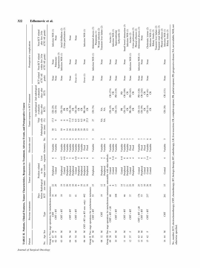
TABLEII.Patients,Clin
ical
Features,Tum
orCha
racteristics,Respo
nseto
Treatment,Adv
erse
Events,an
dPostoperative
Cou
rse
Patient
Previoustreatm
ents
Tum
orcharacteristics
Electrodesused
Tum
orresponse
toECTtreatm
ent
Postoperativ
ecomplications
No.
Age
Sex
Type
Days
before
ECT
Radiological
size
(mm)
Position
related
tothe
major
vessels
Liver
segm
ent
Geometry
No.
Pathological
size
(mm)
Vital
cells
(%)
1stradiological
evaluatio
n(daysafter
ECT)
2ndradiological
evaluatio
n(daysafter
ECT)
ECTrelated
(CTC‐A
Egrade)
Non‐ECTrelated
with
infirst24
hr(CTC‐A
Egrade)
Non‐ECTrelated
afterfirst24
hr(CTC‐A
Egrade)
Group
I:Twostageoperations
formetachronousmetastases:
0167
MCHT
3522
Peripheral
8Variable
530
17.5
CR
(29)
None
None
InfectionNOS(1)
0255
FCHTþBT
3629
Central
1Variable
617
0CR
(54)
None
Pulmonary
hypertension
(3)
None
0369
MCHTþBT
1019
Peripheral
4‐5
Variable
510
0CR
(54)
None
InfectionNOS(1)
Atrialfibrillation(2)
15Central
4‐8
Variable
59
0PR
Colon
perforation(3)
20Peripheral
5Variable
514
0CR
0456
MCHTþBT
2219
Peripheral
5Variable
628
17.5
CR
(30)
None
None
None
10Peripheral
8Variable
523
7.5
CR
0554
MCHTþBT
4126
Central
4‐8
Variable
650
0.5
CR
(26)
Fever
(1)
None
None
9Peripheral
5Variable
420
0.5
CR
18Peripheral
8Variable
535
0.5
CR
0669
MCHTþRT(asRTsens.only)
492
21Peripheral
4Fixed
720
30PR
(44)
Fever
(1)
None
InfectionNOS(1)
16Peripheral
8Fixed
715
27.5
CR
Group
II:Twostageoperations
forsynchronousmetastases:
0759
MCHTþBT
5315
Peripheral
5Variable
521
27CR
(76)
None
InfectionNOS(1)
Abdom
inal
abscess(3)
Pneum
onia
(1)
Wound
infection(2)
0832
MCHT
1912
Peripheral
4Variable
5NA
NA
None
None
Transient
liver
failu
re(2)
14Peripheral
5Variable
5NA
NA
Group
III:One
stageoperations
formetachronousmetastases,untreatablewith
othermethods:
0938
FCHTþBTþLR
686
Central
8Variable
5CR
(19)
CR
(274)
None
None
Ascites(2)
8Central
5–8
Variable
5CR
CR
InfectionNOS(1)
1069
MCHTþBT
108
17Central
5Variable
5CR
(33)
PD
(119)
None
None
Supraventricular
tachycardia(2)
14Central
4–8
Variable
5PR
PD
1144
MCHTþBT
6617
Peripheral
8Variable
5CR
(33)
CR
(189)
None
None
Smallbowel
obstruction(3)
21Peripheral
8Variable
5CR
CR
InfectionNOS(1)
1257
FCHTþBT
3725
Central
4Fixed
7PR
(50)
PD
(163)
None
InfectionNOS(1)
Ascites(2)
18Peripheral
4Fixed
7CR
CR
Colon
perforation(3)
Pleural
effusion
(2)
1363
MCHTþBTþLR
176
25Central
4Variable
5CR
(14)
PD
(128)
None
InfectionNOS(1)
Abdom
inal
abscess(3)
1461
MCHTþBT
5313
Peripheral
8Fixed
7CR
(59)
CR
(242)
None
None
None
10Central
2Fixed
7CR
CR
1562
FCHTþBT
237
26Central
3–4
Variable
6CR
(14)
CR
(41)
None
None
Cholestatic
icterus(2)
18Central
3Variable
6CR
CR
InfectionNOS(1)
Biliaryfistula(3)
Transient
liver
failu
re(2)
Transient
renalfailu
re(3)
Pleural
effusion
(2)
1664
MCHT
261
15Central
4Variable
5CR
(30)
CR
(131)
None
None
InfectionNOS(1)
Biliaryfistula(3)
No.,num
ber;ECT,electrochem
otherapy;C
HT,chemotherapy;B
T,biologictherapy;RT,radiotherapy;LR,liverresection;CR,com
pleteresponse;P
R,partialresponse;PD,progressive
disease;NA,notavailable;NOS,not
otherw
isespecified.
Journal of Surgical Oncology
322 Edhemovic et al.

bleomycin in the tumors [8]. To maximize the safety of patients, thedelivery of electric pulses was synchronized with the absolute refractoryperiod of the heart (see [24] for details) to prevent the electric pulses frombeing delivered during the vulnerable period of the ventricles [28–31].
Safety Assessment
Adverse events were assessed using the National Cancer InstituteCommon Terminology Criteria for Adverse Events (CTC‐AE) version4.0. The ECGwasmonitored continuously during the surgical procedureas well as for 24 hr before and after the surgery using an ambulatory ECGHolter device (SpiderView, ELA Medical, France). Processing of ECGsignals included statistical comparison of average RR and QT intervalsover different time intervals and heart rate variability (HRV) analysis.
Efficacy Assessment Based on Pathology
Tissue for histological analysis was available in seven patients thatwere operated twice (Table II). The samples were assessed semi‐quantitatively by two pathologists independently. One of thepathologists was blinded with respect to clinical information,treatment regimen and outcome. The mean between the two scoreswas calculated. The proportion of residual vital tumor tissue andproportion of regressive changes in relation to total tumor area wereestimated as described by Ribero et al. [32]. Regressive changesincluded infarct‐like tumor necrosis, fibrosis, foamy macrophagesand other reparative changes. Infarct‐like tumor necrosis wasconsidered to be a form of treatment effect as proposed by Changet al. [33].
Efficacy Assessment Based on Radiology
Before and after electrochemotherapy, liver metastases wereevaluated by magnetic resonance (MRI) using a specific hepatocytecontrast agent (gadoliniumethoxybenzyl‐diethylenetriaminepentaaceticacid—Gd‐EOB‐DTPA, Primovist, Bayer, Berlin, Germany) or contrastenhanced computed tomography (CE‐CT) examination. The treatmentresponse was evaluated by CE‐CT or MRI, using the mRECISTcriteria [34,35]. In the eight patients (group III) who did not undergo asecond operation, an additional radiographic follow‐up was performedsubsequently.
Statistical Analysis
All data were entered into a Microsoft Access 2010 database, whichwas used for all calculations except for statistical analysis. For statisticalanalysis, SigmaPlot Ver. 12 software was used (Systat Software, Inc.,San Jose, CA). The pathohistological differences between theelectrochemotherapy treated and non‐treated metastases were statisticallyevaluated by the t‐test after confirming data normality using the Shapiro–Wilk test. A chi‐square test was used for statistical comparison of responseof metastases located near major blood vessels (referred as “central”) andresponse of metastases located away from the major blood vessels (referredas “peripheral”). A two‐tailed P value for the t‐test and P value less than0.05 was considered to be statistically significant.
RESULTS
The clinical features, treatment characteristics and response, adverseevents and postoperative course of the 16 patients with 29metastases arepresented in Table II. Safety assessment was possible in all 16 patients;however, response to the treatment was evaluable in 15 patients (27evaluable metastases)—one patient developed numerous new livermetastases, so evaluation of the response of the treated metastases wasnot possible.
Adverse Events
No electrochemotherapy related serious adverse events occurred. Allobserved adverse events are reported in Table II. Only grade 1 fevercould be attributed to electrochemotherapy. Postoperative complicationswithin and after 24 hr post electrochemotherapy could not be attributedspecifically to electrochemotherapy and were in the range grades 1–3.
Three patients required reoperation: two patients due to colonperforation and one due to small bowel obstruction. None of thesecomplications were related to the electrochemotherapy itself(Supplementary Data II: Data on patients’ complications). All threepatients were successfully reoperated and all 16 patients were dischargedfrom hospital—there was no perioperative mortality.
The median duration of the patient’s hospitalization afterelectrochemotherapy was 14 days (range 7–42); including threepatients (one patient from group I and two patients from group III)that needed prolonged hospitalization, due to their reoperations.
After discharge from hospital, patients were followed up onoutpatients’ basis. Seven out of eight patients from groups I and IIunderwent major hepatic resection as planned at median of 59 days(range 43–84 days) after the electrochemotherapy (one patient was notreoperated due the disease progression). After 90 days, no patient fromgroup III had signs of liver, renal or lung dysfunction, including thosewith serious complications and reoperations. Biliary fistulas in twopatients ceased without intervention.
The treatment of 13 metastases (48%), that were located near or in‐between the major blood vessels of the liver (referred as “central” inTable II), was safe. Neither intraoperatively nor postoperativelybleeding was observed. In some cases, the withdrawal of theelectrodes resulted in mild bleeding, which however was easilystopped by electrocoagulation.
Safety Aspect of Electrochemotherapy in the Context ofChanges in the ECG
The safety aspect of electrochemotherapy of colorectal liver metastaseswas evaluated based on detected changes in ECG signals recorded duringand after the surgical procedure. No significant arrhythmias orpathological morphological changes that would indicate developmentof myocardial ischemia after electrochemotherapy were detected. Theprocedure did not result in new‐onset of abnormal heartbeats (atrial orventricular extrasystoles) or in increased frequency of abnormal heartbeatsin patients who rarely hadminor arrhythmias present in ECG signal beforethe treatment. ECG and HRV analysis; however, revealed somestatistically significant but clinically irrelevant changes in the propertiesof the ECG during and after the surgical procedure. The most obvious onewas a mild increase in heart rate immediately after electrochemotherapy(two patients) and also during the first 24hr after the procedure (threepatients). In addition, there was a mild depression in the low frequencycomponent of the HRV spectrum (three patients).
Pathologic Response Evaluation
Pathologic analysis was performed on metastases treated withelectrochemotherapy during the first operation and resected at thesecond operation (groups I and II). Altogether, 13 liver metastases treatedwith electrochemotherapy were microscopically analyzed and comparedwith 22 non‐treated metastases from the same patients. Pathologicanalysis revealed that metastases which were not treated byelectrochemotherapy had a significantly higher percentage of residualvital tumor tissue, than electrochemotherapy treated metastases. Onaverage, electrochemotherapy treated metastases had 9.9� 12.2%(AM�SD) viable tissue, and control metastases had 34.1� 22.5%(P¼ 0.001, two‐tailed t‐test) (Fig. 1). Typical changes that were observedin the metastases with complete response were infarct‐like necrosis of the
Journal of Surgical Oncology
Electrochemotherapy of Liver Metastases 323

tumor tissue and the surrounding tumor parenchyma, with encapsulationof the treated tissue (fibrous pseudocapsule on the border between thenormal liver tissue and the electrochemotherapy treated area).
Radiologic Response Evaluation
The median interval between the treatment and first radiologicalevaluation was 33 days (range 14–76). Twenty‐seven metastases wereevaluated (Table II), a complete response was observed in 23 (85%). Infour metastases (15%) some enhancements of the treated lesion wereseen, in both phases of liver enhancement, and they were evaluated as apartial response or local tumor progression.
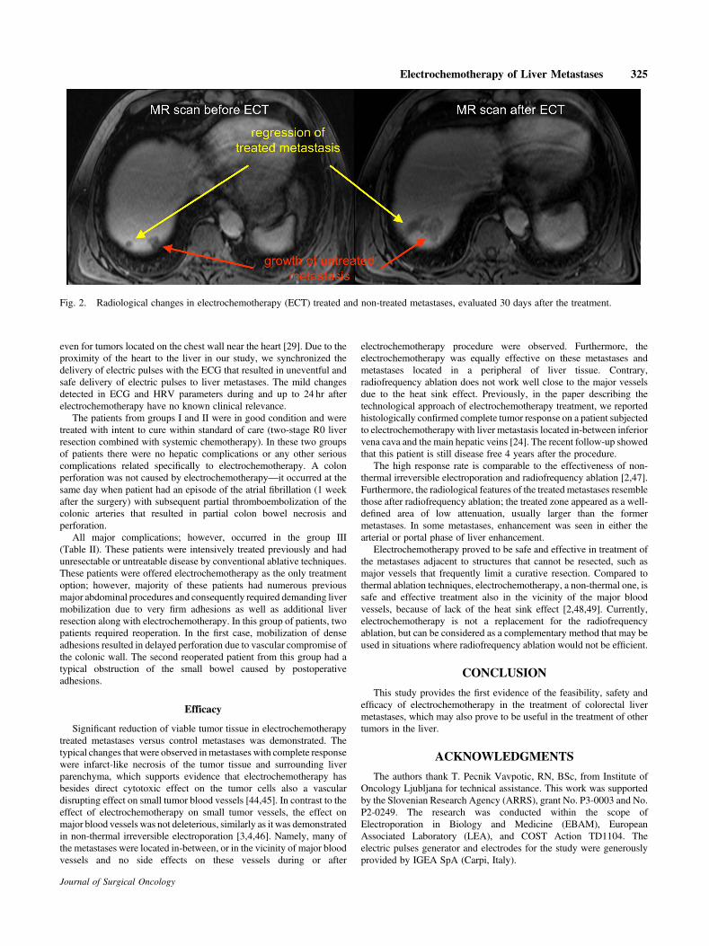
In the group of eight patients (group III) with a single‐stage operation,14 metastases were treated by electrochemotherapy. These patients wereevaluated radiologically twice. On the first follow‐up examination at amedian of 31.5 days (range 14–59) after electrochemotherapy, acomplete response was seen in 12 metastases (86%). There wasperipheral enhancement of the lesions in two metastases, whichsuggested a partial response. At the second follow‐up, at median of147 days (range 41–274) after electrochemotherapy, 10 (71%)metastases were still in complete response, while the other 4progressed (Fig. 2). Response evaluated on a per patient basis wascomplete response for 5 (62.5%) patients and progressive disease for 3(37.5%) patients.
Thirteen metastases were adjacent to major hepatic vessels. A 77%(10 metastases) were in complete response 33 days afterelectrochemotherapy (Table II). There was no difference detected inresponse of metastases located near major blood vessels and metastaseslocated away from the major blood vessels (P¼ 0.244).
DISCUSSION
This translational study shows that electrochemotherapy is feasible,safe, and efficient treatment modality for the colorectal liver metastases.
The simple physicochemical concept of electrochemotherapy procedure,using electric pulses to transiently increase the permeability of the cellmembrane and facilitate the uptake of otherwise poorly permeant buthighly effective cytotoxic drugs, provides a solid basis for itseffectiveness in various tumor types, including colorectal tumors [10–12,18,36,37]. Translation of this treatment approach to the treatment ininternal organs has recently begun. It is based on technological advance,with newly developed electric pulse generators and different sets ofelectrodes for specific organs [22,38].
Feasibility
In this study, we treated 16 patients with colorectal liver metastases,in different anatomical locations in the liver, including 13 metastases inthe close vicinity of major hepatic vessels. Metastases positioned>3 cmdeep in liver parenchyma were treated by long individual electrodesplaced by ultrasound guidance according to the pretreatment plan [24].For more superficially positioned metastases, electrodes with fixedgeometry that were placed all at once were used without pretreatmentplan. Eventually, further possible development of this method shouldprovide percutaneous treatment, as in the case of radiofrequencyablation [39].
Safety
So far, no treatment related adverse events have been reported, eitherin the treatment of superficial tumors, or tumors in internalorgans [12,16,24,40]. Local pain and transient erythema affecting theelectroporated areas are among themost commonly reported side effects.
It is known that application of electric fields can affect implantedelectrical devices (pacemakers) and interfere with cardiacfunction [24,30,31,41–43], therefore such patients were excluded forsafety reasons. Electrochemotherapy of the cutaneous tumors has beendemonstrated to have minimal risk of interfering with cardiac function,
Fig. 1. Pathohistological features of tumors treated by electrochemotherapy in relation to those that were not. (A) Gross picture of two metastases:the large one corresponds to a metastasis treated by chemotherapy only, the small one corresponds to a metastasis treated by electrochemotherapy.The patient was in the group I where the two‐stage operation was done. (B) Gross picture of metastasis treated by electrochemotherapy: completenecrosis of tumor and surrounding liver parenchyma. (C) Histological picture of completely necrotic tumor treated with electrochemotherapy: aninfarct‐like necrosis is in the right part of the picture, vital liver parenchyma in the left. In‐between there is a fibrous pseudocapsule (H&E, 5�). (D)The only focus of residual vital tumor tissue in otherwise completely necrotic electrochemotherapy treated metastasis. An infarct‐like necrosis is inthe upper part of the picture (H&E, 10�). (E) Partial response in metastasis treated with chemotherapy only: an infarct‐like necrosis is present in theupper field of the picture with lager amount of residual vital tumor tissue.
Journal of Surgical Oncology
324 Edhemovic et al.
even for tumors located on the chest wall near the heart [29]. Due to theproximity of the heart to the liver in our study, we synchronized thedelivery of electric pulses with the ECG that resulted in uneventful andsafe delivery of electric pulses to liver metastases. The mild changesdetected in ECG and HRV parameters during and up to 24 hr afterelectrochemotherapy have no known clinical relevance.
The patients from groups I and II were in good condition and weretreated with intent to cure within standard of care (two‐stage R0 liverresection combined with systemic chemotherapy). In these two groupsof patients there were no hepatic complications or any other seriouscomplications related specifically to electrochemotherapy. A colonperforation was not caused by electrochemotherapy—it occurred at thesame day when patient had an episode of the atrial fibrillation (1 weekafter the surgery) with subsequent partial thromboembolization of thecolonic arteries that resulted in partial colon bowel necrosis andperforation.
All major complications; however, occurred in the group III(Table II). These patients were intensively treated previously and hadunresectable or untreatable disease by conventional ablative techniques.These patients were offered electrochemotherapy as the only treatmentoption; however, majority of these patients had numerous previousmajor abdominal procedures and consequently required demanding livermobilization due to very firm adhesions as well as additional liverresection along with electrochemotherapy. In this group of patients, twopatients required reoperation. In the first case, mobilization of denseadhesions resulted in delayed perforation due to vascular compromise ofthe colonic wall. The second reoperated patient from this group had atypical obstruction of the small bowel caused by postoperativeadhesions.
Efficacy
Significant reduction of viable tumor tissue in electrochemotherapytreated metastases versus control metastases was demonstrated. Thetypical changes that were observed inmetastases with complete responsewere infarct‐like necrosis of the tumor tissue and surrounding liverparenchyma, which supports evidence that electrochemotherapy hasbesides direct cytotoxic effect on the tumor cells also a vasculardisrupting effect on small tumor blood vessels [44,45]. In contrast to theeffect of electrochemotherapy on small tumor vessels, the effect onmajor blood vessels was not deleterious, similarly as it was demonstratedin non‐thermal irreversible electroporation [3,4,46]. Namely, many ofthe metastases were located in‐between, or in the vicinity of major bloodvessels and no side effects on these vessels during or after
electrochemotherapy procedure were observed. Furthermore, theelectrochemotherapy was equally effective on these metastases andmetastases located in a peripheral of liver tissue. Contrary,radiofrequency ablation does not work well close to the major vesselsdue to the heat sink effect. Previously, in the paper describing thetechnological approach of electrochemotherapy treatment, we reportedhistologically confirmed complete tumor response on a patient subjectedto electrochemotherapy with liver metastasis located in‐between inferiorvena cava and the main hepatic veins [24]. The recent follow‐up showedthat this patient is still disease free 4 years after the procedure.
The high response rate is comparable to the effectiveness of non‐thermal irreversible electroporation and radiofrequency ablation [2,47].Furthermore, the radiological features of the treated metastases resemblethose after radiofrequency ablation; the treated zone appeared as a well‐defined area of low attenuation, usually larger than the formermetastases. In some metastases, enhancement was seen in either thearterial or portal phase of liver enhancement.
Electrochemotherapy proved to be safe and effective in treatment ofthe metastases adjacent to structures that cannot be resected, such asmajor vessels that frequently limit a curative resection. Compared tothermal ablation techniques, electrochemotherapy, a non‐thermal one, issafe and effective treatment also in the vicinity of the major bloodvessels, because of lack of the heat sink effect [2,48,49]. Currently,electrochemotherapy is not a replacement for the radiofrequencyablation, but can be considered as a complementary method that may beused in situations where radiofrequency ablation would not be efficient.
CONCLUSION
This study provides the first evidence of the feasibility, safety andefficacy of electrochemotherapy in the treatment of colorectal livermetastases, which may also prove to be useful in the treatment of othertumors in the liver.
ACKNOWLEDGMENTS
The authors thank T. Pecnik Vavpotic, RN, BSc, from Institute ofOncology Ljubljana for technical assistance. This work was supportedby the Slovenian Research Agency (ARRS), grant No. P3‐0003 and No.P2‐0249. The research was conducted within the scope ofElectroporation in Biology and Medicine (EBAM), EuropeanAssociated Laboratory (LEA), and COST Action TD1104. Theelectric pulses generator and electrodes for the study were generouslyprovided by IGEA SpA (Carpi, Italy).
Fig. 2. Radiological changes in electrochemotherapy (ECT) treated and non‐treated metastases, evaluated 30 days after the treatment.
Journal of Surgical Oncology
Electrochemotherapy of Liver Metastases 325
REFERENCES
1. Alberts SR: Update on the optimal management of patients withcolorectal liver metastases. Crit Rev Oncol Hematol 2012;84:59–70.
2. Wong SL, Mangu PB, Choti MA, et al.: American Society ofClinical Oncology 2009 clinical evidence review on radiofrequencyablation of hepatic metastases from colorectal cancer. J Clin Oncol2010;28:493–508.
3. Maor E, Ivorra A, Leor J, et al.: The effect of irreversibleelectroporation on blood vessels. Technol Cancer Res Treat 2007;6:307–312.
4. Cheung W, Kavnoudias H, Roberts S, et al.: Irreversibleelectroporation for unresectable hepatocellular carcinoma: Initialexperience and review of safety and outcomes. Technol Cancer ResTreat 2013;12:233–241.
5. Orlowski S, Belehradek J Jr, Paoletti C, et al.: Transientelectropermeabilization of cells in culture. Increase of thecytotoxicity of anticancer drugs. Biochem Pharmacol 1988;37:4727–4733.
6. Sersa G, Cemazar M, Miklavcic D: Antitumor effectiveness ofelectrochemotherapy with cis‐diamminedichloroplatinum(II) inmice. Cancer Res 1995;55:3450–3455.
7. Marty M, Sersa G, Garbay JR, et al.: Electrochemotherapy—Aneasy, highly effective and safe treatment of cutaneous andsubcutaneous metastases: Results of ESOPE (European StandardOperating Procedures of Electrochemotherapy) study. Eur J CancerSuppl 2006;4:3–13.
8. Mir LM,Gehl J, Sersa G, et al.: Standard operating procedures of theelectrochemotherapy: Instructions for the use of bleomycin orcisplatin administered either systemically or locally and electricpulses delivered by the CliniporatorTM bymeans of invasive or non‐invasive electrodes. Eur J Cancer Suppl 2006;4:14–25.
9. Eggermont AM: Treatment of melanoma in‐transit metastasesconfined to the limb. Cancer Surv 1996;26:335–349.
10. Jaroszeski MJ, Dang V, Pottinger C, et al.: Toxicity of anticanceragents mediated by electroporation in vitro. Anticancer Drugs2000;11:201–208.
11. Todorovic V, Sersa G, Flisar K, et al.: Enhanced cytotoxicity ofbleomycin and cisplatin after electroporation in murine colorectalcarcinoma cells. Radiol Oncol 2009;43:264–273.
12. Matthiessen LW, Chalmers RL, Sainsbury DCG, et al.: Manage-ment of cutaneous metastases using electrochemotherapy. ActaOncol 2011;50:621–629.
13. Matthiessen LW, Johannesen HH, Hendel HW, et al.: Electro-chemotherapy for large cutaneous recurrence of breast cancer: Aphase II clinical trial. Acta Oncol 2012;51:713–721.
14. Campana LG, Valpione S, Mocellin S, et al.: Electrochemotherapyfor disseminated superficial metastases from malignant melanoma.Br J Surg 2012;99:821–830.
15. Campana LG, Valpione S, Falci C, et al.: The activity and safety ofelectrochemotherapy in persistent chest wall recurrence from breastcancer after mastectomy: A phase‐II study. Breast Cancer Res Treat2012;134:1169–1178.
16. Sersa G, Cufer T, Paulin SM, et al.: Electrochemotherapy of chestwall breast cancer recurrence. Cancer Treat Rev 2012;38:379–386.
17. Latini A, Bonadies A, Trento E, et al.: Effective treatment ofKaposi’s sarcoma by electrochemotherapy and intravenous bleo-mycin administration. Dermatol Ther 2012;25:214–218.
18. Gargiulo M, Papa A, Capasso P, et al.: Electrochemotherapy fornon‐melanoma head and neck cancers: Clinical outcomes in 25patients. Ann Surg 2012;255:1158–1164.
19. Curatolo P, Quaglino P, Marenco F, et al.: Electrochemotherapy inthe treatment of Kaposi sarcoma cutaneous lesions: A two‐centerprospective phase II trial. Ann Surg Oncol 2012;19:192–198.
20. Testori A, Faries MB, Thompson JF, et al.: Local and intralesionaltherapy of in‐transit melanoma metastases. J Surg Oncol 2011;104:391–396.
21. Mali B, Miklavcic D, Campana LG, et al.: Tumor size andeffectiveness of electrochemotherapy. Radiol Oncol 2013;47:32–41.
22. Miklavcic D, Sersa G, Brecelj E, et al.: Electrochemotherapy:Technological advancements for efficient electroporation‐basedtreatment of internal tumors. Med Biol Eng Comput 2012;50:1213–1225.
23. Jahangeer S, Forde P, Soden D, et al.: Review of current thermalablation treatment for lung cancer and the potential of electro-chemotherapy as a means for treatment of lung tumours. CancerTreat Rev 2013;39:862–871.
24. Edhemovic I, Gadzijev EM, Brecelj E, et al.: Electrochemotherapy:A new technological approach in treatment of metastases in theliver. Technol Cancer Res Treat 2011;10:475–485.
25. Miklavcic D, SnojM, Zupanic A, et al.: Towards treatment planningand treatment of deep‐seated solid tumors by electrochemotherapy.Biomed Eng OnLine 2010;9:10.
26. Pavliha D, Kos B, Zupanic A, et al.: Patient‐specific treatmentplanning of electrochemotherapy: Procedure design and possiblepitfalls. Bioelectrochemistry 2012;87:265–273.
27. Zupanic A, Kos B, Miklavcic D: Treatment planning ofelectroporation‐based medical interventions: Electrochemotherapy,gene electrotransfer and irreversible electroporation. PhysMed Biol2012;57:5425–5440.
28. Reilly JP, editor: Applied bioelectricity: From electrical stimula-tions to electropathology. New York: Springer; 1998.
29. Mali B, Jarm T, Corovic S, et al.: The effect of electroporationpulses on functioning of the heart. Med Biol Eng Comput2008;46:745–757.
30. Ball C, Thomson KR, Kavnoudias H: Irreversible electroporation:A new challenge in ‘out of operating theater’ anesthesia. AnesthAnalg 2010;110:1305–1309.
31. Deodhar A, Dickfeld T, Single GW, et al.: Irreversible electropora-tion near the heart: Ventricular arrhythmias can be preventedwith ECG synchronization. Am J Roentgenol 2011;196:W330–W335.
32. Ribero D, Wang H, Donadon M, et al.: Bevacizumab improvespathologic response and protects against hepatic injury in patientstreated with oxaliplatin‐based chemotherapy for colorectal livermetastases. Cancer 2007;110:2761–2767.
33. Chang HHL, Leeper WR, Chan G, et al.: Infarct‐like necrosis: Adistinct form of necrosis seen in colorectal carcinoma livermetastases treated with perioperative chemotherapy. Am J SurgPathol 2012;36:570–576.
34. Lencioni R, Llovet JM: Modified RECIST (mRECIST) assessmentfor hepatocellular carcinoma. Semin Liver Dis 2010;30:52–60.
35. Park M, Rhim H, Kim Y, et al.: Spectrum of CT findings afterradiofrequency ablation of hepatic tumors. Radiographics 2008;28:379–390.
36. Mali B, Jarm T, Snoj M, et al.: Antitumor effectiveness ofelectrochemotherapy: A systematic review and meta‐analysis.EJSO 2013;39:4–16.
37. Soden DM, Larkin JO, Collins CG, et al.: Successful application oftargeted electrochemotherapy using novel flexible electrodes andlow dose bleomycin to solid tumours. Cancer Lett 2006;232:300–310.
38. Linnert M, Iversen HK, Gehl J: Multiple brain metastases—Currentmanagement and perspectives for treatment with electrochemo-therapy. Radiol Oncol 2012;46:271–278.
39. Crocetti L, Lencioni R, Debeni S, et al.: Targeting liver lesionsfor radiofrequency ablation: An experimental feasibility studyusing a CT‐US fusion imaging system. Invest Radiol 2008;43:33–39.
40. Testori A, Tosti G, Martinoli C, et al.: Electrochemotherapy forcutaneous and subcutaneous tumor lesions: A novel therapeuticapproach. Dermatol Ther 2010;23:651–661.
41. Thomson K: Human experience with irreversible electroporation.In: Rubinsky B, editor. Irreversible electroporation. Berlin Heidel-berg: Springer‐Verlag; 2010.pp. 249–254.
42. Pech M, Janitzky A, Wendler JJ, et al.: Irreversible electroporationof renal cell carcinoma: A first‐in‐man phase I clinical study.Cardiovasc Intervent Radiol 2011;34:132–138.
43. Bagla S, Papadouris D: Percutaneous irreversible electroporation ofsurgically unresectable pancreatic cancer: A case report. J VascInterv Radiol 2012;23:142–145.
Journal of Surgical Oncology
326 Edhemovic et al.
44. Sersa G, Jarm T, Kotnik T, et al.: Vascular disrupting action ofelectroporation and electrochemotherapy with bleomycin in murinesarcoma. Br J Cancer 2008;98:388–398.
45. Jarm T, Cemazar M, Miklavcic D, et al.: Antivascular effects ofelectrochemotherapy: Implications in treatment of bleedingmetastases. Expert Rev Anticancer Ther 2010;10:729–746.
46. Neal RE II, Rossmeisl JH Jr, Garcia PA, et al.: Successful treatmentof a large soft tissue sarcomawith irreversible electroporation. J ClinOncol 2011;29:e372–e377.
47. Charpentier KP: Irreversible electroporation for the ablation of livertumors: Are we there yet? Arch Surg 2012;147:1053–1061.
48. Czymek R, Nassrallah J, Gebhard M, et al.: Intrahepatic radio-frequency ablation versus electrochemical treatment in vivo. SurgOncol 2012;21:79–86.
49. Neal RE II, Kavnoudias H, Cheung W, et al.: Hepatic epithelioidhemangioendothelioma treated with irreversible electroporation andantibiotics. J Clin Oncol 2013;31:e422–e426.
SUPPORTING INFORMATION
Additional supporting information may be found in the online version ofthis article at the publisher’s web‐site.
Journal of Surgical Oncology
Electrochemotherapy of Liver Metastases 327
SUPPLEMENTARY MATERIALS
Supplementary Table I. The list of previous chemotherapy treatments for individual patient.
Patient ID Age Sex Previous chemotherapy
Group one: Two stage operations for metachroneus metastases:
01 67 M XELOX x 7
02 55 F Capecitabine x 8;
XELOX + Bevacizumab x 3
03 69 M XELIRI + Cetuximab x 6;
Cetuximab (cont. maint.)
04 56 M XELOX + Cetuximab x 8
Cetuximab (cont. maint.)
05 54 M XELOX x 6;
Capecitabine x 2;
XELIRI + Bevacizumab x 4
XELIRI x 1
06 69 M 5FU + Leukov. (as sensitizer during RX) x 4
Group two: Two stage operations for synchroneus metastases:
07 59 M FOLFOX x 3;
Cetuximab x 7;
Capecitabine x 2 (conc. RX)
XELOX + Cetuximab x 6
08 32 M FOLFOX x 6
Group three: One stage operations for metachroneus metastases, untreatable with other
methods:
09 38 F XELIRI + Cetuximab x6;
XELIRI + Cetuximab x4;
Cetuximab (cont. maint.);
XELOX + Cetuximab;
Cetuximab (cont. maint.);
XELIRI + Bevacizumab;
With ECT also HIPEC (Oxali + 5FU)

10 69 M XELIRI + Bevacizumab;
Bevacizumab (cont. maint.)
11 44 M XELOX x 5;
Capecitabine x 3;
XELIRI + Bevacizumab x 3
12 57 F FOLFOX + Cetuximab x 13;
Cetuximab x 4;
XELIRI + Bevacizumab x 8;
FOLFOX + Bevacizumab x 3
13 63 M XELIRI + Cetuximab x 7;
Cetuximab x 6;
XELOX x 2;
XELOX + Cetuximab x 2;
Cetuximab x 3;
XELIRI + Cetuximab x 4;
FOLFIRI + Cetuximab x 2;
Cetuxumab x 12
14 61 M XELIRI + Bevacizumab x 4
XELOX + Bevacizumab x 2
FOLFOX + Bevacizumab x 4
15 62 F Capecitabine x 2;
XELOX + Bevacizumab x 8
16 64 M XELOX x 7
SUPPLEMENTARY MATERIALS
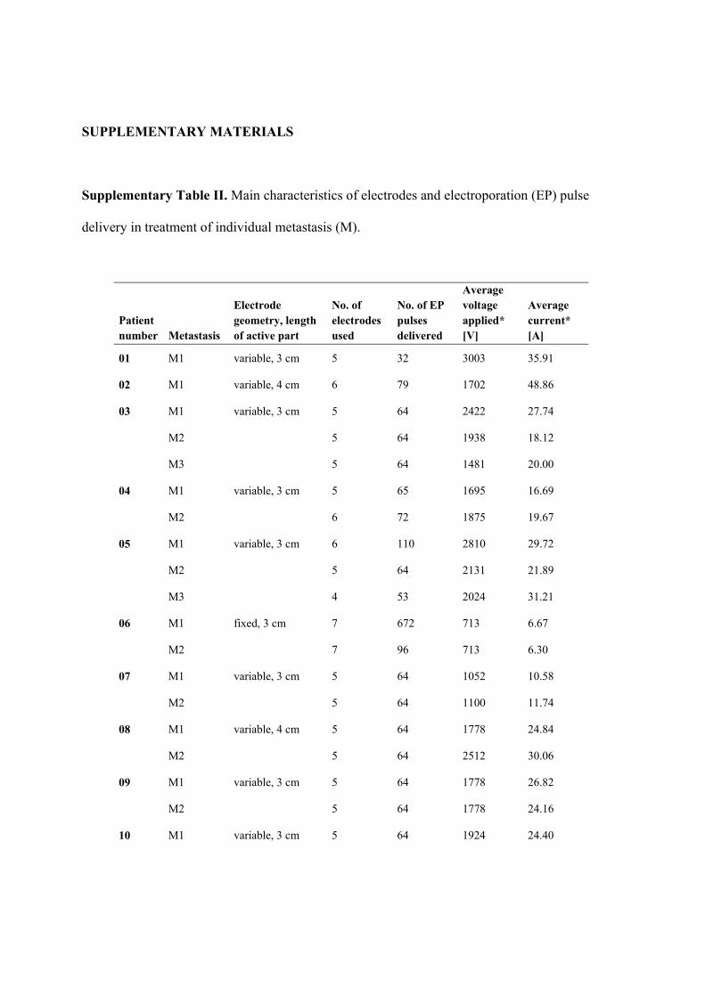
Supplementary Table II. Main characteristics of electrodes and electroporation (EP) pulse
delivery in treatment of individual metastasis (M).
Patient number Metastasis
Electrode geometry, length of active part
No. of electrodes used
No. of EP pulses delivered
Average voltage applied* [V]
Average current* [A]
01 M1 variable, 3 cm 5 32 3003 35.91
02 M1 variable, 4 cm 6 79 1702 48.86
03 M1
M2
M3
variable, 3 cm 5
5
5
64
64
64
2422
1938
1481
27.74
18.12
20.00
04 M1
M2
variable, 3 cm 5
6
65
72
1695
1875
16.69
19.67
05 M1
M2
M3
variable, 3 cm 6
5
4
110
64
53
2810
2131
2024
29.72
21.89
31.21
06 M1
M2
fixed, 3 cm 7
7
672
96
713
713
6.67
6.30
07 M1
M2
variable, 3 cm 5
5
64
64
1052
1100
10.58
11.74
08 M1
M2
variable, 4 cm 5
5
64
64
1778
2512
24.84
30.06
09 M1
M2
variable, 3 cm 5
5
64
64
1778
1778
26.82
24.16
10 M1 variable, 3 cm 5 64 1924 24.40
M2 5 64 1744 17.25
11 M1
M2
variable, 4 cm 5
5
64
64
1633
2124
20.83
20.20
12 M1
M2
fixed, 3 cm 7
7
288
384
713
713
9.02
8.24
13 M1 variable, 3 cm 5 75 2421 46.13
14 M1
M2
fixed, 3 cm 7
7
96
288
718
718
4.59
3.77
15 M1
M2
variable, 3 cm 6
6
104
111
2533
–
31.50
–
16 M1 variable, 3 cm 5 68 2383 31.67
* Average values of voltage and current were evaluated from recorded time course of voltage and current
during electric pulse delivery. The median value of voltage and current delivered within each electric pulse
was first calculated. Then, the median of these median values for all electric pulses delivered on individual
tumor was calculated and used as measure for average value of voltage and current delivered on this tumor.
– data not available

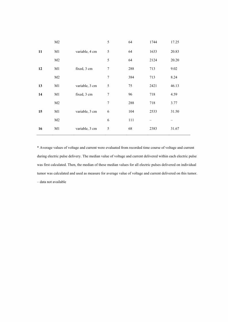
Treatment plan
Example treatment plan of Patient 2
Treatm
Electrod
ment rep
de placement
port: Exam
in the liver
mple cas
se of met
ECTplan
tastasis in the livver

Electroporation pulses applied
1
Chapter 1. Electroporation pulses applied Table 1.1 Electroporation pulses applied
Electrode pair Voltage Predicted current
1-5 2100 V 31 A
2-5 2100 26 A
2-6 2100 V 25 A
1-6 2100 V 26 A
5-6 1700 V 40 A
3-5 2100 V 25 A
3-6 2100 V 29 A
4-5 2100 V 28 A
4-6 2100 V 33 A
The total volume of tumor treated above the reversible electroporation threshold (400 V/cm) was 100 %, the volume of tumor above 600 V/cm was 99 %.
The volume of liver tissue treated above the irreversible electroporation threshold was 27 cm3.
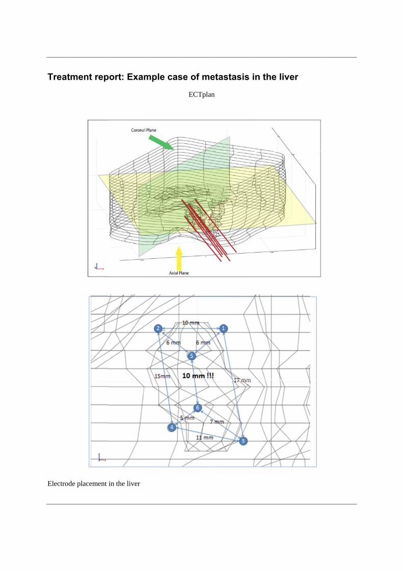
ChapFigure 2
The figutumor.
pter 2. E2.1. Electrod
ure shows the
Electrodde pair contr
e contribution
Electro
e pair cributions
n of each elec
ode pair cont
2
contribu
ctrode pair in
tributions
utions
n the treatmennt to the finaal coverage o
f the

ChapFigure 3
The figu
pter 3. C3.1. Cumulat
ure shows the
Cumulattive coverag
e volume of ti
Cumula
tive covge curves – tu
issue treated
ative coverag
3
erage cumor
above the el
ge curves
curves
lectric field sstrength indiccated on the x
x axis.
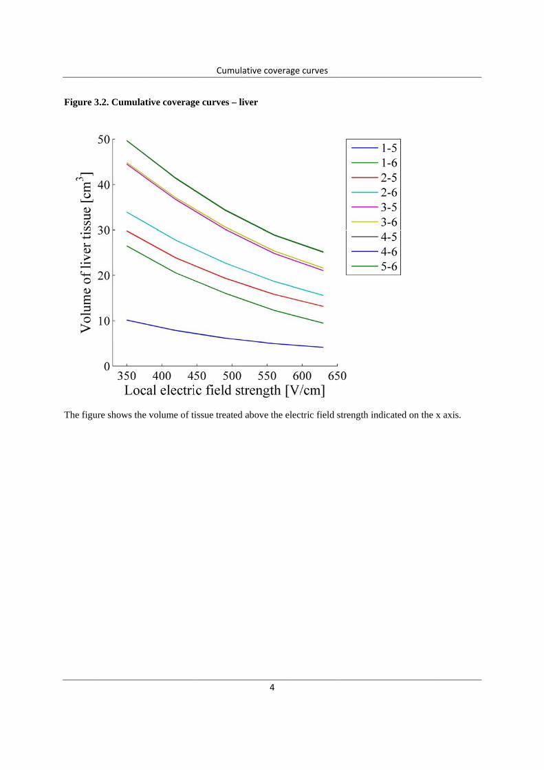
Figure 3
The figu
3.2. Cumulat
ure shows the
tive coverag
e volume of ti
Cumula
ge curves – li
issue treated
ative coverag
4
iver
above the el
ge curves
lectric field sstrength indiccated on the x
x axis.
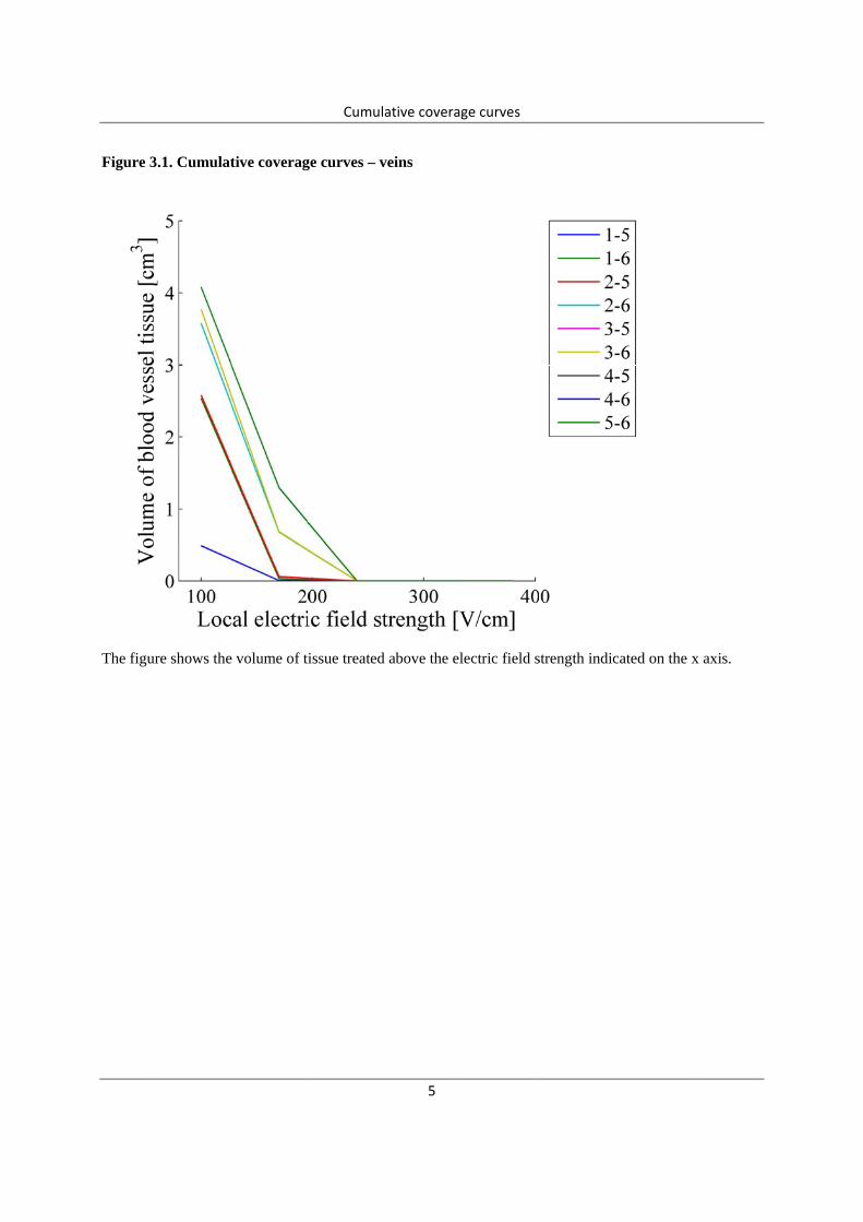
Figure 3
The figu
3.1. Cumulat
ure shows the
tive coverag
e volume of ti
Cumula
ge curves – v
issue treated
ative coverag
5
veins
above the el
ge curves
lectric field sstrength indiccated on the x
x axis.
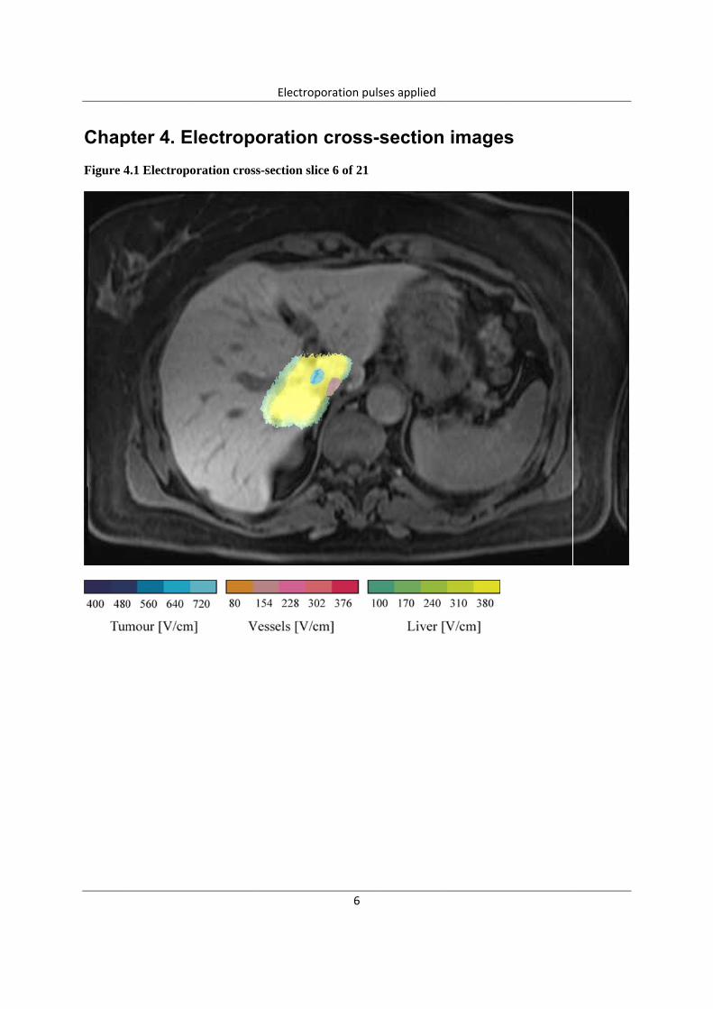
ChapFigure 4
pter 4. E4.1 Electropo
Electroporation cros
Electrop
oration ss-section sli
poration puls
6
cross-sce 6 of 21
ses applied
section images
s
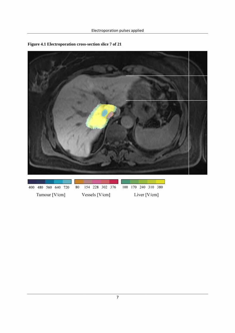
Figure 4
4.1 Electropooration cros
Electrop
ss-section sli
poration puls
7
ce 7 of 21
ses applied
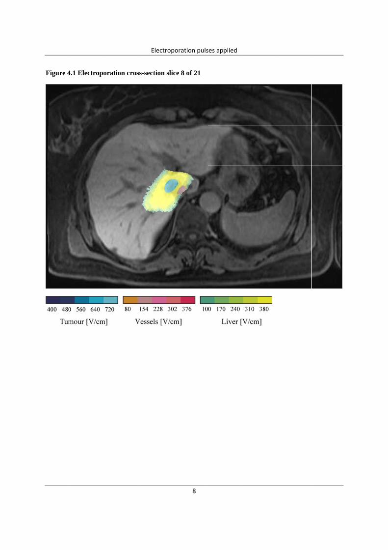
Figure 4
4.1 Electropooration cros
Electrop
ss-section sli
poration puls
8
ce 8 of 21
ses applied
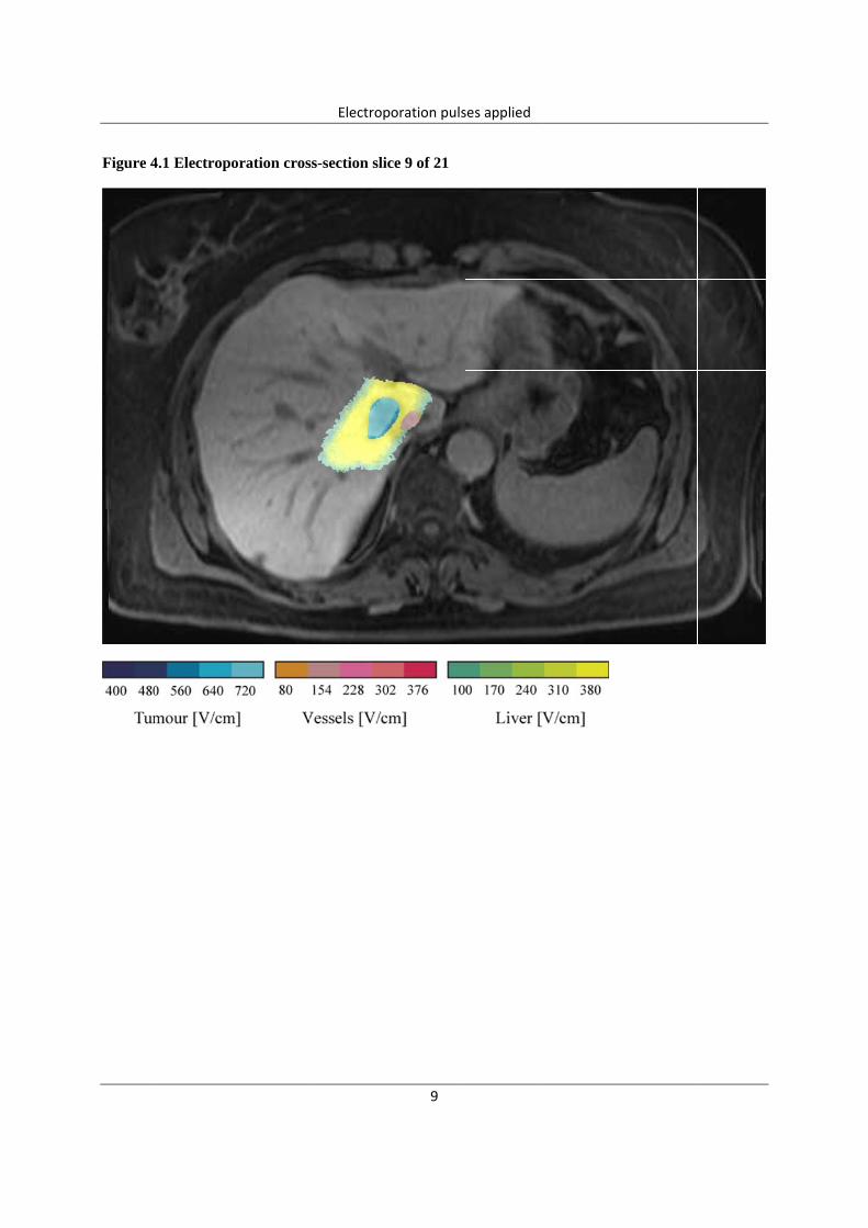
Figure 4
4.1 Electropooration cros
Electrop
ss-section sli
poration puls
9
ce 9 of 21
ses applied

Figure 4
4.1 Electropooration cros
Electrop
ss-section sli
poration puls
10
ce 10 of 21
ses applied
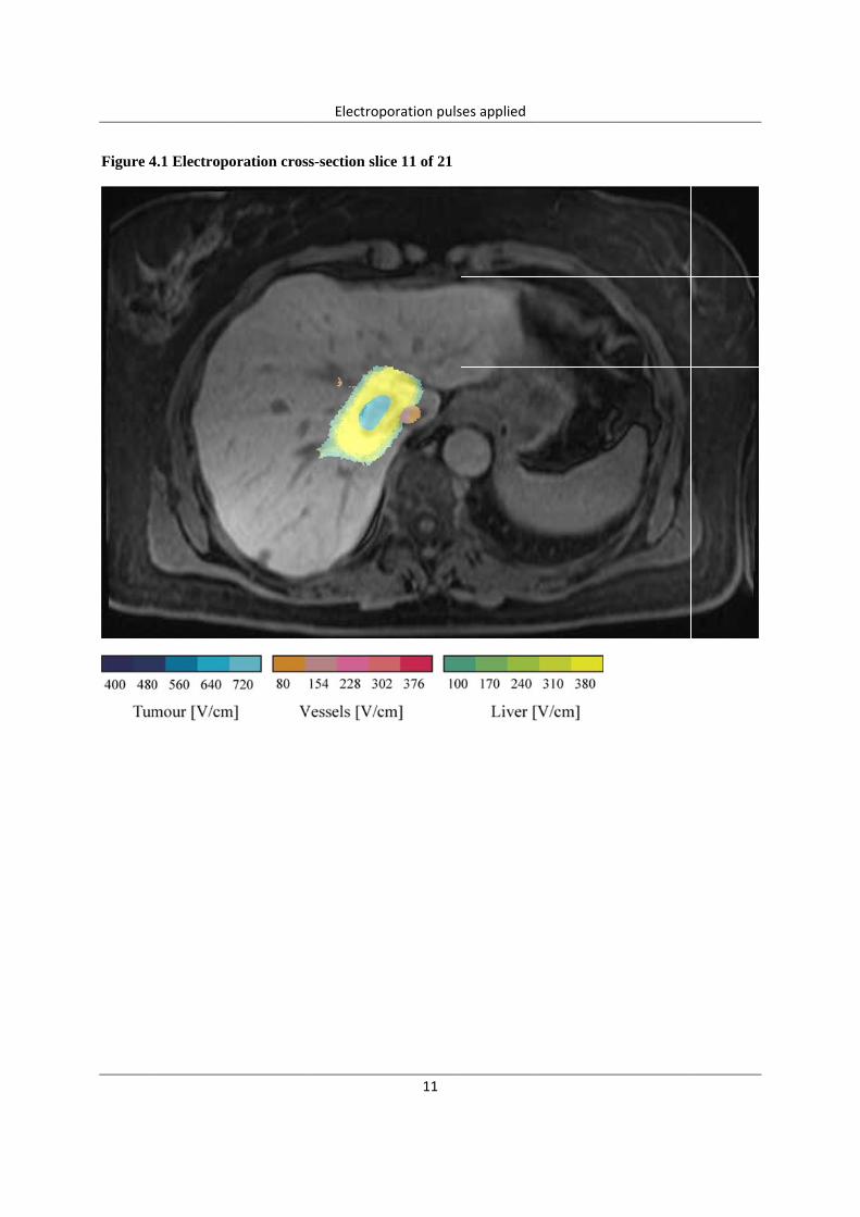
Figure 4
4.1 Electropooration cros
Electrop
ss-section sli
poration puls
11
ce 11 of 21
ses applied
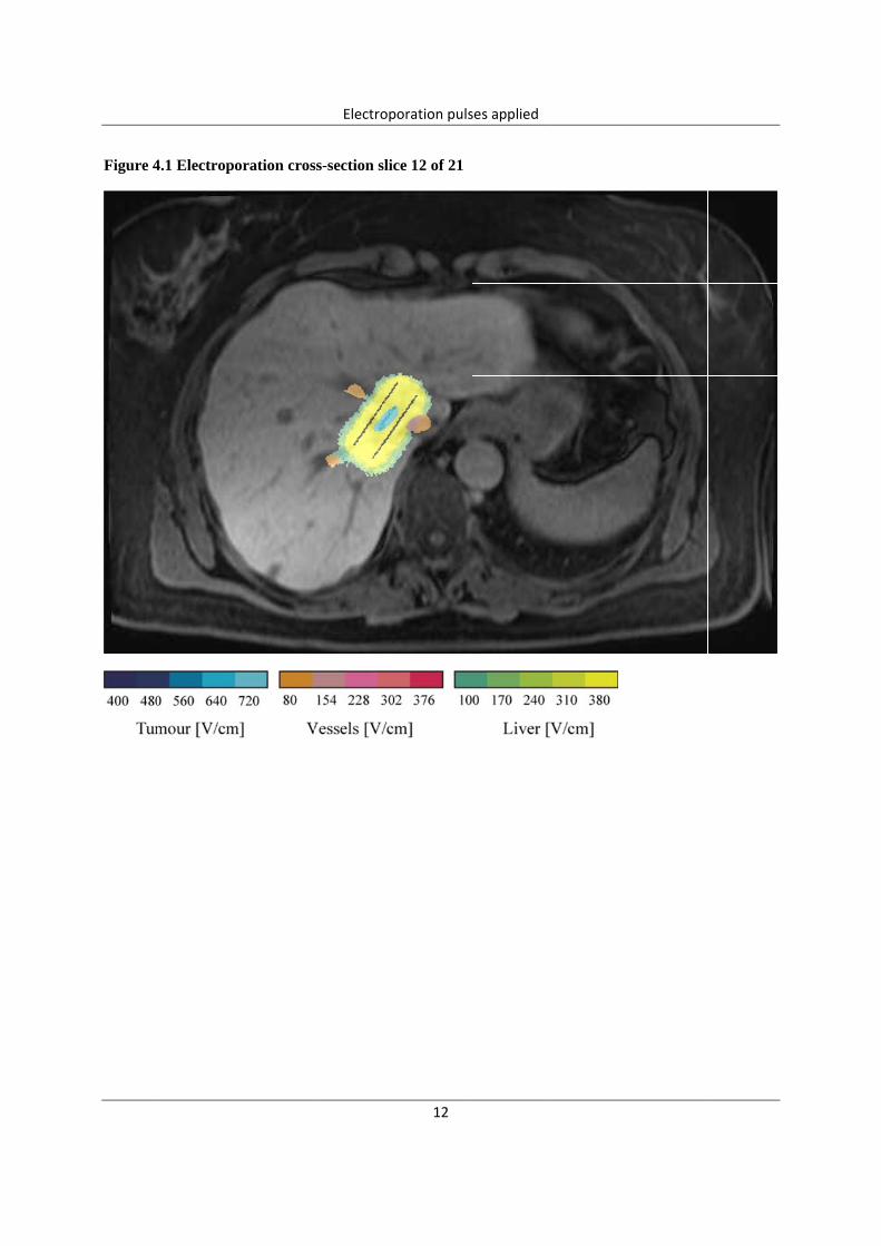
Figure 4
4.1 Electropooration cros
Electrop
ss-section sli
poration puls
12
ce 12 of 21
ses applied
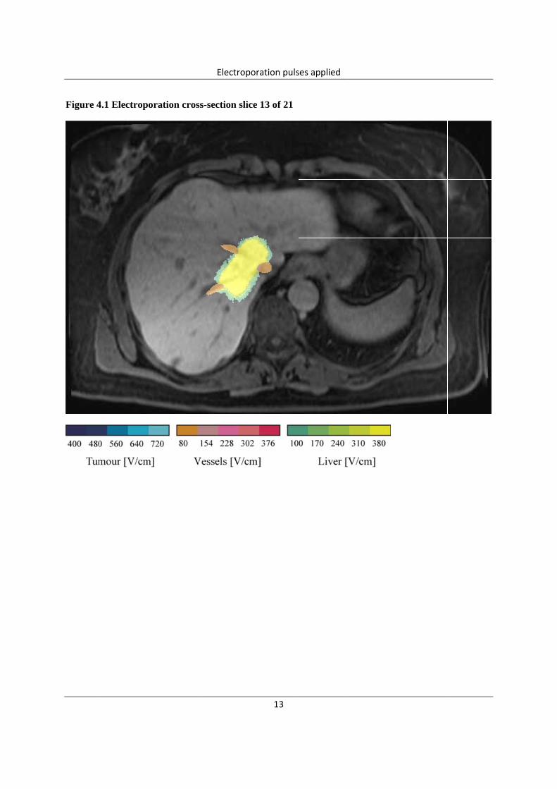
Figure 44.1 Electropooration cros
Electrop
ss-section sli
poration puls
13
ce 13 of 21
ses applied

SUPPLEMENTARY MATERIALS
Supplementary Data II. Additional explanation of the patients with serious complications.
The patients from groups I and II were in good condition with curable disease. They were
treated with intent to cure within standard of care and electrochemotherapy did not influence
their standard treatment plan in any way. During the first operation, a relatively small procedure
on the liver was performed (one or two metastasectomies on the left side and right portal vein
ligation) along with electrochemotherapy. In these two groups of the patients there were no
hepatic complications (ascites, biliary fistula, icterus, pleural effusion …) or any other serious
complications. There was one reoperation indeed in patient # 03 due to the colonic perforation;
however, this was the consequence of an episode of the atrial fibrillation (one week after the
surgery). We assume that this patient suffered partial thromboembolisation of the colonic arteries
resulting in partial colon wall necrosis and perforation which was confirmed during the
reoperation. The intra-abdominal abscess in patient # 07 was drained percutaneously.
All major complications, however, occurred in the group III. These patients were
extensively previously treated and had unresectable disease or untreatable disease with
convenient ablative techniques. These patients were offered electrochemotherpy as the only
treatment option; however, majority of these patients required demanding liver mobilization due
to very firm adhesions and/or some kind of liver resection along with electrochemotherapy.
Illustration of cases with serious complications:
Patient # 11 which required reoperation had a typical obstruction of the small bowel
caused by postoperative adhesions.
Patient # 12 had previously right hemihepatectomy and later metastasectomy due to the
recurrence. Electrochemotherapy was the third operation during which we found colon very
firmly adhered to the resection surface of the previously resected liver. These patients had also
very firm adhesions and scars between liver and diaphragm. During the mobilization of the
2
hepatic flexure, we probably accidentally superficially injured/devascularized the colonic wall,
which resulted in colon perforation one week later. We also had to mobilize the remnant of the
liver from the diaphragm which resulted in both liver and diaphragm injury.
Abdominal abscess in patient # 13 was drained percutaneously.
Patient # 15 had previously surgically untouched liver; however, he had incurable
bilateral disease. One of the metastases on the right side (Sg. 8) was 5 cm in diameter and
ingrowing into the inferior caval vein. One of the two metastases on the left side (Sg. 3 and Sg.
3-4) was in contact with left hepatic vein and therefore untreatable with radiofrequency ablation.
In this patient we performed the right hepatectomy with partial resection of the inferior caval
vein wall and the electrochemotherapy of the both metastases on the left side.
Patient # 16 had three metastases (Sg. 7, Sg. 8 and in Sg. 4a which was in contact with
median hepatic vein). Patients’ performance status and the size of the left liver did not allow the
extended right hemihepatectomy (which would have been the only potentially curable surgical
option), so the metastasectomies from Sg. 7 and Sg. 8 were performed, while the metastasis from
the Sg 4a was treated with electrochemotherapy.
Having in mind all these procedures, as well as the fact that in groups I and II there were
no hepatic complications, we anticipate that additional procedures which had to be performed in
group III caused these complications and not the electrochemotherapy itself.
It is true that without electrochemotherapy none of these patients would have been
operated and none of them would have had these complications; however, it is also true that
without electrochemotherapy the best supportive care would have been their only option.