Embed Size (px)
Citation preview

Research ArticleIntraoperative Ultrasound Staging for Colorectal LiverMetastases in the Era of Liver-Specific MagneticResonance Imaging: Is It Still Worthwhile?
Serena Langella ,1 Francesco Ardito ,2 Nadia Russolillo ,1 Elena Panettieri,2
Serena Perotti ,1 Caterina Mele,2 Felice Giuliante ,2 and Alessandro Ferrero 1
1Department of General and Oncological Surgery, Ospedale Mauriziano “Umberto I”, Torino, Italy2Unit of Hepatobiliary Surgery, Fondazione Policlinico Universitario A. Gemelli IRCCS,Universita Cattolica Del Sacro Cuore, Rome, Italy
Correspondence should be addressed to Felice Giuliante; [email protected]
Received 23 April 2019; Revised 24 July 2019; Accepted 11 August 2019; Published 22 September 2019
Guest Editor: Gyorgy Kovacs
Copyright © 2019 Serena Langella et al. *is is an open access article distributed under the Creative Commons AttributionLicense, which permits unrestricted use, distribution, and reproduction in any medium, provided the original work isproperly cited.
Background. To assess the efficacy of intraoperative ultrasound (IOUS) compared with liver-specific magnetic resonance imaging(MRI) in patients with colorectal liver metastases (CRLMs). Methods. From January 2010 to December 2017, 721 patientsunderwent MRI as a part of preoperative workup within 1 month before hepatectomy and were considered for the study. Earlyintrahepatic recurrence (relapse at cut surface excluded) was assessed 6 months after the resection and was considered as residualdisease undetected by IOUS and/or MRI. IOUS and MRI performance was compared on a patient-by-patient basis. Long-termresults were also studied. Results. A total of 2845 CRLMs were detected byMRI, and themedian number of CRLMs per patient was2 (1–31). Preoperative chemotherapy was administered in 489 patients (67.8%). In 177 patients, 379 new nodules were intra-operatively found and resected. Among 379 newly identified nodules, 317 were histologically proven CRLMs (11.1% of entireseries). *e median size of new CRLMs was 6± 2.5mm. Relationships between intrahepatic vessels and tumors differed betweenIOUS and MRI in 128 patients (17.7%). *e preoperative surgical plan was intraoperatively changed for 171 patients (23.7%).Overall, early intrahepatic recurrence occurred in 8.7% of cases. To assess the diagnostic performance, 24 (3.3%) recurrences at thecut surface were excluded; thus, 5.4% of early relapses were considered for analysis. *e sensitivity of IOUS was superior to MRI(94.5% vs 75.1%), while the specificity was similar (95.7% vs 95.9%). Multivariate analysis at the hepatic dome or subglissonian andmucinous histology revealed predictive factors of metastases missing at MRI. *e 5-year OS (52.1% vs 37.8%, p � 0.006) and DFsurvival (45.1% vs 33%, p � 0.002) were significantly worse among patients with new CRLMs than without. Conclusions. IOUSimproves staging in patients undergoing resection for CRLMs even in the era of liver-specific MRI. Intraoperative detection ofnew CRLMs negatively affects oncologic outcomes.
1. Introduction
Various imaging modalities have been developed in the fieldof liver surgery for accurate detection of colorectal metas-tases (CRLMs) [1]. Nevertheless, additional CRLMs can befound at the time of surgery in up to 25% of patients [2–9].We previously reported [9] that intraoperative ultrasonog-raphy (IOUS) enabled detection of 17.6% of new nodules inpatients undergoing resection for CRLMs. In this series, we
also demonstrated that IOUS provides significant in-formation about vascular relationships between tumors andhepatic vessels. *erefore, surgical plan was modifiedaccording to IOUS findings in 24.6% of cases. *e publisheddata on the impact of intraoperative staging are extremelyheterogeneous because of differences among centers inpreoperative diagnostic workup and surgical policies.Moreover, magnetic resonance imaging (MRI) with liver-specific contrast agent has dramatically improved the
HindawiJournal of OncologyVolume 2019, Article ID 1369274, 8 pageshttps://doi.org/10.1155/2019/1369274
sensitivity of detection of liver tumors [10, 11]. Although theefficacy of this new imaging modality to stage hepatic diseasein patients with CRLMs has been reported in several studies[12, 13], whether IOUS can improve liver staging when MRIis performed as a part of preoperative workup remainsunclear.*e aim of this study is to assess the efficacy of IOUScompared with liver-specific MRI in patients undergoinghepatectomy for CRLMs.
2. Materials and Methods
Between January 2010 and December 2017, 721 consecutivepatients who underwent liver resection for CRLMs at twoinstitutions (Ospedale Mauriziano, Torino, and PoliclinicoGemelli, Roma, Italy) were considered for the study. Eli-gibility criteria were one- or two-stage resection for CRLMs(with or without preoperative chemotherapy), age ≥18 years,written informed consent, preoperative MRI with liver-specific contrast agent performed within 1 month beforehepatectomy, IOUS accomplished by surgeon during theprocedure, postoperative follow-up at least 6 months.
Data from prospectively collected databases were ret-rospectively reviewed. *e collection and registration of theoriginal database were performed according to regulationsand with the approval of the institutional review boards ofthe two hospitals.
Primary endpoint was to compare diagnostic perfor-mance of MRI and IOUS to stage intrahepatic disease. *eperformances of IOUS and MRI were also compared inpatients who did not receive preoperative chemotherapy.Secondly, we evaluated the impact of new CRLMs intra-operatively found on long-term outcomes.
2.1. Preoperative Workup. At diagnosis, all patients wereevaluated with computed tomography (CT) scans and MRI.CTscans were performed with a multislice helical CTusing a3mm collimation and reconstruction at 1 and 2.5mm.Images were acquired using a triphasic hepatic protocolfollowing a noncontrast evaluation of the liver. Images wereobtained 11, 80, and 180 seconds after the start of in-travenous injection of iopromide (Ultravist® 370, BayerHealthCare Pharmaceuticals Inc., Wayne, NJ) at a rate of3.5mL/s. MRI was conducted on a 1.5 T superconductingsystem using a liver-specific contrast agent (EOB-gadoxeticacid disodium, Primovist, Bayer Schering Pharma AG,Berlin, Germany). All MR images were preoperativelyevaluated by radiologists skilled in liver pathology anddiffusion-weighted imaging (DWI). Fluorodeoxyglucosepositron emission tomography was also performed in se-lected cases. After chemotherapy, restaging was accom-plished by MRI and thoracic CT scans or thoracoabdominalCT scans in the presence of extrahepatic disease. Chemo-therapy response was assessed by using the ResponseEvaluation Criteria in Solid Tumors (RECIST) [14].
2.2. Intraoperative Staging. Abdominal exploration andintraoperative liver ultrasonography (Aloka Prosound Alpha7 with 7.5MHz intraoperative miniconvex probe, Aloka Co.,
Tokyo, Japan; Philips HDI® 5000 SonoCT with 8MHz to4MHz intraoperative convex probe ATL Entos CT8-4,Koninklijke Philips Electronics, Eindhoven, Netherlands)were always performed as the first step to assess the site,extent of the disease, and the tumor’s relationships withmajor intrahepatic vessels and to define the extension of therequired resection. *e surgeon conducted IOUS for allpatients according to a standardized protocol. A similartechnique was used for laparoscopic liver ultrasound. *iswas performed with a multifrequency (5–10MHz) flexiblelinear-array laparoscopic transducer (UST-5536-7.5; HitachiAloka Medical) and a Pro Focus 2202 Ultrasound Systemwith Laparoscopic Transducer Type 8666-RF (Bk Medical,Herlev, Denmark).
Contrast-enhanced intraoperative ultrasound (CEIOUS)was additionally performed in selected cases. CEIOUS wasachieved with a convex 2–6MHz harmonic frequencytransducer. In all patients, 2.4mL sulfur hexafluoridemicrobubbles (SonoVue®, Bracco Imaging, Milan, Italy) wasinjected intravenously through a peripheral vein by theanesthesiologist.
All nodules consistent with CRLMs found intraoperativelyby IOUS and/or CEIOUS that were not detected at pre-operative MRI were classified as new lesions. During surgery,MR images were always available on a computer screen, whichallowed a real-time comparisonwith intraoperative findings. Inpatients who underwent chemotherapy, disappeared livermetastases (DLMs) on preoperative MRI that were detectedintraoperatively were not considered new lesions.
2.3. Histopathologic Examination. *e pathologist was in-formed about the site of preoperatively detected CRLMs andnew nodules. Specimens were fixed, embedded in paraffin,and stained with hematoxylin-eosin.*en, 0.5 cm slices weretaken for microscopic examination. Steatosis was estimatedas the percentage of involved hepatocytes and categorized asdefined by Kleiner et al. [15]: no fatty change (<5%), mild(5% to <33%), moderate (33% to <66%), or severe (≥66%).
2.4.Diagnostic PerformanceAnalysis. We conducted patient-by-patient analysis to evaluateMRI and intraoperative stagingperformance (IOUS and CEIOUS). Early intrahepatic re-currences were registered at 6 months after the resection andwere considered residual disease undetected by intraoperativestaging and/or MRI (false negative: FN). Recurrences wereassessed using radiological imaging during the follow-up.Patients were evaluated every 3 months with physical ex-amination, measurement of CEA levels, and abdominal ul-trasonography or thoracoabdominal CT. Local recurrence onthe cut liver surface was not considered an FN. In patient-by-patient analysis, sensitivity was defined as the number ofpatients without FN lesions divided by the total number ofpatients. Conversely, specificity was defined as the number ofpatients without false-positive (FP) lesions divided by the totalnumber of patients. By definition, we considered the positiveand negative predictive values (PPV andNPV, respectively) asthe proportions of positive and negative results in true-positive and true-negative results. *e likelihood ratio was
2 Journal of Oncology
calculated for both positive (LR+, likelihood ratio positive:sensibility/1− specificity) and negative (LR− , likelihood rationegative: 1− sensibility/specificity) results.
2.5. Definitions
Indirect signs to identify liver metastases by IOUS werepresence of bile duct dilatation, distortion, or in-terruption of the venous wall.Types of hepatectomies were classified according to theBrisbane 2000 terminology [16].Were considered mucinous CRLMs, those histologi-cally proven liver metastases comprising more than50% mucinous carcinoma.Local recurrence was defined as intrahepatic relapse atcut surface of the previous hepatectomy.Subglissonian metastasis was defined as lesions within1 cm of the liver surface.
2.6. Statistical Analysis. All statistical analyses were per-formed using IBM SPSS software (v20.1). *e distribution ofvariables was analyzed using the Kolmogorov–Smirnov test.Categorical variables were compared using the chi-square test,Fisher’s exact test, or Pearson’s test as appropriate. Contin-uous variables were compared between groups using theunpaired t-test or Mann–Whitney U test, as appropriate.Continuous variables were presented as median± standarddeviation (SD) or range. Categorical variables were repre-sented as number and percentage in brackets. Diagnosticperformance was evaluated assessing sensitivity, specificity,PPV, NPV, and likelihood ratio. Cohen’s kappa coefficientwas used to assess the interrater reliability of preoperative andintraoperative imaging. *e results have been interpreted asfollows: values≤0 as indicating no agreement and 0.01–0.20 asnone to slight, 0.21–0.40 as fair, 0.41–0.60 as moderate,0.61–0.80 as substantial, and 0.81–1.00 as almost perfectagreement. Uni- and multivariate binary logistic regressionanalyses were performed to assess the predictive factors formissing CRLMs at MRI. After univariate analysis, a p value≤0.05 was considered to include variables in the multivariateanalysis. Receiver operating characteristic curves were plottedto identify the value of preoperative number of metastases andmedian number of neoadjuvant chemotherapy cycle inpredicting missing CRLMs at MRI with a high sensitivity andspecificity. Disease-free survival was measured from the dateof hepatic resection until the date of radiographic detection ofrecurrence, death, or last follow-up. Overall survival wasmeasured from the date of hepatic resection until the date ofdeath or last follow-up. *e Kaplan–Meier method was usedto estimate survival probabilities, which were compared usingthe log-rank test. All p values were two sided, and p≤ 0.05was considered statistically significant.
3. Results
Patients were investigated with a median of 3 (range 2–4)preoperative imaging modalities (US, CTscan, and MRI and
PET). All patients were staged with MRI, and fluorodeox-yglucose positron emission tomography was undertaken in317 (43.9%) patients. A total of 2845 CRLMs were detectedpreoperatively using MRI. *e median number of CRLMsper patients was 2 (1–31). Multiple (more than 3) CRLMswere observed in 358 (49.6) patients. *e median diameterwas 24± 22.05mm. In 56 (7.7%) patients, CRLMs were frommucinous cancer.
Preoperative chemotherapy was administered in 489patients (67.8%). In this subgroup, DLMs were present in 52of 489 (10.6%) patients. Hepatic resections were minor in592 (82.1%) patients. Among these, multiple liver resectionswere required in 517 (87.3%) cases. Minor resections weredistributed as follows: 459 (77.5%) nonanatomical, 60(10.1%) anatomical, and 73 (12.3%) both anatomical andnonanatomical. Two stage procedures were accomplished in30 (4.2%) patients. Redo-resection for recurred CRLMs wasperformed in 50 of 721 patients (6.9%). A laparoscopicapproach was used in 103 (14.3%) patients to perform 11(10.7%) major and 92 (89.3%) minor hepatectomies.
Preoperative and operative data are detailed in Table 1.
3.1. Intraoperative Findings and Management. In 177 pa-tients, 379 (13.3%) new nodules were intraoperatively foundand resected. Among 379 newly identified nodules, 317(83.6%) were histologically proven CRLMs (11.1% of entireseries). *e 62 FP cases (16.4%) were classified by pathol-ogists as hemangiomas (19), focal steatosis (14), biliaryhamartoma (12), granulomatous inflammation (9), fibrosis(6), and focal nodular hyperplasia (2). Furthermore, 38(73%) of 52 DLMs were found intraoperatively (not con-sidered new CRLMs).
*e liver was hyperechoic in half of the patients (363,50.3%). *e median new CRLM size was 6± 2.5mm, andmost were hypoechoic (77.3%). *e new CRLMs were onlyrarely detected by indirect signs (3.8%) or CE-IOUS (5.9%).Features of new CRLMs were summarized in Table 2.
Seventy out of 317 (22%) new lesions were located at thehepatic dome (Segments (Sgs) 8 and 4a). *e remaining newnodules were distributed as follows: nine in Sg 1 (2.8%), 31 inSg 2 (9.7%), 38 in Sg 3 (11.9%), 41 in Sg 4b (12.9%), 43 in Sg 5(13.5%), 45 in Sg 6 (14.2%), and 40 in Sg 7 (12.7%). Twenty-eight new CRLMs were sited within 1 cm from the liversurface.
Vascular relationships between intrahepatic vessels andtumors differed between IOUS and MRI in 128 patients(17.7%). In 31 (4.3%) patients, 46 (1.6%) lesions suspected formetastases at preoperative imaging were not identified orassessed intraoperatively as metastases. Among the 46 le-sions left in situ because of IOUS findings, only two weresubsequently diagnosed as metastases during the follow-up(rate of FN lesion 4.3%). Overall, in 171 (23.7%) patients, thepreoperative surgical plan changed according to intra-operative findings. Commonly, in case of new CRLMs,limited additional resections were required (83%).
Overall, 232 (32.2%) patients were scheduled for upfrontsurgery (without preoperative chemotherapy). Among these,MRI preoperatively identified 504 CRLMs, and the median
Journal of Oncology 3

number of CRLMs per patient was 1 (1–4). In this subset ofpatients, IOUS detected 68 (13.5%) histologically provenCRLMs in 31 patients (13.3%). IOUS also revealed 5 (0.9%) newnodules in 4 (1.7%) patients that—after resection—wereclassified by the pathologist as benign lesions (FP). On the otherhand, all but one CRLM identified at MRI were confirmedintraoperatively. *e median size of new CRLMs was 5mm(1–7). CRLMs newly identified weremainly hypoechoic (27/31,87%). Features of new CRLMs are summarized in Table 2.
3.2. Diagnostic Performance Analysis. Performance of MRIand intraoperative staging was compared on a patient-by-patient basis.
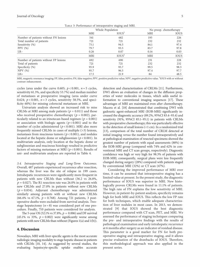
Overall early intrahepatic recurrences were 8.7%. Toassess the diagnostic performance, 24 (3.3%) recurrences atthe cut surface were excluded; thus, 5.4% of early relapseswere considered for analysis. According to the rates of FPand FN patients, IOUS was more sensitive than MRI (94.5%vs 75.1%), while the specificity was similar (95.7% vs 95.9%).
PPV of MRI was 79.7% vs 93.5% of IOUS while NPV was95.7% (MRI) vs 96.5% (IOUS). *e LR− was 0.26 (MRI) vs0.07 (IOUS), and the LR+ was 17.5 (MRI) vs 21.9 (IOUS).Finally, Cohen’s kappa coefficient was 0.73, indicating asubstantial agreement between MRI and IOUS.
Diagnostic performance was also assessed among pa-tients who did not receive preoperative chemotherapy. Inthis subset of patients, IOUS revealed a higher sensitivity(MRI 84%, IOUS 97.4%). Both sensitivity and sensibility(MRI 99.5%, IOUS 98.3%) were improved compared tothose of the whole population with a substantial agreementbetween MRI and IOUS (Cohen’s kappa coefficient� 0.80).All parameters considered for diagnostic performanceanalysis are detailed in Table 3.
3.3. Predictor of Missing CRLMs at MRI. We assessed pre-dictors of missing CRLMs at MRI. Receiver operatingcharacteristic curve analysis revealed a significant predictivevalue of the median number of preoperative chemotherapy
Table 1: Preoperative and operative characteristics of 721 patients with 2845 CRLMs preoperatively assessed by MRI (whole population)and 232 patients who did not receive preoperative chemotherapy.
Characteristics Whole population n� 721 Patients without chemotherapy n� 232Age (years), median± SD 64± 10.8 66± 10.0Male, n (%) 453 (62.2) 149 (64.2)BMI (kg/m2), median± SD 25± 3.34 27± 4.0Preoperative chemotherapy 489 (67.8) —Number of cycles, median± SD 5± 6.5 —Oxaliplatin based, n (%) 358 (49.7) —Irinotecan based, n (%) 208 (28.8) —Biologics, n (%) 309 (42.9) —
Response to chemotherapy∗PR 306 (62.6) —SD 154 (31.5) —PD 29 (5.9) —
Preoperative radiologic workupPET total body, n (%) 317 (44) 107 (46.1)Number of LMs, median (range) 2 (1–31) 1 (1–4)Maximum diameter (mm), median± SD 24± 22.05 19.4± 23.0Types of resection(1) Minor hepatectomy, n(%) 592 (82.1) 194 (83.6)(2) Laparoscopic resection, n(%) 103 (14.3) 62 (26.7)(3) Redo-resection, n(%) 50 (6.9) 28 (18)BMI, body mass index; PR, partial response; SD, stable disease; PD, progression disease. ∗Rates calculated on 489 patients who received preoperativechemotherapy.
Table 2: Details of new CRLMs intraoperatively found in the whole population and in patients who did not receive preoperativechemotherapy.
New CRLM Features Whole population (n� 317) Patients without chemotherapy (n� 31)Diameter (mm), median± SD 6± 2.5 5± 2.3Number per patients, median (range) 1 (1–9) 1 (1–6)US aspectHypoechoic, n (%) 245 (77.3) 27 (87)Hyperechoic, n (%) 46 (14.5) 1 (9.6)Isoechoic, n (%) 11 (3.4) 1 (3.2)
Location subglissonian, n (%) 75 (23.6) 6 (19.3)New CRLMs identified by CE-IOUS, n (%) 19 (5.9) 2 (6.4)New CRLMs identified by indirect signs, n (%) 12 (3.8) 1 (3.2)
4 Journal of Oncology
cycles (area under the curve 0.605; p< 0.001, n� 5 cycles,sensitivity 61.3%, and specificity 53.7%) and median numberof metastases at preoperative imaging (area under curve0.624; p< 0.001, n� 3 cycles, sensitivity 58.5%, and speci-ficity 60%) for missing colorectal metastases at MRI.
Univariate analysis showed an increased risk to missCRLMs at MRI among male patients (p � 0.011) and thosewho received preoperative chemotherapy (p � 0.002), par-ticularly related to an irinotecan-based regimen (p � 0.001)or association with biologic agents (p< 0.001) and to thenumber of cycles administered (p< 0.001). MRI also morefrequently missed CRLMs in cases of multiple (>3) lesions,metastases from mucinous tumors (p< 0.001), and noduleslocated at the hepatic dome or subglissonian (p< 0.001). Inmultivariate analysis, only location at the hepatic dome orsubglissonian and mucinous histology resulted in predictivefactors of missing metastases at MRI (p< 0.001). Results ofuni- and multivariate analyses are reported in Table 4.
3.4. Intraoperative Staging and Long-Term Outcomes.Overall, 487 patients experienced recurrence after resection,whereas the liver was the site of relapse in 199 cases.Intrahepatic recurrences were significantly more frequent inpatients with new CRLMs than without (36.2 vs 26.8%,p � 0.027). *e R1 resection rate was 26.0% in patients withnew CRLMs and 27.0% in patients without new CRLMs(p � 0.834). Adjuvant chemotherapy was administeredsimilarly among patients with or without new CRLMs(66.4% vs 67.1%, p � 0.768). Among 721 patients, 3 post-operative deaths were excluded from survival analysis. Two-stage hepatectomy (n� 8) was considered part of one pro-cedure. Finally, 710 patients were considered for analysis.
*e 5-year OS (52.1% vs 37.8%, p � 0.006) andDF survival(45.1% vs 33%, p � 0.002) were significantly worse amongpatients with newCRLMs thanwithout (Figures 1(a) and 1(b)).
4. Discussion
Nowadays, MRI with liver-specific agents is the most accurateradiologic imagingmodality to stage hepatic disease in patientswith CRLMs [10, 14]. As suggested by several studies, theevaluating hepatocyte-specific uptake enables accurate
detection and characterization of CRLMs [11]. Furthermore,DWI allows an evaluation of changes in the diffusion prop-erties of water molecules in tissues, which adds useful in-formation to conventional imaging sequences [17]. *eseadvantages of MRI are maintained even after chemotherapy.Macera et al. [18] demonstrated that combining DWI withgadoxetic agent-enhanced MRI (EOB-MRI) significantly in-creased the diagnostic accuracy (89.2%, 95%CI 83.4–93.4) andsensitivity (91%, 95%CI 85.1–95.1) in patients with CRLMswith preoperative chemotherapy; this was particularly effectivein the detection of small lesions (<1 cm). In a randomized trial[13], comparison of the total number of CRLM detected atinitial imaging versus the number found intraoperatively andat pathological examination of resected specimens showed thegreatest number of patients with equal assessments (88%) inthe EOB-MRI group (compared with 74% and 62% in con-ventional MRI and CT scan groups, respectively). Diagnosticconfidence was high or very high for 98.3% of patients withEOB-MRI; consequently, surgical plans were less frequentlychanged during surgery (28%) compared with patients stagedby conventional MRI (32%) or CT scan (47%).
Considering the improved performance of MRI overtime, it can be assumed that intraoperative staging has alimited value at present. In the present study, the diagnosticperformance of IOUS was superior to MRI. New histo-logically proven CRLMs were found in 11.1% of patients.*e high rate of FN explains the low sensitivity of MRI.However, in patient-by-patient analysis, specificity remainshigh for both MRI and IOUS. *is reflects the low FP ratefor both techniques, which enable adequate characteriza-tion of liver nodules in most cases. In 2013, we demon-strated [9] that IOUS showed the best diagnosticperformance compared with CT scan, PET, and MRI. Weassessed the performance of staging techniques comparingthe pre- and intraoperative findings with the results ofpathological examination and early intrahepatic recurrenceat 6 months after surgery as an indicator of residual disease.*is parameter is a good marker for FN for both pre-operative staging and intraoperative staging, and it allowsprecise evaluation of the drawbacks of IOUS. *erefore,this methodological approach was also applied in thepresent series.
Table 3: Performance of intraoperative staging and MRI.Whole Population No preop. chemotherapy
MRI IOUS1 MRI IOUSNumber of patients without FN lesions 541 682 195 226Total number of patients 721 721 232 232Sensitivity (%) 75.1 94.5 84.1 97.4PPV (%) 79.7 93.5 83.7 97.8LR− 0.26 0.07 0.16 0.03
MRI IOUS1 MRI IOUSNumber of patients without FP lesions 692 690 231 228Total of patients 721 721 232 232Specificity (%) 95.9 95.7 99.5 98.3NPV (%) 95.7 96.5 97.4 97.7LR+ 17.5 21.9 84 48.5MRI, magnetic resonance imaging; FP, false positive; FN, false negative; PPV, positive predictive value; NPV, negative predictive value. 1IOUS with or withoutcontrast enhancement.
Journal of Oncology 5
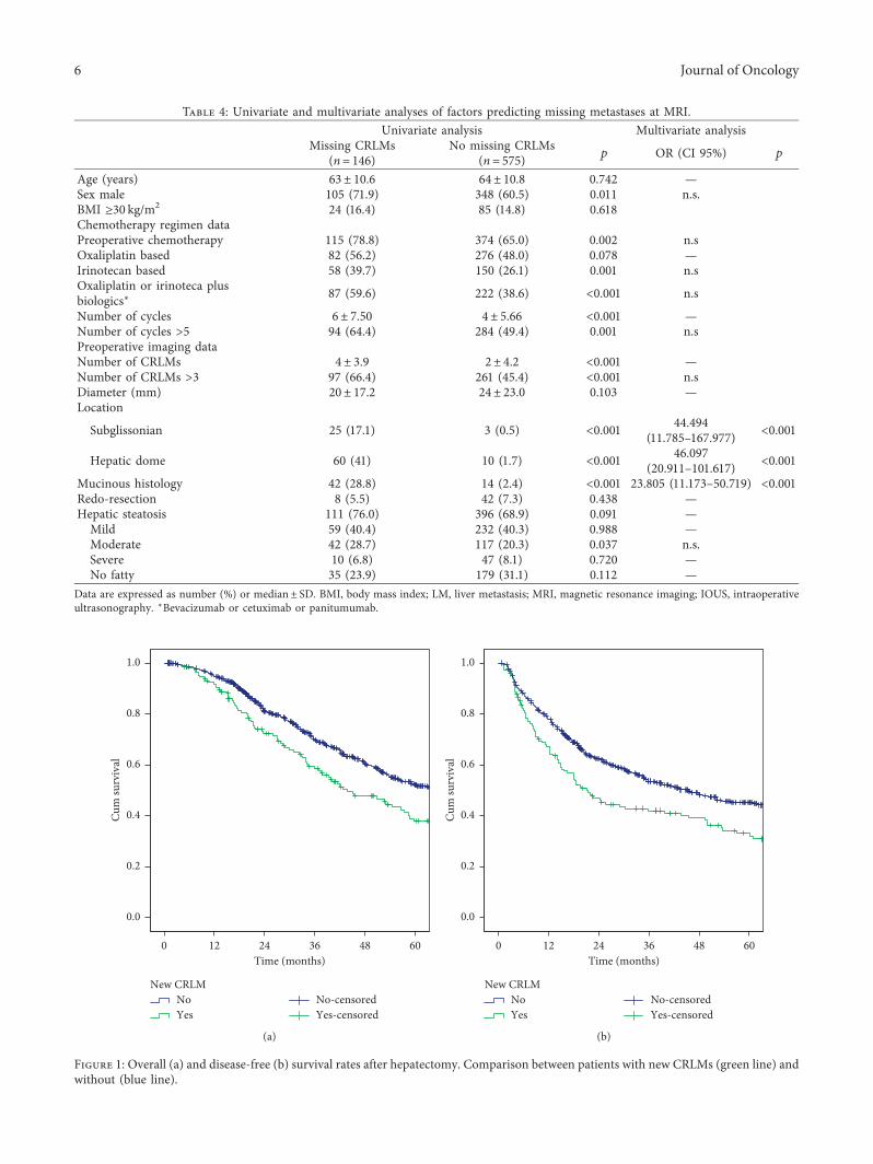
Table 4: Univariate and multivariate analyses of factors predicting missing metastases at MRI.Univariate analysis Multivariate analysis
Missing CRLMs(n� 146)
No missing CRLMs(n� 575) p OR (CI 95%) p
Age (years) 63± 10.6 64± 10.8 0.742 —Sex male 105 (71.9) 348 (60.5) 0.011 n.s.BMI ≥30 kg/m2 24 (16.4) 85 (14.8) 0.618Chemotherapy regimen dataPreoperative chemotherapy 115 (78.8) 374 (65.0) 0.002 n.sOxaliplatin based 82 (56.2) 276 (48.0) 0.078 —Irinotecan based 58 (39.7) 150 (26.1) 0.001 n.sOxaliplatin or irinoteca plusbiologics∗ 87 (59.6) 222 (38.6) <0.001 n.s
Number of cycles 6± 7.50 4± 5.66 <0.001 —Number of cycles >5 94 (64.4) 284 (49.4) 0.001 n.sPreoperative imaging dataNumber of CRLMs 4± 3.9 2± 4.2 <0.001 —Number of CRLMs >3 97 (66.4) 261 (45.4) <0.001 n.sDiameter (mm) 20± 17.2 24± 23.0 0.103 —Location
Subglissonian 25 (17.1) 3 (0.5) <0.001 44.494(11.785–167.977) <0.001
Hepatic dome 60 (41) 10 (1.7) <0.001 46.097(20.911–101.617) <0.001
Mucinous histology 42 (28.8) 14 (2.4) <0.001 23.805 (11.173–50.719) <0.001Redo-resection 8 (5.5) 42 (7.3) 0.438 —Hepatic steatosis 111 (76.0) 396 (68.9) 0.091 —Mild 59 (40.4) 232 (40.3) 0.988 —Moderate 42 (28.7) 117 (20.3) 0.037 n.s.Severe 10 (6.8) 47 (8.1) 0.720 —No fatty 35 (23.9) 179 (31.1) 0.112 —
Data are expressed as number (%) or median± SD. BMI, body mass index; LM, liver metastasis; MRI, magnetic resonance imaging; IOUS, intraoperativeultrasonography. ∗Bevacizumab or cetuximab or panitumumab.
Time (months)60483624120
1.0
0.8
0.6
0.4
0.2
0.0
Yes-censoredNo-censored
YesNo
New CRLM
Cum
surv
ival
(a)
Yes-censoredNo-censored
YesNo
New CRLM
Time (months)60483624120
Cum
surv
ival
1.0
0.8
0.6
0.4
0.2
0.0
(b)
Figure 1: Overall (a) and disease-free (b) survival rates after hepatectomy. Comparison between patients with new CRLMs (green line) andwithout (blue line).
6 Journal of Oncology
It is well known that preoperative chemotherapy maynegatively affect the accuracy of preoperative staging andintraoperative staging [19]. *is is mainly due to the che-motherapy-related changes in liver parenchyma and mod-ification of CRLM features. After chemotherapy, MRI withliver-specific contrast agents guarantees the most accuratepreoperative staging of hepatic disease compared to otherimaging modalities [20]. However, some small CRLMs maydisappear at preoperative MRI after chemotherapy [21]. Inthis series, vanishing metastases at preoperative MRI wereexcluded from the analysis to avoid a possible over-estimation of new CRLMs intraoperatively found. More-over, to overcome the potential bias related to thechemotherapy administration, we also evaluated the di-agnostic performance of MRI and IOUS in patients whounderwent liver resection without preoperative chemo-therapy. In this subset of patients, sensibility and sensitivityof both MRI and IOUS were improved. Nevertheless, IOUSassured the higher sensitivity rate.
To depict the pitfalls in the MRI staging, we assessed thepredictors of “missing” metastases at MRI. Multivariateanalysis demonstrated an increased risk of missing CRLMsat MRI in the case of subglissonian nodules or located athepatic dome, such as for metastases from mucinous tu-mors. *e liver surface can be better assessed intra-operatively [5, 18, 22, 23]; moreover, the evaluation of thehepatic dome duringMRImay be limited by artefacts relatedto respiratory movement. Furthermore, mucinous tumorscould mimic benign lesions, worsening the diagnostic ac-curacy, particularly in the case of small nodules [24, 25]. Inagreement with previous studies [4, 9], we confirmed thatnewly identified CRLMs are more likely to be hypoechoic.Because of steatosis or postchemotherapy changes in thehepatic parenchyma, patients who underwent hepatectomyfor CRLMs often present a “bright” liver. *is conditionenables detection of hypoechoic nodules and reduces thevalue of CE-IOUS staging because the liver is naturallyenhanced [2].*ese findings could explain the high accuracyof IOUS reported in the present series, even if CE-IOUS wasnot performed systematically.
Several surgical series [2–9] have reported the superi-ority of IOUS to stage hepatic disease in CRLMs comparedwith various imaging modalities. *e improvements ofimaging modalities over the years represent a challenge forthe current role of IOUS. As expected, the rate of newCRLMs found by IOUS decreased in the most recent seriesbut remains noteworthy (ranging from 8% to 21%). Un-fortunately, results from published studies cannot be gen-eralized because of extreme variability related to the differentpreoperative workup and technological progress over time.Notably, the present study considered many patientsresected in recent years at two tertiary centers. *is pop-ulation is homogeneous, and the process of MRI with liver-specific contrast agents was similar for all patients. Previouspublished data also showed that the impact on managementis extremely heterogeneous and it is strongly affected bysurgical policies. Our centers shared a parenchymal sparingphilosophy to face CRLMs; this explains the significantimpact of intraoperative findings on changing surgical plans.
In patients with HCC, the intraoperative detection ofnew tumors negatively affects oncologic outcomes [26].However, the impact of new CRLMs on long-term outcomeshas been poorly evaluated. In the present study, we showedthat hepatic recurrences are significantly more frequentamong patients with newly identified CRLMs and theypresent worse OS and DF survival. We previously dem-onstrated that additional CRLMs are more likely to be foundin patients with more aggressive disease. We thereforesuggest considering the new CRLMs during postoperativedecision making along with other known prognostic factors.For example, this subset of patients may benefit from ad-juvant chemotherapy to reduce the risk of relapse. Moreover,because the possibility of reresection for recurred CRLMscan significantly improve survival [27], we also suggest astrict postoperative surveillance to detect and manage he-patic recurrences as early as possible.
*is study presents some limitations, mainly related toits retrospective nature. Even if both IOUS and RM wereperformed by surgeons and radiologists skilled in the field ofliver malignancies, different physicians were involved in thestudy. Nevertheless, this is the largest series to date focusingon this topic and comparing the performance of MRI withliver-specific contrast agents to IOUS.
In conclusion, IOUS improves staging in patients un-dergoing resection for CRLMs even in the era of liver-specific MRI. Intraoperative detection of new CRLMsnegatively affects oncologic outcomes.
Data Availability
*e prospective data used to support the findings of thisstudy are restricted by the ethics committee of MaurizianoHospital in order to protect patient privacy. Data areavailable from corresponding author for researchers whomeet the criteria for access to confidential data.
Disclosure
*is paper is based on a previous communication of pre-liminary results during the 12th Biennial E-AHPBA Con-gress, 23–26 May 2017, Mainz, Germany.
Conflicts of Interest
*e authors declare that they have no conflicts of interest.
Acknowledgments
*e authors thank Dr. Elena Ostuni and Dr. Fabio Forchinofor their contribution to data collection.
References
[1] M. C. Niekel, S. Bipat, and J. Stoker, “Diagnostic imaging ofcolorectal liver metastases with CT, MR imaging, FDG PET,and/or FDG PET/CT: a meta-analysis of prospective studiesincluding patients who have not previously undergonetreatment,” Radiology, vol. 257, no. 3, pp. 674–684, 2010.
Journal of Oncology 7
[2] G. Torzilli, F. Botea, F. Procopio et al., “Use of contrast-en-hanced intraoperative ultrasonography during liver surgeryfor colorectal cancer liver metastases—its impact on operativeoutcome. Analysis of a prospective cohort study,” EuropeanJournal of Cancer Supplements, vol. 6, no. 11, pp. 16–23, 2008.
[3] C. Sietses, M. R. Meijerink, S. Meijer, and M. P. van den Tol,“*e impact of intraoperative ultrasonography on the surgicaltreatment of patients with colorectal liver metastases,” Sur-gical Endoscopy, vol. 24, no. 8, pp. 1917–1922, 2010.
[4] M. G. van Vledder, T. M. Pawlik, S. Munireddy, U. Hamper,M. C. de Jong, and M. A. Choti, “Factors determining thesensitivity of intraoperative ultrasonography in detectingcolorectal liver metastases in the modern era,” Annals ofSurgical Oncology, vol. 17, no. 10, pp. 2756–2763, 2010.
[5] S. Hata, H. Imamura, T. Aoki et al., “Value of visual in-spection, bimanual palpation, and intraoperative ultraso-nography during hepatic resection for liver metastases ofcolorectal carcinoma,” World Journal of Surgery, vol. 35,no. 12, pp. 2779–2787, 2011.
[6] M. D’Hondt, F. Vandenbroucke-Menu, S. Preville-Ratelleet al., “Is intra-operative ultrasound still useful for the de-tection of a hepatic tumour in the era of modern pre-operativeimaging?,” HPB (Oxford), vol. 13, no. 9, pp. 665–669, 2011.
[7] A. M. Lucchese, A. N. Kalil, A. Schwengber, E. Suwa, andG. G. Rolim de Moura, “Usefulness of intraoperative ultra-sonography in liver resections due to colon cancer metastasis,”International Journal of Surgery, vol. 20, pp. 140–144, 2015.
[8] J. Hoareau, A. Venara, J. Lebigot et al., “Intraoperativecontrast-enhanced ultrasound in colorectal liver metastasissurgery improves the identification and characterization ofnodules,”World Journal of Surgery, vol. 40, no. 1, pp. 190–197,2016.
[9] A. Ferrero, S. Langella, F. Giuliante et al., “Intraoperative liverultrasound still affects surgical strategy for patients withcolorectal metastases in the modern era,” World Journal ofSurgery, vol. 37, no. 11, pp. 2655–2663, 2013.
[10] S. Patel, S. Cheek, H. Osman, and D. R. Jeyarajah, “MRI withgadoxetate disodium for colorectal liver metastasis: is it thenew “imaging modality of choice”?,” Journal of Gastrointes-tinal Surgery, vol. 18, no. 12, pp. 2130–2135, 2014.
[11] N. Seo, M.-S. Park, K. Han et al., “Magnetic resonance im-aging for colorectal cancer metastasis to the liver: comparativeeffectiveness research for the choice of contrast agents,”Cancer Research and Treatment, vol. 50, no. 1, pp. 60–70, 2018.
[12] T. D. Vreugdenburg, N. Ma, J. K. Duncan, D. Riitano,A. L. Cameron, and G. J. Maddern, “Comparative diagnosticaccuracy of hepatocyte-specific gadoxetic acid (Gd-EOB-DTPA) enhanced MR imaging and contrast enhanced CT forthe detection of liver metastases: a systematic review andmeta-analysis,” International Journal of Colorectal Disease,vol. 31, no. 11, pp. 1739–1749, 2016.
[13] C. J. Zech, P. Korpraphong, A. Huppertz et al., “Randomizedmulticentre trial of gadoxetic acid-enhanced MRI versusconventional MRI or CT in the staging of colorectal cancerliver metastases,” British Journal of Surgery, vol. 101, no. 6,pp. 613–621, 2014.
[14] P. *erasse, S. G. Arbuck, E. A. Eisenhauer et al., “Newguidelines to evaluate the response to treatment in solid tu-mors. European organization for research and treatment ofcancer, national cancer institute of the United States, nationalcancer institute of Canada,” Journal of the National CancerInstitute, vol. 92, no. 3, pp. 205–216, 2000.
[15] D. E. Kleiner, E. M. Brunt, M. Van Natta et al., “Design andvalidation of a histological scoring system for nonalcoholic
fatty liver disease,” Hepatology, vol. 41, no. 6, pp. 1313–1321,2005.
[16] S. M. Strasberg, J. Belghiti, P.-A. Clavien et al., “*e Brisbane2000 terminology of liver anatomy and resection,”HPB, vol. 2,no. 3, pp. 333–339, 2000.
[17] B. Taouli and D. M. Koh, “Diffusion-weighted MR imaging ofthe liver: a critical look,” Radiology, vol. 254, pp. 47–66, 2010.
[18] A. Macera, C. Lario, M. Petracchini et al., “Staging of co-lorectal liver metastases after preoperative chemotherapy.Diffusion-weighted imaging in combination with Gd-EOB-DTPA MRI sequences increases sensitivity and diagnosticaccuracy,” European Radiology, vol. 23, no. 3, pp. 739–747,2013.
[19] P. J. A. Robinson, “*e effects of cancer chemotherapy on liverimaging,” European Radiology, vol. 19, no. 7, pp. 1752–1762,2009.
[20] C. S. van Kessel, C. F. M. Buckens, M. A. A. J. van den Bosch,M. S. van Leeuwen, R. van Hillegersberg, andH. M. Verkooijen, “Preoperative imaging of colorectal livermetastases after neoadjuvant chemotherapy: a meta-analysis,”Annals of Surgical Oncology, vol. 19, no. 9, pp. 2805–2813,2012.
[21] A. Ferrero, S. Langella, N. Russolillo, L. Vigano’, R. LoTesoriere, and L. Capussotti, “Intraoperative detection ofdisappearing colorectal liver metastases as a predictor ofresidual disease,” Journal of Gastrointestinal Surgery, vol. 16,no. 4, pp. 806–814, 2012.
[22] A. Foroutani, A. M. Garland, E. Berber et al., “Laparoscopicultrasound vs triphasic computed tomography for detectingliver tumors,” Archives of Surgery, vol. 135, no. 8, pp. 933–938,2000.
[23] L. Vigano, A. Ferrero, M. Amisano, N. Russolillo, andL. Capussotti, “Comparison of laparoscopic and openintraoperative ultrasonography for staging liver tumours,”British Journal of Surgery, vol. 100, no. 4, pp. 535–542, 2013.
[24] A. Lacout, M. El Hajjam, C. Julie, P. Lacombe, and J. P. Pelage,“Liver metastasis of a mucinous colonic carcinomamimickinga haemangioma in T2-weighted sequences,” Journal ofMedical Imaging and Radiation Oncology, vol. 52, no. 6,pp. 580–582, 2008.
[25] L. J. Qian, J. Zhu, Z. G. Zhuang, Q. Xia, Q. Liu, and J. R. Xu,“Spectrum of multilocular cystic hepatic lesions: CT and MRimaging findings with pathologic correlation,” Radiographics,vol. 33, no. 5, pp. 1419–1433, 2013.
[26] K. Zhang, N. Kokudo, K. Hasegawa et al., “Detection of newtumors by intraoperative ultrasonography during repeatedhepatic resections for hepatocellular carcinoma,” Archives ofSurgery, vol. 142, no. 12, pp. 1170–1175, 2007.
[27] L. Vigano, N. Russolillo, A. Ferrero, S. Langella, E. Sperti, andL. Capussotti, “Evolution of long-term outcome of liver re-section for colorectal metastases: analysis of actual 5-yearsurvival rates over two decades,” Annals of Surgical Oncology,vol. 19, no. 6, pp. 2035–2044, 2012.
8 Journal of Oncology

Stem Cells International
Hindawiwww.hindawi.com Volume 2018
Hindawiwww.hindawi.com Volume 2018
MEDIATORSINFLAMMATION
of
EndocrinologyInternational Journal of
Hindawiwww.hindawi.com Volume 2018
Hindawiwww.hindawi.com Volume 2018
Disease Markers
Hindawiwww.hindawi.com Volume 2018
BioMed Research International
OncologyJournal of
Hindawiwww.hindawi.com Volume 2013
Hindawiwww.hindawi.com Volume 2018
Oxidative Medicine and Cellular Longevity
Hindawiwww.hindawi.com Volume 2018
PPAR Research
Hindawi Publishing Corporation http://www.hindawi.com Volume 2013Hindawiwww.hindawi.com
The Scientific World Journal
Volume 2018
Immunology ResearchHindawiwww.hindawi.com Volume 2018
Journal of
ObesityJournal of
Hindawiwww.hindawi.com Volume 2018
Hindawiwww.hindawi.com Volume 2018
Computational and Mathematical Methods in Medicine
Hindawiwww.hindawi.com Volume 2018
Behavioural Neurology
OphthalmologyJournal of
Hindawiwww.hindawi.com Volume 2018
Diabetes ResearchJournal of
Hindawiwww.hindawi.com Volume 2018
Hindawiwww.hindawi.com Volume 2018
Research and TreatmentAIDS
Hindawiwww.hindawi.com Volume 2018
Gastroenterology Research and Practice
Hindawiwww.hindawi.com Volume 2018
Parkinson’s Disease
Evidence-Based Complementary andAlternative Medicine
Volume 2018Hindawiwww.hindawi.com
Submit your manuscripts atwww.hindawi.com