Embed Size (px)
Citation preview

Prof James S WolffsohnDeputy Executive Dean, Life and Health Sciences
Intraocular Lenses for Presbyopia

Disclosures

The Intraocular Lens

Defining Presbyopia
Options:• near visual loss (Moshirfar et al., 2017; Zeri et al., 2018)• age-related progressive decline in the crystalline lens’ ability to
accommodate, resulting in the inability to focus on near objects (Abdelkader, 2015; Arines et al., 2017; Benozzi et al., 2012; Fedtke et al., 2017; Moarefi et al., 2017).
• “A refractive condition in which the accommodative ability of the eye is insufficient for near vision work, due to ageing”(Millodot, 2007).
• “Presbyopia is a condition of age rather than ageing and as such is devolved from the lamentable situation where the normal age-related reduction in amplitude of accommodation reaches a point when the clarity of vision at near cannot be sustained for long enough to satisfy an individual’s requirements” (Gilmartin, 1995)
• presbyopia causes the loss of accommodation (Sha et al., 2016)!

Defining Presbyopia
Epidemiological (Holden et al., 2008) Functional - needing a significant optical correction added to the presenting distance refractive correction to achieve a near visual acuity absolute (such as N8 or J1) or relative (such as 1 line of acuity improvement) criteriaObjective - where the significant optical correction is defined (such as ≥1.00 D) and added to the best optical distance correction to achieve a defined near visual acuity. • typically defined as a person aged greater or equal to 35 years who is
unable to read binocularly N8 (or 6/12) at 40 cm or their habitual working distance, and additionally in some studies limited to those whose near vision improves with additional lenses (Cheng et al., 2016; Girum et al., 2017; Kaphle et al., 2016; Muhit et al., 2018; Nsubuga et al., 2016).

Defining Presbyopia
derived from (Gualdi et al., 2017):• Ancient Greek πρέσβυς translated into
Latin (présbus, “old man” ) and • ὤψ (ṓps, “eye” or to “see like)
“presbyopia occurs when the physiologically normal age-related reduction in the eyes focusing range reaches a point, when optimally corrected for distance vision, that the clarity of vision at near is insufficient to satisfy an individual’s requirements.”

IOL Monovision
systematic review and meta-analysis RCT monovision vs multifocal IOLs (9 suitable trials) (Kelava et al., 2017)
monovision with IOLs was inferior in visual outcome to MIOLslaser induced monovision tended towards equivalence, but the data was limited and largely inconclusive
review of wider range of pseudophakic monovision for presbyopia correction - high rate spectacles independence with minimal dysphotopsia side effects (Labiris et al., 2017)poor intermediate vision (Greenstein and Pineda, 2017)some neural activity reduced while other areas compensate with monovision, hence fluid brain adaptation in visual and non-visual areas (Zeri et al., 2018)

MIOLs
available from the late 1980s (Hansen et al., 1990; Keates et al., 1987)concentric refractiveasphericdiffractive optics (largely pupil independent)asymmetric refractive segments – generally less dysphotopsia& good CS (Moore et al., 2017; Venter et al., 2014).
Bifocal – poor intermediate vision (Hutz et al., 2008)Trifocal – less diffractive light lost (~16% vs 18%) and better intermediate (Sheppard et al., 2013; de Medeiros et al., 2017; Vilar et al., 2017)Quadrifocal optic [pan-focal] (diffractive step heights giving focal planes at 40 cm, 60 cm, and 120 cm) – acts as trifocal (Kohnen, 2015; Kohnen et al., 2017)


Extended Depth of Focus’ (EDOF)
General reduction in add power (Rojas and Yeu, 2016)low near addition diffractive (+1.75 D)(Gatinel and Loicq, 2016; Millan and Vega, 2017; Weeber et al., 2015)
visual benefits across all distances - minimal dysphotopsia – high satisfaction (Cochener and Concerto Study, 2016; Kaymak et al., 2016)vs diffractive trifocal IOLs equivalent or slightly better DVA, but reduced NVA, equivalent SA and (low) levels of dysphotopsia (de Medeiros et al., 2017; Monaco et al., 2017; Pedrotti et al., 2016; Ruiz-Mesa et al., 2017a; Ruiz-Mesa et al., 2017b).
aspheric IOL with +SA central 2 mm zone, -SA pericentral 1mm annulus (Bellucci and Curatolo, 2017; Dominguez-Vicent et al., 2016) – no datalight adjustable IOL – EDOF effect (Villegas et al., 2014) pinhole aspheric iris-fixated IOL specifically designed to reduce dysphotopsia and photophobia (Munoz et al., 2015)cubic phase masks - optical transfer function virtually insensitive to defocus suggested (Arines et al., 2017; Mira-Agudelo et al., 2016).


How to select?
based on clinical intuition - more evidence to support appropriate management of complications (Alio et al., 2017)dissatisfaction after largely multifocal and some pseudo AIOLimplantation (n=49), identified residual refractive error and dry eye as principal factors (Gibbons et al., 2016).
adaptive optics potential (Akondi et al., 2017; Dorronsoro et al., 2016; Papadatou et al., 2016; Vinas et al., 2017)
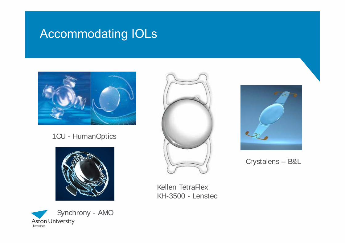
Accommodating IOLs
Crystalens – B&L
1CU - HumanOptics
Kellen TetraFlexKH-3500 - Lenstec
Synchrony - AMO

Dual Optic

AIOLs
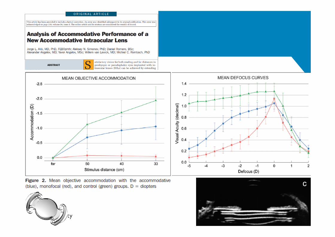
Publications decreasedFew studies, measure accommodationEarly designs - small amount of (?) ciliary muscle driven ‘accommodation’ (Leng et al., 2017), but only for a short period before it is presumed lens fibrosis and capsular shrinkage reduced the lens flexibility (Wolffsohn et al., 2006a; Wolffsohn et al., 2006b). Increased level of spectacle independence, but principally from pseudoaccommodative mechanisms (Pepose et al., 2017a)Newer designs (few clinically tested) include dual optics, shape changing optics and refractive index changing optics (Ben-Nun and Alio, 2005; DeBoer et al., 2016; McCafferty and Schwiegerling, 2015; Tomas-Juan and Murueta-Goyena Larranaga, 2015). In conjunction prevention &/or treatment of capsular contraction to prevent mechanisms inhibition explored (Pepose et al., 2017b).

Inlays
thin, small diameter, biocompatible materials with high fluid & nutrient permeability (Moarefi et al., 2017)femtosecond laser cut flap or pocket (deep) (Moarefi et al., 2017; Moshirfaret al., 2016a) / flocket (Konstantopoulos et al., 2017) epithelium remodels within zone ~2x inlay diameter (Lang et al., 2016), with ~19µm thinning regardless Rx (Steinert et al., 2017) Pulfrich effect (Plainis et al., 2013b), but no VF effect (Atchison et al., 2016). safe (Moshirfar et al., 2017), 2ndry surgical intervention in 12% of thin lens inlays within 3 years (over half explantations)meniscus shaped inlays cause only minimal DVA compromise in implanted eye & provide good NVA, stereopsis & CS (Igras et al., 2016a, b; Jalali et al., 2016; Lin et al., 2016; Linn et al., 2017). similar outcomes implanted pre/post traditional/femto cataract surgery (Ibarz et al., 2017; Stojanovic et al., 2016) & with simultaneous photorefractive keratectomy (PRK)(Moshirfar et al., 2016b). diffractive corneal inlays better performance? (Furlan et al., 2017).

Inlays
thin ‘lens’ which reshapes anterior corneal surface creating -SA (Whang et al., 2017; Whitman et al., 2016a; Whitman et al., 2016b) corneal multifocality (distance vision through a plano central zone surrounded by rings of varying additional power)pinhole design to extends depth-of-focus (Dexl et al., 2015)
Thickness Diameter Implantation Depth
Centration Material Mechanism of Action
Raindrop 32 µm 2 mm 120-200 µm Central over light constricted pupil
Hydrogel Increases central radius of curvature of overlying cornea
Flexivue microlens
15-20 µm 3 mm 280-300 µm Over 1st
Purkinje image
Hydroxyethyl methacrylate & methyl methacrylate + UV blocker
Distance vision through plano central zone surrounded by rings of add power 1.25 to 3.50D in 0.25D steps
KAMRA 5 µm 3.8 mm (1.6mm central aperture)
200-250 µm Over 1st
Purkinje image
Poly-vinylidene Fluoride
Increases depth of focus through pinhole

Optoelectronic adjustable lens technologies
Birefringent liquid crystals frenel lens layers (Srivastava et al., 2015; Wang et al., 2014)flat gradient index lenses (Naumov et al., 1999; Ye et al., 2004)diffractive lenses (Li et al., 2006; Valley et al., 2010)flat lenses using inhomogeneous electric fields (Lin et al., 2005)
demo LC embedded in PMMA contact lens (Milton et al., 2014)graphene electrodes (high electrical conductivity, transparency, flexibility & elasticity properties (Kaur et al., 2016)
Electro-wetting lenses - modulate wetting angle of fluid droplet(s) suspended within annular electrode to change power through electric field - size limited by droplet inertial effects

Other
Alvarez-Lohmann Lenses - complimentary mostly-cubic waveform on two lens elements
fluid lenses - rigid frame holding elastic membrane filled with a transparent refracting fluid (Stevens et al., 2017)

Presbyopia IOLS
Monovision
MIOL
EDOF
AIOL
Inlays
Optoelectronic adjustable lens technologies
Other

Competitors
SpectaclesContact lenses
MonovisionMultifocal designs
Surgical approachesScleral expansionLaser Refractive
Corneal monovisionCorneal collagen shrinkageMultifocal corneal laser profileLenticular ‘softening’
PharmaceuticalsCiliary muscle electrostimulation

Prof James S WolffsohnDeputy Executive Dean, Life and Health Sciences
Intraocular Lenses for Presbyopia