Embed Size (px)
Citation preview

IntracellularIntracellular compartmentscompartments
Protein Sorting and Protein Sorting and TransportTransport

Compartments of an Animal Cell
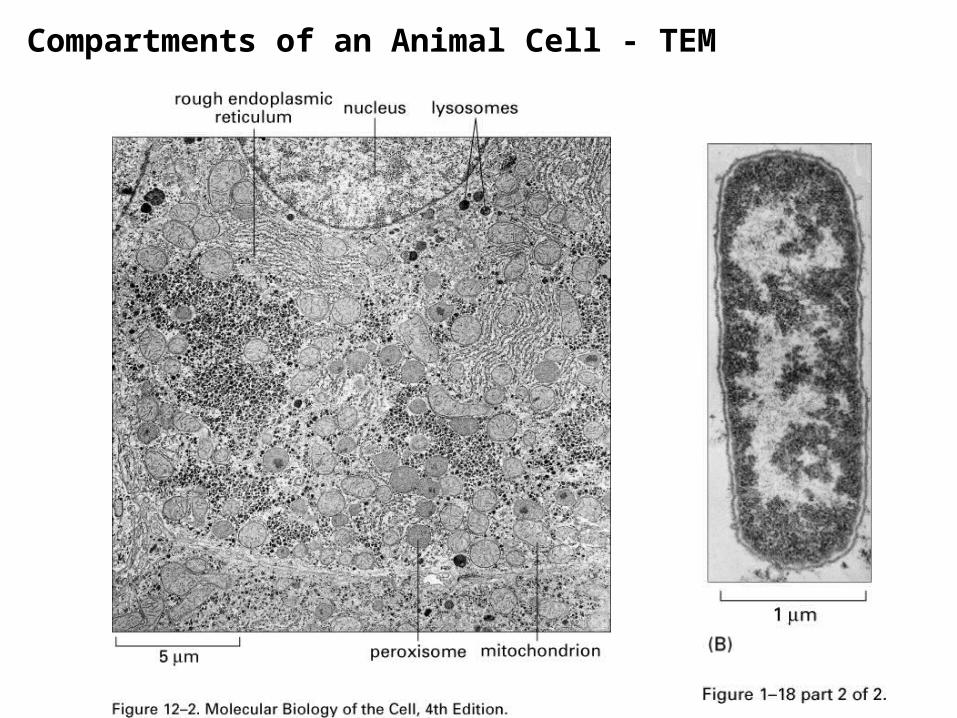
Compartments of an Animal Cell - TEM

Procaryotic CellProcaryotic Cell Eucaryotic CellEucaryotic Cell
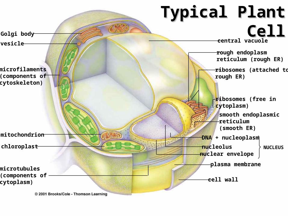
microtubules(components of cytoplasm)
Golgi body
vesicle
microfilaments(components of cytoskeleton)
mitochondrion
chloroplast
central vacuole
rough endoplasm reticulum (rough ER)
ribosomes (attached to rough ER)
ribosomes (free in cytoplasm)
smooth endoplasmic reticulum(smooth ER)
DNA + nucleoplasm
nucleolusnuclear envelope
NUCLEUS
plasma membrane
cell wall
Typical Plant Typical Plant CellCell

Animal Cell Plant Cell
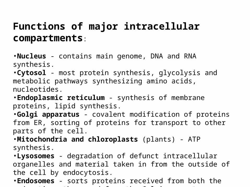
Functions of major intracellular compartments:
•Nucleus - contains main genome, DNA and RNA synthesis.•Cytosol - most protein synthesis, glycolysis and metabolic pathways synthesizing amino acids, nucleotides.•Endoplasmic reticulum - synthesis of membrane proteins, lipid synthesis.•Golgi apparatus - covalent modification of proteins from ER, sorting of proteins for transport to other parts of the cell.•Mitochondria and chloroplasts (plants) - ATP synthesis.•Lysosomes - degradation of defunct intracellular organelles and material taken in from the outside of the cell by endocytosis.•Endosomes - sorts proteins received from both the endocytic pathway and from the Golgi apparatus.•Peroxisomes - oxidize a variety of small molecules.

Three basic modes of Three basic modes of transporttransport
1. Gated transport
2. Transmembrane transport
3. Vesicular transport

Some of the green route illustrated.
Roadmap of protein trafficThe genesis and function of internal compartments depends on the appropriate targeting of proteins
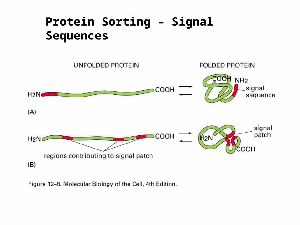
Protein Sorting – Signal Sequences

A simple experiment shows that many sorting signals consist of a continuous stretch of amino acid sequence called a “signal sequence”.
Fusing sorting signals to GFP is particularly good way to do this experiment.

GFP

Cytoplasmic Nuclear
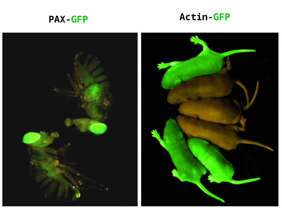
PAX-GFP Actin-GFP
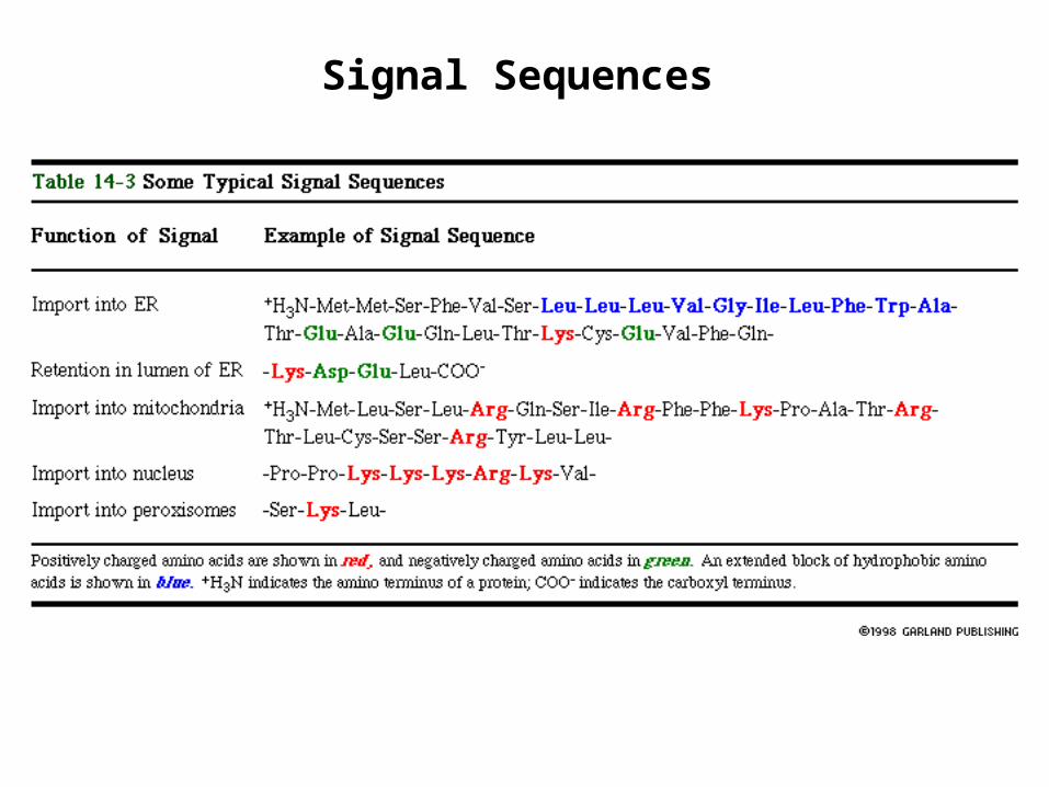
Signal Sequences
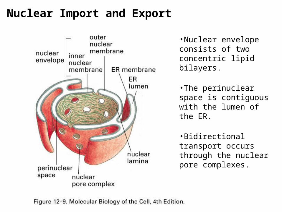
•Nuclear envelope consists of two concentric lipid bilayers.
•The perinuclear space is contiguous with the lumen of the ER.
•Bidirectional transport occurs through the nuclear pore complexes.
Nuclear Import and Export


The nuclear pore complex is an aqueous channel that allows diffusion of small molecules and proteins up to 60kD. Hence, transport of such molecules is passive.
Nuclear Pore
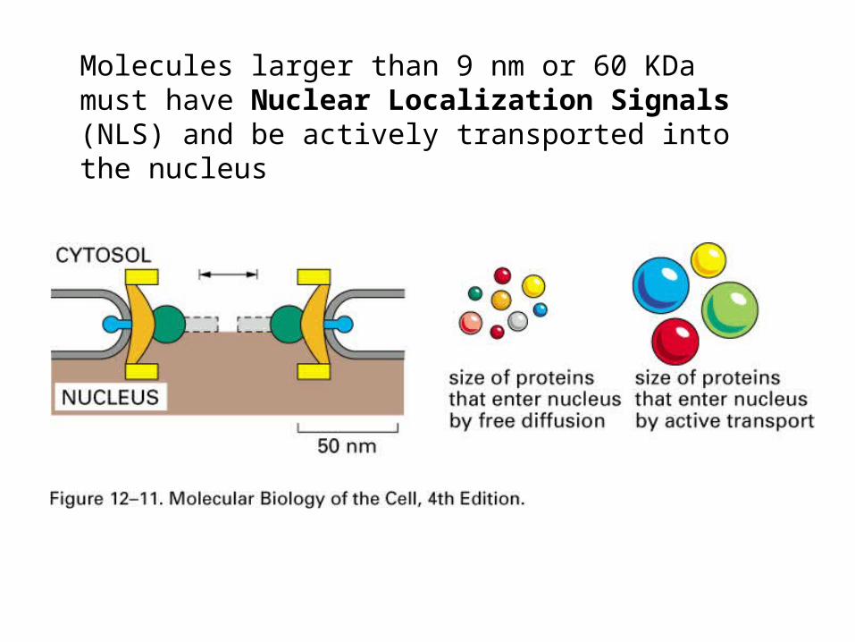
Molecules larger than 9 nm or 60 KDa must have Nuclear Localization Signals (NLS) and be actively transported into the nucleus
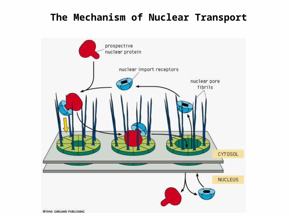
The Mechanism of Nuclear Transport

In most cases, nuclear localization of large proteins relies on a signal sequence called a Nuclear localization signal or NLS.
•NLS can be located anywhere in the primary sequence of the protein.
•Usually arginine and lysine-rich and quite short.
•There are some exceptions where nuclear localization relies on a signal patch.

The NLS directs the protein for transport through the nuclear pore complex, and proteins maintain their tertiary and quaternary structures during transport.
When gold beads are coated with the NLS, the beads can be seen passing through nuclear pore complexes. The maximum size bead that can be transported is 26 nm. Since the gold bead can’t compress, the opening of the pore must be able to expand.
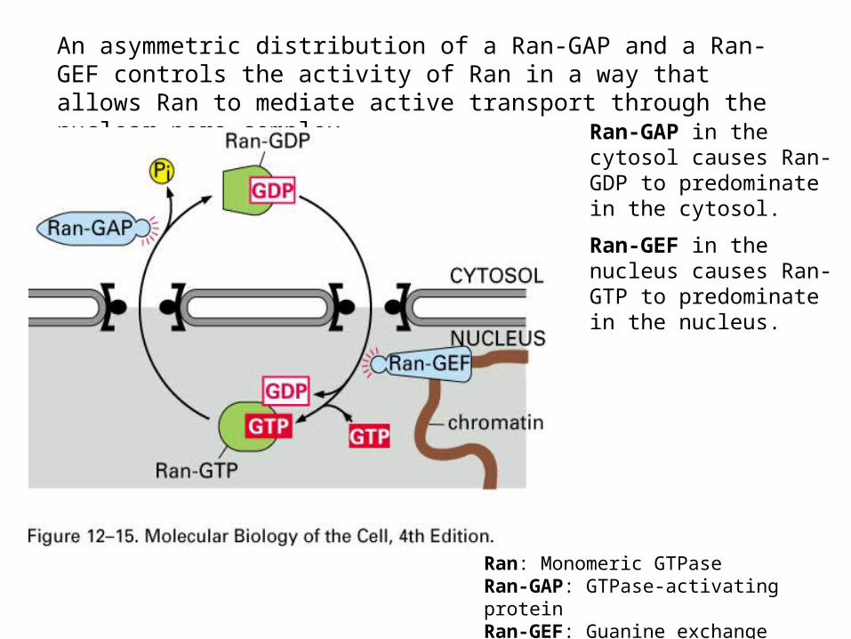
An asymmetric distribution of a Ran-GAP and a Ran-GEF controls the activity of Ran in a way that allows Ran to mediate active transport through the nuclear pore complex.
Ran-GAP in the cytosol causes Ran-GDP to predominate in the cytosol.
Ran-GEF in the nucleus causes Ran-GTP to predominate in the nucleus.
Ran: Monomeric GTPaseRan-GAP: GTPase-activating proteinRan-GEF: Guanine exchange factor

The NLS associates with soluble cytosolic proteins called nuclear import receptors. The nuclear import receptors also bind the nuclear pore complex so they serve to bring the protein containing the NLS to the nuclear pore complex.
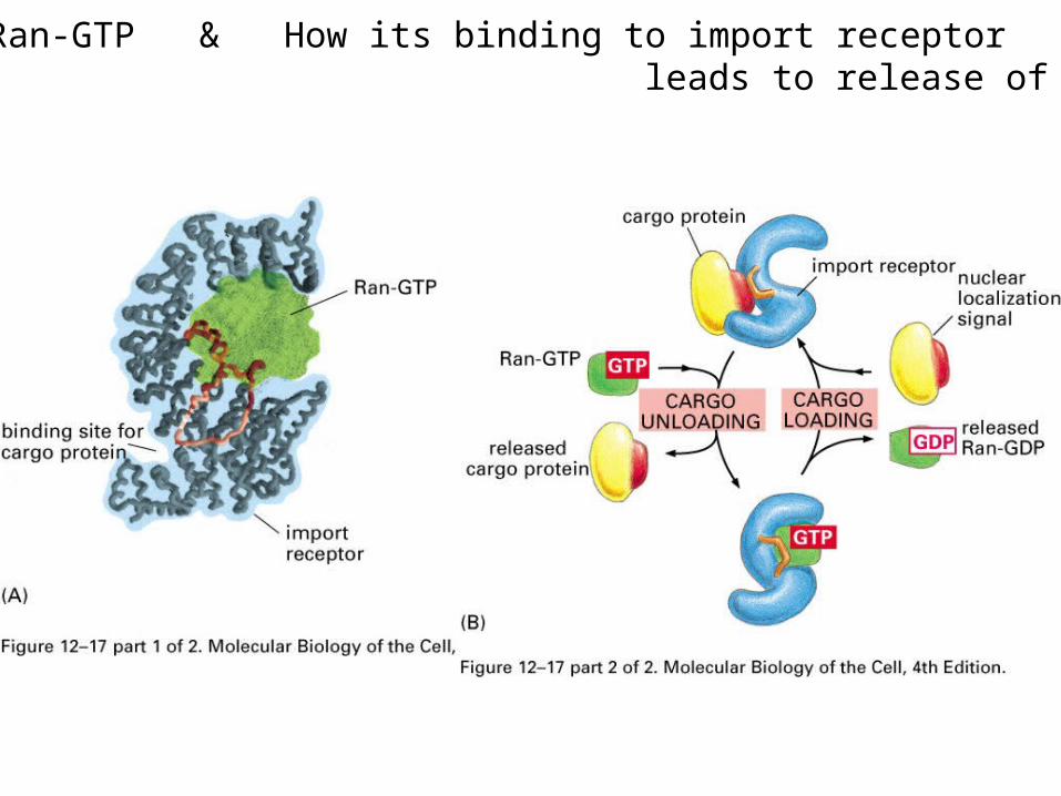
Structure of Ran-GTP & How its binding to import receptor leads to release of cargo protein
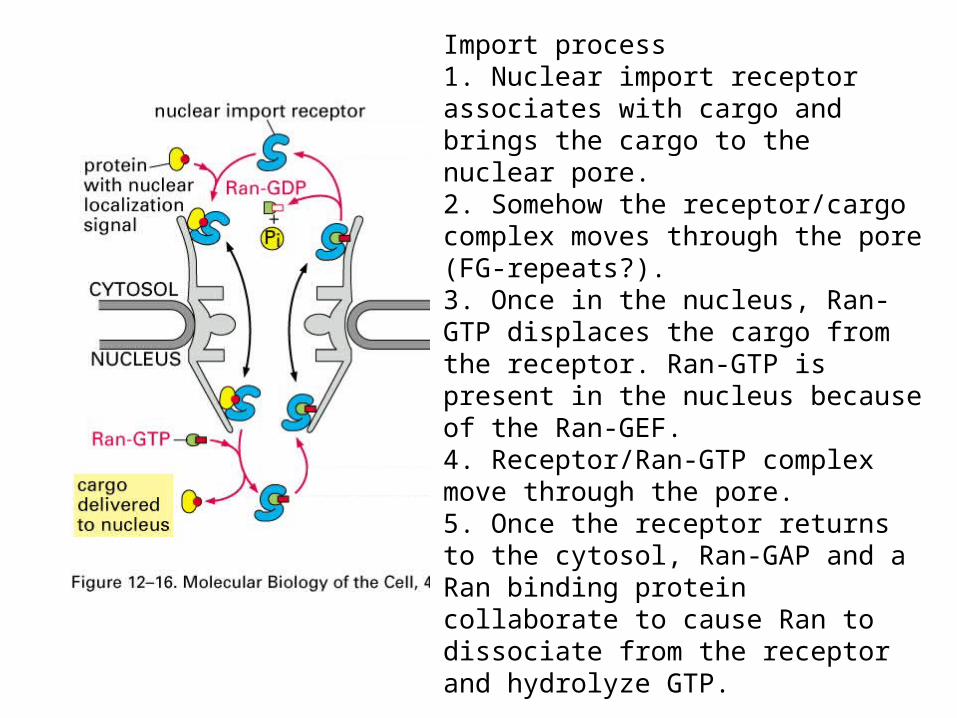
Import process1. Nuclear import receptor associates with cargo and brings the cargo to the nuclear pore.2. Somehow the receptor/cargo complex moves through the pore (FG-repeats?).3. Once in the nucleus, Ran-GTP displaces the cargo from the receptor. Ran-GTP is present in the nucleus because of the Ran-GEF.4. Receptor/Ran-GTP complex move through the pore. 5. Once the receptor returns to the cytosol, Ran-GAP and a Ran binding protein collaborate to cause Ran to dissociate from the receptor and hydrolyze GTP.

The nuclear import players:
•Nuclear import receptor binds the cargo.
•NLS is in the amino acid sequence of the cargo protein.
•Ran-GTP and Ran-GDP are different forms of Ran bound either to GTP or GDP. Ran-GTP causes the NLS to dissociate from the Nuclear import receptor.
•Ran-GAP is distinct from Ran but causes Ran to hydrolyze GTP. Hence, Ran-GAP promotes the conversion of Ran-GTP to Ran-GDP.
•Ran-GEF is distinct from Ran but causes Ran to release GDP and bind a different molecule of GTP. Hence, Ran-GEF promotes the conversion of Ran-GDP to Ran-GTP.

Export of RNA and proteins relies on nuclear export receptors associating with nuclear export signals (NES) found on proteins and RNA-bound proteins.
•Nuclear export receptors are structurally similar to nuclear import receptors.•Nuclear export receptor bind the nuclear export signals and bring the protein to the nuclear pore complex for subsequent transport.•Transport occurs through the same pores through which proteins are imported from the cytosol.•Ran regulates the interaction between the export receptor and the “NES”. The Ran-GTP promotes association of the receptor/cargo complex with the pore in the nucleus and hydrolysis of the GTP on the cytosolic side causes the resulting Ran GDP to dissociate the export receptor from its cargo.
Note - nuclear export receptors do not bind directly to RNA, they bind proteins bound to the RNA.

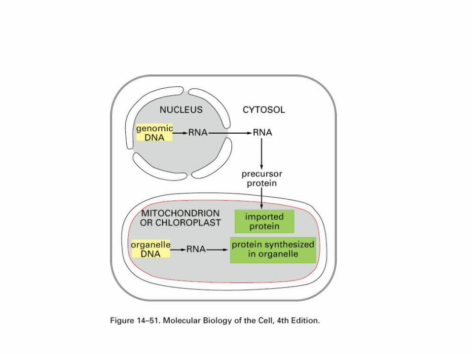
•Organelles specialized for ATP synthesis.•Most, but not all, proteins are encoded by the nuclear genome and synthesized in the cytosol.•Proteins must be transported to one of multiple membranes or compartments.
Transport of Proteins into Mitochondria and Chloroplasts
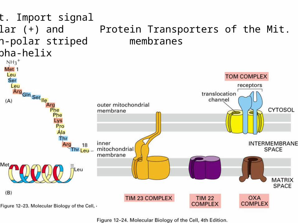
Mit. Import signal Polar (+) and Protein Transporters of the Mit. non-polar striped membranes alpha-helix

Import of the protein into the matrix is directed by an N-terminal signal sequence.•For polypeptides encoded by the nuclear genome, synthesis of the polypeptide is first completed in the cytosol. Transport occurs by a posttranslational mechanism.•Signal sequence at the N-terminus associates with the TOM complex located in the outer mitochondrial membrane. TOM is both a receptor for the signal sequence and a translocase. •The polypeptide is passed from TOM to TIM in the innermembrane. During the transport process, the polypeptide traverses both inner and outer membranes via the two translocators at a point known as a contact site. •The polypeptide is imported and the signal peptide is removed by a signal peptidase.
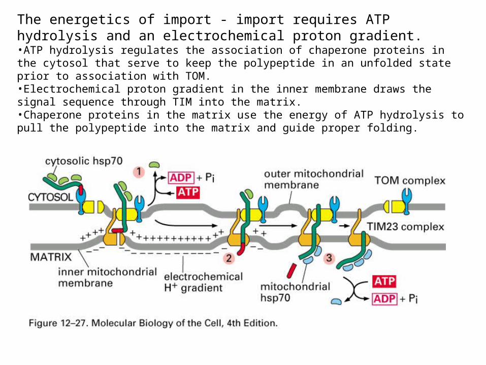
The energetics of import - import requires ATP hydrolysis and an electrochemical proton gradient.•ATP hydrolysis regulates the association of chaperone proteins in the cytosol that serve to keep the polypeptide in an unfolded state prior to association with TOM. •Electrochemical proton gradient in the inner membrane draws the signal sequence through TIM into the matrix. •Chaperone proteins in the matrix use the energy of ATP hydrolysis to pull the polypeptide into the matrix and guide proper folding.
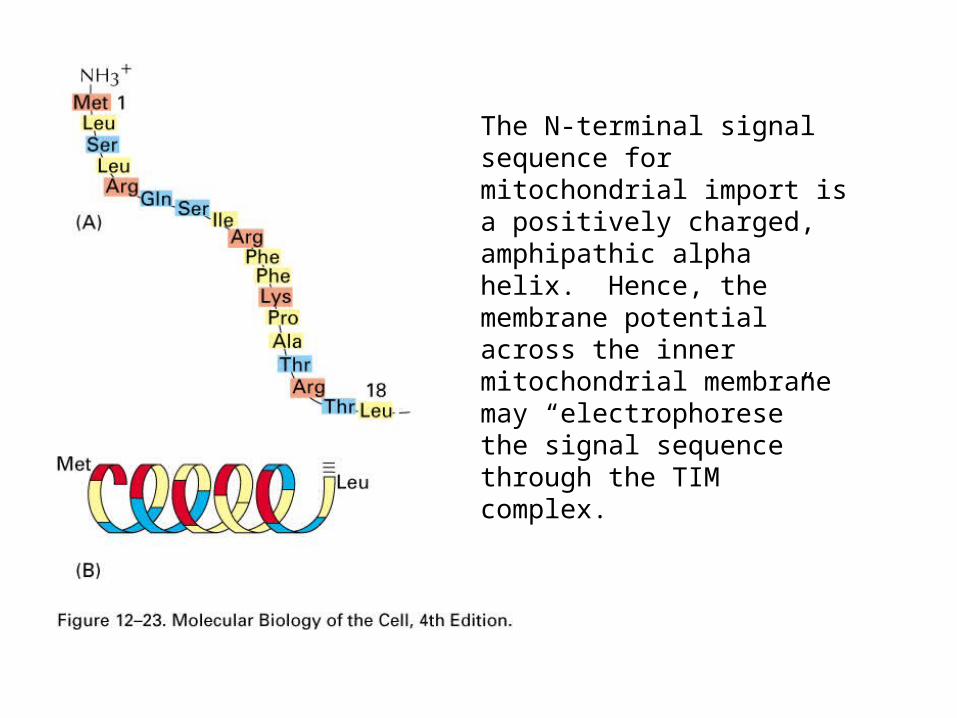
The N-terminal signal sequence for mitochondrial import is a positively charged, amphipathic alpha helix. Hence, the membrane potential across the inner mitochondrial membrane may “electrophorese” the signal sequence through the TIM complex.
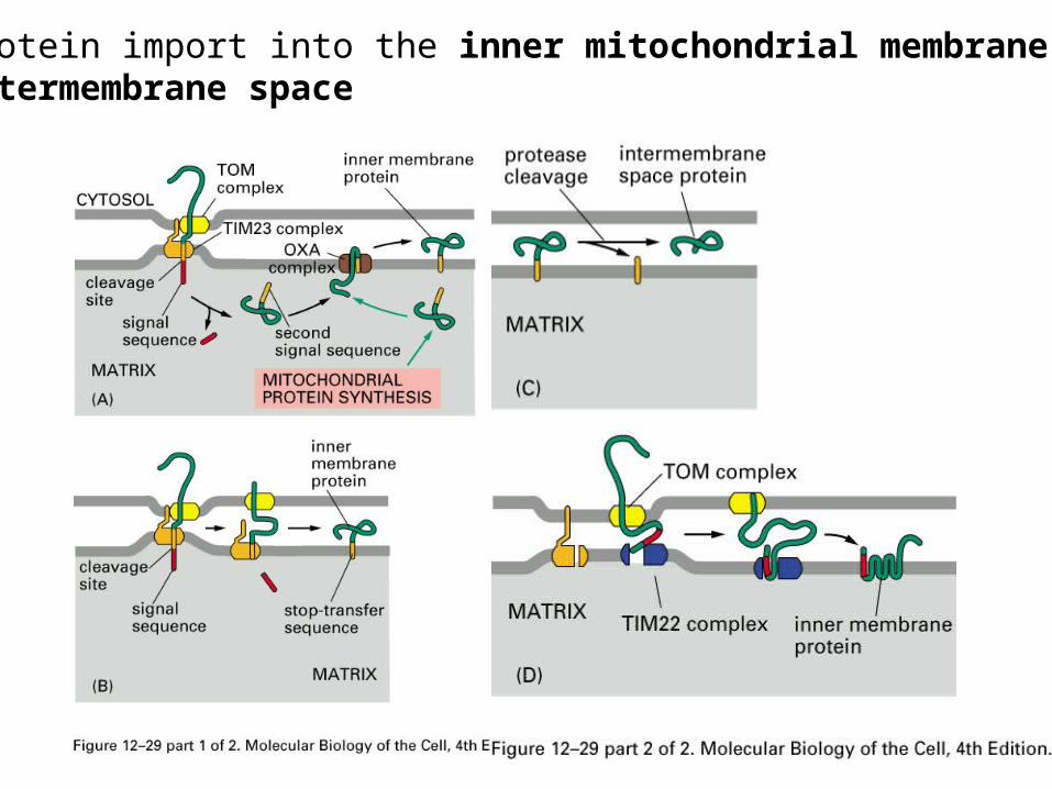
Protein import into the inner mitochondrial membrane orintermembrane space

The human mitochondrial genome

Transport into chloroplasts is similar to mitochondria except there is a 3rd membrane that can be targeted. Targeting the thylakoid membrane involves a second signal sequence.
In the case of chloroplasts, the electrochemical proton gradient is at the thylakoid membrane where this gradient participates in transport. Transport across the chloroplast inner membrane is powered by GTP and ATP hydrolysis.

Comparison of mitochondrial and nuclear import.
Nucleus MitochondriaSignal sequence Short , positively
charged, locatedanywhere
N-terminus,amphipathic alphahelix
Fate of thesignal sequence
Unchanged aftertransport
Removed by signalpeptidase
Energy GTP hydrolysis ATP hydrolysis andelectrochemicalproton gradient
Conformation ofthe transportedprotein
Folded Unf olded

Peroxisomes (which are not part of the endomembrane system) contain enzymes used to incorporate hydrogens with oxygen to make hydrogen peroxide which is then converted to water and oxygen. They function in a variety of situations including detoxifying poisons by transferring hydrogens from them to oxygen. The dense bodies within the peroxisomes are crystallized enzymes.Peroxisomes are formed from phospholipids and enzymes in the cytosol, not by pinching off from the endomembrane system.

Peroxisomes are organelles that perform a variety of oxidation reactions including ones that breakdown fatty acids and toxic molecules that enter the cell from the blood stream.
Genetic defects in peroxisomes often cause neurological problems because a particular lipid found in myelin is produced in the peroxisomes.
Import of proteins involves short signal sequences. The most unusual aspect of the transport process is that oligomeric proteins don’t have to unfold. Very little is known about the transport process.
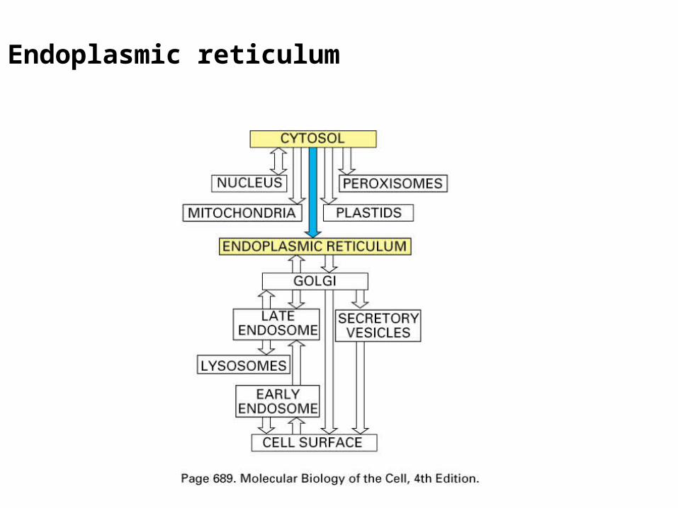
Endoplasmic reticulum
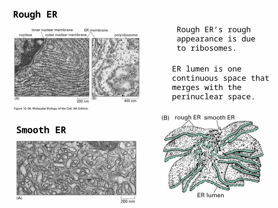
Rough ER’s rough appearance is due to ribosomes.
ER lumen is one continuous space that merges with the perinuclear space.
Rough ER
Smooth ER

Functions of the ER•Starting point for newly synthesized proteins destined for Golgi, Endosome, Lysosomes, Secretory vesicles, and the Plasma membrane (see below). •Establishes orientation of proteins in the membrane.•Site of phospholipid and cholesterol synthesis.•Initiation site for N-linked glycosylation of proteins.•Sequesters Ca++ - sarcoplasmic reticulum in muscle is a specialized ER.

Free and Bound Ribosomes
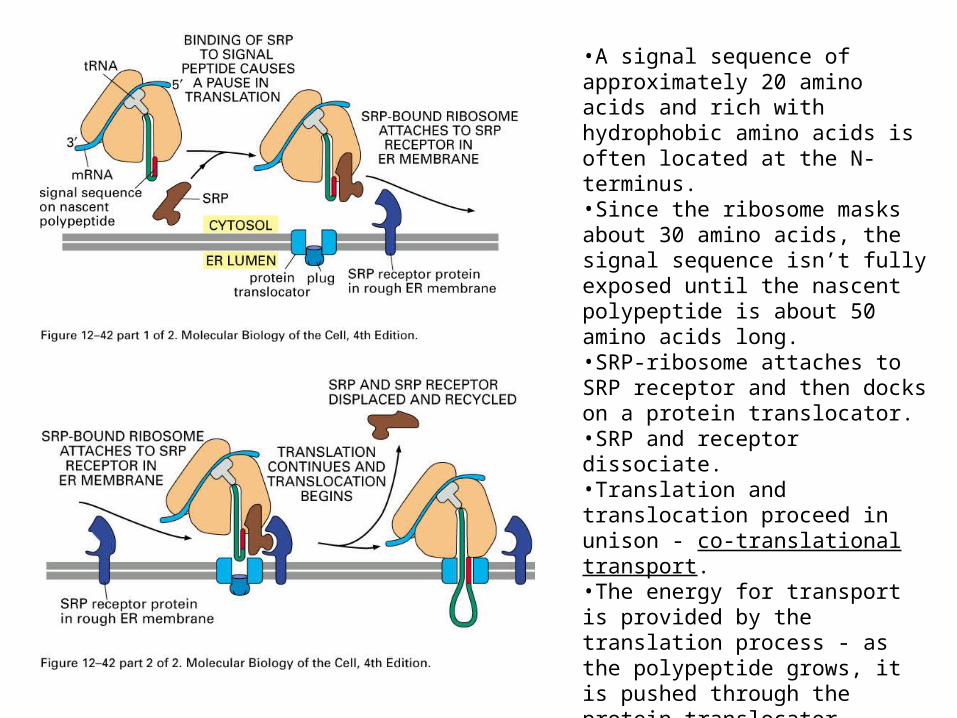
•A signal sequence of approximately 20 amino acids and rich with hydrophobic amino acids is often located at the N-terminus.•Since the ribosome masks about 30 amino acids, the signal sequence isn’t fully exposed until the nascent polypeptide is about 50 amino acids long.•SRP-ribosome attaches to SRP receptor and then docks on a protein translocator.•SRP and receptor dissociate.•Translation and translocation proceed in unison - co-translational transport.•The energy for transport is provided by the translation process - as the polypeptide grows, it is pushed through the protein translocator.
SRP: signal-recognition particleSRP receptor
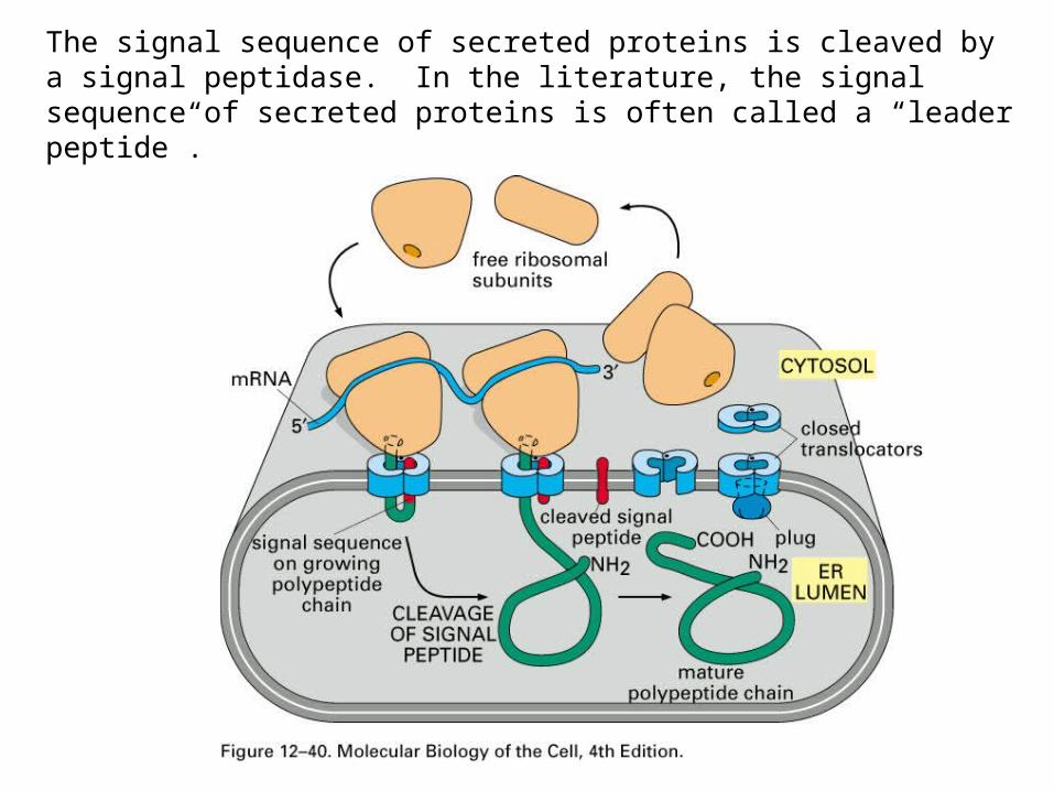
The signal sequence of secreted proteins is cleaved by a signal peptidase. In the literature, the signal sequence of secreted proteins is often called a “leader peptide”.

Translocation of protein across the ER membrane
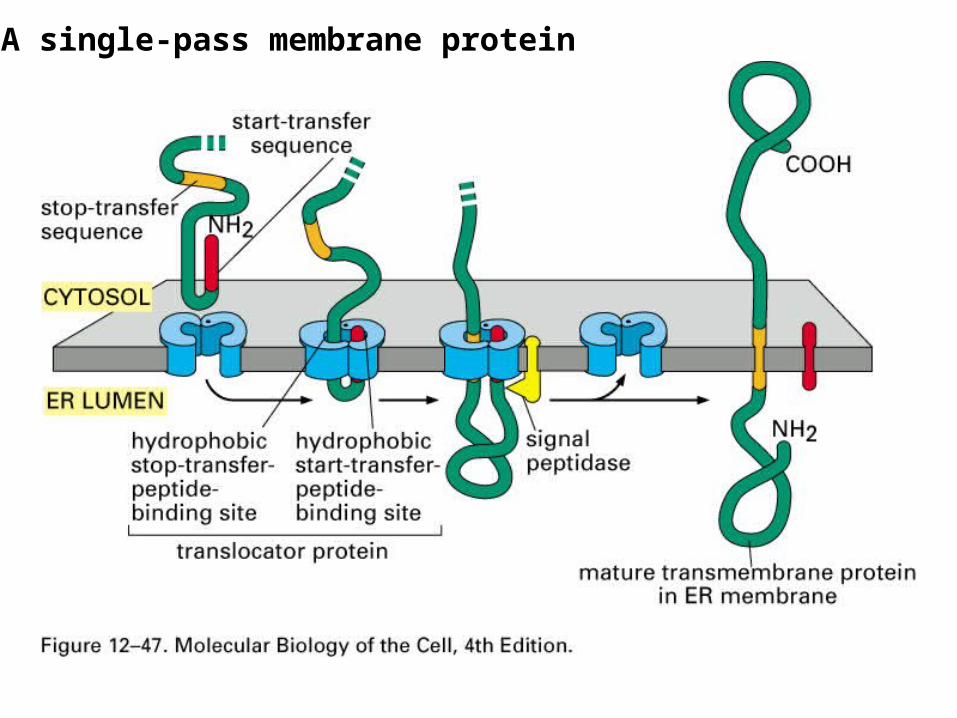
A single-pass membrane protein

Co-translational transport must be able to generate a diverse array of configurations.
For both single-pass and multipass transmembrane proteins, some types will have the N-terminus projecting into the cytosol and others will have the C-terminus projecting into the cytosol.
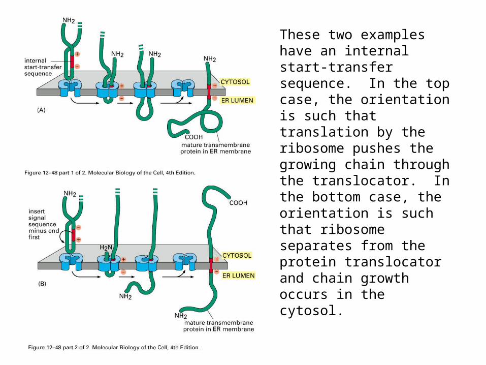
These two examples have an internal start-transfer sequence. In the top case, the orientation is such that translation by the ribosome pushes the growing chain through the translocator. In the bottom case, the orientation is such that ribosome separates from the protein translocator and chain growth occurs in the cytosol.
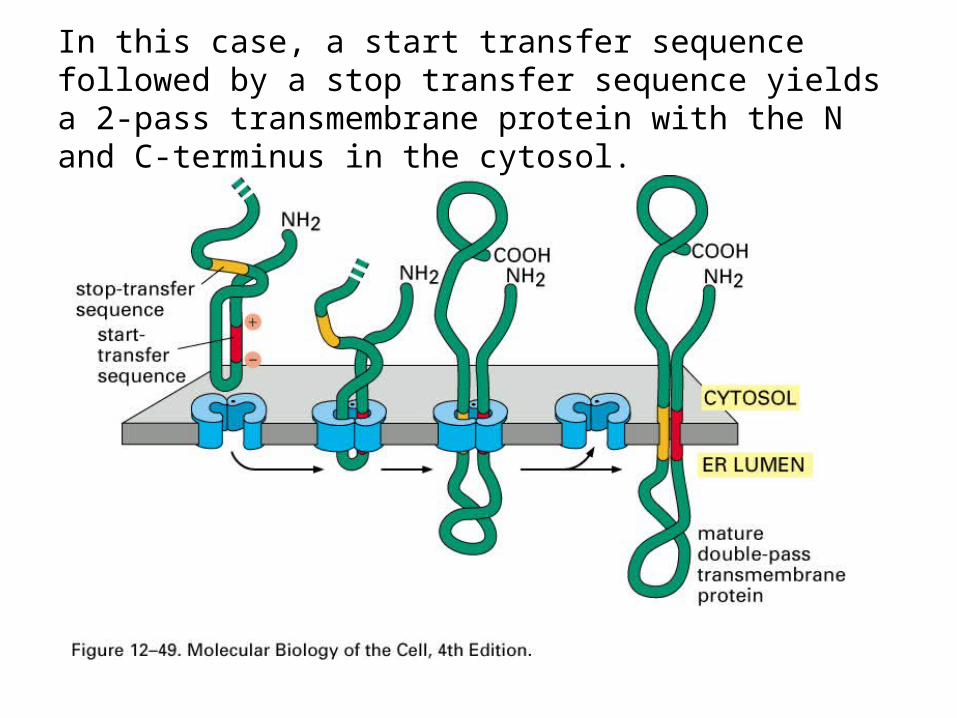
In this case, a start transfer sequence followed by a stop transfer sequence yields a 2-pass transmembrane protein with the N and C-terminus in the cytosol.

Things can get pretty complicated!

GPI anchored integral membrane proteins are generated in the ER.
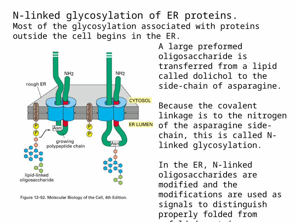
N-linked glycosylation of ER proteins.Most of the glycosylation associated with proteins outside the cell begins in the ER.
A large preformed oligosaccharide is transferred from a lipid called dolichol to the side-chain of asparagine.
Because the covalent linkage is to the nitrogen of the asparagine side-chain, this is called N-linked glycosylation.
In the ER, N-linked oligosaccharides are modified and the modifications are used as signals to distinguish properly folded from unfolded proteins.
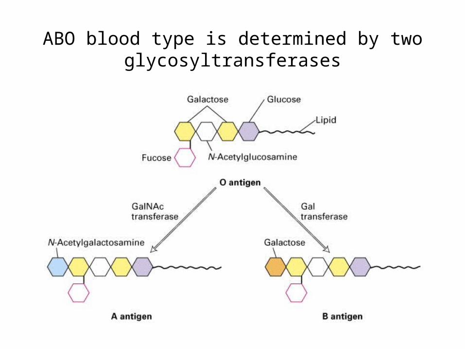
ABO blood type is determined by two glycosyltransferases
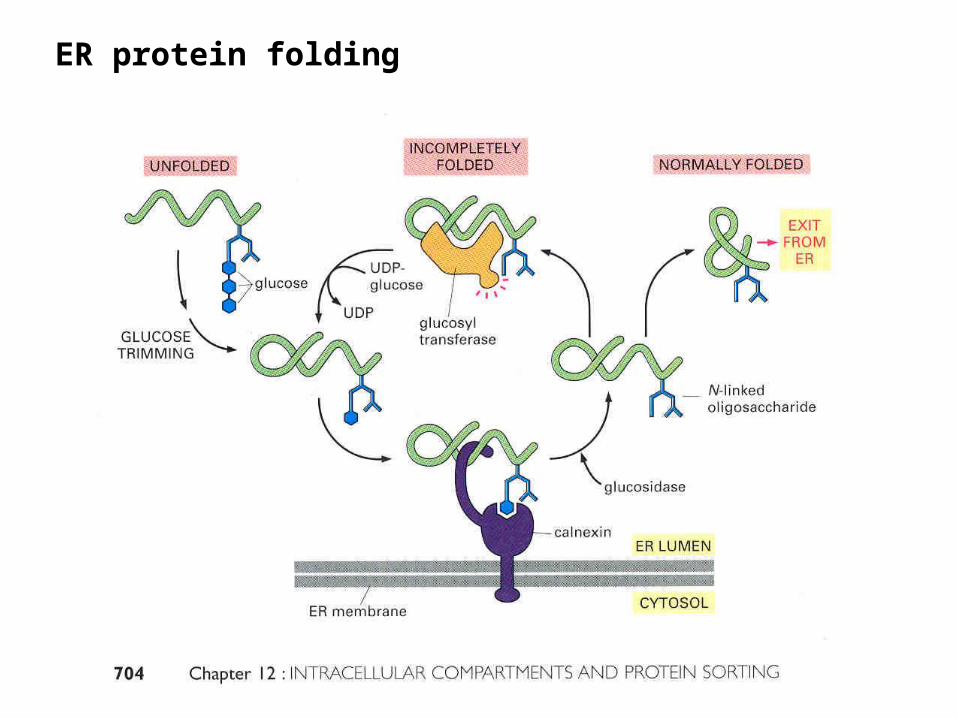
ER protein folding
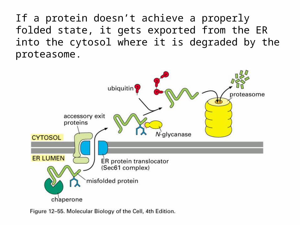
If a protein doesn’t achieve a properly folded state, it gets exported from the ER into the cytosol where it is degraded by the proteasome.

Phospholipids are synthesized in the cytoplasmic leaflet of the ER.

Phospholipid translocators flip-flop the phospholipids.

Transfer of lipids to other organelles.Most lipids for other organelles are synthesized at the ER.•Lateral diffusion will supply the nuclear membrane.•Vesicular transport will supply organelles in the secretory pathway and lysosomes (vesicular transport will be described soon)•Phospholipid exchange proteins deliver phospholipids to the mitochondria, chloroplasts and peroxisomes.
Intracellular Vesicular Transport
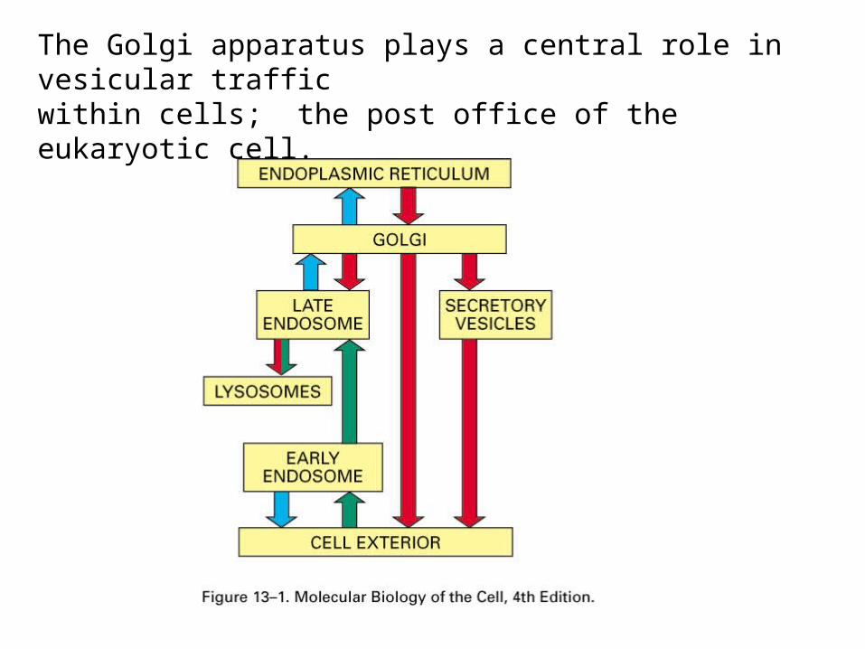
The Golgi apparatus plays a central role in vesicular traffic within cells; the post office of the eukaryotic cell.
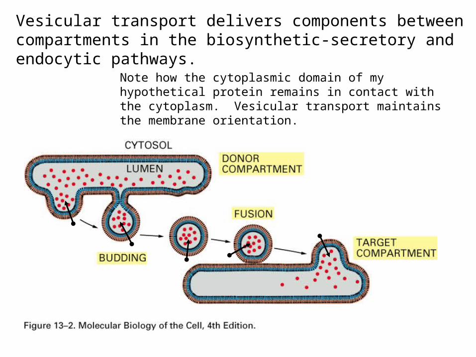
Vesicular transport delivers components between compartments in the biosynthetic-secretory and endocytic pathways.
Note how the cytoplasmic domain of my hypothetical protein remains in contact with the cytoplasm. Vesicular transport maintains the membrane orientation.

Once proteins that don’t normally reside in the ER are properly folded, they are transported to the golgi apparatus.

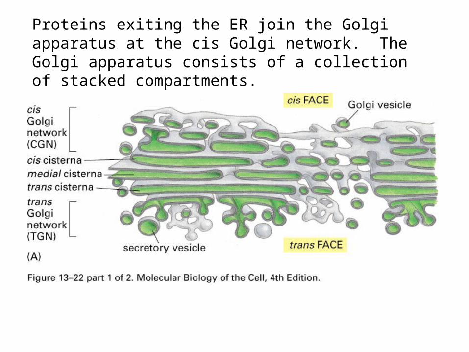
Proteins exiting the ER join the Golgi apparatus at the cis Golgi network. The Golgi apparatus consists of a collection of stacked compartments.
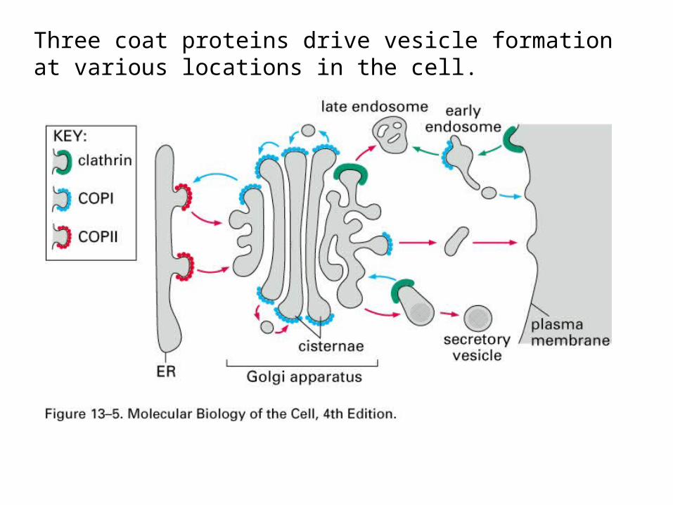
Three coat proteins drive vesicle formation at various locations in the cell.
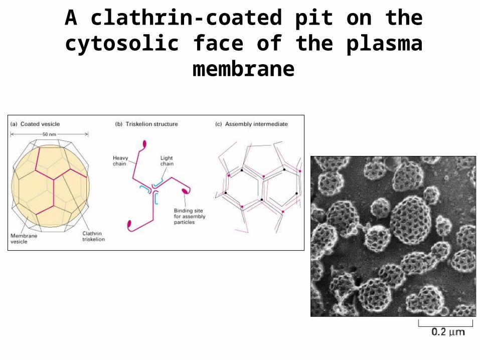
A clathrin-coated pit on the cytosolic face of the plasma
membrane

Clathrin associates via adaptins with receptors in the donor membrane. The receptors bind specific cargo. The clathrin assembles into a cage that encapsulates a region of membrane. Then dynamin causes the membrane to pinch off forming a vesicle.Energy requirements:•GTP hydrolysis by dynamin accompanies pinching off.•ATP hydrolysis by chaperone proteins (not shown).

COPII vesicle formation is mediated by a monomeric GTPase. A GEF in the donor membrane interacts with the GTPase, Sar1, causing GDP/GTP exchange. Sar1-GTP extends a fatty acid tail that inserts into the membrane. COPII assembles on the Sar1 to form a vesicle.
COPI vesicle formation involves a protein called ARF that is analogous to Sar1.
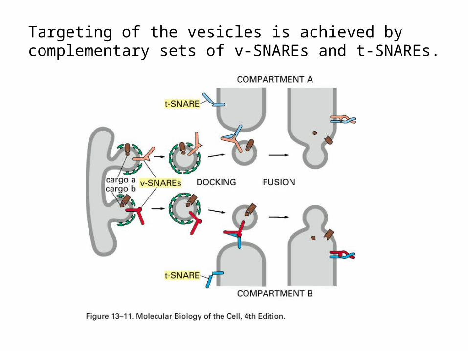
Targeting of the vesicles is achieved by complementary sets of v-SNAREs and t-SNAREs.
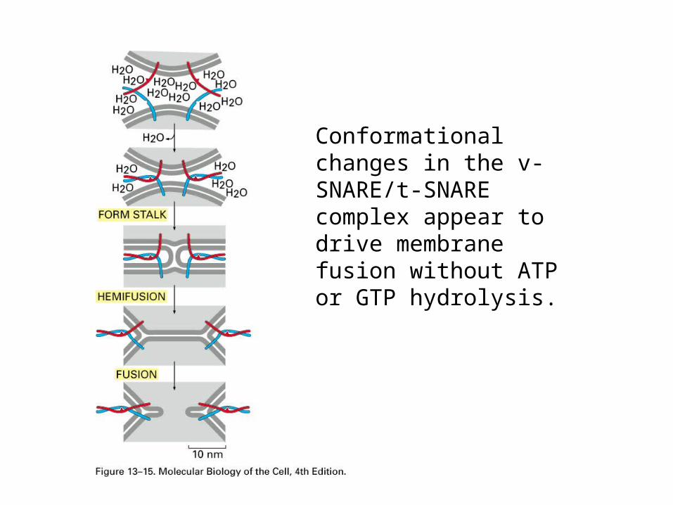
Conformational changes in the v-SNARE/t-SNARE complex appear to drive membrane fusion without ATP or GTP hydrolysis.

After membrane fusion, ATP hydrolysis is used to pry apart the v-SNARE/t-SNARE complex.
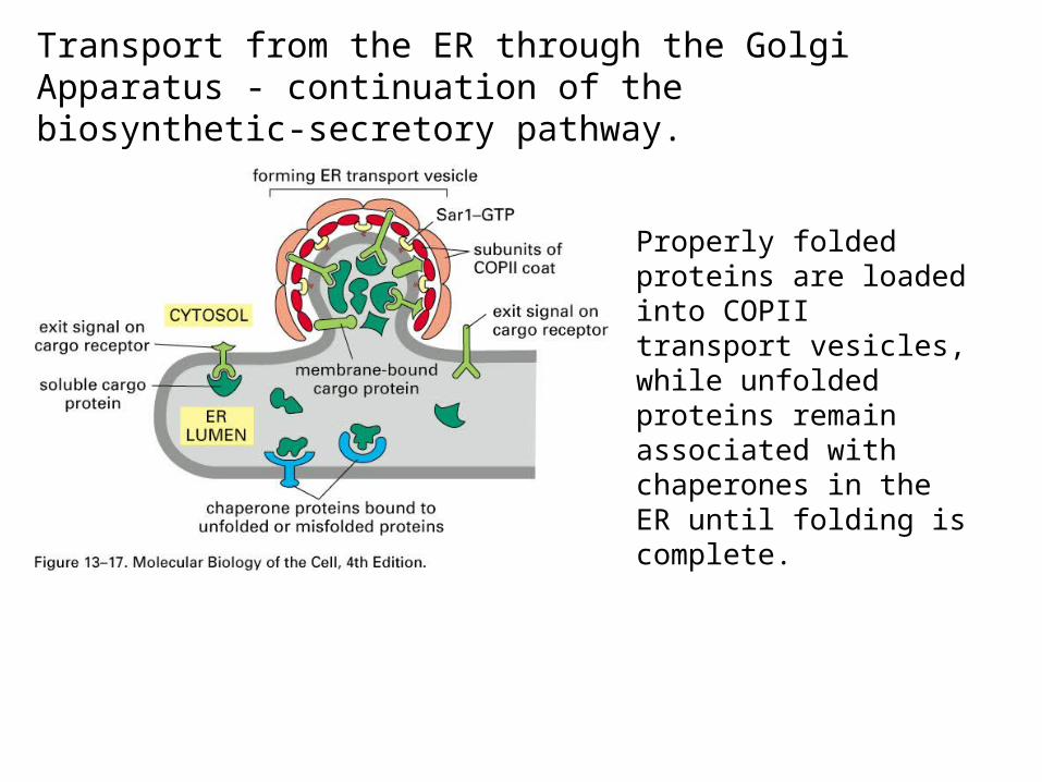
Transport from the ER through the Golgi Apparatus - continuation of the biosynthetic-secretory pathway.
Properly folded proteins are loaded into COPII transport vesicles, while unfolded proteins remain associated with chaperones in the ER until folding is complete.

If a protein doesn’t achieve a properly folded state, it gets exported from the ER into the cytosol where it is degraded by the proteosome.

When the protein is properly folded, COPII coated vesicles transport the proteins via the vesicular tubular cluster (vtc) to the cis-Golgi network.
•The COPII coating is removed (Sar1 hydolyzes GTP) and the vesicles fuse with each other to form the vtc. •The vtc is motored along microtubules that function like railroad tracks.•The vtc fuses with the cis-Golgi network.

Some proteins exiting the ER are returned to the ER by COPI coated vesicles. These proteins are identified by the presence of specific signal sequences that interact with the COPI vesicles or associate with specific receptors.
Examples of retrieved proteins:•v-SNAREs from the ER.•ER chaperones like BiP that are mistakenly transported.
Proteins exiting the ER join the Golgi apparatus at the cis Golgi network. The Golgi apparatus consists of a collection of stacked compartments.

TEMs ofGolgiApparatuses.In ananimalsecretory cell
and
an algal cellChlamydomonas
(trans)Note convexcis face and concave trans face.
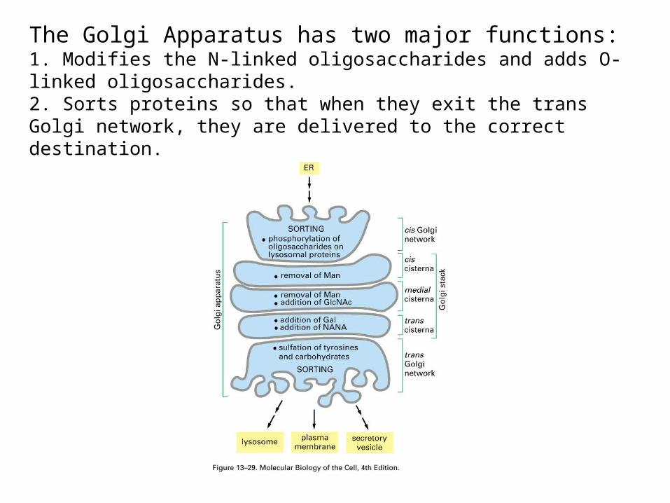
The Golgi Apparatus has two major functions:1. Modifies the N-linked oligosaccharides and adds O-linked oligosaccharides.2. Sorts proteins so that when they exit the trans Golgi network, they are delivered to the correct destination.

Modification of the N-linked oligosaccharides is done by enzymes in the lumen of various Golgi compartments.
1. Sorting in TGN2. Protection from protease digestion3. Cell to cell adhesion via selectins
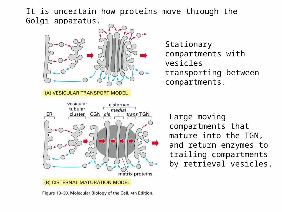
It is uncertain how proteins move through the Golgi apparatus.
Stationary compartments with vesicles transporting between compartments.
Large moving compartments that mature into the TGN, and return enzymes to trailing compartments by retrieval vesicles.

Cytochemical demonstration of different compartments of Golgi
Unstained nucleoside
diphosphatase (trans)
Osmium Acid Golgi Rxn phosphatase(cis) (TGN)

One ultimate destination of some proteins that arrive in the TGN is the lysosome. These proteins include acid hydrolases.
Lysosomes are like the stomach of the cell. They are organelles surrounded by a single membrane and filled with enzymes called acid hydrolases that digest (degrade) a variety of macromolecules. A vacuolar H+ ATPase pumps protons into the lysosome causing the pH to be ~5.

The macromolecules that are degraded in the lysosome arrive by endocytosis, phagocytosis, or autophagy.
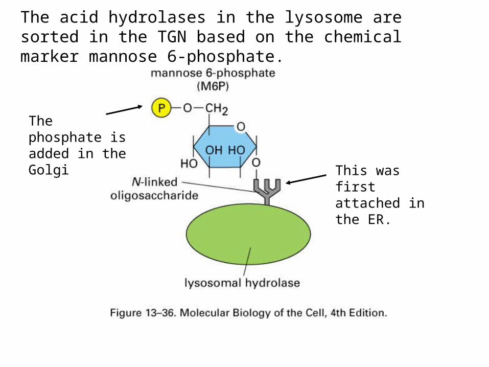
The acid hydrolases in the lysosome are sorted in the TGN based on the chemical marker mannose 6-phosphate.
This was first attached in the ER.
The phosphate is added in the Golgi

Adaptins bridge the M6P receptor to clathrin.
Hydrolases are transported to the late endosome which later matures into a lysosome.
Acidic pH causes hydrolase to dissociate from the receptor.

Endocytosis is a process by which cells take up substances by invaginating the plasma membrane. This process can capture both membrane bound and soluble components.
There are several subclasses of endocytosis:•Phagocytosis takes up large particles and cells.•Pinocytosis continuously takes up small amounts of fluid.•Receptor-mediated endocytosis selectively takes up membrane receptors and associated ligands.
Endocytosis takes up large amounts of the plasma membrane and is balanced by the return of membrane components to the plasma membrane by exocytosis.

Phagocytosis is performed primarily by white blood cells called Macrophages, Neutrophils and Dendritic cells. These cells receptors in the plasma membrane to recognize their targets. For example, macrophages have receptor that recognizes phosphatidylserine which becomes exposed on the surface of dead cells.

Pinocytosis is performed by clathrin coated pits and by caveolae. Clathrin coated pits are precursors to clathrin coated vesicles. Calveolae are deep invaginations in the plasma membrane that are thought to be formed from lipid rafts (regions high in cholesterol and glycolipids) and a transmembrane protein called caveolin.
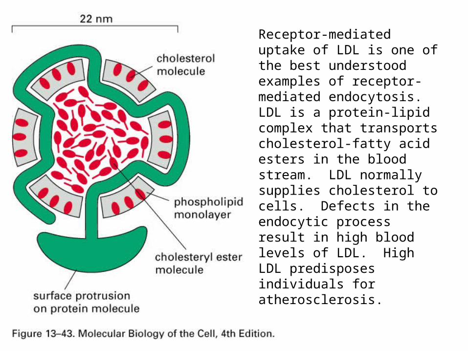
Receptor-mediated uptake of LDL is one of the best understood examples of receptor-mediated endocytosis. LDL is a protein-lipid complex that transports cholesterol-fatty acid esters in the blood stream. LDL normally supplies cholesterol to cells. Defects in the endocytic process result in high blood levels of LDL. High LDL predisposes individuals for atherosclerosis.

Normally, the receptors associate with adaptin.
Some individuals have defects in the cytoplasmic domain recognized by adaptin.
Other genetic defects that result in elevated blood levels of LDL:
•absence of LDL receptor•defective LDL-binding site in the LDL receptor.

Receptor mediated endocytosis involves transmembrane receptors that bind specific ligands. The specificity of receptors allows the cell to control the uptake of particular ligands and the distribution of these ligands in the cell. The early endosome serves as a sorting compartment. The illustration shows the 3 fates of the endocytosed receptors and their ligands.

Multivesicular body
Late endosome
pH 6 induces dissociation
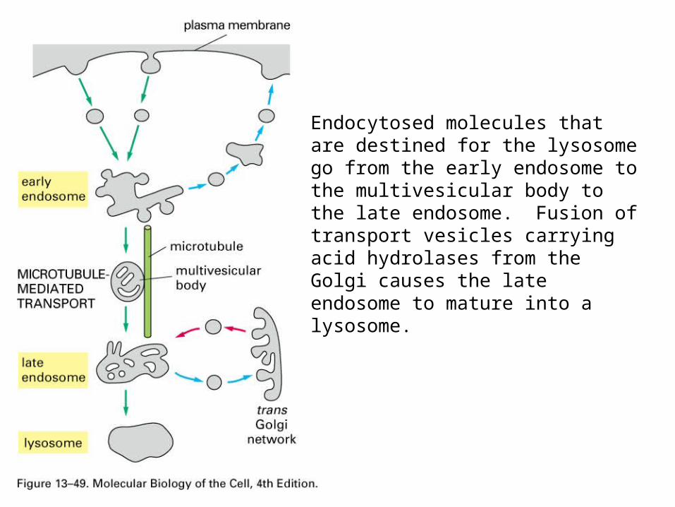
Endocytosed molecules that are destined for the lysosome go from the early endosome to the multivesicular body to the late endosome. Fusion of transport vesicles carrying acid hydrolases from the Golgi causes the late endosome to mature into a lysosome.

In some cases, both the receptor and the ligand are transported to the lysosome. This is the case for EGF and its receptor. EGF triggers a cell to proliferate but the signal is only required for a short time. To limit the response time both the receptor and the ligand are removed from the membrane.

Transcytosis provides a way to deliver proteins across an epithelium.
Transport of antibodies in milk across the gut epithelium of baby rats.
Acidic pH of the gut favor association of antibody with Fc receptor whereas the neutral pH of the extracellular fluid favors dissociation.
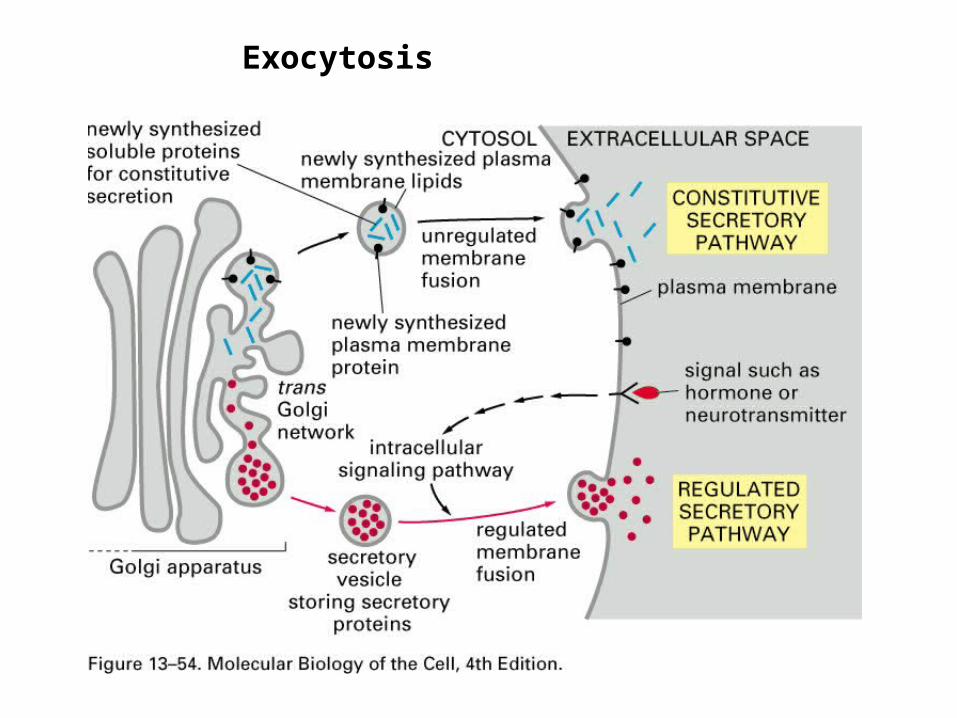
Exocytosis

Secretory vesicles concentrate and store products. Secreted products can be either small molecules or proteins. Proteins originate at the ER. In the Golgi, these proteins aggregate and are packaged into transport vesicles as aggregates.

Removal of the Pre-sequence (not shown), folding and disulfide bond formation occur in ER.
Processing to the final form occurs in the secretory vesicle.
This is an example of a protein that you would not want to treat with mercaptoethanol because reduction of disulfide bonds would inactivate the protein.
Insulin is a good example of a protein that is stored in secretory vesicles until a cell receives an signal to secrete the insulin.

Some proteins are processed in secretory vesicles into multiple small polypeptides. One explanation for this approach is that the small polypeptides are too short to be cotranslationally transported into the ER.
“pre-pro-proteins”

The End