Embed Size (px)
Citation preview
Toivonen et al. Virology Journal 2012, 9:296http://www.virologyj.com/content/9/1/296
RESEARCH Open Access
Intracardiac injection of a capsid-modified Ad5/35results in decreased heart toxicity whencompared to standard Ad5Raine Toivonen1,2,3, Juha Koskenvuo4,5, Mari Merentie6, Mirva Söderström7, Seppo Ylä-Herttuala6 andMikko Savontaus1,2,8*
Abstract
Background: Clinical gene therapy trials for cardiovascular diseases have demonstrated the crucial role of efficientgene delivery and transfection technologies in achieving clinically relevant results. We hypothesized that the use oftropism-modified adenoviruses would improve transduction efficacy and to this end we analyzed the transductionefficiency and toxicity of standard Ad5 and tropism-modified Ad5/35 in combination with ultrasound-guidedintramyocardial gene delivery.
Methods: Ultrasound-guided intracardiac injections were used to deliver 1 × 10¹⁰ pfu/ml Ad5-lacZ and Ad5/35-lacZvectors into mouse left ventricle wall. Since Ad5/35 uses human CD46 as its primary receptor, we used transgenichCD46Ge mice expressing human CD46 at levels comparable to man. Mice were sacrificed 6 or 14 dayspost-injection and immunohistochemistry and X-gal staining were used to detect transgene and viral receptorexpression. Virus-induced cardiac toxicity was evaluated by a pathologist.
Results: The intramyocardial injection was well tolerated and both Ad5-lacZ and Ad5/35-lacZ were able to giverobust transgene expression after a single injection. Interestingly, while Ad5-lacZ was able to generate greatertransgene expression than Ad5/35-lacZ, it also evoked more severe tissue damage with large areas of interstitialinflammatory cell infiltration and myocyte necrosis.
Conclusions: Ultrasound-guided intramyocardial injection is an effective and safe way to deliver vectors to theheart. The observed severe tissue damage of Ad5-lacZ greatly undermines the efficient transgene expression andsuggests that Ad5/35 capsid modification can result in safer adenoviral vectors for cardiovascular gene therapy,although at the cost of some vector transduction efficacy.
Keywords: Adenovirus, Intracardiac injection, CAR, CD46, Targeting
IntroductionCardiovascular disease (CVD) is the leading causes ofdeath in the western world, even after steady decline dueto improved risk control and effective invasive treatmentmethods. Annually over 190 000 people die from CVDin the UK alone and the health care costs are over 3.2billion pounds. As existing methods are clearly inadequate,
* Correspondence: [email protected] Centre for biotechnology, University of Turku, Tykistökatu 6B 5th floor,Turku FIN-20520, Finland2Department of Medical Biochemistry and Molecular Biology, University ofTurku, Turku, FinlandFull list of author information is available at the end of the article
© 2012 Toivonen et al.; licensee BioMed CentrCommons Attribution License (http://creativecreproduction in any medium, provided the or
novel treatment methods, such as gene therapy, are clearlyneeded.Adenoviruses (Ads) are the most utilized gene therapy
vectors. Over 50 different Ad serotypes, divided in sixdifferent subgroups (A-F) have been identified to date[1]. Viruses in different subgroups have varying tissuetropism and recognize different cellular receptors.Coxsackie-adenovirus receptor (CAR) has been identi-fied as the primary attachment receptor for many Ads,including the most utilized serotype 5 (Ad5) [2-4].Group B Ads have been shown to infect cells in CAR-independent manner and the cellular receptor has beenidentified as CD46 for most group B viruses [1,5,6].
al Ltd. This is an Open Access article distributed under the terms of the Creativeommons.org/licenses/by/2.0), which permits unrestricted use, distribution, andiginal work is properly cited.
Toivonen et al. Virology Journal 2012, 9:296 Page 2 of 9http://www.virologyj.com/content/9/1/296
CD46 is a complement regulatory molecule ubiquitous-ly expressed by all nucleated cells in human body [7].Recently it was also reported that a subgroup of Badenoviruses use desmoglein 2 receptor for primaryattachment [8], but serotype 35 was not included inthis group.Several preclinical gene therapy strategies have been
used for cardiovascular disease using different viral vec-tors and non-viral gene transfer and various modes ofadministration [9]. Most clinical cardiovascular genetherapy trials have used the standard Ad5 vector andone of the lessons from these trials has been the key roleof efficient gene transfection and delivery in achievingclinically meaningful therapeutic efficacy.Thus, it hasbeen clear that Ad vectors must be engineered to en-hance their ability to transfect the target tissue. Severalmethods have been developed to transductionally targetAd vectors including the use of bivalent targeting com-plexes and engineering genetic alterations to viral capsidproteins [10]. One targeting method which has recentlybeen successfully used in many applications is to use hy-brid adenoviruses, which are created by interchangingcapsid fibers between serotypes, usually with Ad5 receiv-ing the fiber from another serotype [11]. In this studywe used such a hybrid vector, Ad5/35, which has fibersfrom Group B Ad serotype 35 and thus uses CD46instead of CAR to gain entry into cells. Previous datademonstrate that Ad5/35 hybrid virus is able to effi-ciently transduce many cell types that are relatively re-sistant to Ad5 infection, including hematopoietic stemcells, tumor endothelial cells, and several cancercell types [12-18]. Previously we have reported an ana-lysis of various Ad receptors in both human dilatedcardiomyopathy (DCM) hearts and non-DCM heartsdemonstrating significant differences in expressionpatterns of CAR and CD46, with CAR receptor dem-onstrating higher expression in both DCM and non-DCM hearts [19].In addition to vector development, the method of
gene delivery plays a crucial role in achieving efficientgene transfer to the heart. Various modes of adminis-tration have previously been used to deliver genes tothe heart, including intravascular, intracoronary, intra-myocardial and intramuscular routes. Ultrasound-guided intramyocardial injection of gene delivery vec-tors is an attractive and clinically applicable conceptto directly infect cardiomyocytes. In this study weemployed this method to test the hypothesis thatthe use of tropism-modified adenoviruses would im-prove transduction efficacy for cardiovascular applica-tions and to this end evaluated the transductionalcapability and toxicity of a capsid-modified Ad5/35adenoviral vector in comparison to standard Ad5 vec-tor in the heart.
Materials and methodsHuman CD46 transgenic miceThe transgenic mouse strain hCD46Ge was a generousgift from Dr. Ann-Beth Jonsson, Uppsala University,Sweden. These mice were constructed by using yeastartificial chromosome technique as described earlier[20]. In our studies the homozygous strain was used.The CD46 receptor expression in hCD46Ge mice hasbeen analyzed earlier and it has been shown to be ubi-quitously expressed, with the heart expressing all fourCD46 transcript variants [21]. All animal work was doneby trained scientists with permission from the FinnishLaboratory Animal Board (license number: ESAVI-2010-05655/Ym-23).
Human heart samplesHuman heart samples were used for immunohistochem-istry of adenoviral receptor expression. Samples were akind gift from professor Petri Kovanen, Wihuri Institute,Helsinki, Finland. Heart samples were obtained from leftventricles of organ donors with no history of heart dis-ease and whose donated heart could not be used fortransplantation. Immediately after collection the tissuewas frozen in liquid nitrogen and stored at -70°C. Theuse of human heart samples as control specimens wasapproved by the Institutional ethics committee of theHelsinki University Central Hospital, and the investiga-tion conforms to the principles outlined in the Declar-ation of Helsinki.
Adenoviral vectorsIn this study we used two replication deficient adeno-viral vectors, Ad5-lacZ and Ad5/35-lacZ. Both vectorsharbor Escherichia coli β-galactosidase gene under thecontrol of rous sarcoma virus (RSV) promoter. Theviruses were prepared by cotransfecting 293 cells withshuttle plasmid pAd.RSV.LacZ and backbone plasmidspBHG10 (for Ad5.LacZ), or pAdΔΨF35 (for Ad5/35.LacZ) as previously described [22]. pAdΔΨF35 is basedon pBHG10 (MicrobixBiosystems Inc., Toronto, Canada)and contains the chimeric Ad5/35 fiber gene instead ofAd5 fiber gene [23]. Viruses were isolated from a singleplaque, expanded in 293 cells and purified by doublecesium gradient ultracentrifugation. Viral particles (vp)were measured using standard absorbance method andresulted in vp concentrations of 1.9 × 10¹² for Ad5-lacZand 3.4 × 10¹² for Ad5/35-lacZ. The plaque-formingunits (pfu) were determined by standard agarose-overlayplaque assay on 293 cells resulting in pfu concentrations9.3 × 1010 pfu/ml for Ad5-lacZ and 2.5 × 10¹⁰ pfu/ml forAd5/35-lacZ. The vp/pfu ratios were 20 for Ad5-lacZand 136 for Ad5/35-lacZ. For the in vivo experimentsboth viruses were diluted to 1 × 10¹⁰ pfu/ml, indicating

Toivonen et al. Virology Journal 2012, 9:296 Page 3 of 9http://www.virologyj.com/content/9/1/296
that mice injected with Ad5/35-lacZ received 6.65 timesmore viral particles than Ad5-lacZ mice.
Intracardiac injectionsAll animals received ultrasound-guided injection ofAd5-lacZ (n = 8), Ad5/35-lacZ (n = 9), or PBS (n = 4)into myocardium as previously described [24]. Briefly,the procedure was accomplished under 2.5-3.5% isoflur-ane anesthesia with the use of a Vevo770 (VisualSonics,Toronto, Canada) equipped with a 30-MHz transducer.After obtaining an optimal parasternal long-axis view,the intramyocardial injection was performed via 30-gauge needle by using micromanipulator. A volume of10 μl (1 × 10¹⁰ pfu/ml) was injected in each heart deli-vering 1 × 10⁸ infectious particles of either Ad5-lacZ orAd5/35-lacZ vectors. During the procedure, micewere immobilized to a warm plate, anesthetized withisoflurane and their heart rate, respiration and bodytemperature were monitored. No mice died from theprocedure and all were sacrificed 6 or 14 days post injec-tion. The intramyocardial injection technique was alsovalidated using methylene blue dye injection to visualizeits distribution at myocardial sections after successfulinjection (Additional file 1: Figure S1).
Serum samplesBlood was collected from the submandibular vein attime points 6, 48 and 72 h post injection, into CapijectT-MG serum collection tubes (Terumo, USA). Bloodwas allowed to clot at least 20 min, before centrifuga-tion. Serum was collected by pipetting, snap frozen in li-quid nitrogen, and stored at -20°C before use. CytokinesIL-6 and TNF-α were analyzed with custom Milliplexassay kit (Millipore, USA) according to manufacturer’sinstructions.
Histology, Immunohistochemistry and Image AnalysisEach group of treated animals was divided into two end-point groups 6 days and 14 days post injection. Micewere sacrificed using CO2. Heart, lungs, and liver werecollected. Both frozen (for X-gal staining of reportergene analysis) and paraffin embedded (for histologicaland toxicity analyses) sections were prepared from theinjected mouse hearts. In order to limit the number ofanimals needed in the experiment, frozen sections forreporter gene analysis were performed only from ani-mals sacrificed at 6 days, whereas paraffin embeddedsections for histological and toxicity analyses wereperformed from animals sacrificed at 6 and 14 dayspost injection. Sections were stained with standardHematoxylin / Eosin staining.Five micrometer sections were stained with Vectastain
HRP-kit (Vector laboratories, USA). The sections werefixed onto Superfrost plus slides (O. Kindler GmbH,
Germany) (frozen sections with cold acetone). The en-dogenous peroxidase in frozen sections was inactivatedwith 0.3% H2O2. Anti-CD46 primary antibody(HPA016903, Sigma, USA) was used in 2 μg/ml dilution.To evaluate signal from nonspecific antibody bindingnormal Rabbit IgG sc-2027 (Santa Cruz, USA) was usedin 4 μg/ml dilution. Vectastain ABC-reagent was appliedon the sections (Vector Laboratories, USA), and recep-tors were stained with diaminobenzidine (DAB, Sigma-Aldrich, USA) and counterstained with Mayer’shematoxylin (Sigma-Aldrich, USA). Adenovirus inducedlacZ expression was visualized with X-gal staining infrozen sections fixed with cold acetone and stainedwith 0.2% X-gal (5-bromo-4-chloro-3-indolyl-β-D-galactopyranoside) solution according to supplier’sinstructions (Promega, Madison, WI) or by immunostain-ing with anti-β-galactosidase antibody in paraffin embed-ded sections.The stained sections were photographed using a
microscope and evaluated for tissue damage by patholo-gist, who was blinded to the experimental setting. Weused a grading system which gave 0-2 points for myo-cyte damage, 0-2 points for interstitial inflammation and0-1 points for endocardial involvement resulting in amaximum possible score of 5. The inflammatory re-sponse was quantified by calculating inflammatory areausing ImageJ software (National Institutes of Health,Maryland, USA). To evaluate the spread of adenovirus-mediated transgene expression, the X-gal stained specificareas were measured by counting stained cells usingImageJ software.
Tissue DNA extraction and PCR10 mg of tissue was sectioned from frozen lung and liverof Ad5-lacZ and Ad5/35-lacZ injected mice. DNA wasextracted with standard techniques: Tissue was first sub-jected to proteinase K (400μg/ml) treatment, +50°C o/n.Supernatant was collected after centrifugation 12 000 rpm,5 min. DNA was extracted from the supernatant twice byphenol:chloroform (1:1) extraction and purified with etha-nol precipitation.Purified DNA was used as a template in standard
qualitative PCR with Ad specific primers (forward:CCATTTTCGCGGGAAAACTGA and reverse: AAGCGCCATTCGCCATTC) amplifying RSV promoter areaand beginning of β-galactosidase gene carried by bothAd5-lacZ and Ad5/35-lacZ vectors. DNA purified fromAd5-lacZ vector stock was used as positive control.
Statistical analysisThe results are expressed as mean ± SD or median[interquartile range]. The Shapiro-Wilk procedure wasapplied to determine whether data are normally distribu-ted. Independent sample t-test or Mann-Whitney U- test

Toivonen et al. Virology Journal 2012, 9:296 Page 4 of 9http://www.virologyj.com/content/9/1/296
were used for comparing Ad5-lacZ and Ad5/35-lacZ.Changes in cytokine profiles were evaluated using one-way ANOVA. Statistical analysis was performed usingSPSS 16 (SSPP Inc, Chicago, IL, USA). P-value less than0.05 was considered significant.
ResultsAnimal modelAs CD46 in mice is different from man and Ad35 useshuman CD46 to gain entry into cells, we used transgenichCD46ge mice, where human CD46 is expressed from ayeast artificial chromosome and has similar CD46 ex-pression pattern as humans. In order to establish thevalidity of this model we confirmed the receptor expres-sion by immunostaining with anti-huCD46 antibody.Human CD46 expression was found throughout themouse cardiac tissue at comparable levels to human car-diac tissue, indicating that CD46 expression is compar-able in hCD46ge mice and humans (Figure 1).
Adenoviral transgene expression after intramyocardialinjectionWe sought to compare transduction efficiencies betweenequal amounts of Ad5-lacZ and Ad5/35-lacZ afterclosed-chest echocardiography guided injection into thefree wall of the left ventricle. Echo images show clear li-quid cloud during the injection, this cloud is also
Figure 1 CD46 expression in the heart. A-B show heart tissue of hCD4show positive anti-human CD46 receptor immunostaining on cell surfaces.signal. The scale bar represents 100 μm.
retained in the heart muscle after successful injection(Figure 2), whereas injection into the ventricle cavity isknown to result in rapid clearance of “bubbles” into cir-culation. We first validated this method by injectingmethylene blue to visualize the injected area. The dyewas repeatedly retained in the myocardium after suc-cessful injection (Additional file 1: Figure S1).No mice died from the procedure and no abnormal
behavior of animals was observed during the experi-ment. Expression analyses were performed at 6 days postinjection and they revealed that both Ad5-lacZ and Ad5/35-lacZ viruses were able to give robust transgene ex-pression after a single injection of 1 × 10⁸ pfu of virus(Figure 3). The area of transduced cells after Ad5-lacZinjection comprised approximately 13% of the total leftventricle area (Figure 3A).When comparing the trans-duced areas after Ad5-lacZ and Ad5/35-lacZ injectionwe observed that Ad5-lacZ was able to transduce 2.99times more cells as compared to Ad5/35-lacZ(p = 0.016)(Figure 4A). The infected area with Ad5-lacZ was alsolarger than with Ad5/35-lacZ.
Toxicity after adenoviral injectionIn order to examine toxicity related to the virus injec-tion, the heart and other organs were examined forvirus-related toxicity at 6 and 14 days post injection. Inthe heart there was a clear difference between toxicities
6ge transgenic mice and C-D human heart tissue. Panels A and CPanels B and D are isotype controls showing the level of background

Figure 2 Successful ultrasound guided injection of left ventricle wall. A) Needle (white arrows) was inserted in between the ribs into the leftventricle wall (outlined in blue) of anesthetized mouse. The needle tip (green arrow) was positioned intramyocardially without puncturing the leftventricle lumen. B) A liquid cloud (red circle) becomes visible and is retained in the ventricle wall after successful injection.
Toivonen et al. Virology Journal 2012, 9:296 Page 5 of 9http://www.virologyj.com/content/9/1/296
elicited by the two viruses. At day 6 post injection bothvectors resulted in mild inflammatory reaction. At day14 Ad5-lacZ was found to be significantly more toxicthan Ad5/35-lacZ. Ad5-lacZ vectors induced tissue dam-age of over 11 times larger area than Ad5/35-lacZ vec-tors as measured by infiltration of immune cells(Figure 4B). The difference in the size of the affectedareas was statistically significant (p = 0.04). Ad5-lacZshowed diffuse and severe inflammatory infiltrate,mainly represented by lymphocytes. In addition myocytedamage and necrosis were clearly seen as well as inter-stitial fibrosis (Figures 3B and 3C). In comparison Ad5/
Figure 3 Transgene expression and inflammatory response after intratransgene expression and tissue damage after injection of Ad5-lacZ injectiomagnification image of B shows strong interstitial inflammatory cell infiltrationimage of E, reveals weaker inflammation response and absence of myocyte d(H and I). The sections were HE-stained and panels A, D and G show a light mvisualize transgene expression. The scale bar in panel F represents 200 μm (fo
35-lacZ showed clearly less intense inflammatory infil-trate and no myocyte necrosis (Figures 3E and 3F). At14 days post injection Ad5/35-lacZ transduced heartsections also showed features of resolving infection withevident interstitial fibrosis. The inflammatory reactionwas also quantified by a pathologist with points givenfor myocyte damage, interstitial inflammation and endo-cardial involvement with a maximum possible score of 5(Figure 4C). At 14 days post injection there was a signifi-cant difference between the groups with inflammatorygrade score 3.4 for Ad5-lacZ and 1.0 for Ad5/35-lacZ(p=0.009). Analysis of viral particle/pfu ratios indicated
mural injection of Ad5 and Ad5/35 vectors. Panels representn (A-C), Ad5/35-lacZ injection (D-F) or PBS (G-I). In panel C, a higher, interstitial fibrosis and myocyte necrosis. Panel F, a higher magnificationamage. A mild needle tract reaction is seen in PBS-injected heartsicroscopic view from the same myocardial region stained with X-gal tor A-B, D-E and G-H) and 25 μm (for C, F and I).
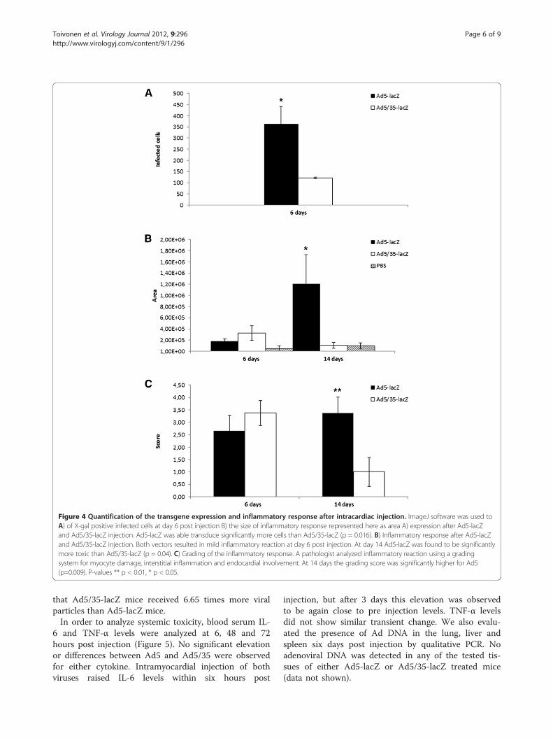
Figure 4 Quantification of the transgene expression and inflammatory response after intracardiac injection. ImageJ software was used toA) of X-gal positive infected cells at day 6 post injection B) the size of inflammatory response represented here as area A) expression after Ad5-lacZand Ad5/35-lacZ injection. Ad5-lacZ was able transduce significantly more cells than Ad5/35-lacZ (p = 0.016). B) Inflammatory response after Ad5-lacZand Ad5/35-lacZ injection. Both vectors resulted in mild inflammatory reaction at day 6 post injection. At day 14 Ad5-lacZ was found to be significantlymore toxic than Ad5/35-lacZ (p = 0.04). C) Grading of the inflammatory response. A pathologist analyzed inflammatory reaction using a gradingsystem for myocyte damage, interstitial inflammation and endocardial involvement. At 14 days the grading score was significantly higher for Ad5(p=0.009). P-values ** p < 0.01, * p < 0.05.
Toivonen et al. Virology Journal 2012, 9:296 Page 6 of 9http://www.virologyj.com/content/9/1/296
that Ad5/35-lacZ mice received 6.65 times more viralparticles than Ad5-lacZ mice.In order to analyze systemic toxicity, blood serum IL-
6 and TNF-α levels were analyzed at 6, 48 and 72hours post injection (Figure 5). No significant elevationor differences between Ad5 and Ad5/35 were observedfor either cytokine. Intramyocardial injection of bothviruses raised IL-6 levels within six hours post
injection, but after 3 days this elevation was observedto be again close to pre injection levels. TNF-α levelsdid not show similar transient change. We also evalu-ated the presence of Ad DNA in the lung, liver andspleen six days post injection by qualitative PCR. Noadenoviral DNA was detected in any of the tested tis-sues of either Ad5-lacZ or Ad5/35-lacZ treated mice(data not shown).

Figure 5 Analysis of serum cytokine levels from 0 to 3 days post injection. No statistically significant elevation of serum cytokine levelswere observed for A) IL-6 or B) TNF-α. Although both viruses raised IL-6 levels within six hours post injection, after 3 days this elevation wasobserved to be again close to normal level. TNF-α levels did not show similar transient change.
Toivonen et al. Virology Journal 2012, 9:296 Page 7 of 9http://www.virologyj.com/content/9/1/296
DiscussionDespite the availability of several promising genes totreat cardiovascular disease, their optimal delivery to theheart remains a challenge. A number of gene deliveryvectors and methods of administration have previouslybeen used for cardiovascular applications with variablesuccess [9]. In this paper we combined two promisingapproaches, hybrid adenoviral vectors and ultrasound-guided intracardiac delivery, and analyzed the differencesin transduction efficiency and toxicity after intracardiacinjection between Ad5 and Ad5/35 vectors. We used atransgenic mouse strain hCD46Ge, which expresses theAd5/35 receptor, human CD46, in a similar pattern as inhumans, to compare the viruses in a situation compar-able to man [21,25].The viral vectors were injected to the left ventricle wall
by using a closed-chest ultrasound guided system. Withthis system vectors can be administered directly into theleft ventricle wall, without traumatic open-chest surgery.The injection leaves only a minor needle track and noother effects to cardiac function were observed. Safety ofthis injection method has been shown in healthy C57BLmice by advancing the needle 1-5 times to myocardiumand also by repeated saline injections without com-promising left ventricular function (unpublished data,Koskenvuo).Transductional targeting has been widely used to im-
prove efficacy and reduce toxicity in various gene ther-apy applications, most notably in cancer gene therapy[26]. Numerous reports have demonstrated markedimprovements in therapeutic efficacy with the use of thehybrid adenovirus technology. For cardiovascular genetherapy, however, reports on transductional targetingapplications for the myocardium have thus far beenlimited. We analyze here for the first time the effectof adenoviral transductional targeting on transgene
expression and toxicity after intramyocardial closed-chest injection. Most previous studies involving adeno-viral activity in the heart are biodistribution studies afterintravenous (i.v.) administration. It has been shown pre-viously that after systemic i.v. administration both Ad5-lacZ and Ad5/35-lacZ genomes are present at relativelyhigh concentrations in the hearts of hCD46Ge mice aswell as baboons and for Ad5/35-lacZ heart is one of themost effectively transduced tissues in both models[27,28]. Interestingly, with the primate model no trans-gene activity was reported in the heart tissue for anyvector despite the presence of viral genomes. Analysis ofgenome copies present in tissues does not discriminatebetween active or inactive viral particles or betweenintracellular genomes and viruses in extracellular space.Unfortunately, only this method of analysis wasemployed by Ganesh et al. (2009). Here we show alsothe expression and activity of viral transgene inhCD46Ge mice, indicating that transgenes from bothvectors are transcribed in our murine model. In theirwork Ganesh et al. (2009) also analyzed vector-mediatedtoxicity by measuring serum IL-6 and liver enzyme levelsand showed decreased overall toxicity with Ad5/35-lacZas compared to Ad5 vector after i.v. administration. Thehighest serum IL-6 levels in our experiment were 87 pg/ml, only 9% of the reported serum IL-6 levels after i.v.administration (Figure 4) [27]. In addition, our analysisof organs other than the heart revealed no signs of tox-icity nor adenoviral DNA, indicating that systemic tox-icity is low in our intracardiac injection model. Othervector systems have previously been used for gene trans-fer into the heart, including adeno-associated virus(AAV). Previous studies have demonstrated high tropismfor the myocardium for AAV serotypes 1,6,8 and 9, mak-ing the AAV system a potentially attractive delivery ve-hicle for cardiovascular gene therapy [29]. The

Toivonen et al. Virology Journal 2012, 9:296 Page 8 of 9http://www.virologyj.com/content/9/1/296
advantages of adenovirus-based approach include higherpayload capacity and long experience from clinicaltrials, and serotype-modification strategies can poten-tially alleviate the strong immune response associatedwith adenovirus treatment.There are also a few previous reports using
ultrasound-guided intramyocardial injection of adeno-virus in mice. Huusko et al. (2010) recently used thismethod to inject Ad5 expressing lacZ or different iso-forms of VEGF, and the inflammatory reaction wasfound to be moderate at 14 days post injection, and thelevel of tissue damage was found to be also dependenton the transgene with VEGF-A giving rise to more in-flammatory reaction than lacZ [30]. Li et al. (2005) usedan Ad5 vector with deletions of the E1, E2a, and E3regions expressing inducible nitric oxide synthase andreported little inflammatory reaction after intramyocar-dial delivery [31].Our previous studies on adenoviral receptor expres-
sion in human heart tissue suggested that the native Ad5capsid configuration would be better suited for cardiacgene transfer than Ad5/35 vectors as CAR had higherlevels of expression than CD46 in both normaland dilated cardiomyopathy hearts [19]. As expected,the injection of unmodified Ad5 vector resultedin significantly more efficient transgene expressionthan administration of hybrid vector Ad5/35 (Figure 3,Figure 4). However, although both vectors elicited an im-mune response, this response was markedly more severeafter Ad5 administration. At 6 days post injection therewas not a significant difference in toxicities between thetwo viruses but by 14 days the Ad5 immune reactionshowed signs of irreversible tissue damage with largeareas of myocyte necrosis, whereas Ad5/35 had featuresof resolving immune reaction. These results are in ac-cordance with a previous report from Nanda et al.,where the addition of Ad5 fiber knob to Ad35-basedvectors was found to substantially increase the immuno-genicity of Ad vectors [32]. LacZ transgene is also im-munogenic and it is theoretically possible that theincreased toxicity of Ad5 observed here could be causedby higher expression of lacZ. However, previous studiesusing different AAV vectors to deliver lacZ to mousehearts have shown a mild immune response and noovert abnormalities in cardiac function despite highlevels of lacZ expression, suggesting that immune re-sponse to lacZ does not play a significant role in ourmodel [33].The results suggest that, although Ad5 is more effect-
ive in transducing cardiac cells after intracardiac injec-tion, this vector clearly has a worse safety profile andsafer gene delivery can be achieved by using Ad5/35 hy-brid vector. The definitive tissue damage after Ad5 injec-tion will undermine the therapeutic effect of the
delivered transgene and probably lead to severe sideeffects, although no adverse effects were evident duringthe 14 day post injection observation period. It must bealso noted that even though Ad5 injection resulted inmore efficient transgene expression, the cardiac tissuewas not refractory to Ad5/35 infection and robust trans-gene expression was also achieved using this vector. It isnotable that due to differences in viral particle/pfu ratiosbetween the two viruses, Ad5/35-lacZ mice received6.65 times more viral particles and still demonstratedless toxicity than A5-lacZ mice.Results described here together with previous studies
suggest that changing Ad5 fiber to those of Ad35increases the safety profile of gene therapy vectors, al-though at the cost of some transduction efficiency.Ultrasound-guided intracardiac injection is a promisingmethod for introducing genes to the heart. Theincreased safety with Ad5/35 vector warrants for furtherdevelopment of hybrid vector system for therapeuticpurposes.
Additional file
Additional file 1: Figure S1. Validation of ultrasound-guidedintramyocardial injection technique for therapeutic purposes usingmethylene blue as an indicator for successful procedure. Left ventricle iscut to five short-axis sections from basis to apex (left to right in Figure).Dye injection produces darker outlook in the anterior myocardium of thethree midventricular slices.
Competing interestsThe authors declare that they have no competing interests.
Authors’ contributionRT planned and performed the experiments, analyzed the data and wrotethe manuscript. JK performed intracardial injections and reviewed themanuscript. MM performed intracardial injections and reviewed themanuscript. MSö analyzed the histology and reviewed the manuscript. SYplanned the experiments and reviewed the manuscript. MSa planned theexperiment, analyzed the data and wrote the manuscript. All authors readand approved the final manuscript.
AcknowledgementsWe thank Sari Pitkänen for her support on preparing histology samples. Weare grateful to Turku University Foundation, Turku University Hospital (EVOfunding) and Finnish Cultural fund for financial support.
Author details1Turku Centre for biotechnology, University of Turku, Tykistökatu 6B 5th floor,Turku FIN-20520, Finland. 2Department of Medical Biochemistry andMolecular Biology, University of Turku, Turku, Finland. 3Turku GraduateSchool of Biomedical Sciences, University of Turku, Turku, Finland. 4ResearchCentre of Applied and Preventive Cardiovascular Medicine, University ofTurku, Turku, Finland. 5Department of Clinical Physiology and NuclearMedicine, Turku University Hospital, Turku, Finland. 6Department ofBiotechnology and Molecular Medicine, A. I. Virtanen Institute for MolecularSciences, University of Kuopio, Kuopio, Finland. 7Department of Pathology,University of Turku and Turku University Hospital, Turku, Finland.8Department of Medicine, University of Turku and Turku University Hospital,Turku, Finland.
Received: 30 April 2012 Accepted: 27 November 2012Published: 29 November 2012

Toivonen et al. Virology Journal 2012, 9:296 Page 9 of 9http://www.virologyj.com/content/9/1/296
References1. Gaggar A, Shayakhmetov DM, Lieber A: CD46 is a cellular receptor for
group B adenoviruses. Nat Med 2003, 9(11):1408–1412.2. Bergelson JM, Cunningham JA, Droguett G, Kurt-Jones EA, Krithivas A, Hong
JS, Horwitz MS, Crowell RL, Finberg RW: Isolation of a common receptorfor Coxsackie B viruses and adenoviruses 2 and 5. Science 1997,275(5304):1320–1323.
3. Tomko RP, Xu R, Philipson L: HCAR and MCAR: the human and mousecellular receptors for subgroup C adenoviruses and group Bcoxsackieviruses. Proc Natl Acad Sci USA 1997, 94(7):3352–3356.
4. Roelvink PW, Lizonova A, Lee JG, Li Y, Bergelson JM, Finberg RW, BroughDE, Kovesdi I, Wickham TJ: The coxsackievirus-adenovirus receptor proteincan function as a cellular attachment protein for adenovirus serotypesfrom subgroups A, C, D, E, and F. J Virol 1998, 72(10):7909–7915.
5. Sirena D, Lilienfeld B, Eisenhut M, Kalin S, Boucke K, Beerli RR, Vogt L, RuedlC, Bachmann MF, Greber UF, Hemmi S: The human membrane cofactorCD46 is a receptor for species B adenovirus serotype 3. J Virol 2004,78(9):4454–4462.
6. Marttila M, Persson D, Gustafsson D, Liszewski MK, Atkinson JP, Wadell G,Arnberg N: CD46 is a cellular receptor for all species B adenovirusesexcept types 3 and 7. J Virol 2005, 79(22):14429–14436.
7. Seya T, Hirano A, Matsumoto M, Nomura M, Ueda S: Human membranecofactor protein (MCP, CD46): multiple isoforms and functions.Int J Biochem Cell Biol 1999, 31(11):1255–1260.
8. Wang H, Li ZY, Liu Y, Persson J, Beyer I, Moller T, Koyuncu D, Drescher MR,Strauss R, Zhang XB, Wahl JK 3rd, Urban N, Drescher C, Hemminki A, FenderP, Lieber A: Desmoglein 2 is a receptor for adenovirus serotypes 3, 7,11 and 14. Nat Med 2011, 17(1):96–104.
9. Rissanen TT, Yla-Herttuala S: Current status of cardiovascular genetherapy. Mol Ther 2007, 15(7):1233–1247.
10. Campos SK, Barry MA: Current advances and future challenges inAdenoviral vector biology and targeting. Curr Gene Ther 2007,7(3):189–204.
11. Gall J, Kass-Eisler A, Leinwand L, Falck-Pedersen E: Adenovirus type 5 and 7capsid chimera: fiber replacement alters receptor tropism withoutaffecting primary immune neutralization epitopes. J Virol 1996,70(4):2116–2123.
12. Amin KM: Ad5 and Ad3 chimeric fiber travels into the cell without theCAR. Cancer Biol Ther 2003, 2(5):516–517.
13. Kanerva A, Mikheeva GV, Krasnykh V, Coolidge CJ, Lam JT, Mahasreshti PJ,Barker SD, Straughn M, Barnes MN, Alvarez RD, Hemminki A, Curiel DT:Targeting adenovirus to the serotype 3 receptor increases gene transferefficiency to ovarian cancer cells. Clin Cancer Res 2002, 8(1):275–280.
14. Segerman A, Mei YF, Wadell G: Adenovirus types 11p and 35p show highbinding efficiencies for committed hematopoietic cell lines and areinfective to these cell lines. J Virol 2000, 74(3):1457–1467.
15. Shayakhmetov DM, Li ZY, Ni S, Lieber A: Targeting of adenovirus vectorsto tumor cells does not enable efficient transduction of breast cancermetastases. Cancer Res 2002, 62(4):1063–1068.
16. Shinozaki K, Suominen E, Carrick F, Sauter B, Kahari VM, Lieber A, Woo SL,Savontaus M: Efficient infection of tumor endothelial cells by acapsid-modified adenovirus. Gene Ther 2006, 13(1):52–59.
17. Sova P, Ren XW, Ni S, Bernt KM, Mi J, Kiviat N, Lieber A: A tumor-targetedand conditionally replicating oncolytic adenovirus vector expressingTRAIL for treatment of liver metastases. Mol Ther 2004, 9(4):496–509.
18. Suominen E, Toivonen R, Grenman R, Savontaus M: Head and neck cancercells are efficiently infected by Ad5/35 hybrid virus. J Gene Med 2006,8(10):1223–1231.
19. Toivonen R, Mayranpaa MI, Kovanen PT, Savontaus M: Dilatedcardiomyopathy alters the expression patterns of CAR and otheradenoviral receptors in human heart. Histochem Cell Biol 2010,133(3):349–357.
20. Mrkic B, Pavlovic J, Rulicke T, Volpe P, Buchholz CJ, Hourcade D, Atkinson JP,Aguzzi A, Cattaneo R: Measles virus spread and pathogenesis ingenetically modified mice. J Virol 1998, 72(9):7420–7427.
21. Kemper C, Leung M, Stephensen CB, Pinkert CA, Liszewski MK, Cattaneo R,Atkinson JP: Membrane cofactor protein (MCP; CD46) expression intransgenic mice. Clin Exp Immunol 2001, 124(2):180–189.
22. Bautista DS, Hitt M, McGrory J, Graham FL: Isolation and characterizationof insertion mutants in E1A of adenovirus type 5. Virology 1991,182(2):578–596.
23. Shayakhmetov DM, Papayannopoulou T, Stamatoyannopoulos G, Lieber A:Efficient gene transfer into human CD34(+) cells by a retargetedadenovirus vector. J Virol 2000, 74(6):2567–2583.
24. Springer ML, Sievers RE, Viswanathan MN, Yee MS, Foster E, Grossman W,Yeghiazarians Y: Closed-chest cell injections into mouse myocardiumguided by high-resolution echocardiography. Am J Physiol Heart CircPhysiol 2005, 289(3):H1307–H1314.
25. Johnstone RW, Loveland BE, McKenzie IF: Identification and quantificationof complement regulator CD46 on normal human tissues. Immunology1993, 79(3):341–347.
26. Bachtarzi H, Stevenson M, Fisher K: Cancer gene therapy with targetedadenoviruses. Expert Opin Drug Deliv 2008, 5(11):1231–1240.
27. Ganesh S, Gonzalez-Edick M, Gibbons D, Waugh J, Van Roey M, Jooss K:Evaluation of biodistribution of a fiber-chimeric, conditionallyreplication-competent (oncolytic) adenovirus in CD46 receptortransgenic mice. Hum Gene Ther 2009, 20(10):1201–1213.
28. Ni S, Bernt K, Gaggar A, Li ZY, Kiem HP, Lieber A: Evaluation ofbiodistribution and safety of adenovirus vectors containing group Bfibers after intravenous injection into baboons. Hum Gene Ther 2005,16(6):664–677.
29. Lyon AR, Sato M, Hajjar RJ, Samulski RJ, Harding SE: Gene therapy:targeting the myocardium. Heart 2008, 94(1):89–99.
30. Huusko J, Merentie M, Dijkstra MH, Ryhanen MM, Karvinen H, Rissanen TT,Vanwildemeersch M, Hedman M, Lipponen J, Heinonen SE, Eriksson U,Shibuya M, Yla-Herttuala S: The effects of VEGF-R1 and VEGF-R2 ligandson angiogenic responses and left ventricular function in mice.Cardiovasc Res 2010, 86(1):122–130.
31. Li Q, Guo Y, Tan W, Stein AB, Dawn B, Wu WJ, Zhu X, Lu X, Xu X, Siddiqui T,Tiwari S, Bolli R: Gene therapy with iNOS provides long-term protectionagainst myocardial infarction without adverse functional consequences.Am J Physiol Heart Circ Physiol 2006, 290(2):H584–H589.
32. Nanda A, Lynch DM, Goudsmit J, Lemckert AA, Ewald BA, Sumida SM, TruittDM, Abbink P, Kishko MG, Gorgone DA, Lifton MA, Shen L, Carville A,Mansfield KG, Havenga MJ, Barouch DH: Immunogenicity of recombinantfiber-chimeric adenovirus serotype 35 vector-based vaccines in miceand rhesus monkeys. J Virol 2005, 79(22):14161–14168.
33. Bish LT, Morine K, Sleeper MM, Sanmiguel J, Wu D, Gao G, Wilson JM,Sweeney HL: Adeno-associated virus (AAV) serotype 9 provides globalcardiac gene transfer superior to AAV1, AAV6, AAV7, and AAV8 in themouse and rat. Hum Gene Ther 2008, 19(12):1359–1368.
doi:10.1186/1743-422X-9-296Cite this article as: Toivonen et al.: Intracardiac injection of a capsid-modified Ad5/35 results in decreased heart toxicity when compared tostandard Ad5. Virology Journal 2012 9:296.
Submit your next manuscript to BioMed Centraland take full advantage of:
• Convenient online submission
• Thorough peer review
• No space constraints or color figure charges
• Immediate publication on acceptance
• Inclusion in PubMed, CAS, Scopus and Google Scholar
• Research which is freely available for redistribution
Submit your manuscript at www.biomedcentral.com/submit