Embed Size (px)
Citation preview
Intra-abdominal Hypertension and Abdominal Compartment Syndrome: A
Potentially Fatal Mix
Daria C. Ruffolo

No Conflict of Interest
Objectives
• Differentiate between intra-abdominal hypertension (IAH) and abdominal compartment syndrome (ACS)
• Identify patient populations at risk for IAH/ACS
• Understand the pathophysiological process of IAH/ACS
• Methods to measure the IAP
• Managemnet of IAH and ACS

Abdominal compartment syndrome:What is it?
A disease process that dramatically increases organ failure and death for medical and surgical
ICU patients
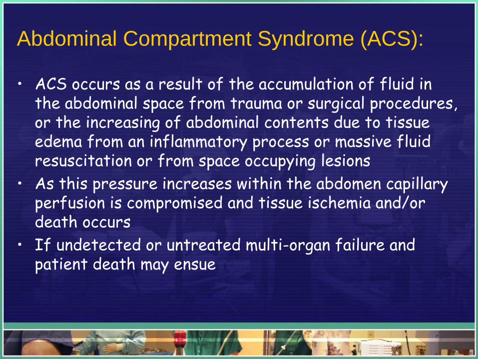
Abdominal Compartment Syndrome (ACS):
• ACS occurs as a result of the accumulation of fluid in the abdominal space from trauma or surgical procedures, or the increasing of abdominal contents due to tissue edema from an inflammatory process or massive fluid resuscitation or from space occupying lesions
• As this pressure increases within the abdomen capillary perfusion is compromised and tissue ischemia and/or death occurs
• If undetected or untreated multi-organ failure and patient death may ensue
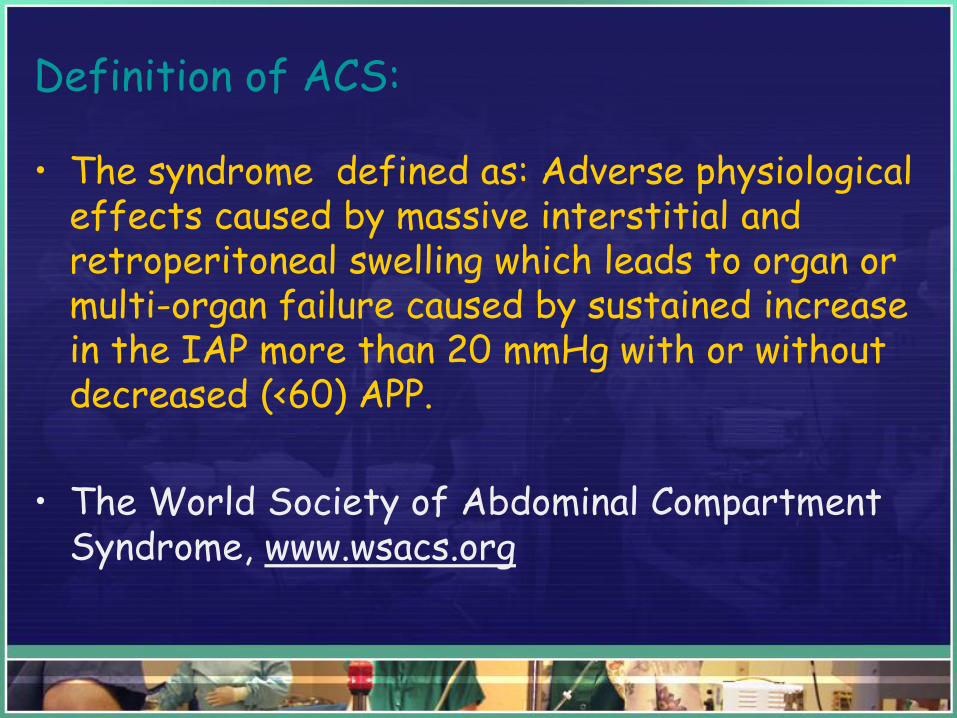
Definition of ACS:
• The syndrome defined as: Adverse physiological effects caused by massive interstitial and retroperitoneal swelling which leads to organ or multi-organ failure caused by sustained increase in the IAP more than 20 mmHg with or without decreased (<60) APP.
• The World Society of Abdominal Compartment Syndrome, www.wsacs.org
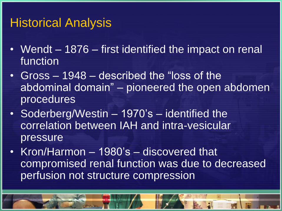
Historical Analysis
• Wendt – 1876 – first identified the impact on renal function
• Gross – 1948 – described the “loss of the abdominal domain” – pioneered the open abdomen procedures
• Soderberg/Westin – 1970’s – identified the correlation between IAH and intra-vesicular pressure
• Kron/Harmon – 1980’s – discovered that compromised renal function was due to decreased perfusion not structure compression
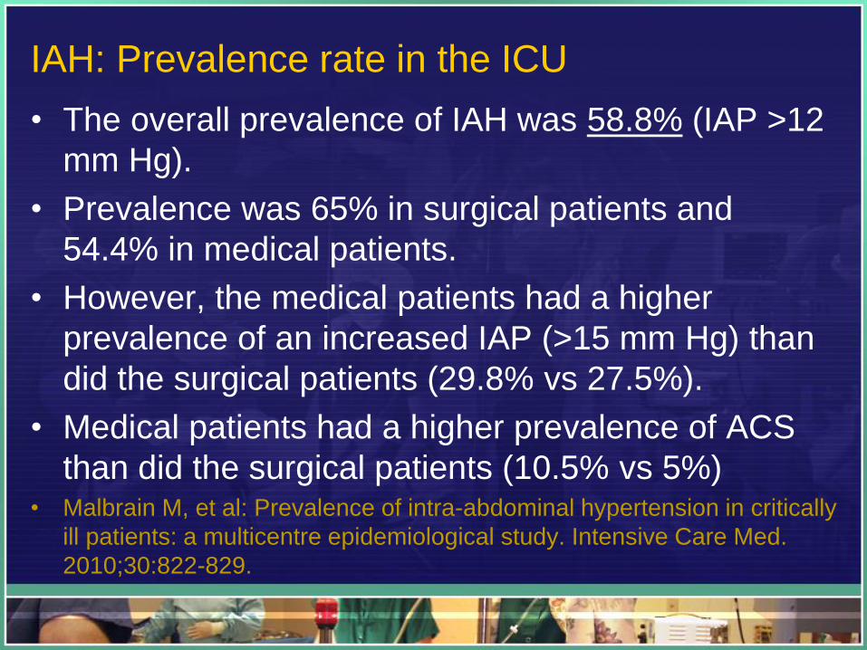
IAH: Prevalence rate in the ICU
• The overall prevalence of IAH was 58.8% (IAP >12
mm Hg).
• Prevalence was 65% in surgical patients and
54.4% in medical patients.
• However, the medical patients had a higher
prevalence of an increased IAP (>15 mm Hg) than
did the surgical patients (29.8% vs 27.5%).
• Medical patients had a higher prevalence of ACS
than did the surgical patients (10.5% vs 5%)• Malbrain M, et al: Prevalence of intra-abdominal hypertension in critically
ill patients: a multicentre epidemiological study. Intensive Care Med.
2010;30:822-829.
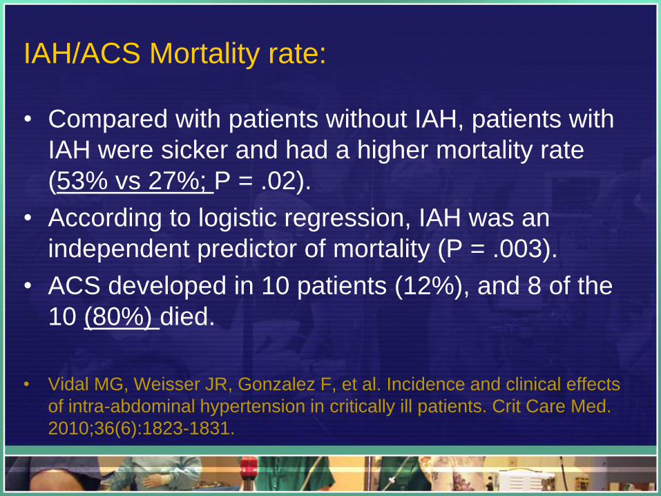
IAH/ACS Mortality rate:
• Compared with patients without IAH, patients with
IAH were sicker and had a higher mortality rate
(53% vs 27%; P = .02).
• According to logistic regression, IAH was an
independent predictor of mortality (P = .003).
• ACS developed in 10 patients (12%), and 8 of the
10 (80%) died.
• Vidal MG, Weisser JR, Gonzalez F, et al. Incidence and clinical effects
of intra-abdominal hypertension in critically ill patients. Crit Care Med.
2010;36(6):1823-1831.
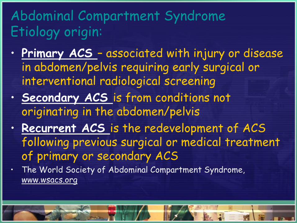
Abdominal Compartment SyndromeEtiology origin:
• Primary ACS – associated with injury or disease in abdomen/pelvis requiring early surgical or interventional radiological screening
• Secondary ACS is from conditions not originating in the abdomen/pelvis
• Recurrent ACS is the redevelopment of ACS following previous surgical or medical treatment of primary or secondary ACS
• The World Society of Abdominal Compartment Syndrome, www.wsacs.org
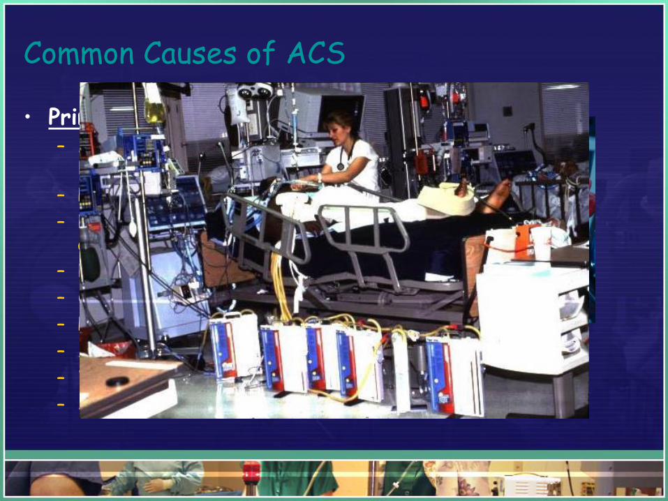
Common Causes of ACS
• Primary causes– Abdominal trauma with
bleeding
– Pancreatitis
– Ruptured abdominal aortic aneurysm
– Retroperitoneal hematoma
– Obstructions/ileus
– Pneumoperitoneum
– Abscesses
– Visceral edema
– Severe colitis
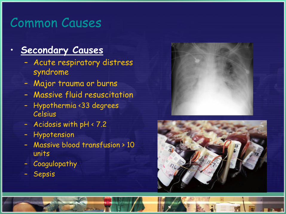
Common Causes
• Secondary Causes– Acute respiratory distress
syndrome
– Major trauma or burns
– Massive fluid resuscitation– Hypothermia <33 degrees
Celsius
– Acidosis with pH < 7.2
– Hypotension
– Massive blood transfusion > 10 units
– Coagulopathy
– Sepsis
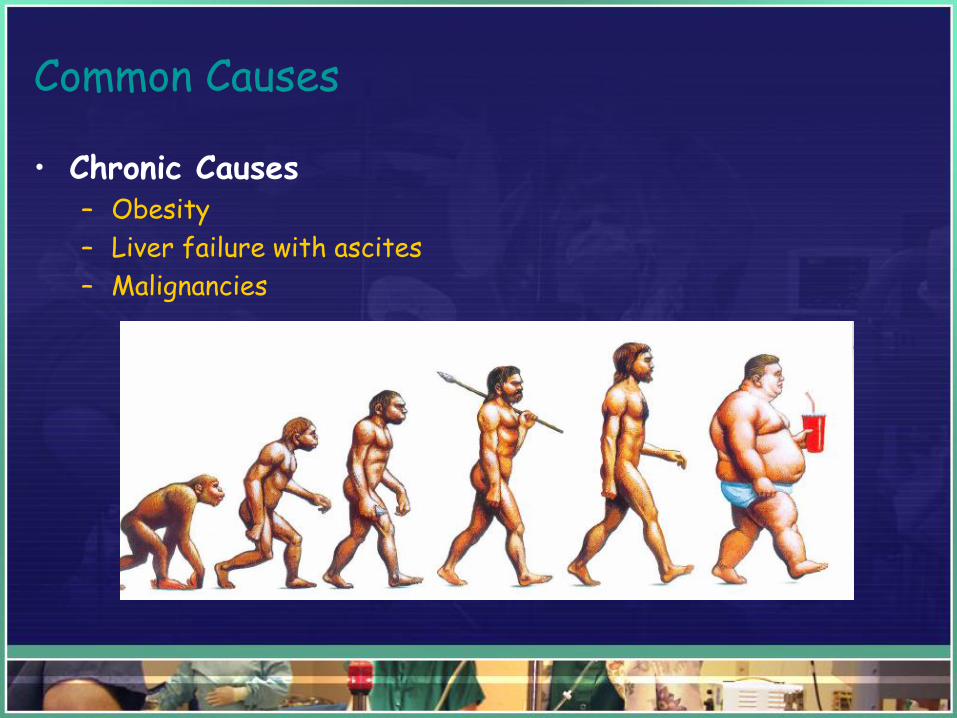
Common Causes
• Chronic Causes– Obesity
– Liver failure with ascites
– Malignancies
What is a normal intra-abdominal pressure (IAP)?
5 – 7 mmHg is normal in a critically ill adult
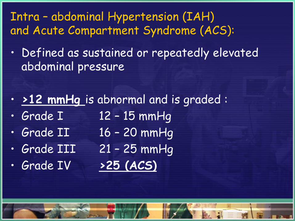
Intra – abdominal Hypertension (IAH) and Acute Compartment Syndrome (ACS):
• Defined as sustained or repeatedly elevated abdominal pressure
• >12 mmHg is abnormal and is graded :
• Grade I 12 – 15 mmHg
• Grade II 16 – 20 mmHg
• Grade III 21 – 25 mmHg
• Grade IV >25 (ACS)
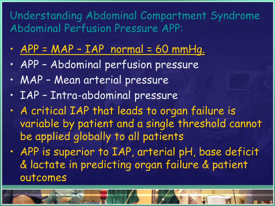
Understanding Abdominal Compartment SyndromeAbdominal Perfusion Pressure APP:
• APP = MAP – IAP normal = 60 mmHg.
• APP – Abdominal perfusion pressure
• MAP – Mean arterial pressure
• IAP – Intra-abdominal pressure
• A critical IAP that leads to organ failure is variable by patient and a single threshold cannot be applied globally to all patients
• APP is superior to IAP, arterial pH, base deficit & lactate in predicting organ failure & patient outcomes
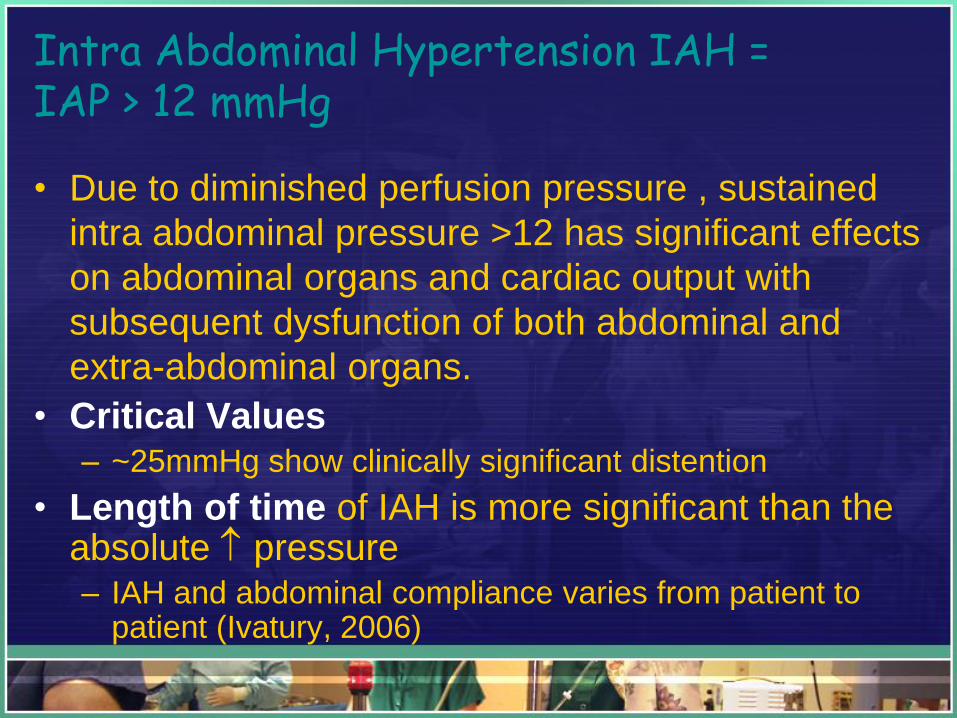
Intra Abdominal Hypertension IAH =IAP > 12 mmHg
• Due to diminished perfusion pressure , sustained
intra abdominal pressure >12 has significant effects
on abdominal organs and cardiac output with
subsequent dysfunction of both abdominal and
extra-abdominal organs.
• Critical Values– ~25mmHg show clinically significant distention
• Length of time of IAH is more significant than the absolute pressure– IAH and abdominal compliance varies from patient to
patient (Ivatury, 2006)
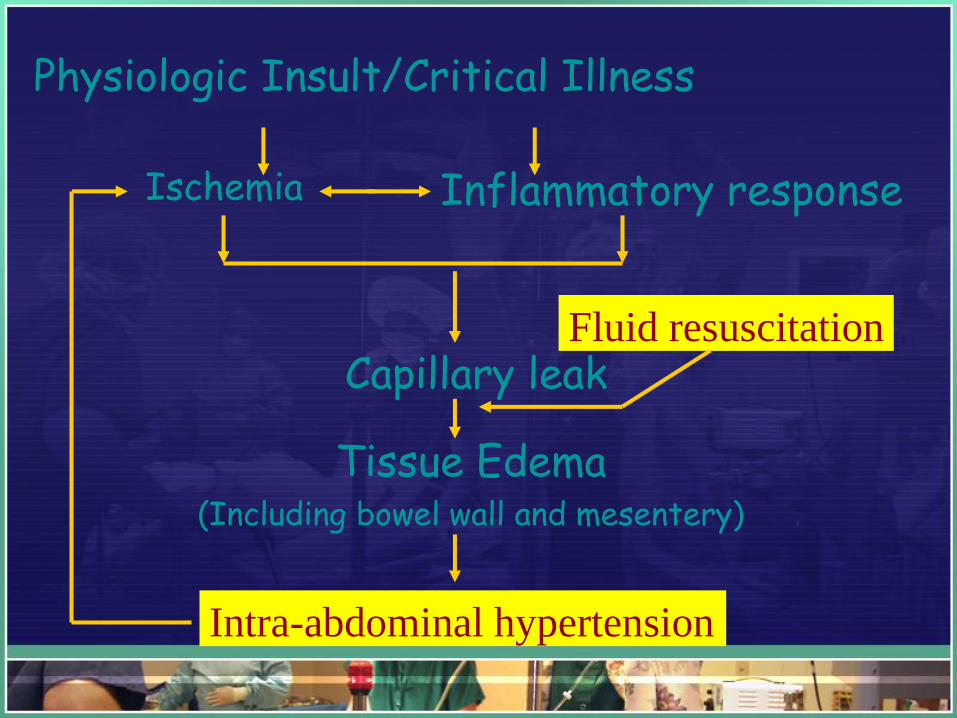
Ischemia Inflammatory response
Capillary leak
Tissue Edema (Including bowel wall and mesentery)
Intra-abdominal hypertension
Fluid resuscitation
Physiologic Insult/Critical Illness
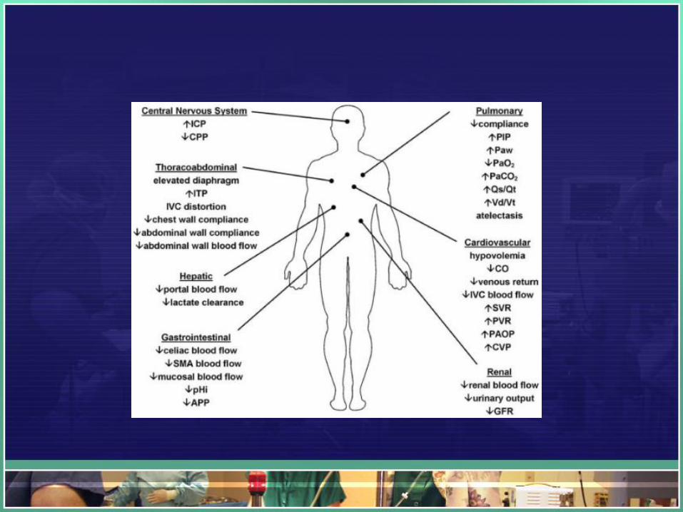
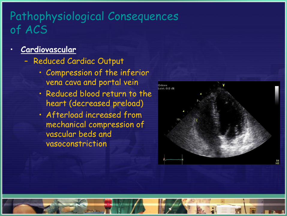
Pathophysiological Consequencesof ACS
• Cardiovascular
– Reduced Cardiac Output
• Compression of the inferior vena cava and portal vein
• Reduced blood return to the heart (decreased preload)
• Afterload increased from mechanical compression of vascular beds and vasoconstriction
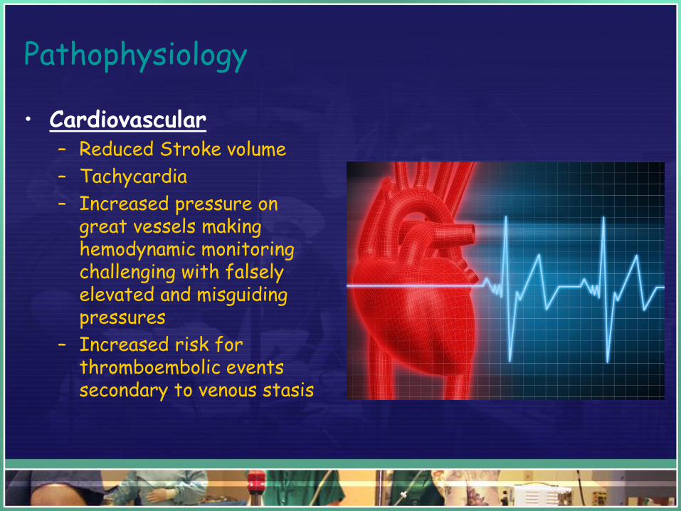
Pathophysiology
• Cardiovascular– Reduced Stroke volume
– Tachycardia
– Increased pressure on great vessels making hemodynamic monitoring challenging with falsely elevated and misguiding pressures
– Increased risk for thromboembolic events secondary to venous stasis
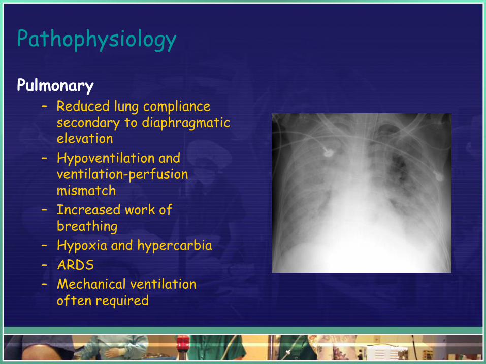
Pathophysiology
Pulmonary– Reduced lung compliance
secondary to diaphragmatic elevation
– Hypoventilation and ventilation-perfusion mismatch
– Increased work of breathing
– Hypoxia and hypercarbia
– ARDS
– Mechanical ventilation often required
Pathophysiology
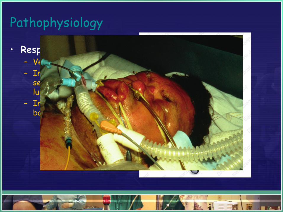
• Respiratory (MV)– Ventialtion difficulty
– Increased peak airway secondary to decreased lung compliance
– Increased risk of barotrauma
Pathophysiology
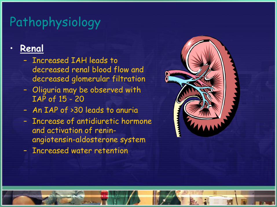
• Renal– Increased IAH leads to
decreased renal blood flow and decreased glomerular filtration
– Oliguria may be observed with IAP of 15 - 20
– An IAP of >30 leads to anuria
– Increase of antidiuretic hormone and activation of renin-angiotensin-aldosterone system
– Increased water retention
Pathophysiology
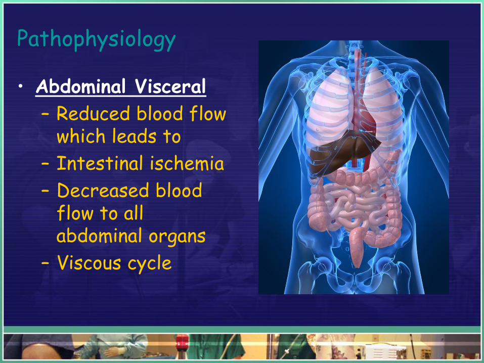
• Abdominal Visceral
– Reduced blood flow which leads to
– Intestinal ischemia
– Decreased blood flow to all abdominal organs
– Viscous cycle
Pathophysiology
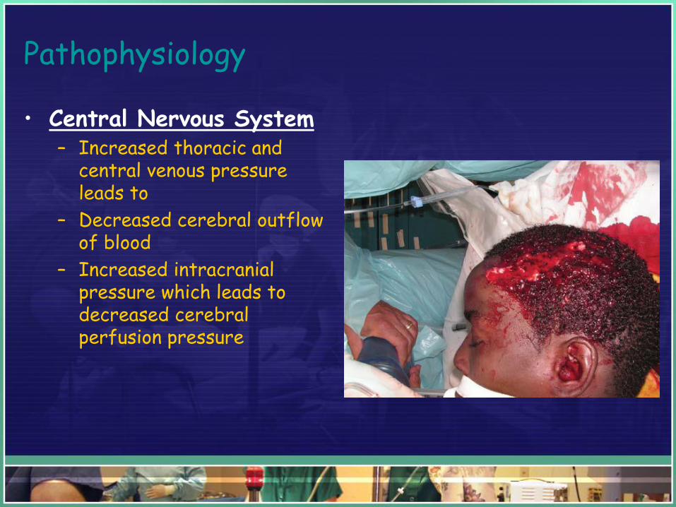
• Central Nervous System– Increased thoracic and
central venous pressure leads to
– Decreased cerebral outflow of blood
– Increased intracranial pressure which leads to decreased cerebral perfusion pressure
Measuring Intra-Abdominal Pressure
Be prepared to measure IAP often, even with very
low level of suspicion.
Importance of accurate measurement
• Physical examination yields low levels of detection of IAH/ACS
• Early detection and intervention reduces morbidity and mortality.
• Diagnosis is dependent on frequent and accurate measurement of IAP (watching trends)
• Cost effective, safe and accurate
• Be prepared to measure IAP often, even with very
low level of suspicion.
When to measure IAP
• New ICU admission with
• Evidence of clinical deterioration and
• Pt has two risk factors for IAH/ACS– Decreased abdominal wall compliance
– Increased intra-luminal contents• ileus, gastroparesis, obstruction
– Increased abdominal contents• Pneumoperitoneum, hemoperitoneum, ascities, liver
dysfunction
– Capillary Leak/fluid resuscitation
Types of Measurements
• Direct Pressure via intraperitoneal catheters
• Indirect Pressure – Gastric Measure
– IVC
– Rectal
– Urinary bladder pressure – Gold Standard
Intestinal Mucosal pH
• Significantly Reduced in IAH
• Measurement of pHi may help in early detection
of spanchnic hypoperfusion in patients with
IAH
– IAH & intestinal ischemia may lead to bacterial
translocation & free oxygen radical production
sepsis MODS
• gastric tonometry
Sieh et al 2001
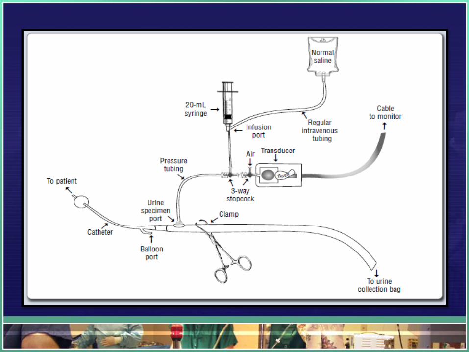
Urinary Bladder Pressure: easy and cheap:
• Most technically reliable
• Correlate closely with pressures measured directly in the abdominal cavity
• Reliably and reproducible
• Transduced through a Foley catheter
• Open (intermittent measuring) or closed (continuous measuring) Systems
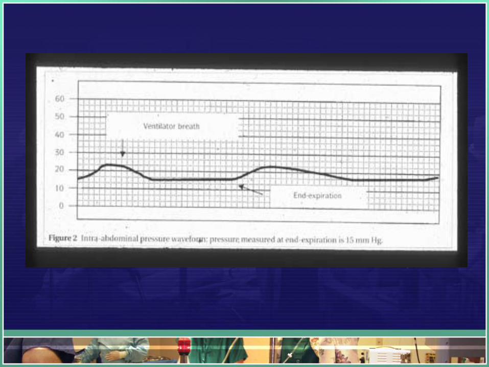
• Position patient flat & supine
• Read Mean pressure
• End Expiration
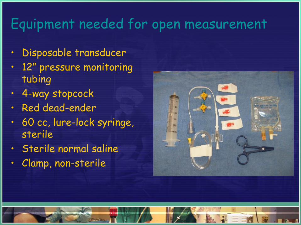
Equipment needed for open measurement
• Disposable transducer
• 12” pressure monitoring tubing
• 4-way stopcock
• Red dead-ender
• 60 cc, lure-lock syringe, sterile
• Sterile normal saline
• Clamp, non-sterile
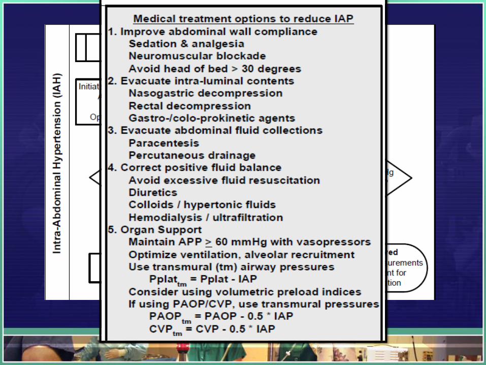
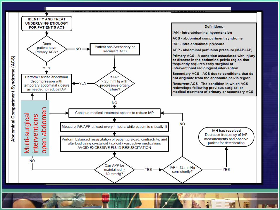
Management Considerations:
• Early detection via frequent monitoring of at risk patients.
• Screen for IAH/ACS in new ICU admissions with new or progressive organ failure: easy and cheap.
• Look for trends of increasing abdominal pressures.
• Preserve organ perfusion and treat clinical conditions with grades I & II IAH
Management Considerations for IAH/ACS
1. Medical management to all patients
2. Early surgical consultations for ACS at risk patients
3. Early intervention for ACS or Grade III
The World Society of Abdominal Compartment Syndrome, www.wsacs.org
Multi-surg
ical
inte
rventions
open a
bdom
en
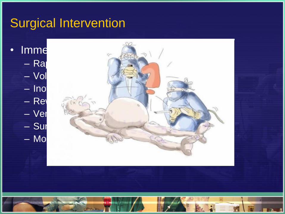
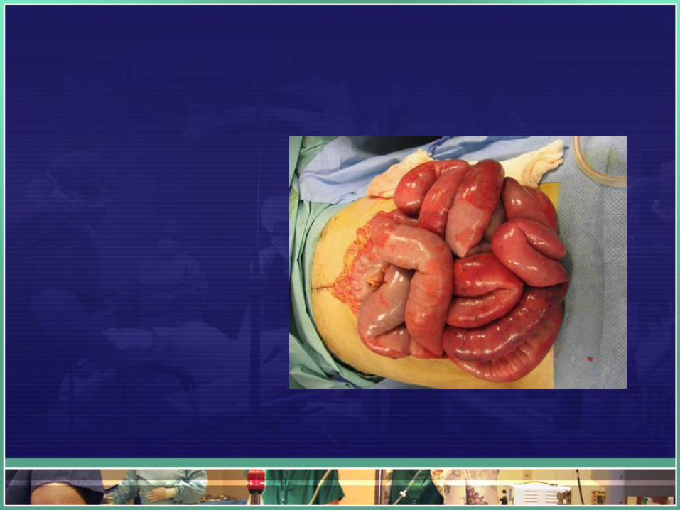
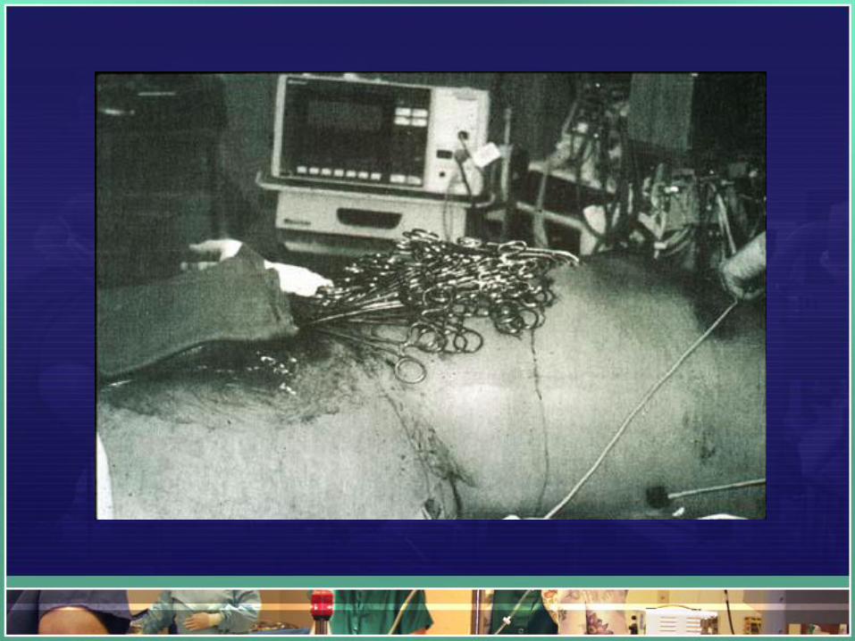
Surgical Intervention
• Immediate– Rapid surgical decompression OR vs Bedside
– Volume resuscitation “washout cocktail”
– Inotropic support - DO2/VO2 optimization
– Rewarming
– Ventilation support
– Survey of all organ systems
– Monitor for recurrence
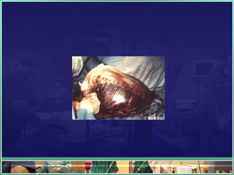
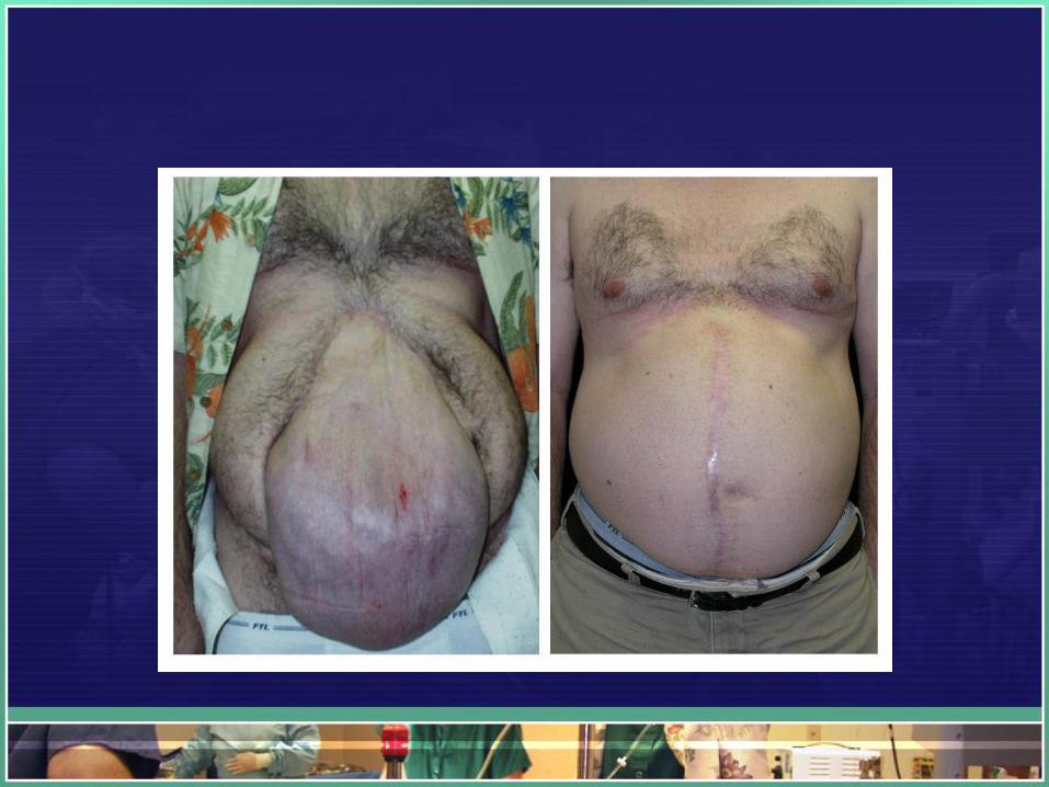
Management Considerations
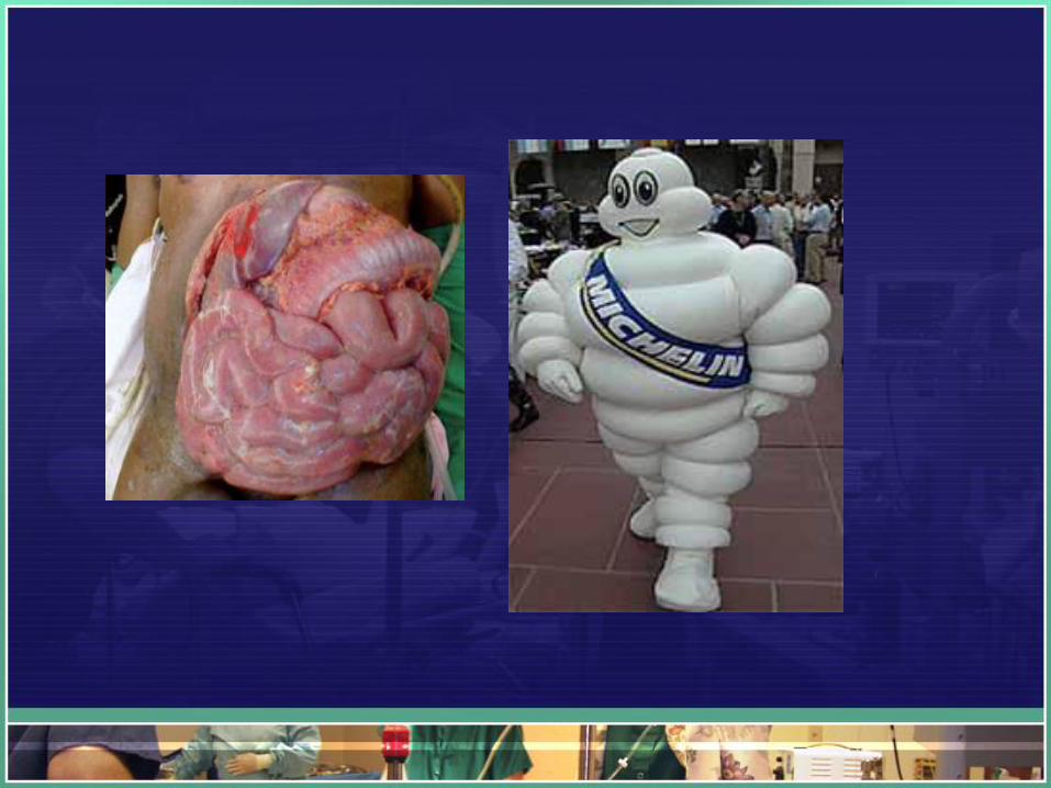
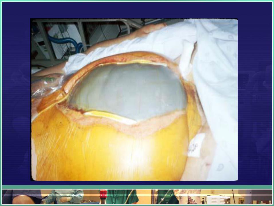
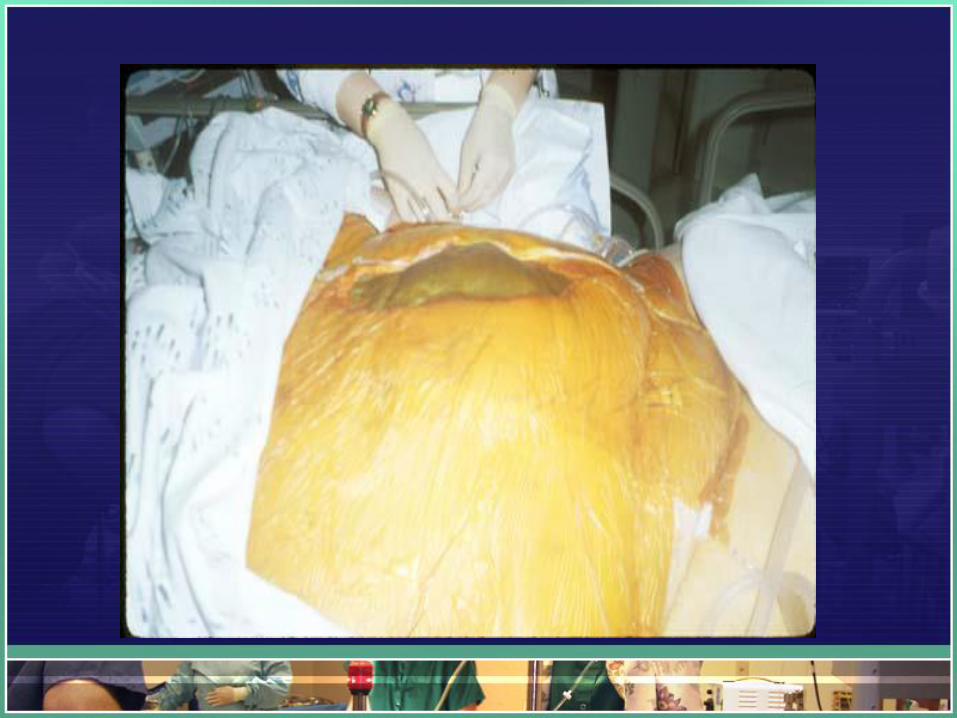
• Anticipate patient to return with an alternative surgical closure or “open” abdomen.
• The abdominal contents will not be sutured into the abdominal cavity
• Alternative closures vary from surgeon to surgeon
Examples:
The “Bogata Bag” – A 3 L IV bag, open and sterilized and applied to the abdominal opening
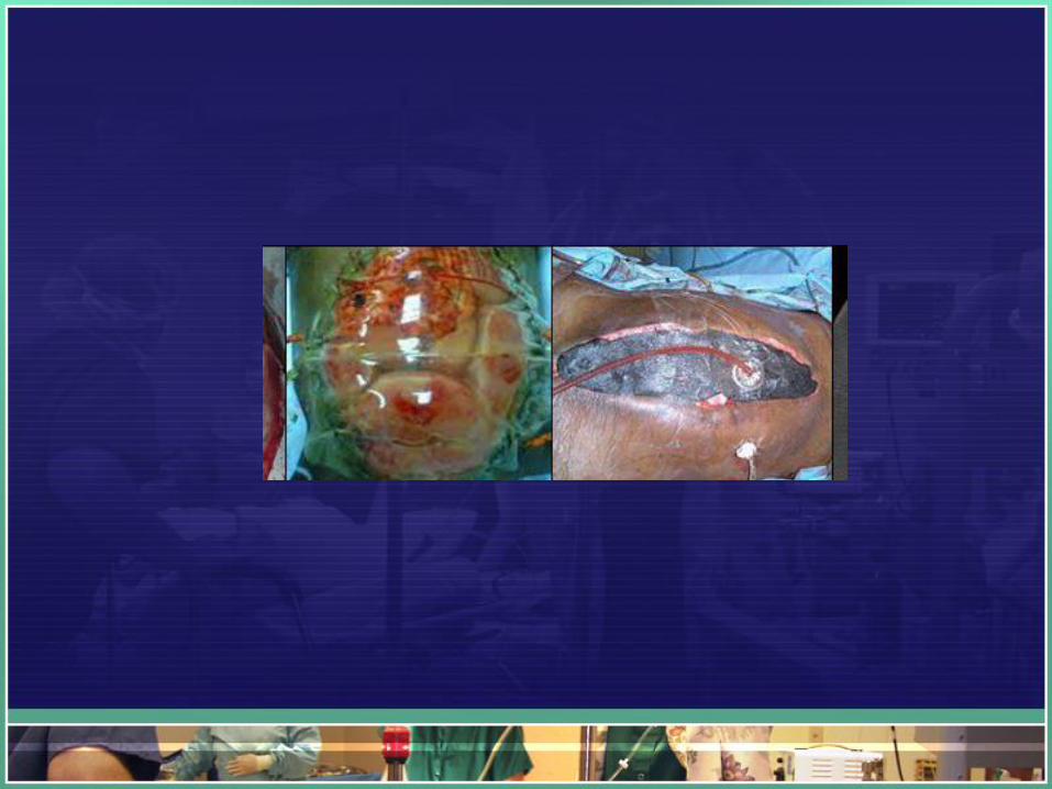
Management Considerations
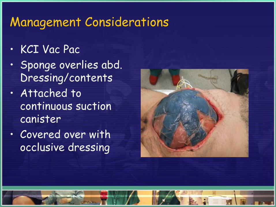
• KCI Vac Pac
• Sponge overlies abd. Dressing/contents
• Attached to continuous suction canister
• Covered over with occlusive dressing
Management Considerations
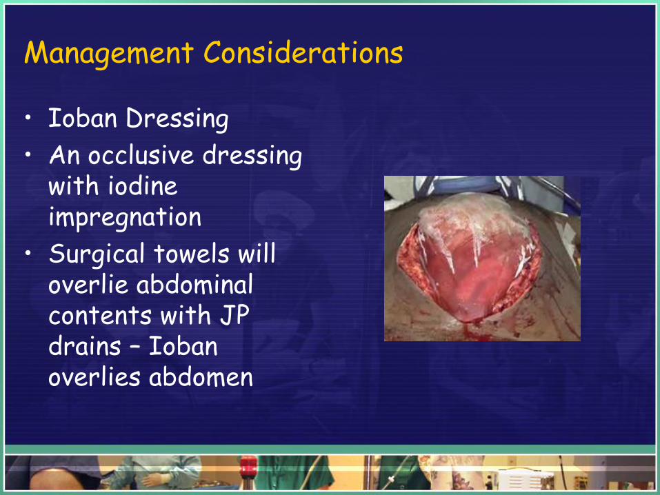
• Ioban Dressing
• An occlusive dressing with iodine impregnation
• Surgical towels will overlie abdominal contents with JP drains – Ioban overlies abdomen
Case Study
• A healthy 25vyeasr old was taken to a level I
trauma center after a 10mfoot fall from his window
and then being struck by an air conditioner that fell
on top of him.
• ASSESSMENT:
• GCS 6
• RTS 8
Case Study
• INJURIES:
- R scapula fx
- R rib fx 4-9
- L inferior and superior rami fx
- Multiple temporal and occipital subdural hematomas/contusions
- Grade II splenic laceration/Grade III liver laceration
Case Study
• COURSE OF CARE:
-Initial fluid resuscitation of 11.5 liters
-Intubation with mechanical ventilation
-Pulmonary artery catheter was placed
-Ventriculostomy was placed
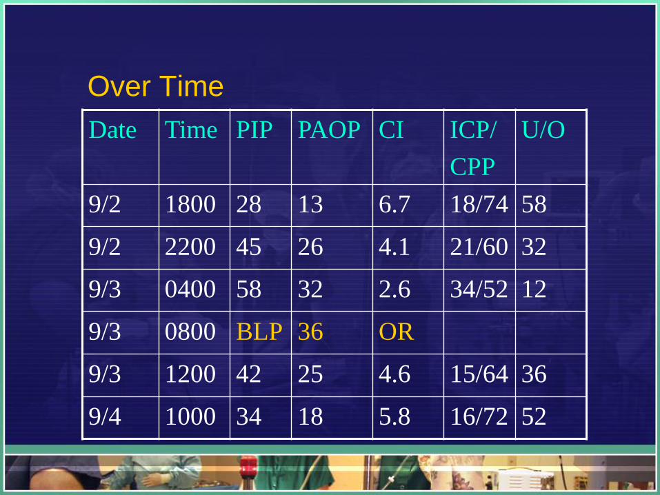
Over Time
Date Time PIP PAOP CI ICP/
CPP
U/O
9/2 1800 28 13 6.7 18/74 58
9/2 2200 45 26 4.1 21/60 32
9/3 0400 58 32 2.6 34/52 12
9/3 0800 BLP 36 OR
9/3 1200 42 25 4.6 15/64 36
9/4 1000 34 18 5.8 16/72 52
Case Study
• OUTCOME:
-Closure utilizing vacuum seal
-Definitive closure with mesh 2 weeks later
-Regained consciousness, following command, MAE,
to rehab and returned one year later to pre-injury
state for mesh removal with ventral hernia repair

Case Study
• 22 year old man that fell 25 feet at a construction site striking his right flank on 2 x 4’s.
• On admission:
– Primary Survey• unremarkable
– Secondary Survey• facial, hand lacerations
• Large contussed area over right flank
• Vital signs – HR 112, BP 102/62, T 368 po
• Labs – ABGs 7.32, 34,352, 24, -5, 100% NRB, Hg 12.2, Crit 38.4
• 2 liter crystalloid resuscitation
Studies
• CXR – fracture of ribs 4-7 on the right side
• Pelvis – WNL
• Cervical spine – WNL
• FAST – free fluid
• CT – abdomen - Grade IV liver laceration
• Angiography with embolization of liver vascular
bleeding
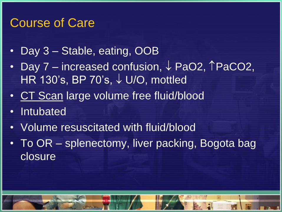
Course of Care
• Day 3 – Stable, eating, OOB
• Day 7 – increased confusion, PaO2, PaCO2,
HR 130’s, BP 70’s, U/O, mottled
• CT Scan large volume free fluid/blood
• Intubated
• Volume resuscitated with fluid/blood
• To OR – splenectomy, liver packing, Bogota bag
closure
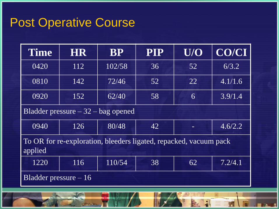
Post Operative Course
Time HR BP PIP U/O CO/CI
0420 112 102/58 36 52 6/3.2
0810 142 72/46 52 22 4.1/1.6
0920 152 62/40 58 6 3.9/1.4
Bladder pressure – 32 – bag opened
0940 126 80/48 42 - 4.6/2.2
To OR for re-exploration, bleeders ligated, repacked, vacuum pack
applied
1220 116 110/54 38 62 7.2/4.1
Bladder pressure – 16
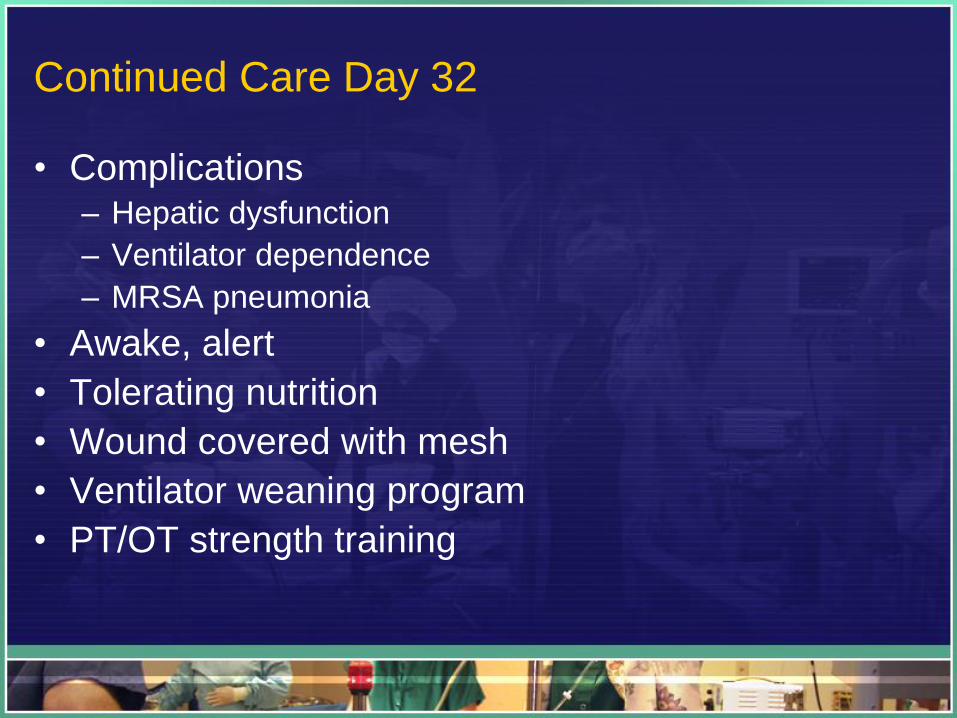
Continued Care Day 32
• Complications– Hepatic dysfunction
– Ventilator dependence
– MRSA pneumonia
• Awake, alert
• Tolerating nutrition
• Wound covered with mesh
• Ventilator weaning program
• PT/OT strength training
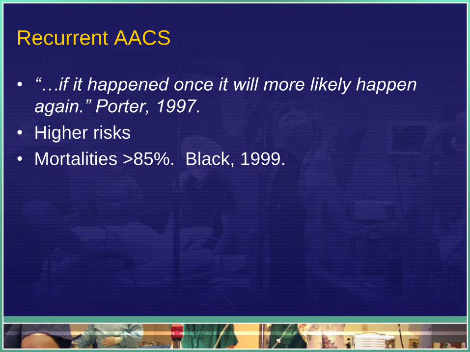
Recurrent AACS
• “…if it happened once it will more likely happen
again.” Porter, 1997.
• Higher risks
• Mortalities >85%. Black, 1999.
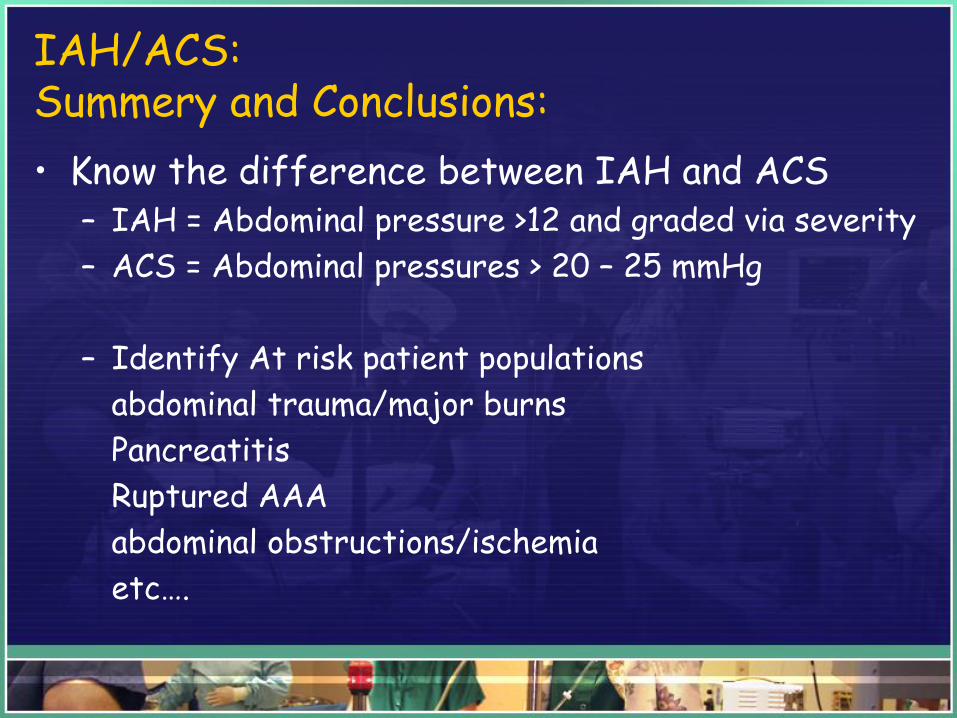
IAH/ACS:Summery and Conclusions:
• Know the difference between IAH and ACS– IAH = Abdominal pressure >12 and graded via severity
– ACS = Abdominal pressures > 20 – 25 mmHg
– Identify At risk patient populations
abdominal trauma/major burns
Pancreatitis
Ruptured AAA
abdominal obstructions/ischemia
etc….
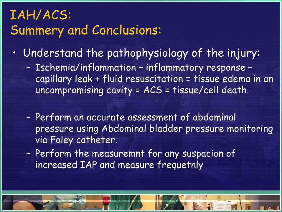
IAH/ACS:Summery and Conclusions:
• Understand the pathophysiology of the injury:– Ischemia/inflammation – inflammatory response –
capillary leak + fluid resuscitation = tissue edema in an uncompromising cavity = ACS = tissue/cell death.
– Perform an accurate assessment of abdominal pressure using Abdominal bladder pressure monitoring via Foley catheter.
– Perform the measuremnt for any suspacion of increased IAP and measure frequetnly
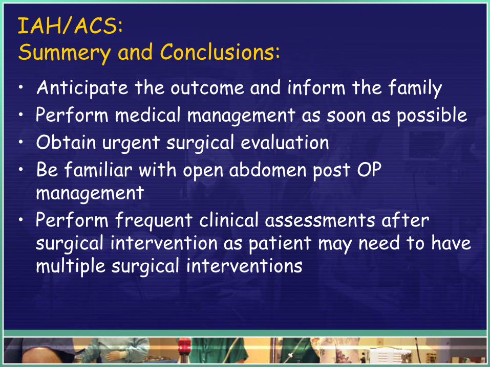
IAH/ACS:Summery and Conclusions:
• Anticipate the outcome and inform the family
• Perform medical management as soon as possible
• Obtain urgent surgical evaluation
• Be familiar with open abdomen post OP management
• Perform frequent clinical assessments after surgical intervention as patient may need to have multiple surgical interventions

Ancoro Imparo
Michelangelo age 87
Thank You!www.abdominalcompartmentsyndrome.org
The World Society of Abdominal Compartment Syndrome, www.wsacs.org


![Original Article Open components separation and underlay ... · ma, pancreatitis, peritonitis, abdominal vascu-lar emergencies, and abdominal compartment syndrome (ACS) [4]. Patients](https://img.pdfslide.us/doc/110x75/5f0b59517e708231d430136f/original-article-open-components-separation-and-underlay-ma-pancreatitis-peritonitis.jpg)