Embed Size (px)
Citation preview

Acute Pancreatitis

Acute pancreatitis is the most ter r ible of all
the calamities that occur in connection to the
abdominal viscera. The suddenness of its
onset, the illimitable agony which
accompanies it, and the mor tality attendant
upon it, render it the most formidable of
catastrophes.
B. Moynihan,
1925
There’s nothing cute about it

Epidemiology
● Mean age of initial attack in 60s
● Fatalities are now less than 5% (down from 15-20%).
● Severe acute pancreatitis (AP) occurs in 10-20% and up to 25% die.

Pathophysiology
● Pathogenesis due to inappropriate activation of trypsinogen to trypsin, autodigestion of the pancreatic tissue with the result of pancreatic and fat necrosis and a necrotizing vasculitis.
● Active pancreatic enzyme is released in to the bloodstream and the inflammatory cascade is activated resulting in systemic inflammatory response syndrome (SIRS). This marks the early phase of pancreatitis (the first 7-14days).

Course
● The first phase is characterized by organ dysfunction and failure, usually not associated with infection.
● The second phase which begins after around 7-14 days is characterized by the septic complications that accompany a devitalized necrotic pancreas.
● Infection of the necrotic pancreas occurs in 8-12% of the patients with AP and in 30-40% of patients with necrotizing pancreatitis (>30% necrotic).

Findings
● Abdominal Pain
● Upper abdomen
● Sudden in occurrence and without a prodrome.
● Classically dull, boring, worse when supine
● Often radiating, bandlike to the lower thoracic region of the back
● Physical signs such as Gray-Turner’s (flank echymosis) or Cullen’s (periumbilical echymosis) are only found in 3% (mortality 37%)

Herbert L. Fred, MD and Hendrik A. van Dijk. Permission (Reusing this file) This work is licensed under a Creative Commons Attribution License
Cullen’s Sign

Herbert L. Fred, MD and Hendrik A. van Dijk. Permission (Reusing this file) This work is licensed under a Creative Commons Attribution License
Grey Turner’s Sign
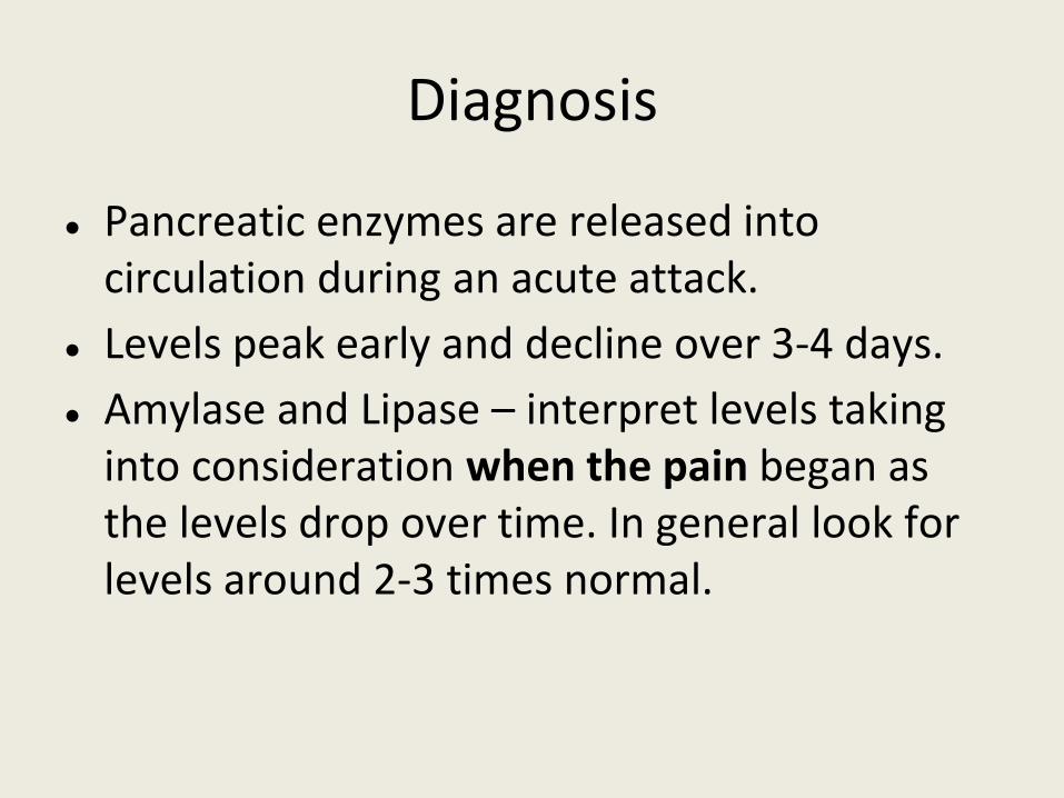
Diagnosis
● Pancreatic enzymes are released into circulation during an acute attack.
● Levels peak early and decline over 3-4 days.
● Amylase and Lipase – interpret levels taking into consideration when the pain began as the levels drop over time. In general look for levels around 2-3 times normal.

Etiology
● Most common causes are:
● Gallstones (more common in women)
● Alcohol (more common in men)
● Idiopathic (same in both) ● Probably due to occult microlithiasis.

Gallstones
● Increased serum ALT up to 3 times normal is indicative of gallstone pancreatitis.
● Normal LFTs do not exclude gallstone pancreatitis (15-20% of patients with biliary AP have normal enzymes).
● If a Biliary etiology is suspected first investigation should be an ultrasound of the abdomen which can demonstrate a dilated biliary tree and look for stones or other pathology.
● If the ultrasound is normal and the suspicion is high then an MRCP or endoscopic ultrasound can be done to look for microlithiasis or other causes of duct obstruction.
● Urgent ERCP if cholangitis present (24h), if suspected stones then early ERCP (within 72h)+

Alcohol
● Acinar cell is the main target damaged by alcohol.
● No accepted explanation for why some patients are predisposed to alcoholic pancreatitis .

Other ● Drug-induced – 2%. Normally mild. Azathioprine, sulfonamides, tetracycline, valproic acid,
didanosine, methyldopa, estrogens, lasix, 6-MP, pentamidine, steroids, octreotide.
● Post-ERCP – around 5%. Risks include female gender, a peri-ampulary diverticulum, and procedure related factors(time to cannulate…)
● Trauma – abdominal trauma causes a rise in amylase and lipase in 17% of cases and acute pancreatitis in 5%. More often due to penetrating injury.
● Infections - <1%, tend to be milder than biliary and alcohol induced AP. Viral most common (EBV, coxsackie, VZV, measles).
● Hypercalcemia - <1% of cases, due to TPN, vitamin D excessive dose, familial hypocalciuric hypercalcemia.
● Hypertriglyceridemia – usually not until TG reach 1000mg/dL, measure as soon as possible after hospitalization because fasting and IV fluid resuscitation drop TG quickly. Can be due to uncontrolled DM, alcohol, obesity.
● Developmental pancreatic – pancreas divisum, sphincter of Oddi dysfunction
● Tumor – 14% of patients with pancreatic tumors present with pancreatitis
● Postoperative – maybe due to transient hypotension or pancreatic trauma. Has a higher complication rate than other etiologies.
● Autoimmune - extremely rare cause of AP. Radiologic and histologic diagnosis with an elevated IgG4 level. Occurs in young patients with IBD, primary sclerosing cholangitis, primary biliary cirrhosis and Sjogren’s syndrome. Therapy is steroids.
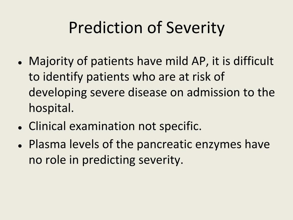
Prediction of Severity
● Majority of patients have mild AP, it is difficult to identify patients who are at risk of developing severe disease on admission to the hospital.
● Clinical examination not specific.
● Plasma levels of the pancreatic enzymes have no role in predicting severity.
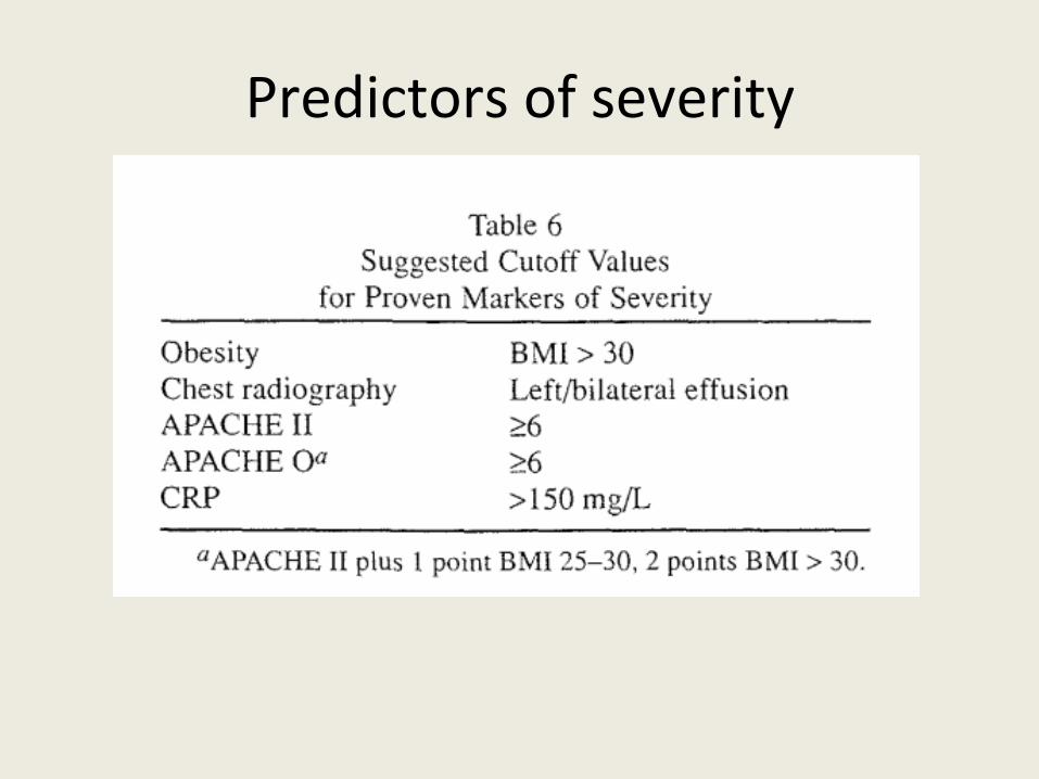
Predictors of severity

Ranson’s criteria
At admission
Age >55years
White blood cell count > 16000 cells/mm
Blood glucose >200mg/dL
Serum AST >250IU/L
Serum LDH > 350IU/L
At 48hours
Calcium <8mg/dL
Hematocrit fall >10%
Oxygen PO2 <60mmHg
GUN increases by 5 or more mg/dL after hydration
Base deficit (24-HCO3) >4mEq/L
Sequestration of fluid >6L
Risk factors <3: 1% mortality, 3-4: 15% mortality, 5-6: 40% mortality, >7: 100% mortality

Severity of Pancreatitis
Ranson's criteria APACHE 2 MODS CT- scan based severity scales Balthazar. (e.g. no necrosis or fluid collections: interstitial or mild pancreatitis - mortality <1%,
no necrosis but fluid collections: exudative pancreatitis mort <8%. Extensive necrosis: mortality >= 25%. )

Necrotic and Infected Pancreatitis
● Contrast-enhanced CT scan is the test of choice of the diagnosis of pancreatic necrosis.
● Full extent of necrosis takes at least 4 days after onset of symptoms – the longer the delay before CT scan the less likely it is to underestimate.
● Secondary infection is the most common cause of death in severe pancreatitis*.
● Mortality increases from 5-25% in patients with sterile necrosis up to 28% with infected necrosis.
● Infection occurs in 10-50% of necrotic pancreatitis.
● FNA is the only reliable method of diagnosing infected necrosis other than surgery.
● Infection is usually due to enteric flora such as E. coli
* World J. Surg. 21, 130-135, 1997

Necrotic and Infected Pancreatitis
● Prophylactic antibiotic therapy still debated.
● Trend towards improved survival and infection but not statistically significant on meta-analysis.
● Limit use to antibacterials.
● Antibiotics used should have good pancreatic penetration such as Imipenem.
● Beta lactams better than quinolones.
Antibiotic therapy for prophylaxis against infection of pancreatic necrosis in acute pancreatitis (Review) 15
Copyright © 2010 The Cochrane Collaboration.

Therapy
1. Aggressive fluid resuscitation
2. Pain control
3. Oxygen
4. Early feeding
5. Watch for infectious complications

Aggressive fluid resuscitation
Aggressive fluid resuscitation within 24 hours improves organ failure rates and decreases hospital stay++
for those with severe volume depletion 500-1000mL each hour for several hours
for those without signs of extracellular fluid loss 250-300/hour.
Reevaluate every 1-4 hours
Watch for Pulmonary Edema
Fluid needs of 5L or more are not uncommon
++Clinical Gastroenterology and Hepatology 2008;6:1070-1076

Pain control
● Uncontrolled pain can cause hemodynamic instability.
● Pain control requires opiates such as Fentanyl, morphine.
● Can use PCA pump if patient is cooperative and alert.
Oxygen
Oxygen to keep sat above 95%
* Gut 2005;54;1-9
Feeding
● Severe pancreatitis is a very catabolic state.
● Feeding may aid in preserving gut barrier and function, attenuates inflammatory response**.
● Early post-pyloric feeding with NJ with elemental or semi-elemental feeds.
● Not really beneficial in mild pancreatitis .
● If unable to feed due to ileus for >5days consider TPN*.
** Gut 1998 , 42:431-435 * Gut 2005;54;1-9

Infectious complications
● Antibiotic use (preferably imipenem) in necrotic pancreatitis if there are signs of infection* such as positive cultures, leukocytosis, fever, organ failure.
● Prophylactic antibiotics are likely ineffective in reducing complications**
● Especially true for patients with <30% necrosis
● Necrostomy if patient fails to improve after antibiotics and attempts at percutaneous drainage.
●endoscopic US guided drainage
●CT guided drainage

fin