Embed Size (px)
Citation preview

Clinical Science (1972) 43, 731-742.
INTESTINAL STRUCTURE AND FUNCTION
AFTER SMALL BOWEL BY-PASS I N THE RAT
M. H. GLEESON, J. CULLEN AND R. H. DOWLING
Departments of Medicine and Morbid Anatomy, Royal Postgraduate Medical School, London
(Received 14 April 1972)
SUMMARY
1. To study further the influence of luminal nutrition on small bowel structure and function, segments of rat jejunum and ileum were completely excluded from intestinal continuity by Thiry-Vella by-pass operations. The effect of partial deprivation of luminal nutrition was also studied in jejunal segments that had been surgically transposed to a distal position in the intestinal tract.
2. Macroscopically, by-passed jejunum and ileum both became narrowed and atrophic, whereas the intestine in continuity showed hypertrophic changes similar to those seen after intestinal resection.
3. In by-passed intestine the pattern of villi changed from mucosal ridges to ‘fingers’ and ‘leaves’. Although villous height and total mucosal thickness were both reduced, light microscopy showed that even 6 months after exclusion from intestinal continuity villous morphology was still retained.
4. Epithelial-cell migration rate and turnover time were diminished in both jejunum and ileum after by-pass. In intestine in continuity the migration rate was increased, but the turnover time remained the same as in controls.
5. In by-passed jejunum, in vivo glucose absorption diminished progressively with time, but did not change in excluded ileal segments.
6. After ileo-jejunal transposition, the jejunum showed no major morphological changes, but glucose absorption became significantly depressed.
7. These results further support the concept that intraluminal nutrition exerts a major influence on cell turnover, villous morphology and absorptive function in the small intestine.
Key words : intestinal by-pass, intestinal morphology, intestinal glucose absorption, cell turnover, intra-luminal nutrition, rat.
In the normal intestine, because of jejunal absorption, relatively little nutrient or unabsorbed
Correspondence: Dr R. Hermon Dowling, Department of Medicine, Royal Postgraduate Medical School, Ducane Road, London, W.12.
73 1

732 M . H . Gleeson, J . Cullen and R. H . Dowling
food remains in the chyme when it reaches the ileum (Borgstrom, Dahlqvist, Lund & Sjovall, 1957). After proximal resection, however, and when the positions of jejunum and ileum have been transposed, the ileum receives food directly from stomach and duodenum. In both these situations the ileum shows marked hypertrophy (Booth, Evans, Menzies & Street, 1959; Dowling & Booth, 1967; Nygaard, 1967) and enhanced absorption (Dowling & Booth, 1967). Based on this and other evidence, it was postulated that intraluminal nutrition is an important factor in regulating mucosal growth and absorptive capacity in the small bowel (Dowling, 1967).
If this hypothesis is correct, intestine deprived of intraluminal nutrition should become atrophic. (The terms ‘hypertrophy’ and ‘atrophy’ are used to described the macroscopic appearance of the intestine. At a cellular level, the terms ‘hyper-’ and ‘hypo-plasia’ are more appropriate.) To test this theory, major segments of either proximal or distal small bowel were excluded from normal intestinal continuity by chronic Thiry-Vella fistulae. The macro- scopic, dissecting and light microscopic appearance of the excluded segments of jejunum and ileum were compared with comparable segments in control animals at different times after surgery. The structural changes found were related to the migration rate and turnover time of the intestinal epithelium, as measured radioautographically with tritiated thymidine, and to glucose absorption measured in vivo in both control and by-passed intestine.
The effects of complete exclusion of topical nutrition from the intestinal lumen as in Thiry- Vella by-passed segments were also compared with partial deprivation of intraluminal nutrition, as in distally sited jejunum after ileo-jejunal transposition.
This paper describes our findings and extends preliminary reports of this work (Gleeson, Cullen, Collins & Dowling, 1969; Gleeson, Peters, White & Dowling, 1970). The results provide further evidence for the hypothesis that intraluminal nutrition is of major importance in regulating mucosal growth and absorptive function in the small bowel.
MATERIALS A N D METHODS
Animals and diets An inbred strain of female albino Wistar rats was used, weighing 150-220 g at the time of
surgery. The animals were starved for 24 h before operation but were allowed free access to water. After surgery, glucose (5 g/lW ml) was given by mouth for 36 h before they resumed a standard pellet diet (Diet 41B as supplied by Messrs E. Dixon and Sons, Crane Mead Mills, Ware, Herts.).
Surgical procedures (a) Small bowel by-pass. The technique was similar to that used for small bowel resection
(Dowling & Booth, 1967) except that the excluded segment was retained in the abdominal cavity as a Thiry-Vella fistula. Groups of rats were subjected to by-pass of between half and two-thirds of either proximal or distal small bowel. Under Penthrane anaesthesia (Methoxy- flurane, Abbott Laboratories, Queenborough, Kent), the abdomen was opened through a mid-line incision.
In the proximal by-pass group, the intestine was divided 2-3 cm below the ligament of Treitz and again approximately two-thirds of the way down the small bowel. Intestinal

Efect of by-pass on rat small bowel 733 continuity was re-established with an end-to-end anastomosis between the jejunal stump and the remaining ileum, using a continuous 510 atraumatic black silk suture. The severed ends of the by-passed segment of intestine (with intact blood and nerve supply) were brought to the abdominal surface through separate stab wounds, and chronic Thiry-Vella fistulae created with double muco-cutaneous stomata.
For the distal by-pass group the same procedure was followed except that the intestine was divided about 2 cm above the ileo-caecal valve and approximately two-thirds of the way up the small bowel from the first division site. The by-passed Thiry-Vella segment in t h i s case was therefore distal small intestine.
An equal number of rats, matched for age, sex and weight, were subjected to simple division and resuture of the intestine and served as controls.
(b) Ileo-jejunal transposition. This operation was successfully carried out in six animals. The technique has been described (Dowling & Booth, 1967) but, briefly, the positions of the jejunum and ileum were interchanged so that the jejunum came to occupy a distal site in the intestinal tract and received chyme after ileal absorption.
Intestinal morphology Macroscopic features. At various times after creating the Thiry-Vella fistulae, the abdomen
was re-opened to examine the structural and/or functional changes in both by-passed intestine and the bowel which remained in continuity.
Microscopic features. Tissue blocks of everted intestine were pinned flat on cork and fixed in 10% (w/v) formol saline. The blocks were taken from the proximal third of by-passed jejunum, from the distal third of by-passed ileum and from comparable sites in control animals. In a small number of rats blocks were also taken from the intestine which remained in con- tinuity.
Dissecting microscopy. Changes found in overall villous morphology in by-passed jejunum and ileum were noted on dissecting microscopy and compared with both adult and weanling small bowel mucosa in control rats.
Light microscopy. Histological sections (4 pm thick) were cut at right angles to the long axis of the intestine (the adult rat has transverse villous ridges), and stained with Haematoxylin and Eosin. A micrometer eye-piece was used to measure villous height (from the villus tips to crypt mouths) and total mucosal thickness (from villus tips to muscularis mucosa, thus including the sub-villus crypt layer) on the ten tallest villi in each section, and the mean reading (pm) recorded. In the proximal by-pass group six rats were studied at less than 6 weeks after creating the Thiry-Vella fistulae, eight rats between 6 and 10 weeks and eight rats between 10 weeks and 7 months, and the results compared with eighteen control animals. Normal ileal morphology was studied in nineteen control rats and compared with results in eight rats studied at less than 10 weeks and in eight rats between 10 weeks and 7 months after their original distal by-pass.
Intestinal function Absorptive function. As an index of absorptive function glucose absorption was measured
from a Krebs-Ringer bicarbonate buffer (Krebs & Henseleit, 1932) containing 21 mM-glucose and polyethylene glycol (mol. wt. 4OOO) as a non-absorbable indicator. A modification (Dowling & Booth, 1967) of an in vivo recirculation perfusion system (Sheff & Smyth, 1955)

734 M. H . Gleeson, J . Cullen and R. H . Dowling
was used to measure absorption in 20-40 cm segments of by-passed intestine and in comparable segments of control animals as previously described (Dowling & Booth, 1967).
Glucose absorption was measured in excluded jejunal segments of twenty-two animals with proximal by-pass and in the ileum of fourteen rats with distal by-pass at times ranging from 2 to 25 weeks after surgery. Absorption by comparable jejunal and ileal segments was also measured in a matched number of control rats. The same technique was used to measure glucose absorption in six rats with ileo-jejunal transposition between 3 and 8 months after surgery, except that absorption was measured in both jejunal and ileal segments simul- taneously.
Cell turnover. Since villous morphology and absorptive function may both be related to epithelial cell turnover, the migration rate and turnover time of the intestinal epithelium was measured radioautographically using tritiated thymidine (Leblond & Stevens, 1948).
The animals were studied 3 months after surgery, when maximal changes in villous mor- phology and absorptive function had become established. [3H]Thymidine (75 pCi/lOO g body wt.) was injected intraperitoneally and the animals were then divided into two groups. The first group was killed 2 h, and the second group 23 h, after thymidine administration. At 2 h the dividing crypt cell population is labelled but these labelled cells have not yet commenced migration along the villus : at 23 h the cells have invariably migrated some distance along the villus, but the leading edge of labelled cells has not yet reached the villus tip (Loran & Althausen, 1960).
The tissue blocks (from sites comparable to those chosen for the histological studies) were again fixed in formol-saline, and embedded in paraffin. Sections 4 pm thick were cut, mounted on slides, dipped in Ilford Nuclear Emulsion K5, and stored in sealed light-proof boxes for 4 weeks at 4°C. They were then developed in Johnson contrast FF developer and lightly stained with Haematoxylin and Eosin.
Cell turnover time and migration rate were measured in nine control rats (both jejunum and ileum), in seven animals with proximal by-pass and in four rats with excluded ileum. In addition, intestine in continuity was studied in eight rats with proximal by-pass. For technical reasons only two sections of jejunum in continuity 23 h after thymidine were suitable for measurement.
The following measurements were made with a micrometer eye-piece on ten villi in each section: (1) Base of crypt to tips of villi (distance A). (2) Base of crypt to leading edge of labelled cells 2 h after [jHIthymidine (distance B). (3) Base of crypt to leading edge of labelled cells 23 h after [3H]thymidine (distance C).
Epithelial cell turnover time and migration rate were then calculated as follows : A-B = ‘effective’ villous height, that is the total distance to be travelled by migrating
C-B = migration rate of cells in (pm/21 h) and hence migration rate/h is calculated. 21 x [(A-B)/(C-B)] = cell turnover rate (h) assuming a constant migration rate, the time
cells (pm).
required by dividing cells migrating from the crypts to reach the villous tips.
RESULTS Intestinal morphology
Macroscopic features. The appearance of excluded intestine at times less than 4 weeks after by-pass was variable, but frequently the excluded intestine appeared normal or even enlarged.

Efect of by-pass on rat small bowel
FIG. 1 . Macroscopic appearance of small bowel after by-pass. The control jejunum (3), is con- trasted with the narrow and contracted appearance of by-passed jejunum (2) 8 weeks after proximal Thiry-Vella by-pass. The enlarged and dilated ileum (1) after proximal by-pass is also shown. The scale is graduated in inches.
(Facing p . 734)

M. H. Gleeson, J . Cullen and R. H. Dowling
FIG. 2. Dissecting microscope appearances of jejunal and ileal mucosa in control and by-passed rat intestine. (a) Normal adult jejunum, x 90. (b) Normal adult ileum, x 90. (c) By-passed jejunum in adult rat, x 90. (d) By-passed ileum in adult rat, x 50. (e) Weanling rat jejunum, x 90.

Efect of by-pass on rat small bowel 735 However, 4 weeks after surgery, by-passed intestine appeared narrow and contracted (Fig. 1). By contrast, the appearance of the intestine which remained in continuity was similar to that seen in the residual intestine following small bowel resection (Dowling & Booth, 1967). After by-pass the whole of the intestine which remained in continuity was dilated and hyper- trophied, but the enlargement was greater near the anastomosis and tapered more distally. Again, as was found after resection, the ileal hyperplasia after proximal by-pass was much more marked than jejunal hyperplasia following ileal exclusion. Microscopic features. Dissecting microscopy. On dissecting microscopy the villi of the normal
adult rat jejunum appear mainly as long, transverse ridges with a few broad leaves (Fig. 2a). In the normal ileum (Fig. 2b) the ridges are shorter and the predominant villous morphology is broad leaves with occasional tongue-shaped villi.
The villous architecture changed in by-passed intestine. In by-passed jejunum the ridges became noticeably shorter and there were many more tongue-like and occasional finger-like villi (Fig. 2c). The changes in excluded ileum were more striking, most of the villi becoming finger-shaped or tongue-like (Fig. 2d). In both by-passed segments, therefore, villous archi- tecture tended to revert to the appearance seen in the normal weanling rat intestine where the villi are finger-like (Fig. 2e).
There were no significant changes in the dissecting microscopic appearance ofjejunum which remained in continuity following distal by-pass. However, in the ileum which remained in continuity when the jejunum was excluded, the villous morphology became indistinguishable from the normal adult rat jejunum, with a uniform appearance of broad villous ridges.
Light microscopy. Despite the dissecting microscopic changes of the villi and despite an undoubted reduction in mucosal wet weight, protein and DNA content/unit length of by- passed intestine (Gleeson, Dowling & Peters, 1972) there were remarkably few changes in mucosal structure that were detectable by light microscopy. However, histological sections from by-passed segments of intestine often showed increased thickness of the circular and longitudinal muscle layers, presumably a result of contraction of the intestinal lumen.
The results of histological measurements of villous height and total mucosal thickness are shown in Fig. 3. In by-passed jejunum measurements of villous height and total mucosal thickness were both reduced when compared with control jejunum although the differences were not statistically signilkant. The mean villous height in by-passed jejunum was 354 ( f SEM 20) pm compared with 393 (+24) pm in control intestine. The corresponding figures for total mucosal thickness were 537 (* 27) and 567 ( f 33) pm. In the distal by-pass group the mean villous height (240f 13 pm) and total mucosal thick-
ness (360f 18 pm) of excluded ileum were both significantly less than the corresponding measurements in control ileum (287 f 15 and 420 f 16 pm respectively) and these differences were both statistically significant (P< 0.05). In a small number of animals measurements of total mucosal thickness and villous height
were made in the intestine which remained in continuity after the by-pass. For the jejunum in continuity (five animals) the mean total mucosal thickness was 655 ( 5 32) and villous height 442 ( f 27) pm. Again, as in the ileal remnant following proximal resection, the most striking changes were seen in the ileum which remained in continuity after excluding the upper small bowel. In ileum in continuity (seven animals) both total mucosal thickness (mean 576143) pm and villous height (396+ 38) pm were significantly greater than corresponding control values (P<O.Ool).
736 M. H. Gleeson, J . Cullen and R. H. Dowling
(a 1
Control By - poss
600
500
400
5 300
200
100
0
(b)
Control By- pass
t [ , I , r =2.43 PC0.05
FIG. 3. Mean (k SEM) villous height (cross-hatched columns) and total mucosal thickness (cross-hatched+ stippled columns) in control and by-passed jejunum (a) and ileum (b).
Control
0 1
By-POSS
<6 weeks . 0 .
- . 0 . - .
t = 0.75 P<0.5 N S
6 -9 weeks
0%
c-4 . - - . .
t = 1.37 P<0.2 N S
10-25 weeks
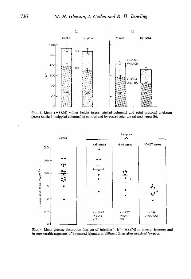
Fm. 4. Mean glucose absorption (mg cm of intestine-' h-' +SEM) in control jejunum and in comparable segments of by-passed jejunum at different times after proximal by-pass.
Efect of by-pass on rat small bowel 737
Intestinal function Absorptive function. The results of glucose absorption from both control and by-passed
jejunum at different times following surgery are shown in Fig. 4. The mean glucose absorption in control jejunum was 1-98 (kO.09) mg cm-' h-'. For the first 6 weeks after by-pass jejunal glucose absorption was not significantly different from control values (2.13 f 0.19 mg cm- ' h-I). Between 6 and 9 weeks the mean glucose absorption was reduced (1-74+0.13 mg cm-' h-') but this difference was not statistically significant. However, after 10 weeks of exclusion from normal continuity, glucose absorption from by-passed jejunum had fallen almost to ileal values, the mean of 1-26+0.07 mg cm-' h-' being less than control jejunal absorption
By contrast, glucose absorption from by-passed ileum did not change with time. Even after a 6 month interval absorption was not significantly different from control ileal values, the mean
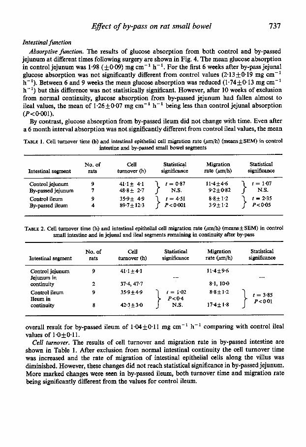
TABLE 1. Cell turnover time (h) and intestinal epithelial cell migration rate (pm/h) (means* SEM) in control intestine and by-passed small bowel segments
(P< 0001).
No. of Cell Statistical Migration Statistical Intestinal segment rats turnover (h) significance rate @/h) significance
Control jejunum 9 41-1+ 41 By-passed jejunum 7 48.8k 2.1 Control ileum 9 35.9f 4.9 By-passed ileum 4 89.7 f 12.3
} t=&Y 11-4f46 9.2 f 0.82 8.8 + 1.2 t = 4.51
P < 0.001 3.92 1.2 t = 2.35 P<005
} tkoT
TABLE 2. Cell turnover time (h) and intestinal epithelial cell migration rate (pm/h) (meansf SEM) in control small intestine and in jejunal and ileal segments remaining in continuity after by-pass
No. of cell Statistical Migration Statistical Intestinal segment rats turnover (h) significance rate @/h) significance
~
Control jejunum 9 41.1 +41 11-4 f 9.6 - - Jejunum in
continuity 2 37.4, 47.7 8.1, 10.0
t = 3-85 P<001
8.8f1.2
17.4f 1.8
Control ileum 9 35-9+49 Ileum in continuity 8 42.3 f 3.0
overall result for by-passed ileum of 1-04+0*11 mg cm-' h-' comparing with control ileal values of 1.0f0.11.
Cell turnover. The results of cell turnover and migration rate in by-passed intestine are shown in Table 1. After exclusion from normal intestinal continuity the cell turnover time was increased and the rate of migration of intestinal epithelial cells along the villus was diminished. However, these changes did not reach statistical significance in by-passed jejunum. More marked changes were seen in by-passed ileum, both turnover time and migration rate being significantly different from the values for control ileum.

738 M. H . Gleeson, J . Cullen and R. H . Dowling
Table 2 shows the results for cell turnover and migration rate in the residual intestine remaining in continuity after by-pass. Again, in the two animals suitable for study, there was no obvious difference between normal jejunum and the slightly hyperplastic jejunum in continuity. However, in the markedly enlarged ileum in continuity, the mean cell migration rate was doubled, increasing from 8.8 2 1.2 pm/h in control to 17-4Ifi 1.8 pm/h in the hyper- plastic intestine, and even though the distance to be travelled along the enlarged villus was greater, the turnover time was not significantly different from the controls.
Control Jejunum
0
0.0
0 0
Transposed jejunum
0 .
0 - H _L
0
t = 2.30
P< 0.05
I
Control ileum
0
0
- *
Transposed ileum
0
43
0
f =4.33 P<O.Ol
I
FIG. 5. Mean glucose absorption (mg cm of intestine- h- f SEM) in control jejunum compared with transposed jejunum and in control ileum compared with transposed ileum.
Eflects of ileo-jejunal transposition The morphological changes in transposed ileum 1 month after ileo-jejunal transposition
were described by Dowling & Booth (1967) and are confirmed in the present study, 3-8 months post-operatively.
Macroscopically, transposed ileum appeared dilated and hyperplastic. This was confirmed by histological measurements which showed significant increases in total mucosal thickness and villous height when compared to controls.
Transposed jejunum, however, was unchanged in its macroscopic appearance and histo- logical measurements were not significantly different from control jejunum, even 8 months after the original operation.
The results of glucose absorption studies are shown in Fig. 5. As found previously, 1 month after transposition there was a marked and significant increase in glucose absorption from the hyperplastic transposed ileum, the mean absorption of 1.92+0.15 mg cm-' h-' comparing with control ileal values of 0.92+0.15.
No reduction in jejunal glucose absorption was observed in previous studies after 1 month of transposition, but the present studies showed that 3-8 months after operation, mean glucose

Efect of by-pass on rat small bowel 739 absorption had fallen from 2.19+0.12 mg cm-I h-l for control jejunum to 1.64f0.17 in the transposed jejunum, and this difference was statistically significant (P -= 0.05).
DISCUSSION
The results in this paper give additional support for the hypothesis that intraluminal nutrition plays an important role in regulating mucosal growth, villous morphology and absorptive function in the small intestine.
There have been few previous studies of intestinal structure after its exclusion from continuity. Several groups have studied intestinal segments defunctionalized as self-emptying blind loops, but with conflicting results. Riecken, Menge, Bloch & Schaumloffel(l970) found that 2 months after surgery the mucosa of self-emptying blind jejunal loops had become hypo- plastic, but Altmann & Leblond (1970) found hyperplasia in self-emptying ileal loops draining into the colon. Such self-emptying blind loops could conceivably be influenced by ascending bacterial ‘infection’ and clearly these experimental models are not strictly comparable to Thiry-Vella fistulae where there is no direct contact with the bowel in continuity.
In the present studies no bacteriological investigations were done, and it will be of interest to see the influence of bacteria, mechanical trauma and individual nutrients on mucosal structure and function in by-passed intestine.
Villous morphology has previously been studied in Thiry-Vella segments of by-passed rat jejunum 4-12 months after surgery (Chacko, Mathan & Baker, 1968). These authors found that in the by-passed segments the adult pattern of transverse mucosal ridges reverted towards a more foetal pattern with linger- and tongue-like villi. The present study confirms these findings and extends them by examining not only by-passed jejunum, but also both excluded ileum and the intestine which remained in continuity after the by-pass. In fact, the reversion to a foetal type of villous pattern was more pronounced in by-passed ileum than in jejunum. In contrast, the ileum in continuity following proximal by-pass became indistinguishable from adult rat jejunum with transverse mucosal ridges.
Although in our experience the intestinal mucosa does eventually become hypoplastic after small bowel by-pass (Gleeson et al., 1972) it is remarkably resistant to change. Clarke, Miller & Shields (1967) found no changes in villous morphology of Thiry-Vella loops 4 weeks after surgery in the dog. Similarly, studies of human ileum excluded from intestinal continuity as urinary conduits show that villous architecture is preserved for long periods (Joseph & Thomas, 1958 ; Shoemaker, Tedeschi & Gotzinger, 1965). Goldstein, Melamed, Grabstald & Sherlock (1967) did, however, find progressive villous atrophy in ileal ‘bladders’ but clearly the long-term effects of urine on the intestinal mucosa makes comparison with the present study difficult.
There have been no previous studies of intestinal absorption after Thiry-Vella by-pass in the rat. However, absorptive function has been measured in proximal and distal intestinal segments excluded from normal continuity as short, self-emptying blind loops (Riecken et nl., 1970). Although their experimental design was not strictly comparable with the present study, overall the results of Riecken et al. (1970) agree with the present findings. They studied struc- ture and function at only one period of time (2 months) after by-pass and showed reduced villous height and diminished glucose absorption in the self-emptying jejunal blind loops. The present study clearly indicates that the absorptive capacity of by-passed jejunum for
B

740 M. H. Gleeson, J . Cullen and R. H. Dowling
glucose is maintained for up to 6 weeks after surgery. Between 6 and 9 weeks absorption was impaired, but the maximal reduction in glucose absorption was not apparent until 10 weeks or longer after proximal by-pass. This contrasts with the relatively rapid onset of enhanced glucose absorption seen in the remaining intestine following small bowel resection, which was found as soon as 2 weeks after surgery, and was maximal at 1 month (Dowling & Booth, 1967).
The difference in the degree of response between proximal and distal small bowel after by-pass again contrasts with the response of jejunum and ileum after small bowel resection (Booth et al., 1959; Dowling & Booth, 1967). After resection the ileum shows a much greater degree of mucosal hyperplasia and enhanced absorption than the jejunum. But after by-pass the reduced absorption was maximal in the by-passed jejunum, and even prolonged periods of exclusion from normal continuity did not affect ileal glucose absorption.
The present ileo-jejunal transposition studies confirm the observation (Dowling & Booth, 1967) that when the ileum is transposed to a proximal situation, it absorbs glucose as efficiently as the normal jejunum. By contrast the absorptive capacity of transposed jejunum, receiving chyme which has been partially absorbed by the proximally sited ileum, is ultimately depressed, although not to ileal values.
The present results also demonstrate that intraluminal chyme influences the rate of epithelial cell migration. The ileum in continuity had a markedly increased cell migration rate, and in spite of the increase in villous length to be traversed by migrating cells, the turnover time was not significantly different from normal. In fact, Loran & Althausen (1960) actually noted a decrease in cell turnover time after small bowel resection. If, as has been suggested, villous height is regulated by the rate of cell production with a steady state between cell production in the crypts and cell loss at the tips of the villi (Creamer, 1967) it remains possible that once an enlarged villus has been produced, a new steady state is achieved in which the duration of cell turnover reverts to its previous value. This can obviously be produced by an increased rate of epithelial cell migration along an increased villous length.
Although the present discussion has emphasized intraluminal nutrition, many other physiological factors may influence mucosal structure and absorptive function. For example, as a result of studies in the rat Loran & Althausen (1968) and Tilson & Wright (1970) have postulated that there is a circulating intestinal growth hormone, while Altmann & Leblond (1970) have suggested that the pylorus and duodenum may produce villus-enlarging factors. We have shown that in man a glucagon-like hormone produced by a renal tumour caused marked trophic changes in the small bowel mucosa (Gleeson, Bloom, Polak, Henry & Dowling, 197 1). However, the present results showing that intestinal hyperplasia and hypoplasia may occur simultaneously argue against the general effect of a circulating intestinal growth hormone. Whether locally produced hormones, perhaps stimulated by intraluminal contents, may stimulate cell turnover and mucosal growth remains conjectural.
Considering the present results and the evidence of previous publications (for review, see Dowling, 1970) the following theory about intraluminal nutrition may be proposed. Nutrients, derived from the intestinal lumen, maintain cell turnover rate, villous height and glucose absorption at a high rate in the jejunum. In the ileum, because of proximal absorption, rela- tively little nutrition remains in the intestinal lumen and as a result cell turnover, villous size and absorptive capacity are considerably less than in proximal small intestine. The cell migration rate and absorptive capacity of the normal ileum may therefore represent the basal

Efect of by-pass on rat small bowel 741 value for the intestine. However, when stimulated by a relative increase in luminal nutrition (as occurs, for example, following proximal small bowel resection or after ileo-jejunal trans- position), the ileum retains the potential to increase its cell turnover and absorptive capacity up to values found in normal jejunum. By contrast, when the jejunum is deprived of its normal complement of intraluminal nutrition, the cell proliferation rate gradually reduces, villous height decreases, mucosal mass diminishes and absorptive function falls to basal ileal values.
ACKNOWLEDGMENTS
We thank Mrs June White for technical assistance, and Mrs Judy Mepham and Mr Brian Chalk from the Research Unit of the Department of Morbid Anatomy for their help in pre- paring histological sections and radioautographs, Mr Bill Brackenbury from the Department of Medical Illustration for the dissecting microphotographs and Mrs Hazel Creed for typing the manuscript. Thanks are also due to the M.R.C. for financial support to the Intestinal Malabsorption Group (R.H.D.).
REFERENCES
ALTMANN, G.G. & LEBLOND, C.P. (1970) Factors influencing villus size in the small intestine of adult rats as revealed by transposition of intestinal segments. American Journal of Anatomy, 127, 15-36.
BOOTH C.C., EVANS, K.T., MENZIES, T. &STREET, D.F. (1959) Intestinal hypertrophy following partial resection of the small bowel in the rat. British Journal of Surgery, 46,403-410.
BORGSTROM, B., DAHLQVIST, A., L m , G. & SJOVALL, J. (1957) Studies of intestinal digestion and absorption in the human. Journal of Clinical Investigation, 36, 1521-1536.
CHACKO, C.J.G., MATHAN, V.I. & BAKER, S.J. (1968) Changes in the mucosal pattern of isolated loops of jejunum in albino rats. A dissection microscope study. British Journal of Experimental Pathology, 49,4043.
CLARKE, A.M., MILLER, M. & SHIELDS, R. (1967) Intestinal transport of sodium, potassium and water in the dog during sodium depletion. Gastroenterology, 52, 846-858.
CREAMER, B! (1967) The turnover of the epithelium of the small intestine. British Medical Bulletin, 23,226-230. DOWLING, R.H. (1967) Compensatory mechanisms in the small intestine. British Medical Bulletin, 23,275-278. DOWLING, R.H. (1970) Small bowel resection and by-pass-recent developments and effects. Modern Trendr
in Gastro-enterology (Ed. by W. I. Card and B. Creamer), Vol. 4, pp. 73-104. Butterworths, London. DOWLING, R.H. & BOOTH, C.C. (1967) Structural and functional changes following small intestinal resection
in the rat. Clinical Science, 32, 139-149. GLEESON, M.H., BLOOM, S.R., POLAK, J.M., HENRY, K. & DOWLING, R.H. (1971) Endocrine tumour in kidney
affecting small bowel structure, motility and absorptive function. Gut, 12, 773-782. GLEESON, M.H., CULLEN, J., COLLINS, J. & DOWLING, R.H. (1969) Structural and functional changes in rat
jejunum and ileum after surgical exclusion from normal intestinal continuity. (Abstract). Gut, 10, 1057. GLEESON, M.H., DOWLING, R.H. & PETERS, T.J. (1972) Biochemical changes in intestinal mucosa after experi-
mental small bowel by-pass in the rat. Clinical Science, 43, 743-757. GLEESON, M.H., PETERS, T.J., WHITE, J. & DOWLING, R.H. (1970) Digestive and absorptive function of proximal
and distal small bowel in the rat following exclusion from normal continuity. Abstracts of the Fourth World Congress of Gastroenterology, 12th-18th July 1970, Copenhagen, p. 331.
GOLDSTEIN, M.J., MELAMED, M.R., GRABSTALD, H. & SHERLOCK, P. (1967) Progressive villous atrophy of the ileum used as a urinary conduit. Gastroenterology, 52, 859-864.
JOSEPH, J. & THOMAS, G.A. (1958) The behaviour of autografts of ileum transplanted into urinary bladder h rabbits. Journal of Anatomy, 92, 551-558.
KREBs, H.A. & HENSELEIT, K. (1932) Untersuchungen iiber die Harmstoffbildung im Tierkorper. Hoppe Seyler's Zeitshrift fur physiologische Chemie, 210, 33-36.
LEBLOND, C.P. & STEVENS, C.E. (1948) The constant renewal of the intestinal epithelium in the albino rat. Anatomical Record, 100,357-378.

742 M. H . Gleeson, J. Cullen and R. H. Dowling LORAN, M.R. & ALTHAUSEN, T.L. (1960) Cellular proliferation of intestinal epithelia in the rat two months after
partial resection of the ileum. Journal of Biophysical and Biochemical Cytology, 7, 667-671. JARAN, M.R. & ALTHAUSEN, T.L. (1968) The humoral effect of intestinal resection on cellular proliferation
and maturation in parabiotic rats. Gastrointestinal Radiation Injury (Ed. by M. F. Sullivan), pp. 127-139. Excerpta Medica Foundation, Amsterdam.
NYGAARD, K. (1967) Resection of the small intestine in rats. 111. Morphological changes in the intestinal tract. Acta Chirurgica Scandinavica, 133, 233-248.
RIECKEN, E.-O., MENGE, H., BLOCH, R. & SCHAUML~FFEL, E. (1970) Transport studies, morphological, histo- chemical and morphometric findings on the excluded jejunal loop in the rat. Abstracts of the Fourth World Congress of Gastroenterology, 12th-18th July, 1910, Copenhagen, p. 448.
SHEFP, M.F. & SMYTH, D.H. (1955) An apparatus for the study of in vivo intestinal absorption in the rat. Journal of Physiology, 128, 6 7 ~ .
SHOEMAKER, W.C., TEDESCHI, C.G. & GOTZINGER, P.J. (1965) Morphological changes of intestinal graft used in bladder reconstruction. Archives of Surgery, 72, 525-529.
TILSON, M.D. & WRIGHT, H.K. (1970) Adaptation of functioning and by-passed segments of ileum during compensatory hypertrophy of the gut. Surgery, 67, 687-693.