Embed Size (px)
Citation preview
INTERPRETATION OF POLYSOMNOGRAPHY
DR ANKAN BANDYOPADHYAY
SR PULMONARY MEDICINE
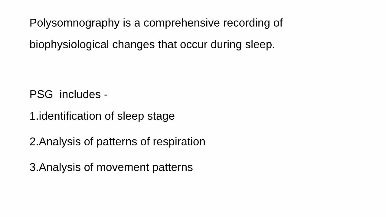
Polysomnography is a comprehensive recording of
biophysiological changes that occur during sleep.
PSG includes -
1.identification of sleep stage
2.Analysis of patterns of respiration
3.Analysis of movement patterns
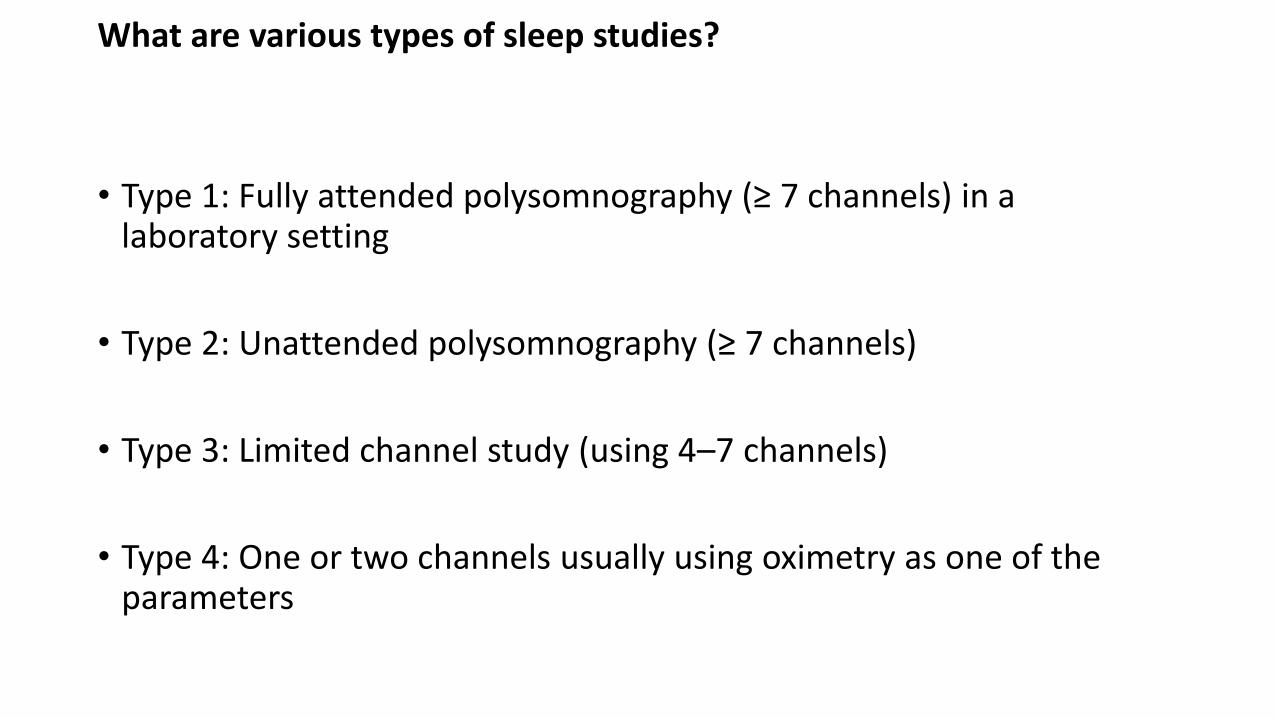
What are various types of sleep studies?
• Type 1: Fully attended polysomnography (≥ 7 channels) in a laboratory setting
• Type 2: Unattended polysomnography (≥ 7 channels)
• Type 3: Limited channel study (using 4–7 channels)
• Type 4: One or two channels usually using oximetry as one of the parameters
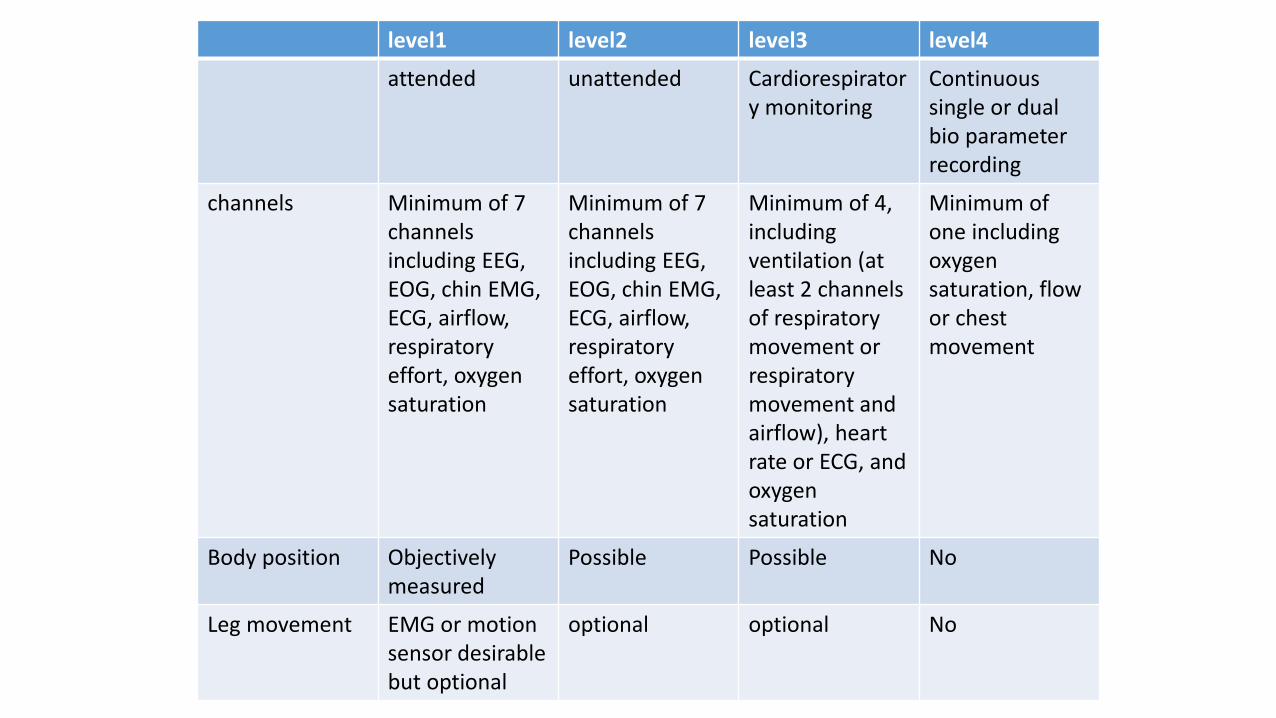
level1 level2 level3 level4
attended unattended Cardiorespiratory monitoring
Continuous single or dual bio parameter recording
channels Minimum of 7channelsincluding EEG,EOG, chin EMG,ECG, airflow,respiratoryeffort, oxygensaturation
Minimum of 7channelsincluding EEG,EOG, chin EMG,ECG, airflow,respiratoryeffort, oxygensaturation
Minimum of 4,includingventilation (atleast 2 channelsof respiratorymovement orrespiratorymovement andairflow), heartrate or ECG, andoxygensaturation
Minimum of one including oxygen saturation, flow or chest movement
Body position Objectivelymeasured
Possible Possible No
Leg movement EMG or motion sensor desirable but optional
optional optional No
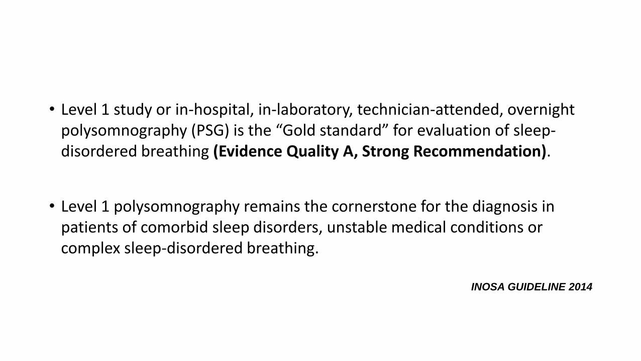
• Level 1 study or in-hospital, in-laboratory, technician-attended, overnight polysomnography (PSG) is the “Gold standard” for evaluation of sleep-disordered breathing (Evidence Quality A, Strong Recommendation).
• Level 1 polysomnography remains the cornerstone for the diagnosis in patients of comorbid sleep disorders, unstable medical conditions or complex sleep-disordered breathing.
INOSA GUIDELINE 2014
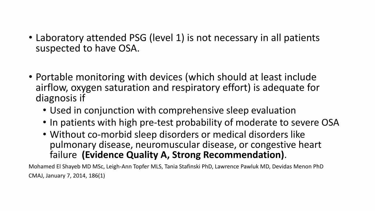
• Laboratory attended PSG (level 1) is not necessary in all patients suspected to have OSA.
• Portable monitoring with devices (which should at least include airflow, oxygen saturation and respiratory effort) is adequate for diagnosis if
• Used in conjunction with comprehensive sleep evaluation• In patients with high pre-test probability of moderate to severe OSA• Without co-morbid sleep disorders or medical disorders like
pulmonary disease, neuromuscular disease, or congestive heart failure (Evidence Quality A, Strong Recommendation).
Mohamed El Shayeb MD MSc, Leigh-Ann Topfer MLS, Tania Stafinski PhD, Lawrence Pawluk MD, Devidas Menon PhD
CMAJ, January 7, 2014, 186(1)
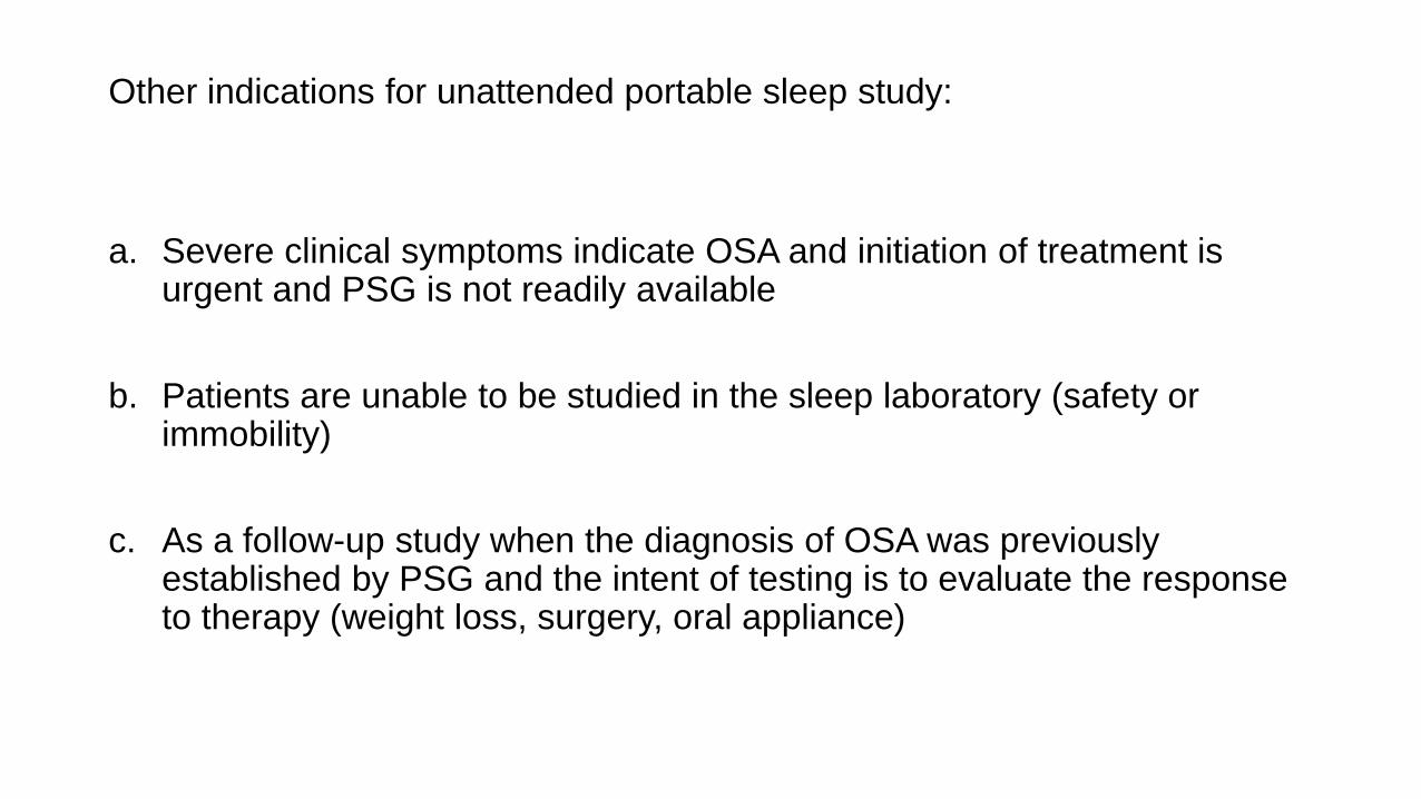
Other indications for unattended portable sleep study:
a. Severe clinical symptoms indicate OSA and initiation of treatment is urgent and PSG is not readily available
b. Patients are unable to be studied in the sleep laboratory (safety or immobility)
c. As a follow-up study when the diagnosis of OSA was previously established by PSG and the intent of testing is to evaluate the response to therapy (weight loss, surgery, oral appliance)
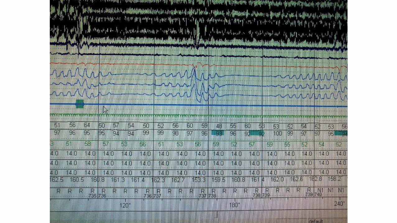
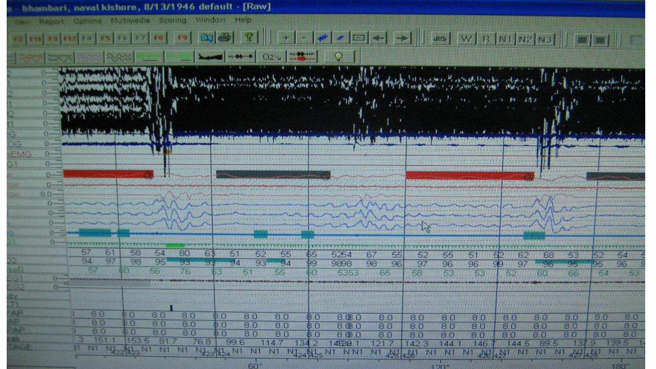
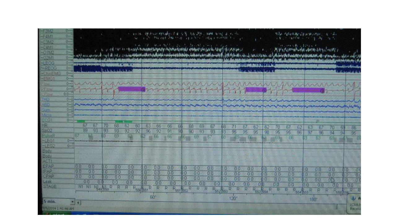
INTERPRETATION AND MONITORING OF RESPIRATORY EVENTS
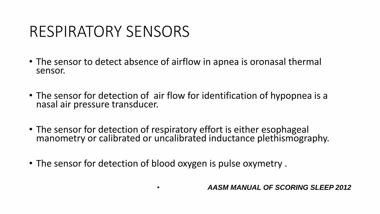
RESPIRATORY SENSORS
• The sensor to detect absence of airflow in apnea is oronasal thermal sensor.
• The sensor for detection of air flow for identification of hypopnea is a nasal air pressure transducer.
• The sensor for detection of respiratory effort is either esophageal manometry or calibrated or uncalibrated inductance plethismography.
• The sensor for detection of blood oxygen is pulse oxymetry .
• AASM MANUAL OF SCORING SLEEP 2012
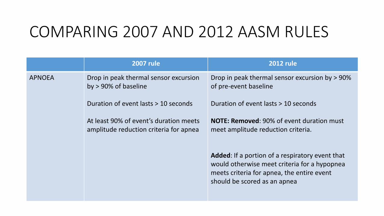
COMPARING 2007 AND 2012 AASM RULES
2007 rule 2012 rule
APNOEA Drop in peak thermal sensor excursion by > 90% of baseline
Duration of event lasts > 10 seconds
At least 90% of event’s duration meets amplitude reduction criteria for apnea
Drop in peak thermal sensor excursion by > 90% of pre-event baseline
Duration of event lasts > 10 seconds
NOTE: Removed: 90% of event duration must meet amplitude reduction criteria.
Added: If a portion of a respiratory event that would otherwise meet criteria for a hypopnea meets criteria for apnea, the entire eventshould be scored as an apnea
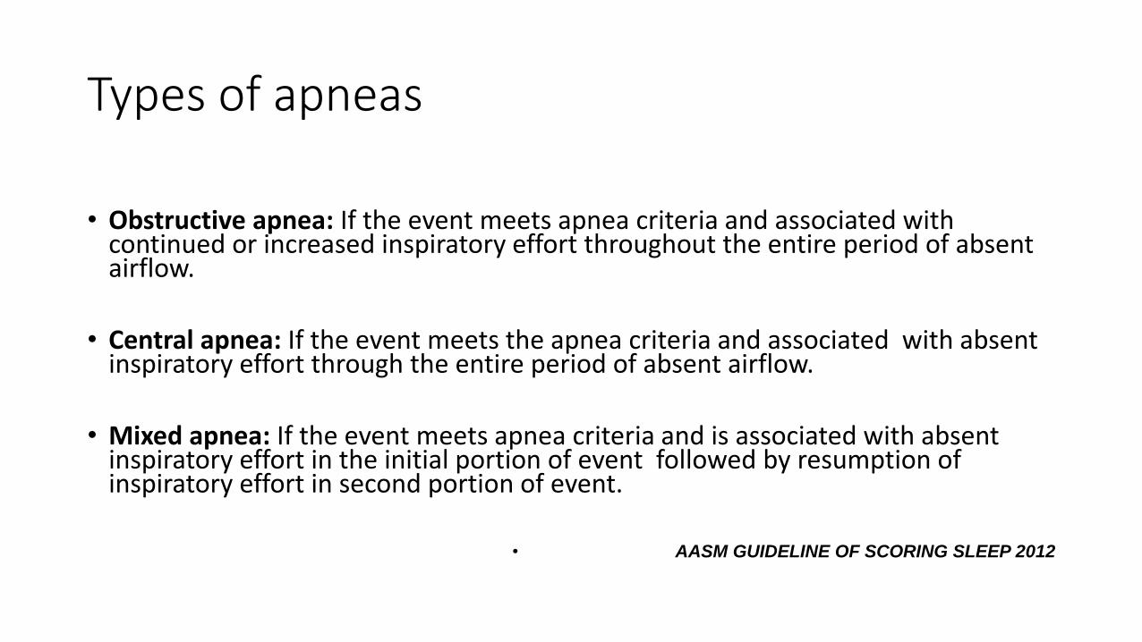
Types of apneas
• Obstructive apnea: If the event meets apnea criteria and associated with continued or increased inspiratory effort throughout the entire period of absent airflow.
• Central apnea: If the event meets the apnea criteria and associated with absent inspiratory effort through the entire period of absent airflow.
• Mixed apnea: If the event meets apnea criteria and is associated with absent inspiratory effort in the initial portion of event followed by resumption of inspiratory effort in second portion of event.
• AASM GUIDELINE OF SCORING SLEEP 2012
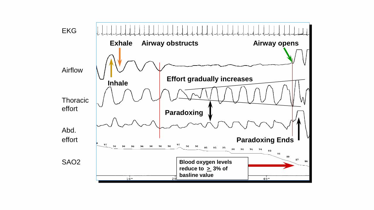
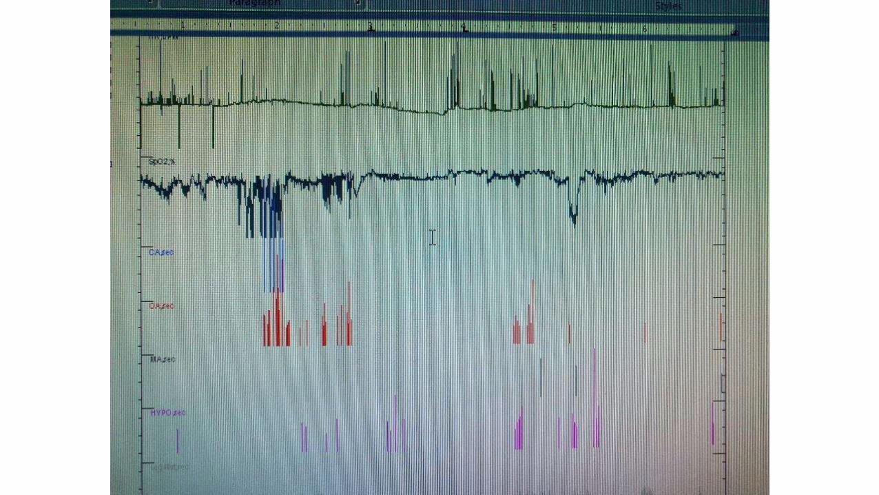
Blood oxygen levels
reduce to > 3% of
basline value
Inhale
Exhale Airway obstructs Airway opens
Paradoxing
Paradoxing Ends
EKG
Airflow
Thoracic
effort
Abd.
effort
SAO2
Effort gradually increases
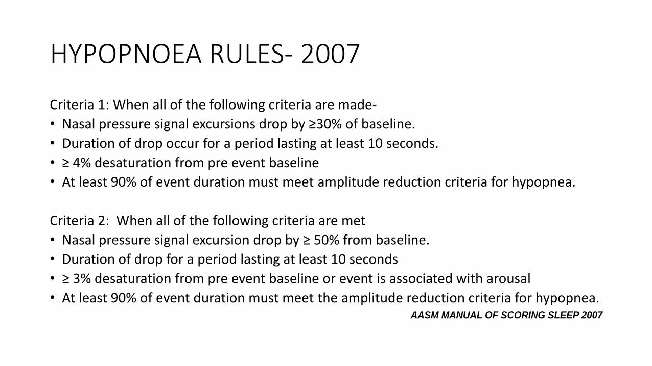
HYPOPNOEA RULES- 2007
Criteria 1: When all of the following criteria are made-
• Nasal pressure signal excursions drop by ≥30% of baseline.
• Duration of drop occur for a period lasting at least 10 seconds.
• ≥ 4% desaturation from pre event baseline
• At least 90% of event duration must meet amplitude reduction criteria for hypopnea.
Criteria 2: When all of the following criteria are met
• Nasal pressure signal excursion drop by ≥ 50% from baseline.
• Duration of drop for a period lasting at least 10 seconds
• ≥ 3% desaturation from pre event baseline or event is associated with arousal
• At least 90% of event duration must meet the amplitude reduction criteria for hypopnea.AASM MANUAL OF SCORING SLEEP 2007
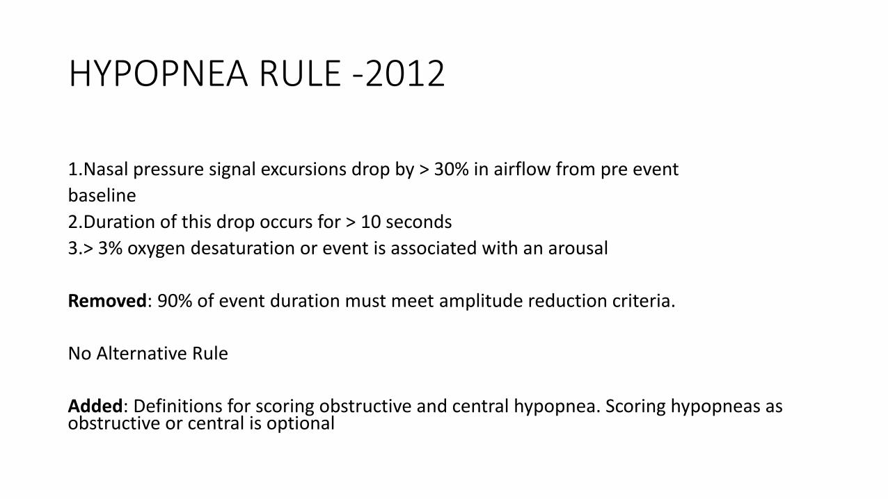
HYPOPNEA RULE -2012
1.Nasal pressure signal excursions drop by > 30% in airflow from pre event
baseline
2.Duration of this drop occurs for > 10 seconds
3.> 3% oxygen desaturation or event is associated with an arousal
Removed: 90% of event duration must meet amplitude reduction criteria.
No Alternative Rule
Added: Definitions for scoring obstructive and central hypopnea. Scoring hypopneas as obstructive or central is optional
CHEYNE STROKES RESPIRATION
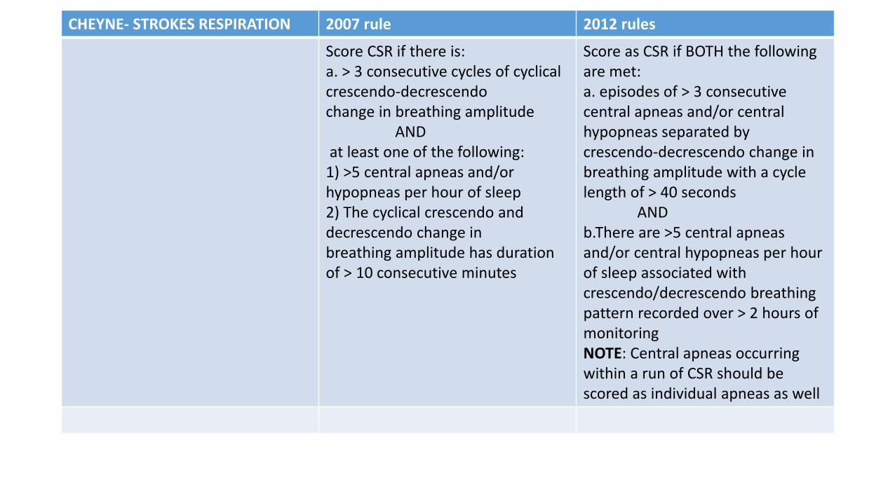
CHEYNE- STROKES RESPIRATION 2007 rule 2012 rules
Score CSR if there is:a. > 3 consecutive cycles of cyclical crescendo-decrescendochange in breathing amplitude
ANDat least one of the following:
1) >5 central apneas and/or hypopneas per hour of sleep2) The cyclical crescendo and decrescendo change inbreathing amplitude has duration of > 10 consecutive minutes
Score as CSR if BOTH the following are met:a. episodes of > 3 consecutive central apneas and/or central hypopneas separated by crescendo-decrescendo change in breathing amplitude with a cycle length of > 40 seconds
ANDb.There are >5 central apneas and/or central hypopneas per hour of sleep associated with crescendo/decrescendo breathing pattern recorded over > 2 hours of monitoringNOTE: Central apneas occurring within a run of CSR should bescored as individual apneas as well
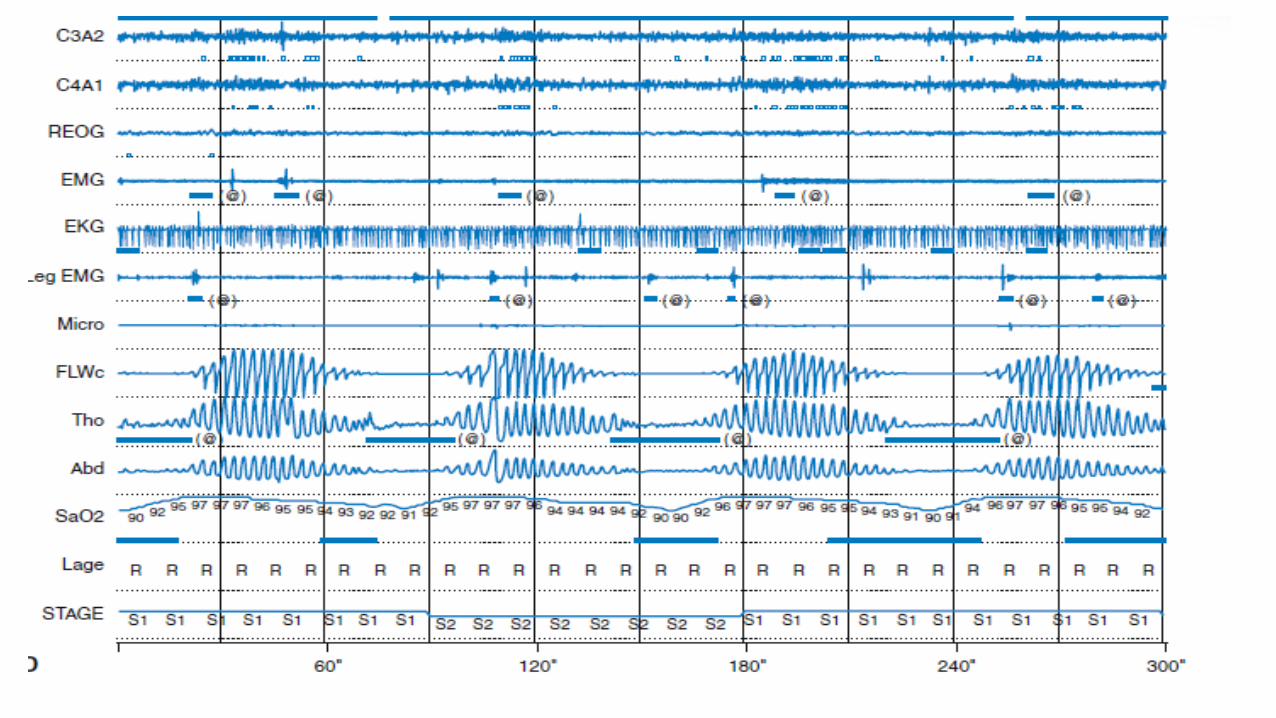
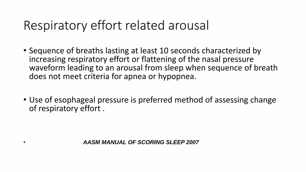
Respiratory effort related arousal
• Sequence of breaths lasting at least 10 seconds characterized by increasing respiratory effort or flattening of the nasal pressure waveform leading to an arousal from sleep when sequence of breath does not meet criteria for apnea or hypopnea.
• Use of esophageal pressure is preferred method of assessing change of respiratory effort .
• AASM MANUAL OF SCORING SLEEP 2007
HYPOVENTILATION RULE
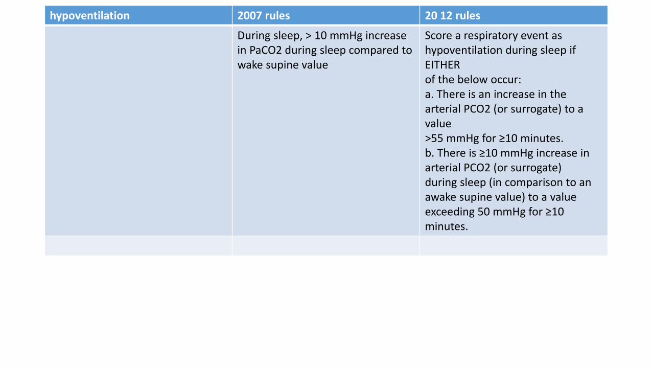
hypoventilation 2007 rules 20 12 rules
During sleep, > 10 mmHg increase in PaCO2 during sleep compared to wake supine value
Score a respiratory event as hypoventilation during sleep if EITHERof the below occur:a. There is an increase in the arterial PCO2 (or surrogate) to a value>55 mmHg for ≥10 minutes.b. There is ≥10 mmHg increase in arterial PCO2 (or surrogate)during sleep (in comparison to an awake supine value) to a valueexceeding 50 mmHg for ≥10 minutes.
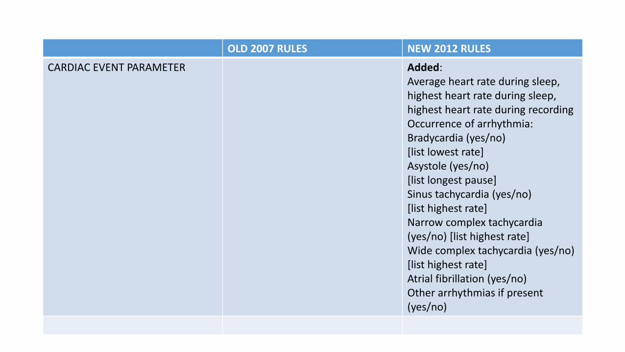
OLD 2007 RULES NEW 2012 RULES
CARDIAC EVENT PARAMETER Added:Average heart rate during sleep, highest heart rate during sleep,highest heart rate during recordingOccurrence of arrhythmia:Bradycardia (yes/no)[list lowest rate]Asystole (yes/no)[list longest pause]Sinus tachycardia (yes/no)[list highest rate]Narrow complex tachycardia (yes/no) [list highest rate]Wide complex tachycardia (yes/no) [list highest rate]Atrial fibrillation (yes/no)Other arrhythmias if present (yes/no)
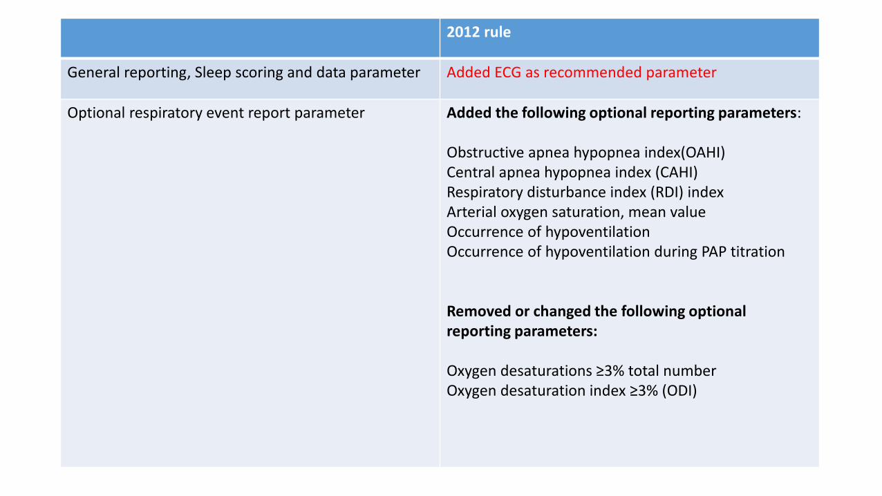
2012 rule
General reporting, Sleep scoring and data parameter Added ECG as recommended parameter
Optional respiratory event report parameter Added the following optional reporting parameters:
Obstructive apnea hypopnea index(OAHI)Central apnea hypopnea index (CAHI)Respiratory disturbance index (RDI) indexArterial oxygen saturation, mean valueOccurrence of hypoventilationOccurrence of hypoventilation during PAP titration
Removed or changed the following optional reporting parameters:
Oxygen desaturations ≥3% total numberOxygen desaturation index ≥3% (ODI)
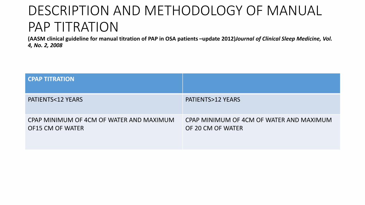
DESCRIPTION AND METHODOLOGY OF MANUAL PAP TITRATION(AASM clinical guideline for manual titration of PAP in OSA patients –update 2012)Journal of Clinical Sleep Medicine, Vol. 4, No. 2, 2008
CPAP TITRATION
PATIENTS<12 YEARS PATIENTS>12 YEARS
CPAP MINIMUM OF 4CM OF WATER AND MAXIMUM OF15 CM OF WATER
CPAP MINIMUM OF 4CM OF WATER AND MAXIMUM OF 20 CM OF WATER
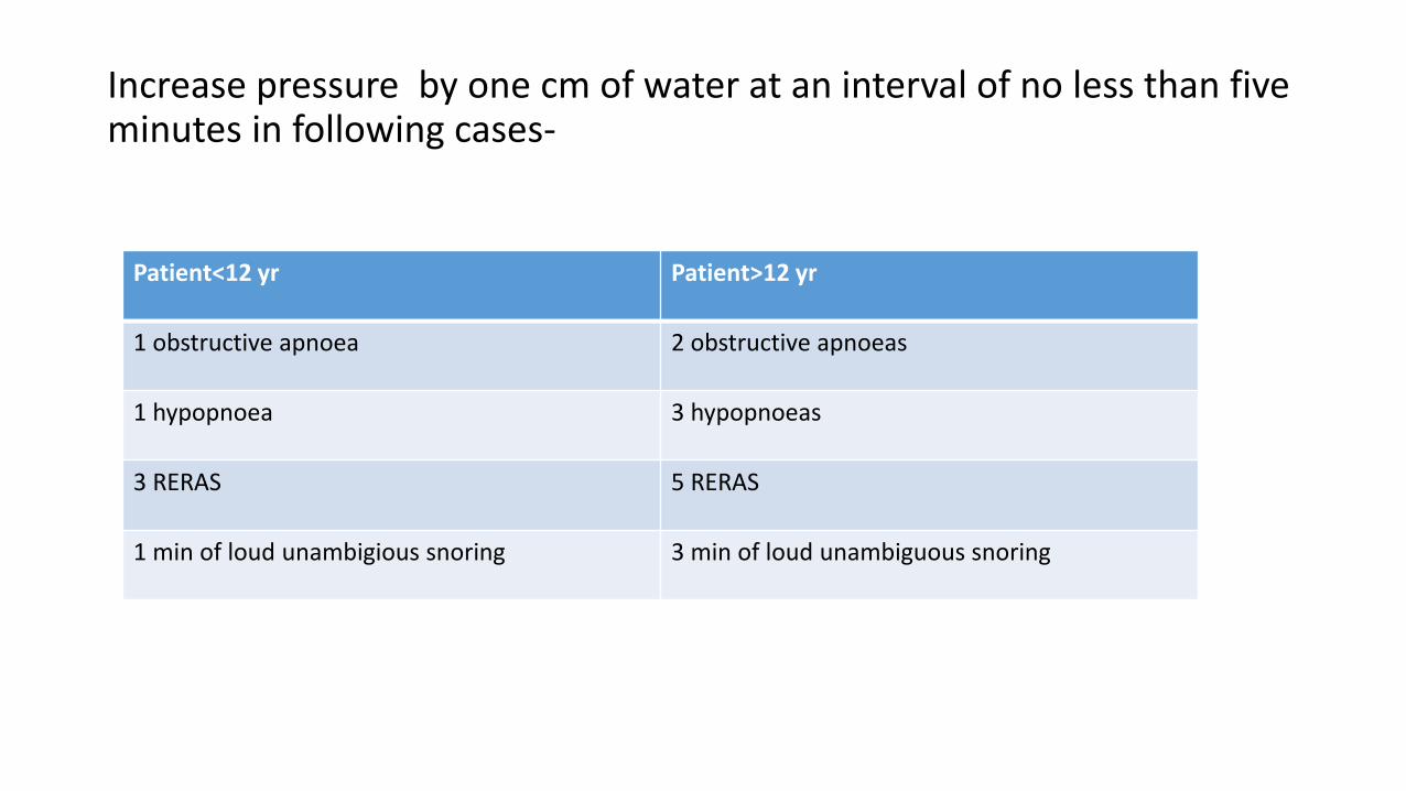
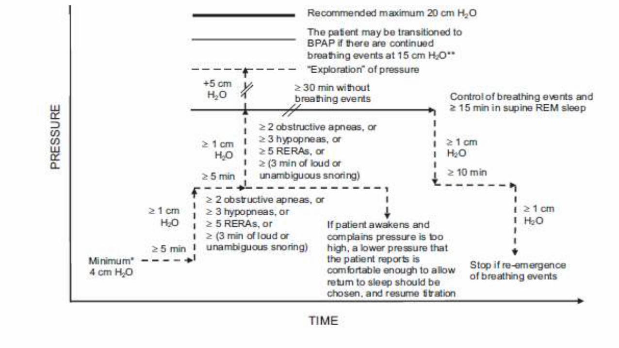
Increase pressure by one cm of water at an interval of no less than five minutes in following cases-
Patient<12 yr Patient>12 yr
1 obstructive apnoea 2 obstructive apnoeas
1 hypopnoea 3 hypopnoeas
3 RERAS 5 RERAS
1 min of loud unambigious snoring 3 min of loud unambiguous snoring
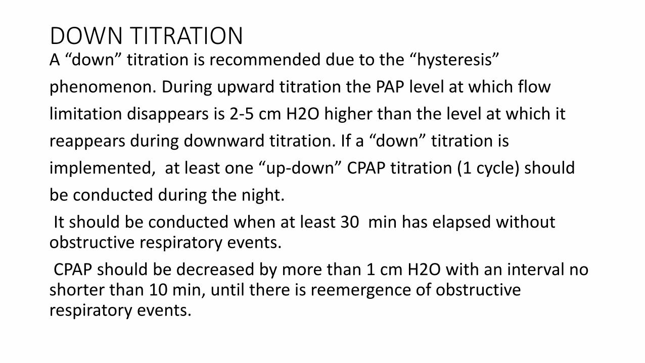
DOWN TITRATIONA “down” titration is recommended due to the “hysteresis”
phenomenon. During upward titration the PAP level at which flow
limitation disappears is 2-5 cm H2O higher than the level at which it
reappears during downward titration. If a “down” titration is
implemented, at least one “up-down” CPAP titration (1 cycle) should
be conducted during the night.
It should be conducted when at least 30 min has elapsed without obstructive respiratory events.
CPAP should be decreased by more than 1 cm H2O with an interval no shorter than 10 min, until there is reemergence of obstructive respiratory events.
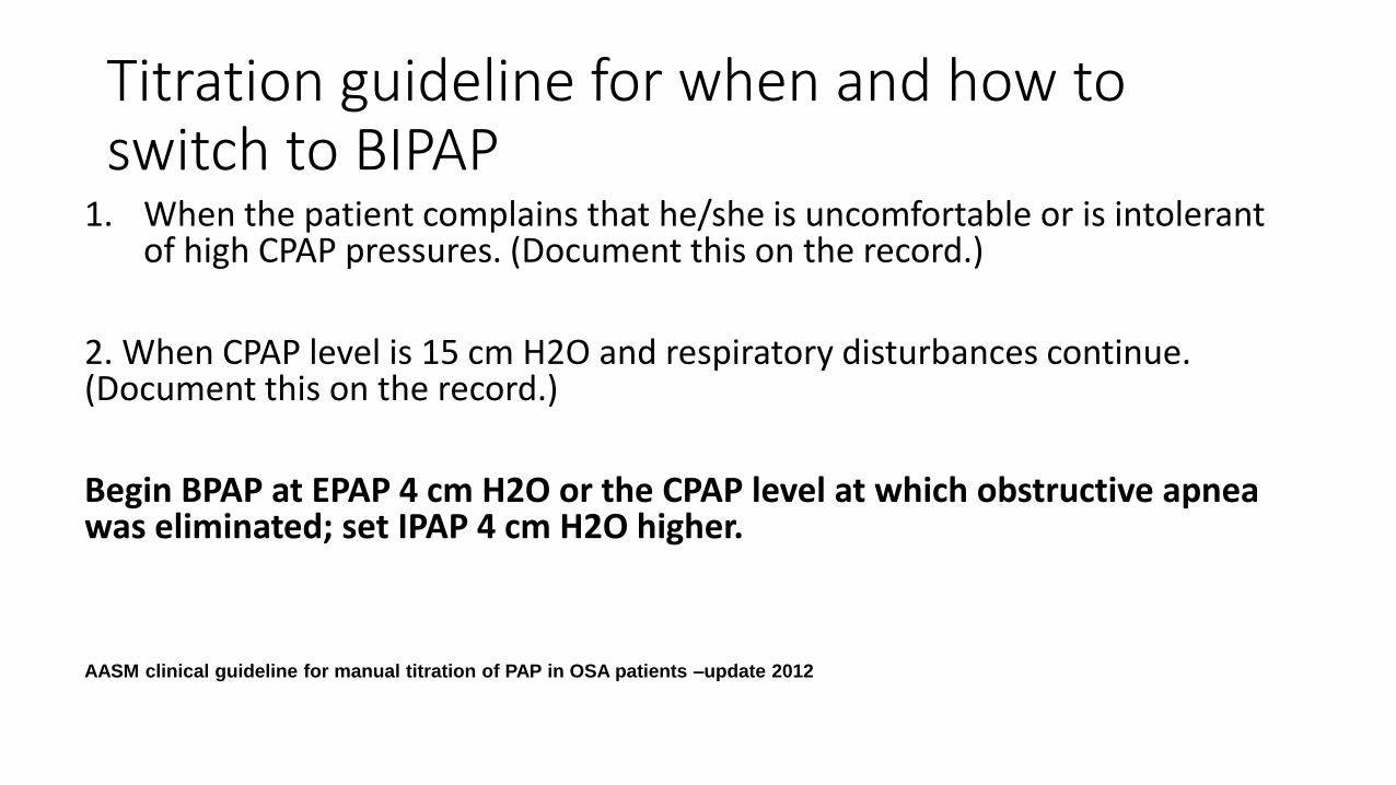
Titration guideline for when and how to switch to BIPAP
1. When the patient complains that he/she is uncomfortable or is intolerant of high CPAP pressures. (Document this on the record.)
2. When CPAP level is 15 cm H2O and respiratory disturbances continue. (Document this on the record.)
Begin BPAP at EPAP 4 cm H2O or the CPAP level at which obstructive apnea was eliminated; set IPAP 4 cm H2O higher.
AASM clinical guideline for manual titration of PAP in OSA patients –update 2012
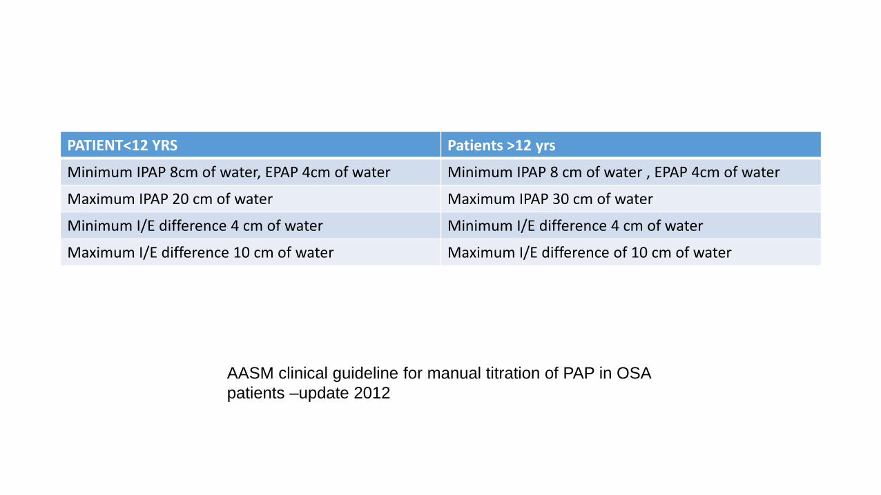
PATIENT<12 YRS Patients >12 yrs
Minimum IPAP 8cm of water, EPAP 4cm of water Minimum IPAP 8 cm of water , EPAP 4cm of water
Maximum IPAP 20 cm of water Maximum IPAP 30 cm of water
Minimum I/E difference 4 cm of water Minimum I/E difference 4 cm of water
Maximum I/E difference 10 cm of water Maximum I/E difference of 10 cm of water
AASM clinical guideline for manual titration of PAP in OSA
patients –update 2012
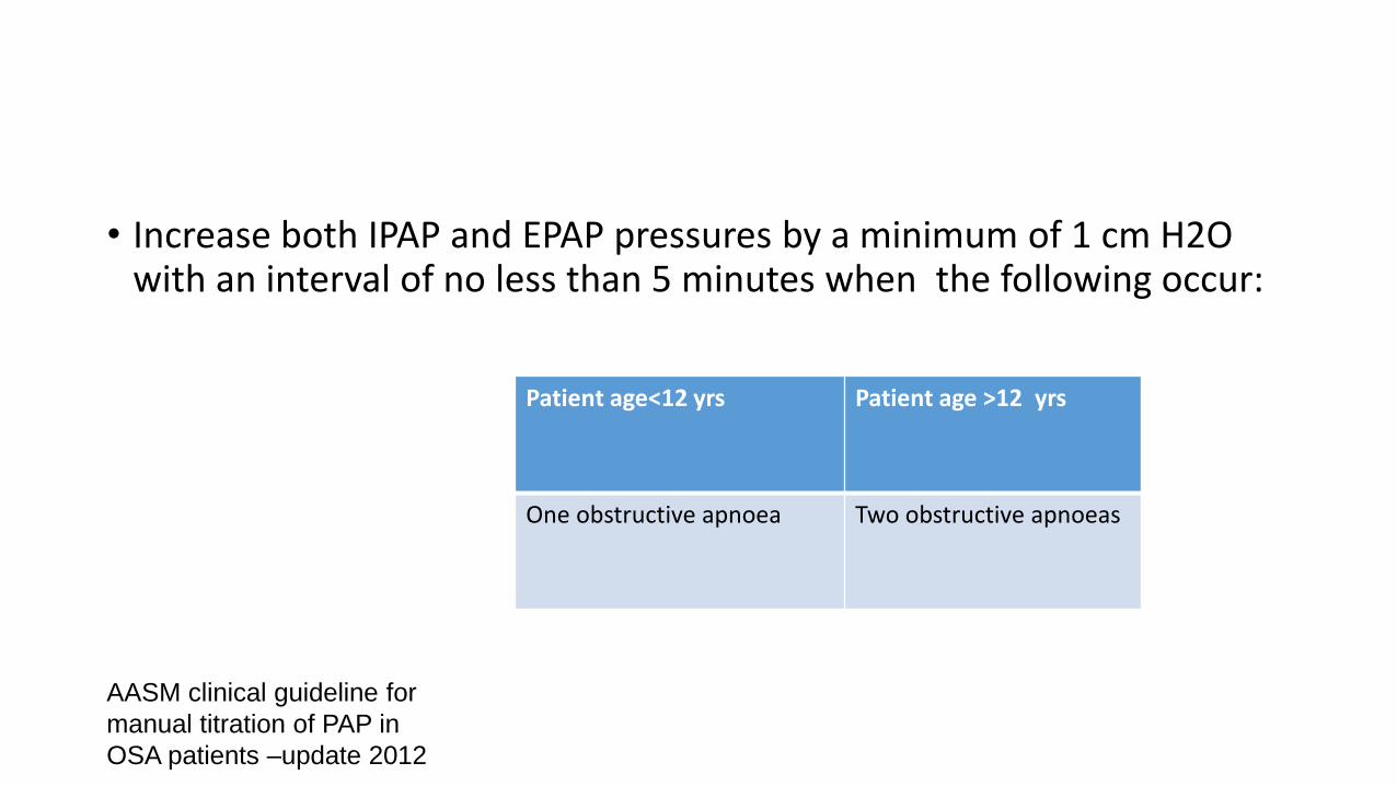
• Increase both IPAP and EPAP pressures by a minimum of 1 cm H2O with an interval of no less than 5 minutes when the following occur:
Patient age<12 yrs Patient age >12 yrs
One obstructive apnoea Two obstructive apnoeas
AASM clinical guideline for
manual titration of PAP in
OSA patients –update 2012
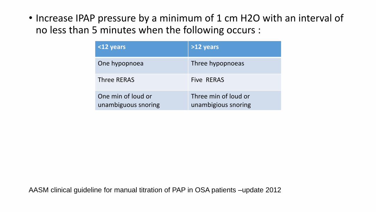
• Increase IPAP pressure by a minimum of 1 cm H2O with an interval of no less than 5 minutes when the following occurs :
<12 years >12 years
One hypopnoea Three hypopnoeas
Three RERAS Five RERAS
One min of loud or unambiguous snoring
Three min of loud or unambigious snoring
AASM clinical guideline for manual titration of PAP in OSA patients –update 2012
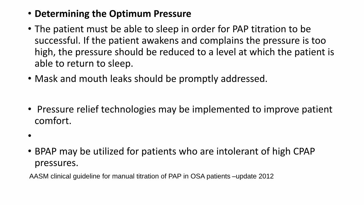
• Determining the Optimum Pressure
• The patient must be able to sleep in order for PAP titration to be successful. If the patient awakens and complains the pressure is too high, the pressure should be reduced to a level at which the patient is able to return to sleep.
• Mask and mouth leaks should be promptly addressed.
• Pressure relief technologies may be implemented to improve patient comfort.
•
• BPAP may be utilized for patients who are intolerant of high CPAP pressures.
AASM clinical guideline for manual titration of PAP in OSA patients –update 2012
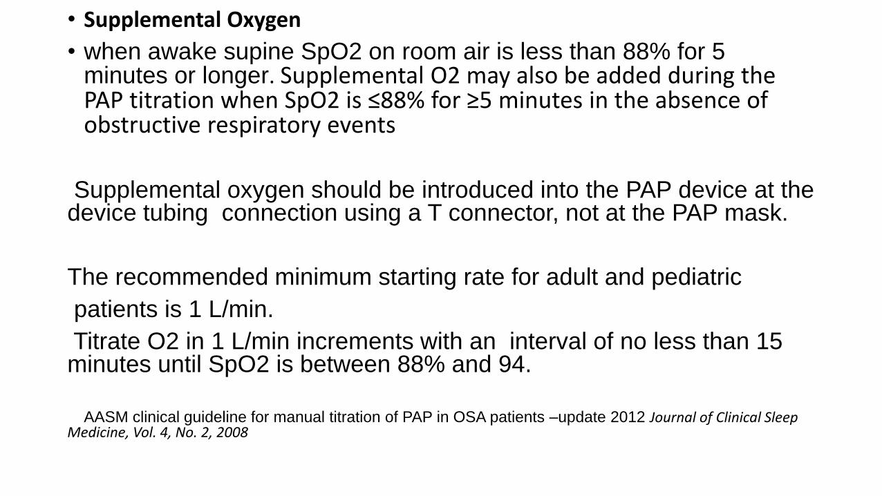
• Supplemental Oxygen
• when awake supine SpO2 on room air is less than 88% for 5 minutes or longer. Supplemental O2 may also be added during the PAP titration when SpO2 is ≤88% for ≥5 minutes in the absence of obstructive respiratory events
Supplemental oxygen should be introduced into the PAP device at the device tubing connection using a T connector, not at the PAP mask.
The recommended minimum starting rate for adult and pediatric
patients is 1 L/min.
Titrate O2 in 1 L/min increments with an interval of no less than 15 minutes until SpO2 is between 88% and 94.
AASM clinical guideline for manual titration of PAP in OSA patients –update 2012 Journal of Clinical Sleep Medicine, Vol. 4, No. 2, 2008
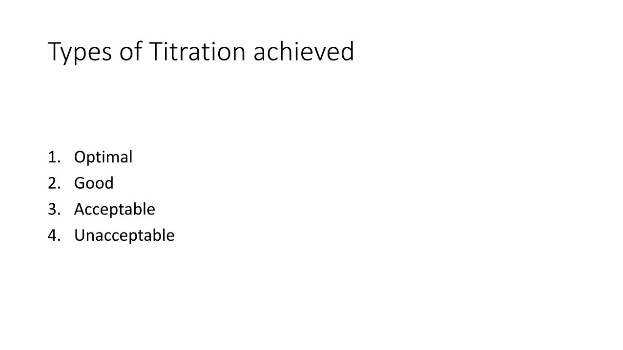
Types of Titration achieved
1. Optimal
2. Good
3. Acceptable
4. Unacceptable
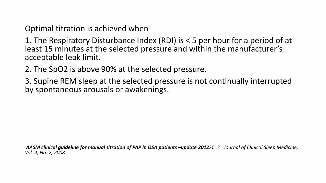
Optimal titration is achieved when-
1. The Respiratory Disturbance Index (RDI) is < 5 per hour for a period of at least 15 minutes at the selected pressure and within the manufacturer’s acceptable leak limit.
2. The SpO2 is above 90% at the selected pressure.
3. Supine REM sleep at the selected pressure is not continually interrupted by spontaneous arousals or awakenings.
AASM clinical guideline for manual titration of PAP in OSA patients –update 20122012 Journal of Clinical Sleep Medicine, Vol. 4, No. 2, 2008
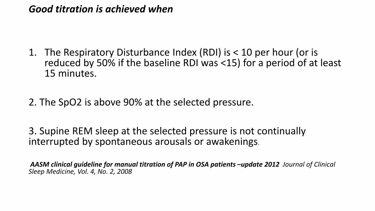
Good titration is achieved when
1. The Respiratory Disturbance Index (RDI) is < 10 per hour (or is reduced by 50% if the baseline RDI was <15) for a period of at least 15 minutes.
2. The SpO2 is above 90% at the selected pressure.
3. Supine REM sleep at the selected pressure is not continually interrupted by spontaneous arousals or awakenings.
AASM clinical guideline for manual titration of PAP in OSA patients –update 2012 Journal of Clinical Sleep Medicine, Vol. 4, No. 2, 2008
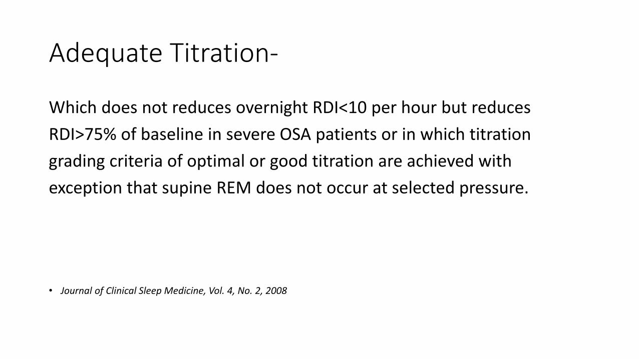
Adequate Titration-
Which does not reduces overnight RDI<10 per hour but reduces
RDI>75% of baseline in severe OSA patients or in which titration
grading criteria of optimal or good titration are achieved with
exception that supine REM does not occur at selected pressure.
• Journal of Clinical Sleep Medicine, Vol. 4, No. 2, 2008
Split-Night Studies
• Split-night studies must be performed using algorithms identical to those used for full-night PAP titration and should include greater than 3 hours of titration time.
• Split-night studies should not be performed in children less than 12 years old.
• Due to the reduced titration time available during split-night studies, increasePAP pressures by a minimum of 2 cm H2O with an interval of no less than 5minutes.
Journal of Clinical Sleep Medicine, Vol. 4, No. 2, 2008
AASM clinical guideline for manual titration of PAP in OSA patients –update 2012

INTERPRETATION OF EEG
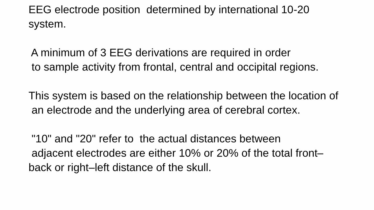
EEG electrode position determined by international 10-20
system.
A minimum of 3 EEG derivations are required in order
to sample activity from frontal, central and occipital regions.
This system is based on the relationship between the location of
an electrode and the underlying area of cerebral cortex.
"10" and "20" refer to the actual distances between
adjacent electrodes are either 10% or 20% of the total front–
back or right–left distance of the skull.
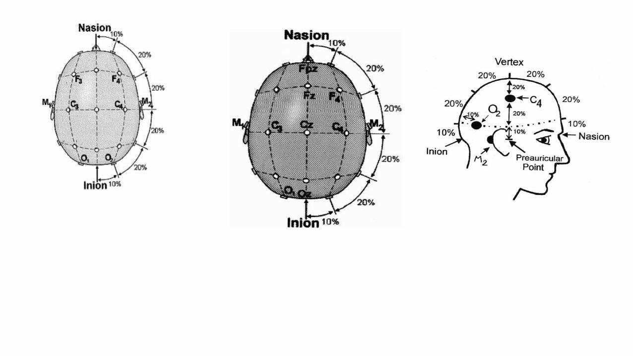
• Recommended derivations are F4-M1,C4-M1,O2-M1.
• M1 and M2 refer to right and left mastoid process
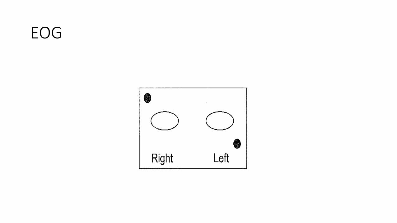
EOG
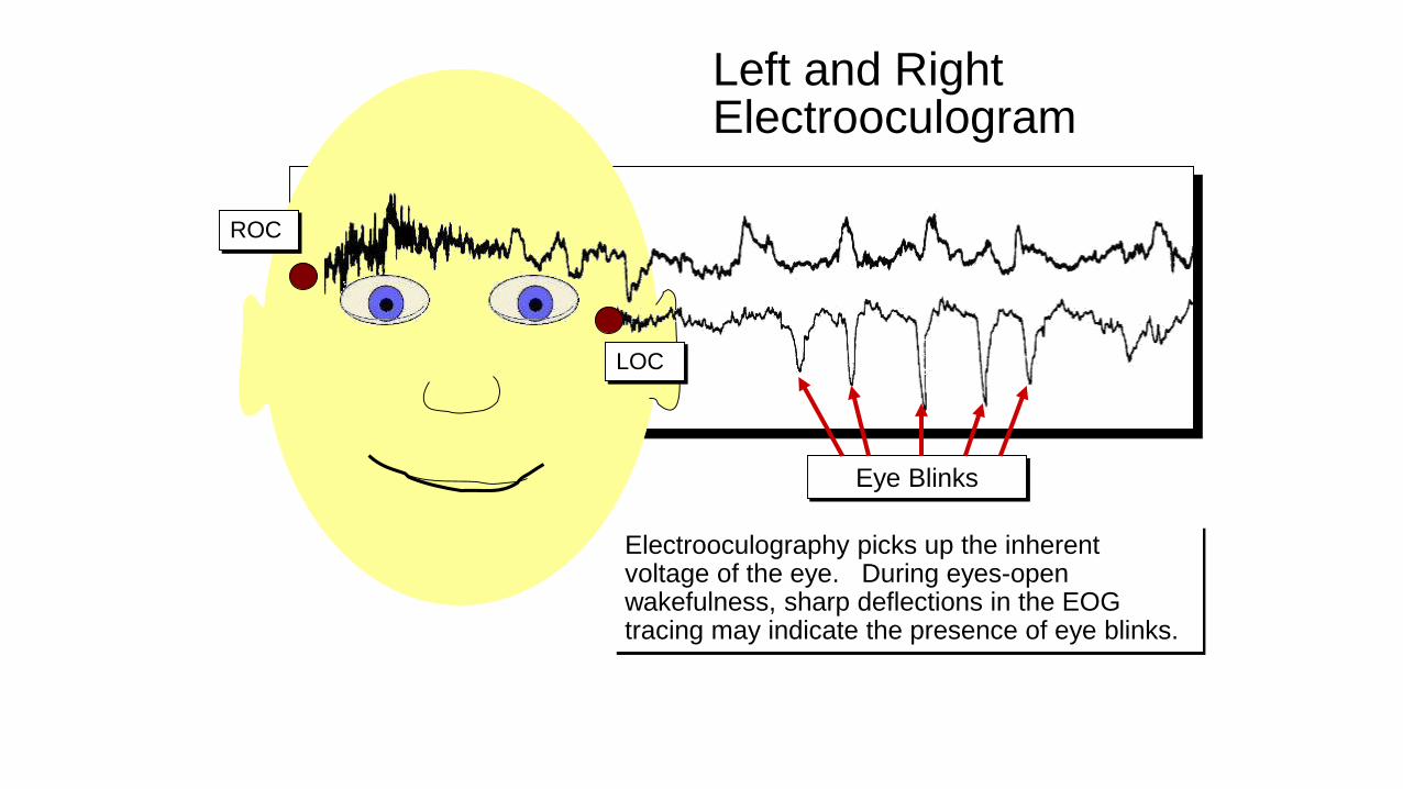
ROC
LOC
Left and Right Electrooculogram
Eye Blinks
Electrooculography picks up the inherent voltage of the eye. During eyes-open wakefulness, sharp deflections in the EOG tracing may indicate the presence of eye blinks.
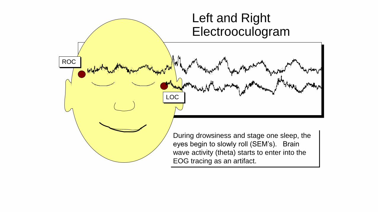
ROC
LOC
Left and Right Electrooculogram
During drowsiness and stage one sleep, the
eyes begin to slowly roll (SEM’s). Brain
wave activity (theta) starts to enter into the
EOG tracing as an artifact.
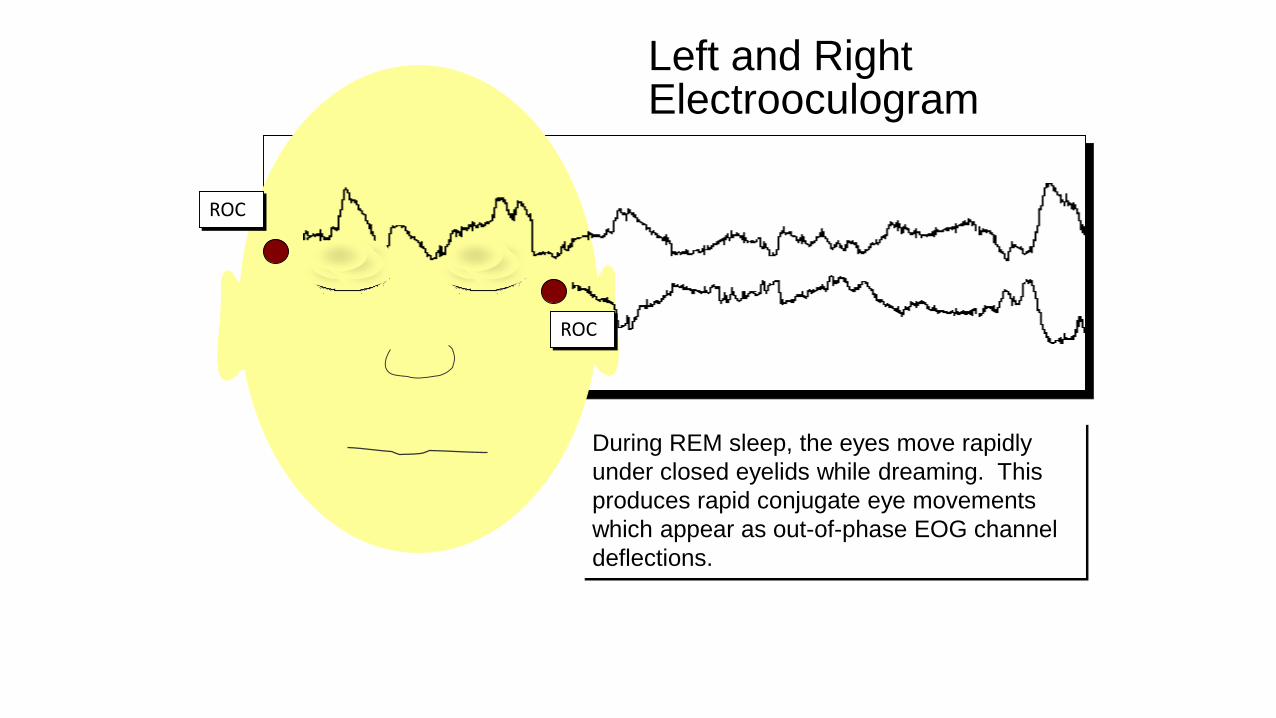
ROC
ROC
Left and Right Electrooculogram
During REM sleep, the eyes move rapidly
under closed eyelids while dreaming. This
produces rapid conjugate eye movements
which appear as out-of-phase EOG channel
deflections.
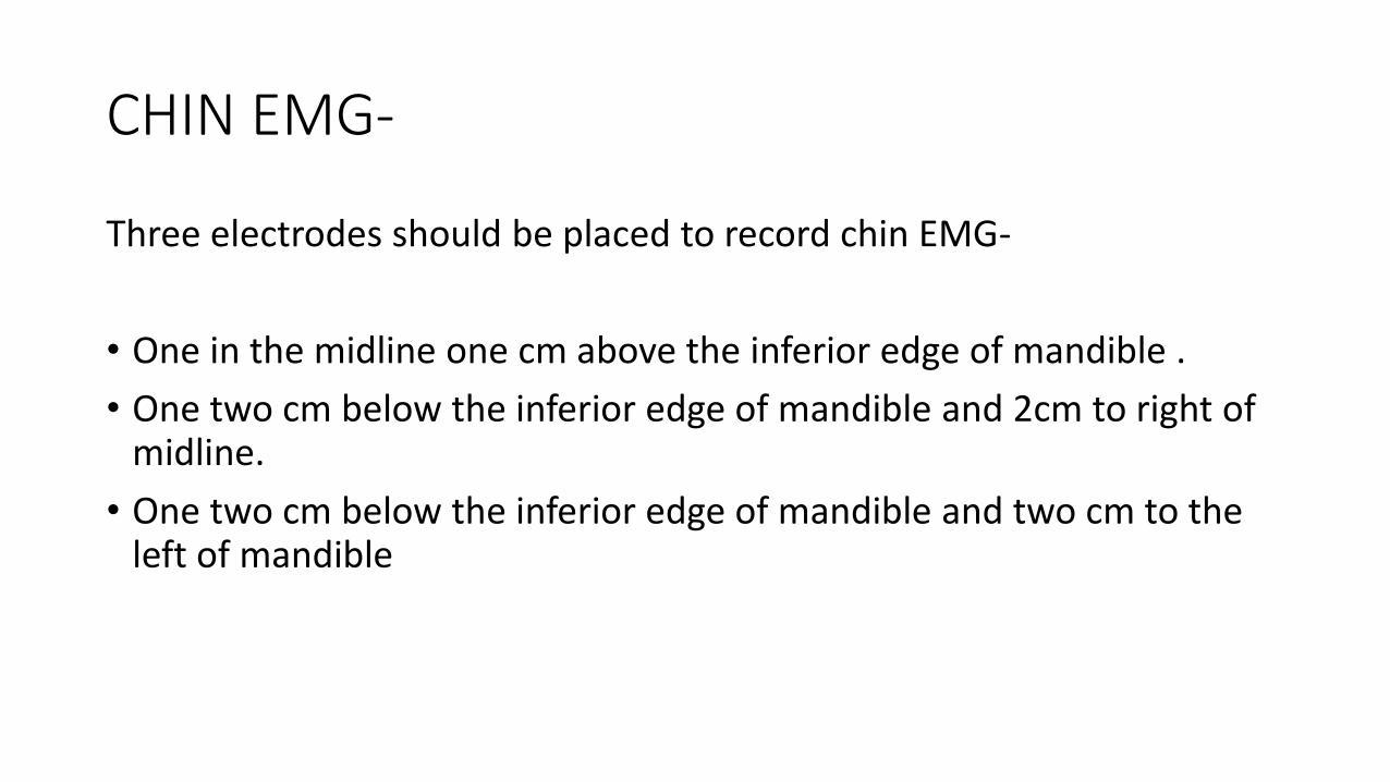
CHIN EMG-
Three electrodes should be placed to record chin EMG-
• One in the midline one cm above the inferior edge of mandible .
• One two cm below the inferior edge of mandible and 2cm to right of midline.
• One two cm below the inferior edge of mandible and two cm to the left of mandible
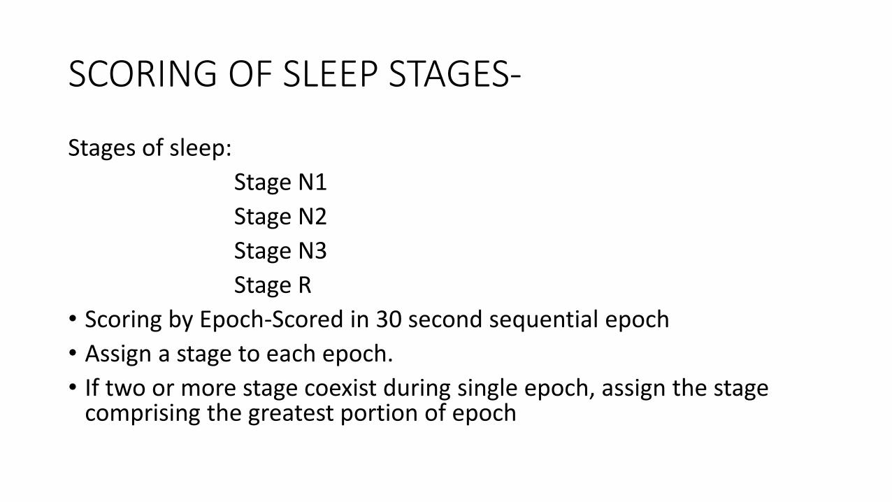
SCORING OF SLEEP STAGES-
Stages of sleep:
Stage N1
Stage N2
Stage N3
Stage R
• Scoring by Epoch-Scored in 30 second sequential epoch
• Assign a stage to each epoch.
• If two or more stage coexist during single epoch, assign the stage comprising the greatest portion of epoch
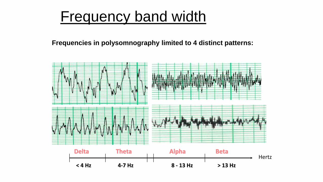
Frequency band width
Frequencies in polysomnography limited to 4 distinct patterns:
Delta
< 4 Hz
HertzTheta
4-7 Hz
Alpha
8 - 13 Hz
Beta
> 13 Hz
Delta
Theta
Alpha
Beta
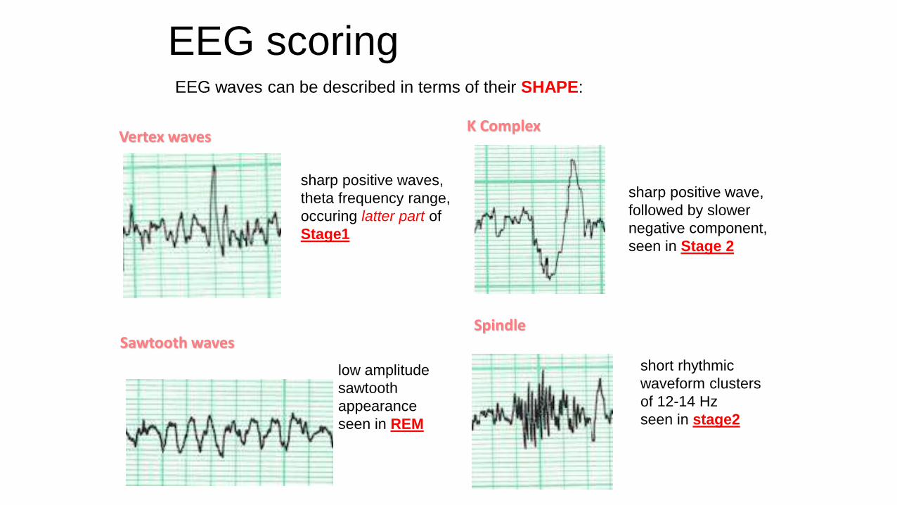
EEG scoringEEG waves can be described in terms of their SHAPE:
Vertex waves
sharp positive waves,
theta frequency range,
occuring latter part of
Stage1
K Complex
sharp positive wave,
followed by slower
negative component,
seen in Stage 2
Sawtooth waves
low amplitude
sawtooth
appearance
seen in REM
Spindle
short rhythmic
waveform clusters
of 12-14 Hz
seen in stage2
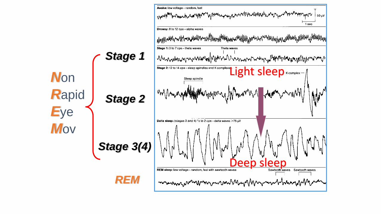
Light sleep
Deep sleep
Stage 1
Stage 2
Stage 3(4)
REM
Non
Rapid
Eye
Mov
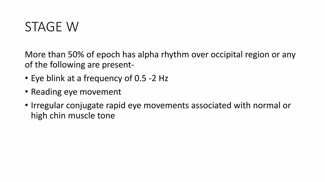
STAGE W
More than 50% of epoch has alpha rhythm over occipital region or any of the following are present-
• Eye blink at a frequency of 0.5 -2 Hz
• Reading eye movement
• Irregular conjugate rapid eye movements associated with normal or high chin muscle tone
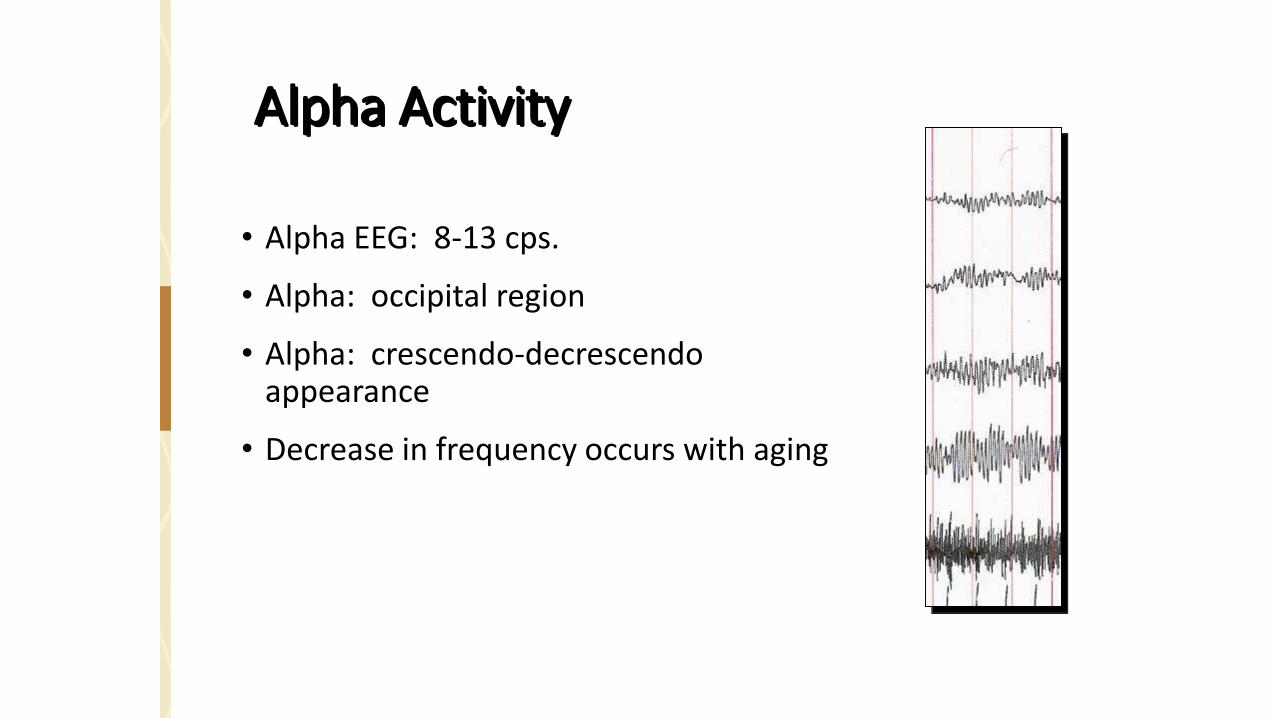
Alpha Activity
• Alpha EEG: 8-13 cps.
• Alpha: occipital region
• Alpha: crescendo-decrescendo appearance
• Decrease in frequency occurs with aging
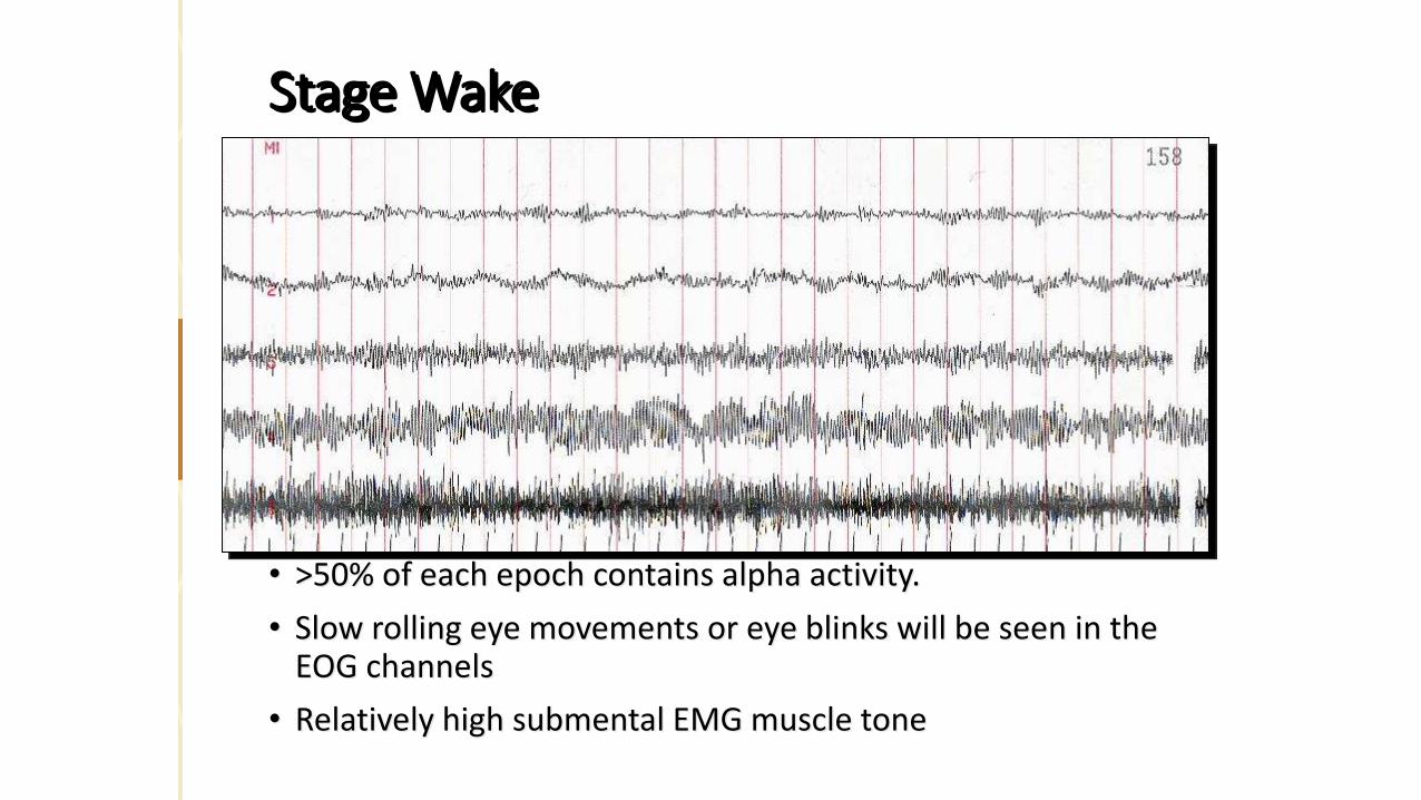
• >50% of each epoch contains alpha activity.
• Slow rolling eye movements or eye blinks will be seen in the EOG channels
• Relatively high submental EMG muscle tone
Stage Wake
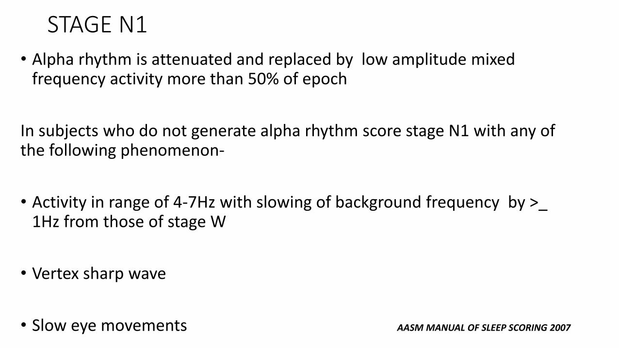
STAGE N1
• Alpha rhythm is attenuated and replaced by low amplitude mixed frequency activity more than 50% of epoch
In subjects who do not generate alpha rhythm score stage N1 with any of the following phenomenon-
• Activity in range of 4-7Hz with slowing of background frequency by >_ 1Hz from those of stage W
• Vertex sharp wave
• Slow eye movements AASM MANUAL OF SLEEP SCORING 2007
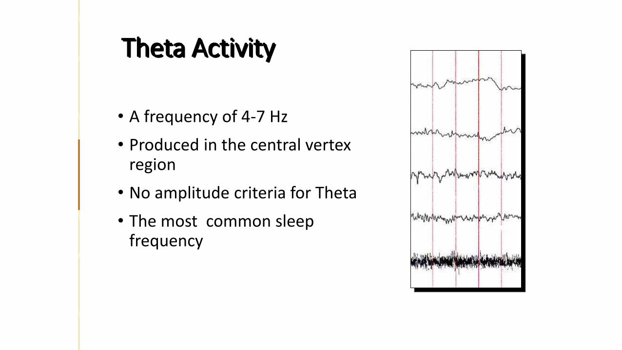
Theta Activity
• A frequency of 4-7 Hz
• Produced in the central vertex region
• No amplitude criteria for Theta
• The most common sleep frequency
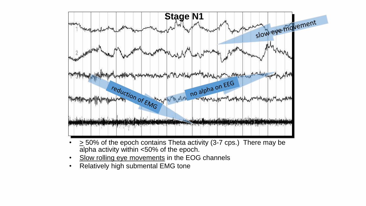
• > 50% of the epoch contains Theta activity (3-7 cps.) There may be alpha activity within <50% of the epoch.
• Slow rolling eye movements in the EOG channels
• Relatively high submental EMG tone
Stage N1
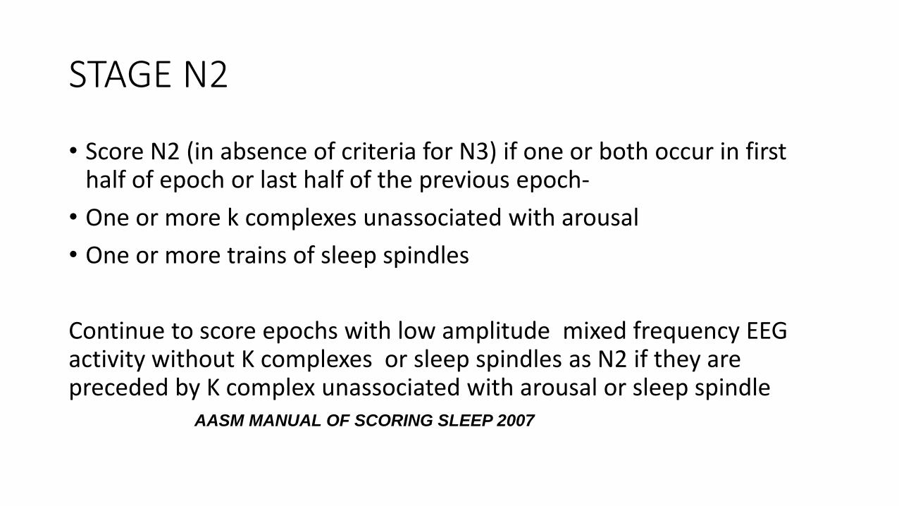
STAGE N2
• Score N2 (in absence of criteria for N3) if one or both occur in first half of epoch or last half of the previous epoch-
• One or more k complexes unassociated with arousal
• One or more trains of sleep spindles
Continue to score epochs with low amplitude mixed frequency EEG activity without K complexes or sleep spindles as N2 if they are preceded by K complex unassociated with arousal or sleep spindle
AASM MANUAL OF SCORING SLEEP 2007
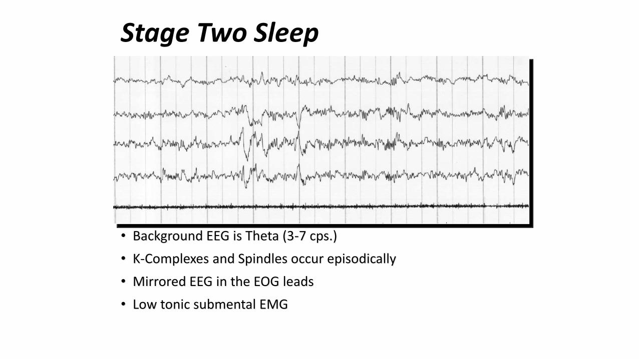
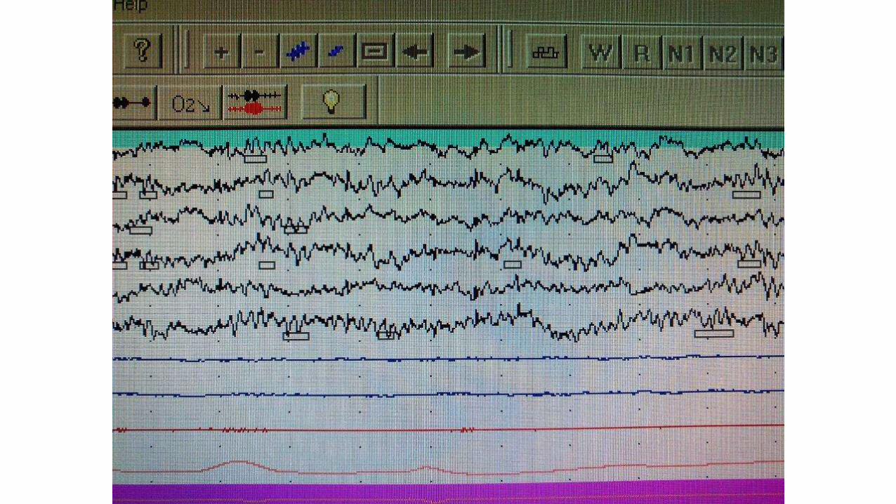
• Background EEG is Theta (3-7 cps.)
• K-Complexes and Spindles occur episodically
• Mirrored EEG in the EOG leads
• Low tonic submental EMG
Stage Two Sleep
K Complex: A well delineated negative sharp wave immediately followed by a
positive component standing out from the background EEG, with total duration
> or equal to 5 seconds, usually maximal in amplitude when recorded using
frontal derivations
Sleep Spindle: A train of distinct waves with frequency 11-16 Hz with a duration of
> and equal to 0.5 seconds usually maximal in amplitude using central
derivations
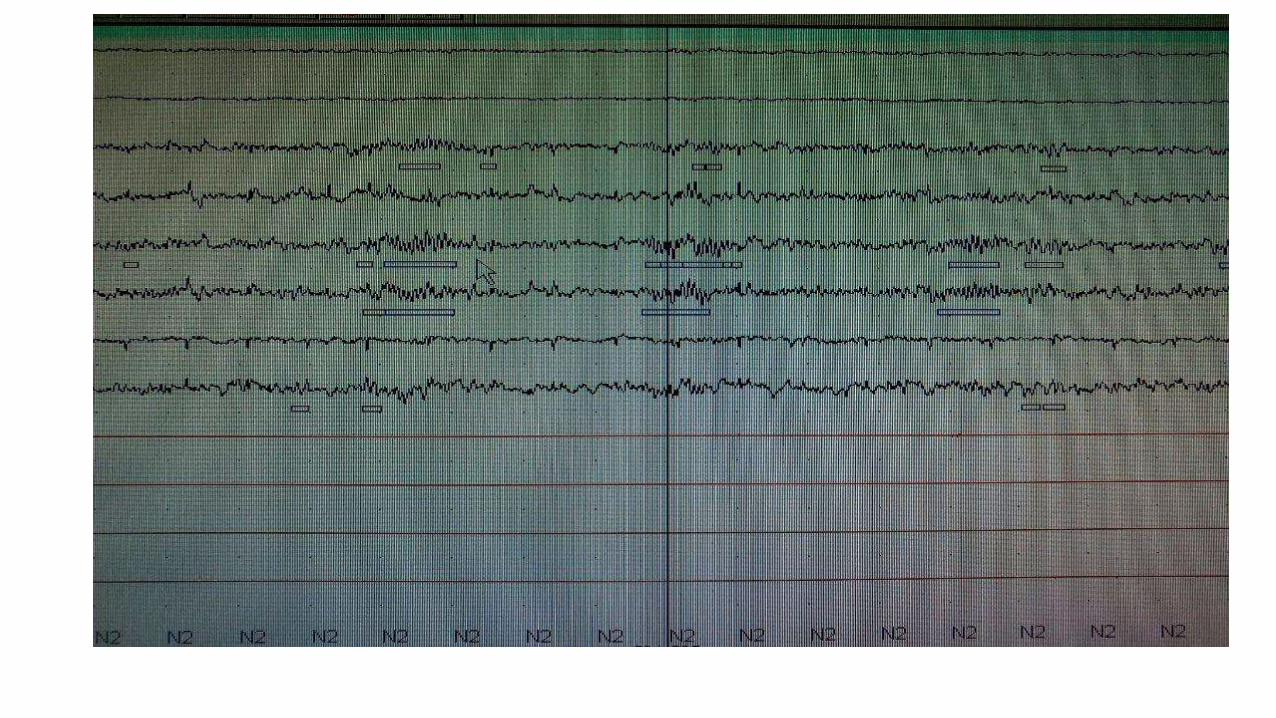
End of N2 sleep when one of the following events occur-
• Transition to stage W or N3 or stage R
• Major body movement followed by slow eye movements and low amplitude mixed frequency EEG without nonarousal associated K complexes or sleep spindles
• AASM MANUAL OF SCORING SLEEP 2007
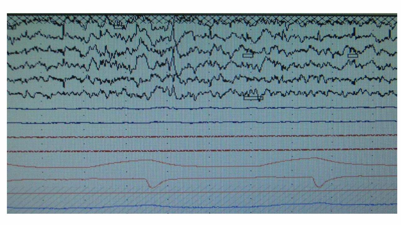
• STAGE N3- When 20% or more of an epochs consists of slow wave activity irrespective of age.
• Sleep spindles may persist in stage N3 sleep.
• Eye movements are not typically seen in N3 sleep.
• In N3 stage the chin EMG is of variable amplitude often lower than in stage N2 sleep and sometimes as low as in stage R sleep.
• AASM MANUAL OF SCORING SLEEP 2007
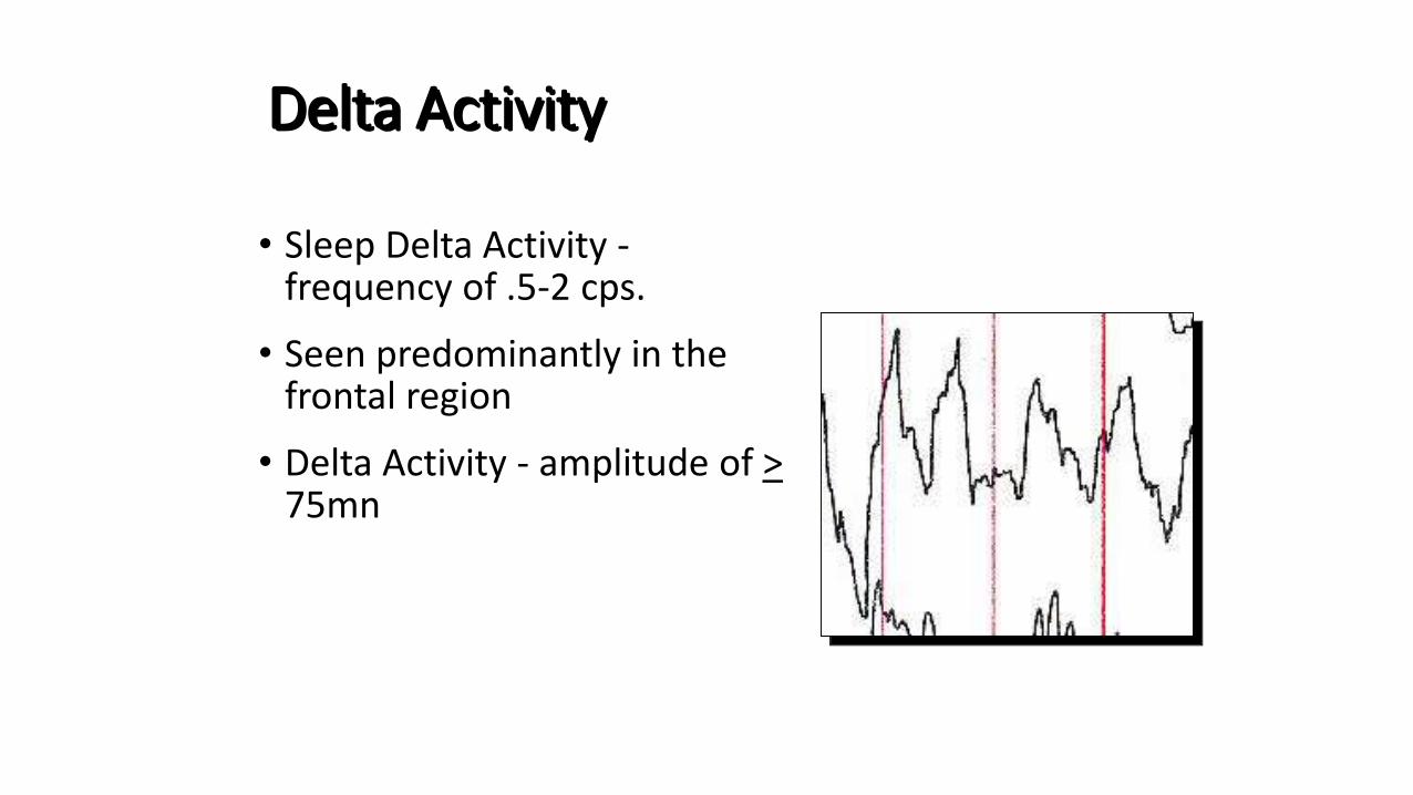
Delta Activity
• Sleep Delta Activity -frequency of .5-2 cps.
• Seen predominantly in the frontal region
• Delta Activity - amplitude of >75mn
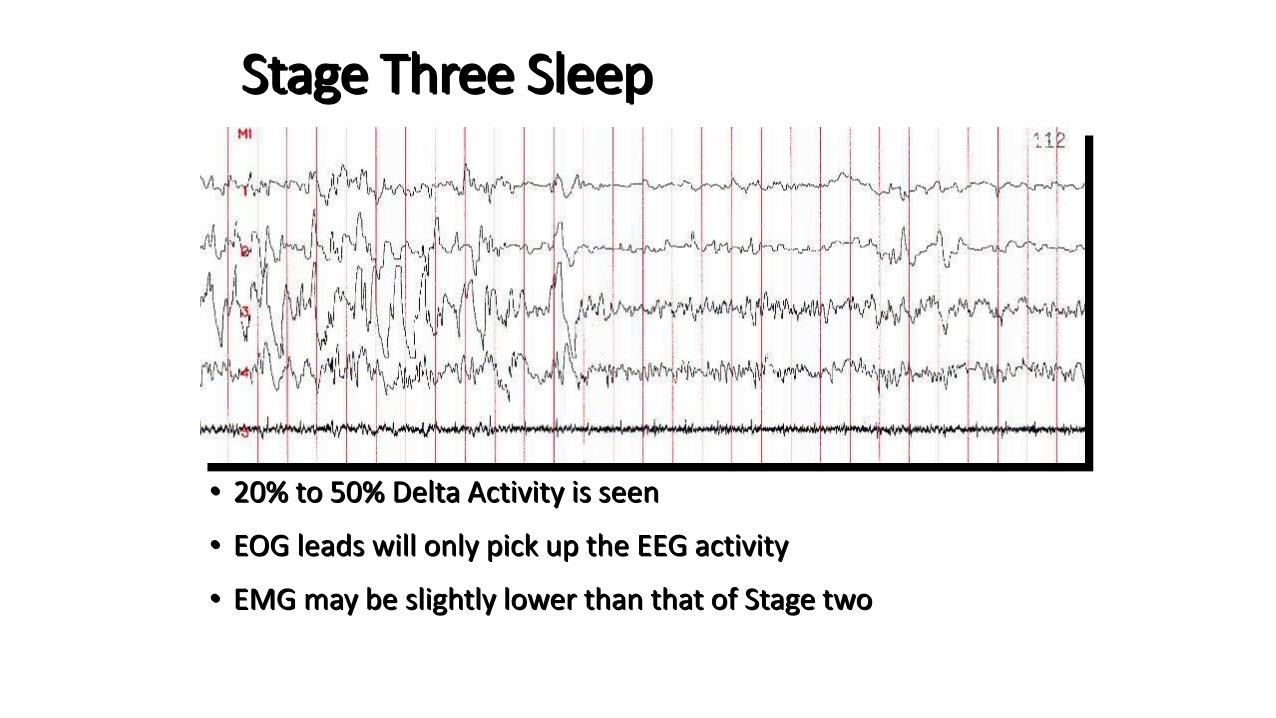
Stage Three Sleep
• 20% to 50% Delta Activity is seen
• EOG leads will only pick up the EEG activity
• EMG may be slightly lower than that of Stage two
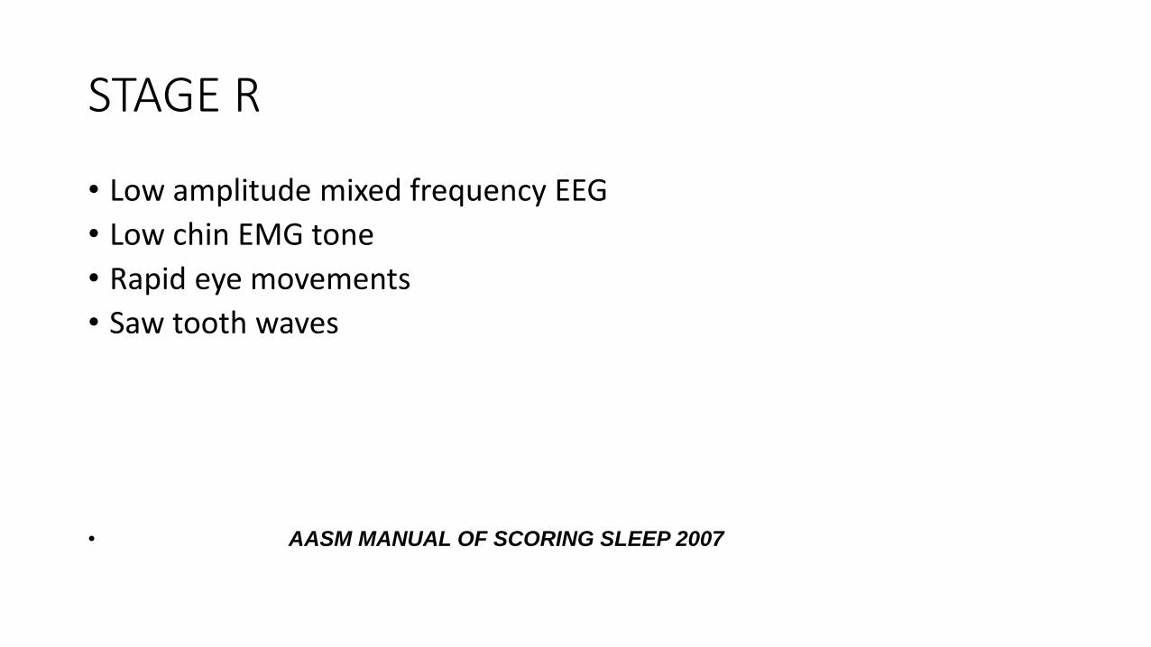
STAGE R
• Low amplitude mixed frequency EEG
• Low chin EMG tone
• Rapid eye movements
• Saw tooth waves
• AASM MANUAL OF SCORING SLEEP 2007
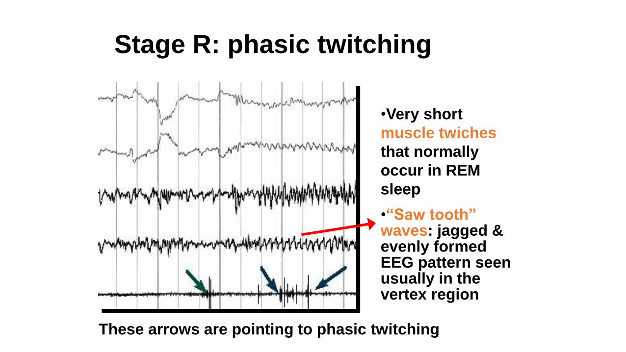
Stage R: phasic twitching
•Very short
muscle twiches
that normally
occur in REM
sleep
•“Saw tooth” waves: jagged & evenly formed EEG pattern seen usually in the vertex region
These arrows are pointing to phasic twitching
MAJOR BODY MOVEMENTS-
• Movement and muscle artifact obscuring EEG for>half of epoch to the extent that sleep stage can not be determined
Score an epoch with major body movement as follows-
• If alpha rhythm is present for part of epoch score as stage W.
• Otherwise score the epoch as the same stage as the epoch follows it.
• AASM MANUAL OF SCORING SLEEP 2007
PULSE TRANSIT TIME
• Pulse transit time (PTT) is the time taken for the arterial pulse pressure
wave to travel from the aortic valve to a peripheral site. For convenience, it
is usually measured from the R wave on the electrocardiogram to the pulse
wave arrival at the finger.
• Pulse transit time is inversely proportional to blood pressure, and the falls
in blood pressure which occur with inspiration (pulsus paradoxus)
correspond to rises (lengthening) in pulse transit time.
• Pulse transit time may, therefore, provide a clinically useful
noninvasive and quantitative measure of inspiratory effort in patients
with sleep-related breathing disorders.
Eur Respir J., 1995, 8, 1669–1674