Embed Size (px)
Citation preview

POSEIDO. 2014;2(4) Sandwich osteotomy material in posterior mandible
233
ISSN 2307-5295, Published by the POSEIDO Organization & Foundation
under a Creative Commons Attribution-NonCommercial-NoDerivatives 4.0 International (CC BY-NC-ND 4.0) License
Research article Interpositional graft associated with alveolar osteotomy for posterior mandible ridge augmentation (sandwich osteotomy) using hydroxyapatite or autogenous bone: histological evaluation Karen Bechara,1 Alexandre M. Dottore,1 Paulo Y. Kawakami,1 Alessandra Cassoni,2 Gabriela Giro,1 Jose Augusto Rodrigues,2 Leandro Chambrone,1 Adriano Piattelli,3 Giovanna Iezzi,3 and Jamil Awad Shibli.1,* 1 Department of Periodontology and Oral Implantology, Dental Research Division, University of Guarulhos - UnG, São Paulo, Brazil 2 Department of Restorative Dentistry, Dental Research Division, University of Guarulhos - UnG, São Paulo, Brazil 3 Department of Medical, Oral and Biotechnological Sciences, University of Chieti-Pescara, Chieti, Italy *Corresponding author: Jamil Awad Shibli, [email protected] Submitted on April 10th, 2014; accepted after minor corrections on April 25th, 2014.
Abstract Background and objectives. The influence of interpositional graft material on bone behavior associated with alveolar osteotomy for posterior mandible ridge augmentation (osteotomy with sandwich technique) is not fully understood. Therefore, this study evaluated, histologically, the impact of 2-inlay grafts material in posterior mandible. Materials and Methods. Alveolar augmentation osteotomies were performed bilaterally in 9 partially edentulous mandibular patients in a split-mouth design. The alveolar segmental osteotomies were assigned in 2 groups: test group, interpositional hydroxyapatite (HA), and control group, interpositional intra-oral autogenous bone graft. After 6 months of healing, a bone core was retrieved from each side for histological evaluation before implant placement. Results. Ground sections depicted more newly-formed bone for autogenous group (p<0.05) and more residual-grafted material in the HA group (p<0.05). Discussion and conclusions. The results of this split mouth design suggest that both intra-oral autogenous bone and HA as an interpositional graft material to vertically augment posterior atrophic mandibles could be used. Keywords. Alveolar bone grafting, bone substitutes, dental implants, hydroxyapatites, osteotomy.
1. Introduction The resorption of the alveolar process may preclude implant placement, mainly in the
atrophic posterior mandible. Reconstruction of the alveolar process with bone augmentation prior to implant placement will facilitate the latter, but the result is influenced by the quality and quantity of the regenerated bone [1-3].
234 Research article: Bechara K, et al. (2014)
ISSN 2307-5295, Published by the POSEIDO Organization & Foundation
under a Creative Commons Attribution-NonCommercial-NoDerivatives 4.0 International (CC BY-NC-ND 4.0) License
Alveolar osteotomy represents an elegant and efficient treatment of option for the preimplant bone ridge reconstruction of the severely resorbed posterior mandible [4,5]. This surgical method implies in general to fill the osteotomy cavity with a material – autologous bone or any other filling biomaterial. Interpositional or inlay grafts as a “sandwich” involve the placement of graft material within a 3 to 5-walled cancellous compartment [6]. This procedure allows that the recipient sites contains and stabilizes the graft material, and the circulating of blood flow between the osteotomized bony blocks providing cells, soluble regulators and nourishment [7].
Nevertheless, at the present, few data are available about long-term stability of dental implants inserted in grafted sites and about differences to bone native sites. Moreover, after implants osseointegration and bone remodeling, the grafted bone-implant system is different to that present at implant insertion surgery time, and the stability is quite probably dependent on the quality of the bone-implant interface [8]. Bone quality and quantity at the implantation sites are routinely evaluated using the imaging techniques, but their resolution is not high enough to analyze bone microarchitecture [9].
Therefore, the aim of this study was to evaluate, by histologic analysis, the influence of bone density on stability of dental implants after vertical ridge augmentation of the atrophic posterior mandible with different interpositional graft material.
2. Material and Methods 2.1. Patient Population
This prospective study reports on patients who were consecutively treated with vertical augmentation on posterior mandible using alveolar osteotomy. The Institutional Clinical Research Ethics Committee of Guarulhos University (CEP #168/11) approved the experimental protocol. Briefly, nine healthy non-smokers (6 females and 3 males, mean age 55 years) presenting bilateral partial edentulism in the posterior mandible with a residual bone height between 4 to 6mm were enrolled in this study. The edentulous ridges, in a split mouth design, were assigned in 2 groups: a control group consisting of n = 9 alveolar osteotomy that received an interpositional inlay autogenous bone graft from lateral oblique line, and a test group consisting of n = 9 alveolar osteotomy that received an interpositional inlay resorbable non-ceramic hydroxylapatite - HA (OsteoGen powder and pellets, Impladent Ltd, Holliswood, NY, USA). Tossing a coin was used to determine which posterior mandible was assigned as control or test.
2.2. Alveolar osteotomy All subjects received oral prophylaxis treatment before surgery. Panoramic
radiographs and dental volume tomography – DVT - (ICat, KaVo Dental GmbH, Biberach, Germany) were taken of all patients. The surgical procedure involved an elliptical incision of 10-12mm from the ridge bone in the labiobuccal gingiva of the edentulous area. A full thickness flap was raised without detaching the lingual and the crestal mucoperiosteum to expose the labiobuccal cortical bone of the posterior atrophic mandible and the mental nerve. Two vertical and one horizontal osteotomy were made with a surgical burr and saws. The horizontal osteotomy was located at least 2mm below the ridge bone and 2mm above the mandibular canal. The osteotomized segment was then raised in the coronal direction, sparing the lingual periosteum. In the control group, the intra-oral autogenous bone was shaped to fit between the mandible and the cranial fragment. Titanium osteosynthesis screws
POSEIDO. 2014;2(4) Sandwich osteotomy material in posterior mandible
235
ISSN 2307-5295, Published by the POSEIDO Organization & Foundation
under a Creative Commons Attribution-NonCommercial-NoDerivatives 4.0 International (CC BY-NC-ND 4.0) License
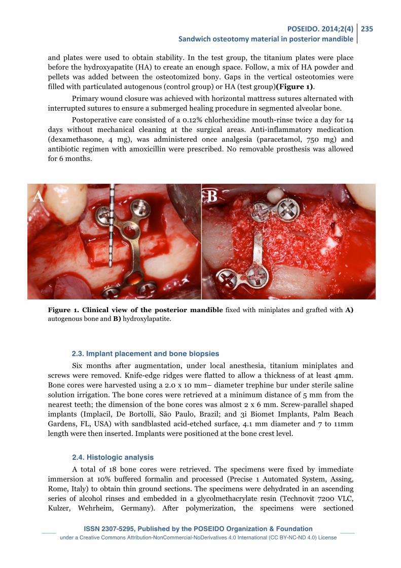
and plates were used to obtain stability. In the test group, the titanium plates were place before the hydroxyapatite (HA) to create an enough space. Follow, a mix of HA powder and pellets was added between the osteotomized bony. Gaps in the vertical osteotomies were filled with particulated autogenous (control group) or HA (test group)(Figure 1).
Primary wound closure was achieved with horizontal mattress sutures alternated with interrupted sutures to ensure a submerged healing procedure in segmented alveolar bone.
Postoperative care consisted of a 0.12% chlorhexidine mouth-rinse twice a day for 14 days without mechanical cleaning at the surgical areas. Anti-inflammatory medication (dexamethasone, 4 mg), was administered once analgesia (paracetamol, 750 mg) and antibiotic regimen with amoxicillin were prescribed. No removable prosthesis was allowed for 6 months. Figure 1. Clinical view of the posterior mandible fixed with miniplates and grafted with A) autogenous bone and B) hydroxylapatite.
2.3. Implant placement and bone biopsies Six months after augmentation, under local anesthesia, titanium miniplates and
screws were removed. Knife-edge ridges were flatted to allow a thickness of at least 4mm. Bone cores were harvested using a 2.0 x 10 mm– diameter trephine bur under sterile saline solution irrigation. The bone cores were retrieved at a minimum distance of 5 mm from the nearest teeth; the dimension of the bone cores was almost 2 x 6 mm. Screw-parallel shaped implants (Implacil, De Bortolli, São Paulo, Brazil; and 3i Biomet Implants, Palm Beach Gardens, FL, USA) with sandblasted acid-etched surface, 4.1 mm diameter and 7 to 11mm length were then inserted. Implants were positioned at the bone crest level.
2.4. Histologic analysis A total of 18 bone cores were retrieved. The specimens were fixed by immediate
immersion at 10% buffered formalin and processed (Precise 1 Automated System, Assing, Rome, Italy) to obtain thin ground sections. The specimens were dehydrated in an ascending series of alcohol rinses and embedded in a glycolmethacrylate resin (Technovit 7200 VLC, Kulzer, Wehrheim, Germany). After polymerization, the specimens were sectioned
236 Research article: Bechara K, et al. (2014)
ISSN 2307-5295, Published by the POSEIDO Organization & Foundation
under a Creative Commons Attribution-NonCommercial-NoDerivatives 4.0 International (CC BY-NC-ND 4.0) License
longitudinally along the major axis of the implants with a high-precision diamond disc at about 150 µm and ground down to about 30 µm. One to two slides were obtained for each bone biopsy.
The slides were stained with basic fuchsin and toluidine blue. The slides were observed under a light microscope. Histomorphometry of newly formed bone, remaining particles and/or non-vital bone and marrow space were carried out on the whole sample at low magnification (25x). These evaluations were performed using a light microscope connected to a high-resolution video camera and interfaced to a monitor and personal computer. This optical system was associated with a digitizing pad and a histometry software package with image-capture functionalities (Image-Pro Plus 4.5, Media Cybernetics Inc., Immagini & Computer Snc, Milan, Italy).
2.5. Statistical analysis The mean and standard deviation of histometric variables were calculated for each
site and then for each group. Wilcoxon test was used to calculate the differences between groups. The unit of analysis was the patient and the level of significance was 0.05.
3. Results 3.1. Clinical Assessment and vertical bone gain None of the patients presented complications following implant placement. All
mandibular sites showed optimal bone graft integration without signs of inflammation. The vertical bone gain was 6.5±1.6mm and 7.0±1.12mm for autogenous and HA groups respectively (p>0.05).
3.2. Histological Findings After 6 months, histological evaluation revealed the presence of mature bone with compact areas in varied degree in both groups. The compact bone exhibits incremental basophilic lines mixed with interposed reversion lines. The medullary spaces were scarce and almost filled with a well-vascularized connective tissue with no signs of inflammation or foreign body reaction (Figure 2). The spaces were interposed with areas of fibrosis that were sometimes dense. In some cases, particles of the implanted material, seen as irregular vacuolated amorphous masses of basophilic tendency or as discretely eosinophilic amorphous masses, could be found mainly in the HA group (Figure 3). The bone formation process was characterized by the presence of osteoblasts, and the harvesian system was well preserved. The inflammatory infiltrated is on average non-significant with prevalence of monuclear cells. In some situations of HA particles were present, close to the bony wall with the absence of osteogenic activity. The Table presents the histometric data of biopsy cores. The percentage of newly-formed bone was higher in the autogenous group (p<0.05) while the mean of remaining particles was higher for ncHA group (p<0.05). The percentage of marrow-space and bone density (% of newly-formed bone + % of remaining particles and/or non-vital particles) was similar for both groups (p>0.05).

POSEIDO. 2014;2(4) Sandwich osteotomy material in posterior mandible
237
ISSN 2307-5295, Published by the POSEIDO Organization & Foundation
under a Creative Commons Attribution-NonCommercial-NoDerivatives 4.0 International (CC BY-NC-ND 4.0) License
Figure 2. Lower view of histologic ground section of autogenous group (25x; Toluidine blue and acid fuchsin staining).
Figure 3. Ground section of histology of hydroxylapatite group (25x). Note the presence of extended basophilic areas (Toluidine blue and acid fuchsin staining).
Table. Mean and standard deviation of histometric variables of bone biopsies retrieved from grafted mandibular sites with autogenous or ncHA. Mann-Whitney Test (*p<0.05; NS= Non-significant).
Histological variables (%) Autogenous (n=9) HA (n=9)
New Bone* 78.50 ± 9.26 61.50 ± 13.28
Graft Material* 15.62 ± 8.27 29.72 ± 13.20
Marrow Space NS 5.87 ± 2.6 9.18 ± 5.98%
238 Research article: Bechara K, et al. (2014)
ISSN 2307-5295, Published by the POSEIDO Organization & Foundation
under a Creative Commons Attribution-NonCommercial-NoDerivatives 4.0 International (CC BY-NC-ND 4.0) License
4. Discussion There are few studies that evaluate the alveolar osteotomy in posterior mandible [1,5]
and the behavior of the grafted material. In general, this technique is considered as an efficient and elegant therapeutic solution for the preimplant reconstruction of the severely resorbed posterior mandible. Publications showed a good success rate, even if the surgery remains very delicate and requires an experienced practitioner [4,6]. However, because of the relative small number of publications on this topic and cases using this approach, the choice of the best material to use in sandwich was not really discussed or investigated thoroughly, each surgeons having its own habits or experience.
This study aimed to evaluated, histometrically, the bone behavior on mandibular sites grafted with intra-oral autogenous bone and non-ceramic hydroxylapatite (HA) using “sandwich” osteotomy. The results of this study demonstrated that both bone grafts were able to provide both vertical bone gain and bone anchorage for implant placement after 6 months healing.
Successful bone grafting requires that the surgeon select the optimal bone grafting material from a number of alternatives [7]. A synthetic bioactive resorbable graft material having osteoconductive biochemical and biomechanical qualities similar to the host bone provides the mean to improve compromised bone topography for ride preservation and augmentation or to enhance the bony site for implant placement and subsequent prosthetic rehabilitation [10].
Histomorphometric measurements of bone cores retrieved 6 months after augmentation depicted more residual-grafted material in the HA group. Felice et al. [1] demonstrated that 10 to 13% more residual particles of deproteinezed bovine bone when compared with extra-oral autogenous bone, after 4 months healing. The residual-graft in non-ceramic hydroxyapatite group observed in our study was an expected finding, because non-ceramic hydroxyapatite tends to resorb slowly, while the grafted autogenous bone remodels faster, mainly in this surgical technique (“sandwich” osteotomy). While a faster remodeling process of the graft could be theoretically advantageous because allow that dental implant surface being placed at surgical site with more vital bone available, from a clinical point of view, it did not appear that this characteristic provide any beneficial effect, at least, after 6 months healing in our study.
5. Conclusion The use of alveolar osteotomy with an interpositional graft (sandwich technique) for posterior mandible ridge augmentation is becoming an important therapeutic option, even if the technique remains sensible and operator-dependent. As a conclusion, this split mouth study suggested that both intra-oral autogenous bone and ncHA could be used as an interpositional graft material to vertically augment posterior atrophic mandibles, at least after 6 months follow-up. The investigations on the materials to use in this approach are still very recent, and many configurations have to be tested thoroughly. Disclosure of interests
The authors have no conflict of interest to report.

POSEIDO. 2014;2(4) Sandwich osteotomy material in posterior mandible
239
ISSN 2307-5295, Published by the POSEIDO Organization & Foundation
under a Creative Commons Attribution-NonCommercial-NoDerivatives 4.0 International (CC BY-NC-ND 4.0) License
Acknowledgements The authors want to thank Intra-Lock Brazil (São Paulo, Brazil) and Impladent Ltd
(Holliswood, NY, USA), for providing the hydroxyapatite; Implacil de Bortolli Implants (São Paulo, Brazil), for providing the dental implants. Author Contributions JAS was in charge of the elaboration of the study proposal and the financial support of the study, and he participated to the elaboration of the manuscript and the treatment planning of each case. JAR, AC, LC and GG were in charge of the statistical analysis and the financial support for the study. KB, AD, PYK were in charge of the treatment planning of each case, the implant placement surgery and they participated to the elaboration of the manuscript. AP, GI and GG were in charge of histological procedures. JAR and AC also participated to the elaboration of the study design and proposal. References [1] Felice P, Marchetti C, Iezzi G, Piattelli A, Worthington H, Pellegrino G, Esposito M. Vertical ridge augmentation of the atrophic posterior mandible with interpositional bloc grafts: bone from the iliac crest vs. bovine anorganic bone. Clinical and histological results up to one year after loading from a randomized-controlled clinical trial. Clin Oral Implants Res. 2009;20(12):1386-93. [2] Block MS, Haggerty CJ. Interpositional osteotomy for posterior mandible ridge augmentation. J Oral Maxillofac Surg. 2009;67(11 Suppl):31-9. [3] Sammartino G, Bernard JP. A clinical round table about the treatment of the severely resorbed posterior mandible. Part 1: challenges, endeavor and perspectives. POSEIDO. 2013;1(2):65-7. [4] Bormann KH, Suarez-Cunqueiro MM, von See C, Kokemuller H, Schumann P, Gellrich NC. Sandwich osteotomy for vertical and transversal augmentation of the posterior mandible. Int J Oral Maxillofac Surg. 2010;39(6):554-60. [5] Mazor Z, Lorean A. Preimplant reconstruction of the severely resorbed posterior mandible using the Sandwich technique with piezosurgical osteotomy and Leukocyte- and Platelet-Rich Fibrin (L-PRF): a 5-year follow-up with histological controls. POSEIDO. 2013;1(2):117-24. [6] Lopez-Cedrun JL. Implant rehabilitation of the edentulous posterior atrophic mandible: the sandwich osteotomy revisited. Int J Oral Maxillofac Implants. 2011;26(1):195-202. [7] Smiler D, Soltan M. The bone-grafting decision tree: a systematic methodology for achieving new bone. Implant Dent. 2006;15(2):122-8. [8] Scarano A, Degidi M, Iezzi G, Petrone G, Piattelli A. Correlation between implant stability quotient and bone-implant contact: a retrospective histological and histomorphometrical study of seven titanium implants retrieved from humans. Clin Implant Dent Relat Res. 2006;8(4):218-22. [9] Roze J, Babu S, Saffarzadeh A, Gayet-Delacroix M, Hoornaert A, Layrolle P. Correlating implant stability to bone structure. Clin Oral Implants Res. 2009;20(10):1140-5. [10] Valen M, Ganz SD. A synthetic bioactive resorbable graft for predictable implant reconstruction: part one. J Oral Implantol. 2002;28(4):167-77. This article can be cited as: Bechara K, Dottore AM, Kawakami PY, Cassoni A, Giro G, Rodrigues JA, Chambrone L, Piattelli A, Giovanna Iezzi G, Shibli JA. Interpositional graft associated with alveolar osteotomy for posterior mandible ridge augmentation (sandwich osteotomy) using hydroxyapatite or autogenous bone: histological evaluation. POSEIDO. 2014;2(4):233-9.