Embed Size (px)
Citation preview

biomolecules
Article
Interplay between Endoplasmic Reticulum (ER) Stressand Autophagy Induces Mutant p53H273 Degradation
Alessia Garufi 1,2, Giulia Federici 1, Maria Saveria Gilardini Montani 3, Alessandra Crispini 4,Mara Cirone 3 and Gabriella D’Orazi 1,*
1 Department of Research and Advanced Technologies, IRCCS Regina Elena National Cancer Institute,00144 Rome, Italy; [email protected] (A.G.); [email protected] (G.F.)
2 University “G. D’Annunzio”, School of Medicine, 66100 Chieti, Italy3 Department of Experimental Medicine, Sapienza University of Rome, laboratory affiliated to Istituto Pasteur
Italia-Fondazione Cenci Bolognetti, 00161 Rome, Italy;[email protected] (M.S.G.M.); [email protected] (M.C.)
4 Department of Chemistry and Chemical Technologies, laboratory MAT_IN LAB, Calabria University,87036 Rende, Italy; [email protected]
* Correspondence: [email protected]
Received: 29 December 2019; Accepted: 29 February 2020; Published: 3 March 2020�����������������
Abstract: The unfolded protein response (UPR) is an adaptive response to intrinsic and externalstressors, and it is mainly activated by the accumulation of misfolded proteins at the endoplasmicreticulum (ER) lumen producing ER stress. The UPR signaling network is interconnected withautophagy, the proteolytic machinery specifically devoted to clearing misfolded proteins in order tosurvive bioenergetic stress and/or induce cell death. Oncosuppressor TP53 may undergo inactivationfollowing missense mutations within the DNA-binding domain (DBD), and mutant p53 (mutp53)proteins may acquire a misfolded conformation, often due to the loss of the DBD-bound zinc ion,leading to accumulation of hyperstable mutp53 proteins that correlates with more aggressive tumors,resistance to therapies, and poorer outcomes. We previously showed that zinc supplementationinduces mutp53 protein degradation by autophagy. Here, we show that mutp53 (i.e., Arg273)degradation following zinc supplementation is correlated with activation of ER stress and of theIRE1α/XBPI arm of the UPR. ER stress inhibition with chemical chaperone 4-phenyl butyrate (PBA)impaired mutp53 downregulation, which is similar to IRE1α/XBPI specific inhibition, reducing cancercell death. Knockdown of mutp53 failed to induce UPR/autophagy activation indicating that theeffect of zinc on mutp53 folding was likely the key event occurring in ER stress activation. Recentlydiscovered small molecules targeting components of the UPR show promise as a novel anticancertherapeutic intervention. However, our findings showing UPR activation during mutp53 degradationindicate that caution is necessary in the design of therapies that inhibit UPR components.
Keywords: p53; mutp53H273; autophagy; endoplasmic reticulum (ER) stress; IRE1α/XBP1; zincsupplementation; 4-PBA; ST-083010; cancer therapy
1. Introduction
Tumor suppressor p53 plays a central role in tumor prevention and response to therapies.The presence of a functional p53 pathway is incompatible with neoplastic growth and, for this reason,p53 is the most frequently mutated gene in tumors [1]. The majority of p53 mutations are missensemutations (i.e., R175H, R248Q, and R273H) that are mainly within the DNA-binding domain (DBD),leading to the synthesis of p53 proteins that are unable to bind target gene promoters; however, somemutant (mut) p53 proteins can physically bind other transcription factors, profoundly remodeling the
Biomolecules 2020, 10, 392; doi:10.3390/biom10030392 www.mdpi.com/journal/biomolecules

Biomolecules 2020, 10, 392 2 of 15
cancer cell transcriptome and proteome [2]. Mouse models of different hotspot mutp53 proteins andclinical data from germline and sporadic cancers have established that some mutp53 proteins not onlyabolish the wild-type (wt) p53 tumor suppressive function, but can become oncogenic, promotinginvasion, metastasis, and chemoresistance [3,4]. Mutp53 proteins may acquire a misfolded and partiallydenatured conformation, forming hyperstable micro- and macro-aggregates that cannot undergodegradation, with their accumulation in tumors [5]. This aggregation may result from loss or alterationof DBD-bound zinc, which is necessary for the thermodynamic stability of the DBD and is neededfor the wtp53 oncosuppressor function [6]. Preventing mutp53 accumulation provides an importantchemopreventive and chemotherapeutic anticancer strategy and, in the last few years, many smallmolecules have been identified to induce mutp53 downregulation and/or reactivation of wtp53 (whichis inhibited by mutp53 as a dominant negative effect) [7,8]. Our previous studies showed that zincsupplementation modifies the equilibrium between p53 mutant and wild-type conformation, positivelyreactivating wtp53 functions of some of the most frequently p53 mutated residues, such as Arg175 andArg273 [9–12]. The reactivation of wtp53 conformation results in the re-establishment of canonicalDNA binding activity and the transcription of target genes, inducing apoptosis and inhibition of tumorgrowth, in vitro and in animal models [9–12]. We also showed that a curcumin-based zinc complex [13]induces mutp53 degradation through autophagy, in part depending on the wtp53-induced target geneDRAM (damage-regulated autophagy modulator) [14–16], and in line with the notion that mutp53blocks autophagy while wtp53 induces it [17].
Macroautophagy (hereafter indicated as autophagy), is a dynamic catabolic process that degradescytoplasmic components, unfolded proteins, and damaged organelles by enwrapping them intolysosomes assisting cells to cope with stress load [18,19]. Autophagy can promote cell survival ordeath. The process occurs at both the basal level and in response to stress, and it is linked to theendoplasmic reticulum (ER) stress. ER, the principal intracellular organelle responsible for proteinfolding, localization, and post-translational modifications, undergoes stress when unfolded proteinsaccumulate into it due to intracellular and extracellular insults (i.e, glucose deprivation, hypoxia,acidosis, inhibition of protein glycosylation, disturbance of intracellular Ca2+ stores, oncogenic mutation,etc.). ER stress triggers the unfolded protein response (UPR) that, besides inducing autophagy, restoresER homeostasis via the reduction of global protein synthesis and the activation of chaperones anddegradation processes [20]. UPR is orchestrated by three main sensors, namely inositol-requiringenzyme 1α (IRE1α), activating transcription factor 6 (ATF6), and protein kinase RNA-like ER kinase(PERK), which regulate many signaling pathways (autophagy, apoptosis, antioxidant response,inflammation, etc.), and ultimately dictate cell survival or death decision [21]. The latter occurs whenthe adaptive UPR response to stress is overwhelmed [22]. Under basal condition, the ER luminalchaperone BiP/GRP78 protein binds to these UPR molecules from the ER lumen and suppresses theirbasal activity. During ER stress, misfolded proteins accumulate in the ER lumen remove BiP from IRE1α,PERK, and ATF-6, leading to activation of downstream signaling [23]. The IRE1α signaling pathwayinduces expression of the transcription factor Xbp1s, which increases the expression of ER chaperonsand ER mass, stimulates lipid biogenesis, degrades unfolded proteins to enhance the secretory functionof ER, and triggers autophagy [24,25]. The activation of PERK can induce the pro-apoptotic function ofUPR through the eIF2α-ATF4-CHOP axis, but it can also induce autophagy, together with IRE1α/XBPIand ATF6 arms [26]. UPR and autophagy are, thus, interconnected processes that share commonproperty to promote the adaption of cells to stress [27] and, for this reason, the dysregulation of one ofthese processes strongly influences the other. Autophagy has been proposed as an innovative targetfor anticancer therapies, although its manipulation in cancer is still debated [28]. In addition, targetingspecific components of the UPR signaling network is also becoming a novel potential anticancerstrategy [21]. Therefore, a better understanding of the interplay between UPR and autophagy mayhelp in elucidating the molecular mechanisms that tip the balance towards cell death or survival.Based on these premises, we asked whether ER stress/UPR activation was involved in zinc-inducedmutp53 downregulation.

Biomolecules 2020, 10, 392 3 of 15
2. Materials and Methods
2.1. Cell Culture and Reagents
The human U373MG (expressing R273H p53 mutation) and T98G (expressing M237I p53 mutation)glioblastoma cell lines, and the HT29 (expressing R273H p53 mutation) and RKO (carrying wild-typep53) colon cancer cell lines were maintained in RPM1-1640 (Life Technologies-Invitrogen); the humanHCT116 (carrying wild-type p53) colon cancer cell line was maintained in the Dulbecco modified Eagle’smedium (DMEM) (Life Technologies-Invitrogen). All were supplemented with 10% heat-inactivatedfetal bovine serum (FBS) (Corning, NY, USA; 35-079) plus L-glutamine and streptomycin (100 µg/mL)(Corning, NY, USA; 30-002), in 5% CO2 at 37 ◦C.
The following reagents were used: A heteroleptic pentacoordinated (bpy-9)Zn(curc, Cl) complexcontaining a 4,4’-disubstituted-2,2’-bipyridine as the main ligand, and curcumin (curc) and chloride(Cl) as ancillary ligands (Zn(II)-curc) [13], dissolved in DMSO and used at 100 µg/mL, as reportedin [12]; the inhibitor of wtp53 transactivation function pifithryn α (PFT-α) (Enzo Life Sciences, Lausen,Switzerland, BML-GR325), dissolved in DMSO and used at 30 µM [15,29]; the ER stress inhibitor4-Phenylbutyric acid (4-BPA) (Sigma-Aldrich, #P21005) [30], dissolved in filtered sterile water and usedat 2.5 mM; the ER stress inducer Tunicamycin (Tn) (Sigma-Aldrich, #T7765), dissolved in DMSO andused at 1 µg/mL; and the inhibitor of XBP1 cleavage STF-083010 (Sigma-Aldrich, #SML0409) (hereafterindicated as STF) [31,32], dissolved in DMSO and used at 60 µM.
2.2. Viability Assay and Cell Death/PI Staining
Subconfluent cells were plated in triplicate in six-well plates and, the day after, were treated withZn (II)-curc (100 µg/mL) for 24 h. Both adherent and floating cells were collected and cell viabilitywas assessed by Trypan blue (Sigma-Aldrich, #72571) exclusion counting blue (dead)/total cells with aNeubauer hemocytometer using light microscopy.
Cell death was detected by cytofluorimetric analysis of propidium iodide (PI)-stained cells, aspreviously reported [12]. Briefly, both floating and adherent cells were fixed in 80% ethanol and stainedin a PBS solution containing PI (62.5 mg/mL; Sigma-Aldrich, #P4864) and RNase A (1.125 mg/mL;Sigma-Aldrich, #R6148). Samples were acquired with a FACScan instrument (Becton Dickinson) andthe percentage of cells in the sub-G1 compartment was calculated using ModFit LT software (BectonDickinson).
2.3. Endoplasmic Reticulum (ER) Staining
Subconfluent cells were seeded on coverslips in 35 mm Petri dishes and, the day after, were treatedwith Zn (II)-curc (100 µg/mL) for 16 h. After treatment, cells were fixed with 3.7% paraformaldehyde(Thermo Fisher Scientific, #50-980-487) for 10 min at room temperature (RT) and stained withER Staining Kit-Red Fluorescence (Abcam, #ab139482) following the manufacturer’s instructions.Immunofluorescence was visualized by an Olympus BX53 microscope equipped with epifluorescence,and photographs were taken (×40 objective) using a cooled camera device (ProgRes MF). ImageJ(NIH) software [33] (http://imagej.nih.gov) was used to calculate the relative fluorescence from 40×magnification images and normalized to cell size from phase-contrast images. At least 25 cells wereanalyzed in duplicate for each group at the same exposure time.
2.4. RNA Extraction and Semi-Quantitative Reverse Transcription (RT)-PCR Analysis
Cells were harvested in TRIzol Reagent (Invitrogen, #15596026) and total RNA was isolatedfollowing the manufacturer’s instructions. The first strand cDNA was synthesized from 2 µg of totalRNA with a MuLV reverse transcriptase kit (TermoFisher Scientific, #28025013). Semi-quantitativeReverse-Transcribed (RT)-PCR was carried out by using Hot-Master Taq polymerase (Geneaid BiotechLtd., New Taipei City, Taiwan, #TQ050) with 2 µL cDNA reaction and genes specific oligonucleotidesunder the conditions of linear amplification. The primers used for amplification of Xbp1s were as

Biomolecules 2020, 10, 392 4 of 15
follows: Xbp1s-for GGAGTTAAGACAGCGCT; Xbp1s-rev TGTTCTGGAGGGGTGAC. PCR productswere run on a 2% agarose gel and visualized with ethidium bromide. The housekeeping 28S gene,used as the internal standard, was amplified from the same cDNA reaction mixture. Densitometricanalysis was applied to quantify mRNA levels compared to control gene expression.
2.5. siRNA Interference
Subconfluent cells were plated in 35 mm Petri dishes and, the day after, were transfected withcontrol pSuper and pSuper-p53 (for p53 interference, si-p53) vectors [34] using the LipofectaminePlusreagent (Invitrogen, #11514-015), according to the manufacturer’s instructions. Twenty-four hoursafter transfection, cells were trypsinized and replated for the indicated experiments.
2.6. Western Blotting
Total cell extracts were prepared by incubation in a lysis buffer (50 mM Tris–HCl, pH 7.5, 150 mMNaCl, 5 mM EDTA, 150 mM KCl, 1 mM dithiothreitol, and 1% Nonidet P-40) (all from Sigma-Aldrich)and a mix of protease inhibitors (cOmpleteTM, Mini Protease Inhibitor Cocktail, Merck, Life Science S.r.l.,Milan, Italy, #11836153001). Then, 15–30 µg of protein lysate was subjected to electrophoresis on 9–18%SDS-PAGE gradient gels (Bio-Rad, #456-1095), according to the manufacturer’s instructions. The gelswere transferred to a polyvinylidene difluoride (PVDF) membrane (Immobilon-P, Millipore, #IPVH00010) for 2 h in Tris-glycine buffer. Membranes were blocked in PBS-0.1% Tween 20 solution containing3% BSA (Sigma-Aldrich) before probing with the following specific primary antibodies: mousemonoclonal anti-p53 (1:1000) (DO-1, (1:100) (Santa Cruz Biotechnology Inc., Heidelberg, Germany;sc-126,); rabbit polyclonal anti-BiP (1:1000) (Cell Signaling, C50B12 #3177); rabbit polyclonal anti-IRE1alpha (p-Ser724) (1:1000) (Novus Biologicals, #NB100-2323SS); rabbit polyclonal anti-IRE1 alpha(1:1000) (Novus Biologicals, #NB100-2324); rabbit polyclonal anti XBP1 (1:1000) (Novus Biologicals,#NBP1-77681SS); and rabbit polyclonal anti-LC3B (1:1000) (Sigma-Aldrich, #L7543). Mouse monoclonalanti-β-actin (1:10,000) (Sigma Aldrich, #A5441) was used as a protein loading control. Primaryantibodies were detected with the following horseradish peroxidase-labeled secondary antibodies: goatpolyclonal anti-mouse IgG-horseradish peroxidase (HRP, BioRad.; #172-1011) and anti-rabbit IgG-HRP(BioRad; #172-1019). Enzymatic signals were visualized by chemiluminescence (ECL Detection system,Amersham GE Healthcare, Milan, Italy, #RPN2106), according to the manufacturer’s protocol.
2.7. Statistical Analysis
Each experiment was performed at least three times. Results are expressed as values of mean ±standard deviation (S.D.). Statistical significance was determined using Student’s t-tests for two samplecomparison. Difference was considered statistically significant when the p-value was at least <0.05.
3. Results
3.1. ER Stress Sis Activated by Zn(II)-Curc in mutp53H273 Cancer Cells
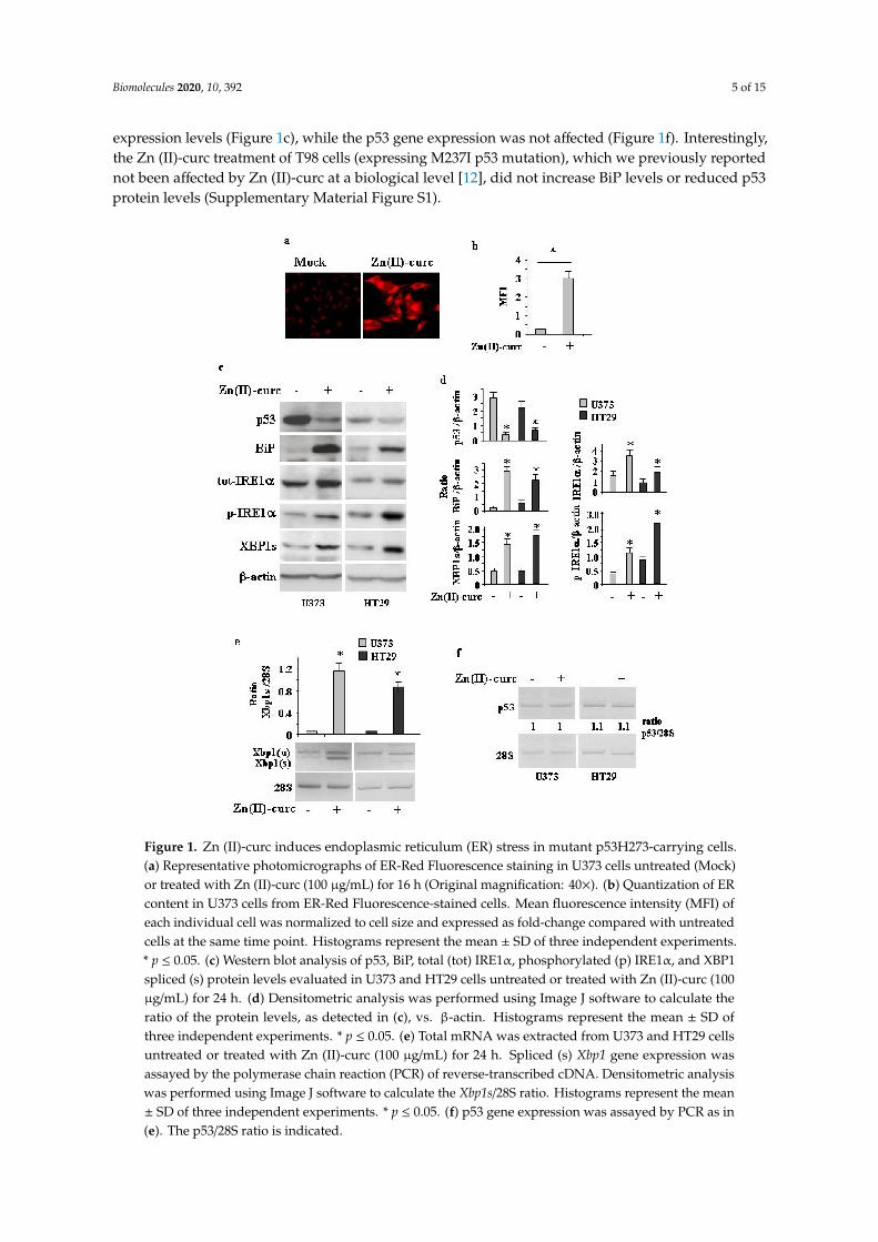
As Zn (II)-curc may induce mutp53 protein degradation through autophagy [14,15], and autophagycan be induced by UPR activation [27], we investigated the effect of Zn(II)-curc on ER stress/UPRactivation. To address this issue, we used several approaches. U373 cells were treated with 100µM Zn (II)-curc, the dose that induces mutp53 degradation and autophagy [14,15]. ER analysisby specific fluorescent red labelling shows that Zn (II)-curc caused a marked increase in ER size(Figure 1a,b). This increase correlated with the augmented expression of BiP/Grp78 as well as withthe IRE1α and p-IRE1α protein levels in both U373 and HT29 cells (Figure 1c,d), while the otherarms of the UPR were not affected in this experimental condition (not shown). Western blot andsemiquantitative RT-PCR evidenced spliced (s) Xbp1 mRNA (Figure 1c–e), in agreement with thenotion that activated IRE1α functions as an endoribonuclease, splicing a 26 base pair intron from Xbp1mRNA [35]. The Zn (II)-curc-induced activation of IRE1α correlated with the reduction of mutp53
Biomolecules 2020, 10, 392 5 of 15
expression levels (Figure 1c), while the p53 gene expression was not affected (Figure 1f). Interestingly,the Zn (II)-curc treatment of T98 cells (expressing M237I p53 mutation), which we previously reportednot been affected by Zn (II)-curc at a biological level [12], did not increase BiP levels or reduced p53protein levels (Supplementary Material Figure S1).
Figure 1. Zn (II)-curc induces endoplasmic reticulum (ER) stress in mutant p53H273-carrying cells.(a) Representative photomicrographs of ER-Red Fluorescence staining in U373 cells untreated (Mock)or treated with Zn (II)-curc (100 µg/mL) for 16 h (Original magnification: 40×). (b) Quantization of ERcontent in U373 cells from ER-Red Fluorescence-stained cells. Mean fluorescence intensity (MFI) ofeach individual cell was normalized to cell size and expressed as fold-change compared with untreatedcells at the same time point. Histograms represent the mean ± SD of three independent experiments.* p ≤ 0.05. (c) Western blot analysis of p53, BiP, total (tot) IRE1α, phosphorylated (p) IRE1α, and XBP1spliced (s) protein levels evaluated in U373 and HT29 cells untreated or treated with Zn (II)-curc (100µg/mL) for 24 h. (d) Densitometric analysis was performed using Image J software to calculate theratio of the protein levels, as detected in (c), vs. β-actin. Histograms represent the mean ± SD ofthree independent experiments. * p ≤ 0.05. (e) Total mRNA was extracted from U373 and HT29 cellsuntreated or treated with Zn (II)-curc (100 µg/mL) for 24 h. Spliced (s) Xbp1 gene expression wasassayed by the polymerase chain reaction (PCR) of reverse-transcribed cDNA. Densitometric analysiswas performed using Image J software to calculate the Xbp1s/28S ratio. Histograms represent the mean± SD of three independent experiments. * p ≤ 0.05. (f) p53 gene expression was assayed by PCR as in(e). The p53/28S ratio is indicated.
Biomolecules 2020, 10, 392 6 of 15
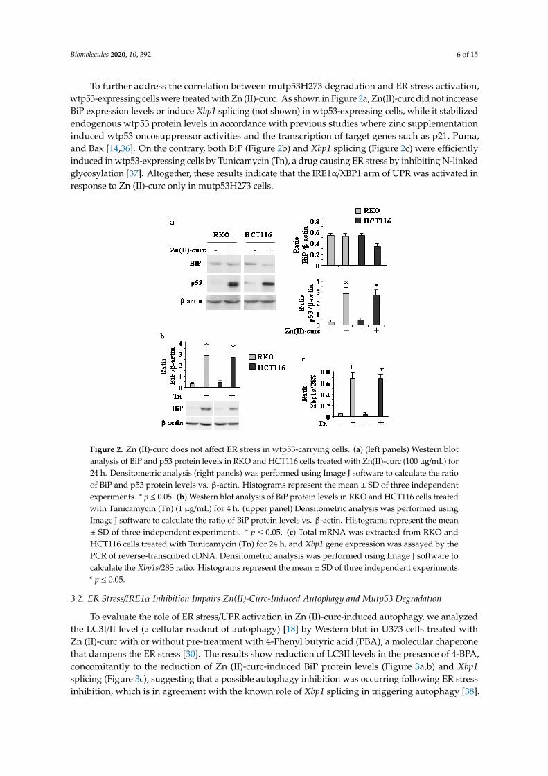
To further address the correlation between mutp53H273 degradation and ER stress activation,wtp53-expressing cells were treated with Zn (II)-curc. As shown in Figure 2a, Zn(II)-curc did not increaseBiP expression levels or induce Xbp1 splicing (not shown) in wtp53-expressing cells, while it stabilizedendogenous wtp53 protein levels in accordance with previous studies where zinc supplementationinduced wtp53 oncosuppressor activities and the transcription of target genes such as p21, Puma,and Bax [14,36]. On the contrary, both BiP (Figure 2b) and Xbp1 splicing (Figure 2c) were efficientlyinduced in wtp53-expressing cells by Tunicamycin (Tn), a drug causing ER stress by inhibiting N-linkedglycosylation [37]. Altogether, these results indicate that the IRE1α/XBP1 arm of UPR was activated inresponse to Zn (II)-curc only in mutp53H273 cells.
Figure 2. Zn (II)-curc does not affect ER stress in wtp53-carrying cells. (a) (left panels) Western blotanalysis of BiP and p53 protein levels in RKO and HCT116 cells treated with Zn(II)-curc (100 µg/mL) for24 h. Densitometric analysis (right panels) was performed using Image J software to calculate the ratioof BiP and p53 protein levels vs. β-actin. Histograms represent the mean ± SD of three independentexperiments. * p ≤ 0.05. (b) Western blot analysis of BiP protein levels in RKO and HCT116 cells treatedwith Tunicamycin (Tn) (1 µg/mL) for 4 h. (upper panel) Densitometric analysis was performed usingImage J software to calculate the ratio of BiP protein levels vs. β-actin. Histograms represent the mean± SD of three independent experiments. * p ≤ 0.05. (c) Total mRNA was extracted from RKO andHCT116 cells treated with Tunicamycin (Tn) for 24 h, and Xbp1 gene expression was assayed by thePCR of reverse-transcribed cDNA. Densitometric analysis was performed using Image J software tocalculate the Xbp1s/28S ratio. Histograms represent the mean ± SD of three independent experiments.* p ≤ 0.05.
3.2. ER Stress/IRE1α Inhibition Impairs Zn(II)-Curc-Induced Autophagy and Mutp53 Degradation
To evaluate the role of ER stress/UPR activation in Zn (II)-curc-induced autophagy, we analyzedthe LC3I/II level (a cellular readout of autophagy) [18] by Western blot in U373 cells treated withZn (II)-curc with or without pre-treatment with 4-Phenyl butyric acid (PBA), a molecular chaperonethat dampens the ER stress [30]. The results show reduction of LC3II levels in the presence of 4-BPA,concomitantly to the reduction of Zn (II)-curc-induced BiP protein levels (Figure 3a,b) and Xbp1splicing (Figure 3c), suggesting that a possible autophagy inhibition was occurring following ER stressinhibition, which is in agreement with the known role of Xbp1 splicing in triggering autophagy [38].

Biomolecules 2020, 10, 392 7 of 15
Concomitantly, we verified the effect of ER stress inhibition on mutp53 levels and found that mutp53protein was no longer downregulated in the presence of 4-BPA (Figure 3a,b).
Figure 3. ER stress inhibition impairs Zn (II)-curc-induced autophagy and mutp53 degradation.(a) Western blot analysis of BiP, LC3I/II, and p53 protein levels in U373 cells untreated or treatedwith Zn(II)-curc (100 µg/mL) for 24 h, with or without 1 h pre-treatment with 4-BPA (2.5 mM).(b) Densitometric analysis was performed using Image J software to calculate the ratio of the proteinlevels, as detected in (a), vs. β-actin. Histograms represent the mean ± SD of three independentexperiments. * p ≤ 0.05. (c) Total mRNA was extracted from U373 cells untreated or treated as in (a).Spliced (s) and unspliced (u) Xbp1 gene expression were assayed by the PCR of reverse-transcribedcDNA. Densitometric analysis was performed using Image J software to calculate the Xbp1s/28S ratio.Histograms represent the mean ± SD of three independent experiments. * p ≤ 0.05.
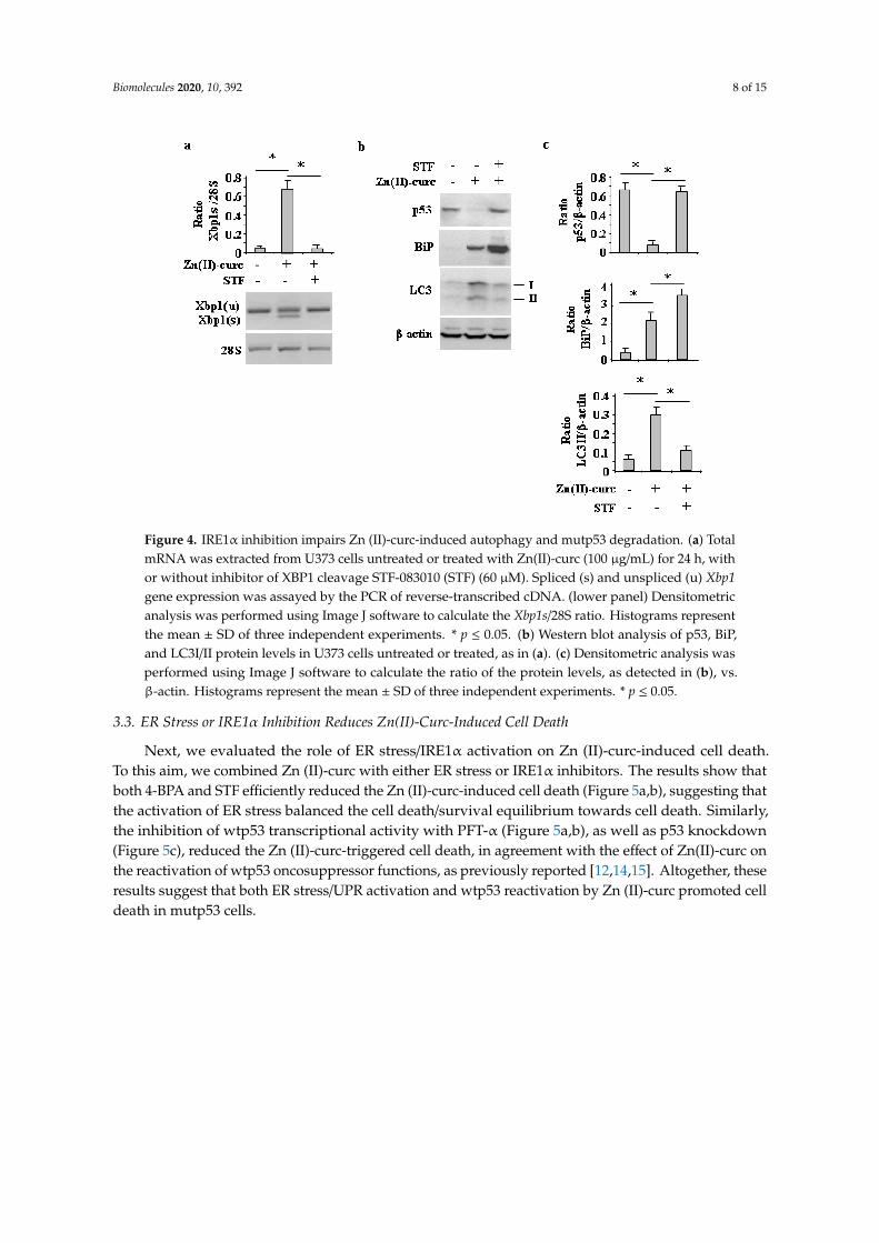
Similarly, the inhibition of IRE1α-Xbp1 by the small molecule STF [31,32], which indeedcounteracted the Zn (II)-curc-induced Xbp1 splicing (Figure 4a) and slightly increased BiP levels(Figure 4b,c), impaired the Zn (II)-curc-induced increase of LC3II levels and counteracted theZn (II)-curc-induced mutp53 downregulation (Figure 4b,c). Altogether, these results suggestthat the Zn (II)-curc-induced IRE1α-Xbp1 arm of the UPR was able to promote autophagy andmutp53 downregulation.
Biomolecules 2020, 10, 392 8 of 15
Figure 4. IRE1α inhibition impairs Zn (II)-curc-induced autophagy and mutp53 degradation. (a) TotalmRNA was extracted from U373 cells untreated or treated with Zn(II)-curc (100 µg/mL) for 24 h, withor without inhibitor of XBP1 cleavage STF-083010 (STF) (60 µM). Spliced (s) and unspliced (u) Xbp1gene expression was assayed by the PCR of reverse-transcribed cDNA. (lower panel) Densitometricanalysis was performed using Image J software to calculate the Xbp1s/28S ratio. Histograms representthe mean ± SD of three independent experiments. * p ≤ 0.05. (b) Western blot analysis of p53, BiP,and LC3I/II protein levels in U373 cells untreated or treated, as in (a). (c) Densitometric analysis wasperformed using Image J software to calculate the ratio of the protein levels, as detected in (b), vs.β-actin. Histograms represent the mean ± SD of three independent experiments. * p ≤ 0.05.
3.3. ER Stress or IRE1α Inhibition Reduces Zn(II)-Curc-Induced Cell Death
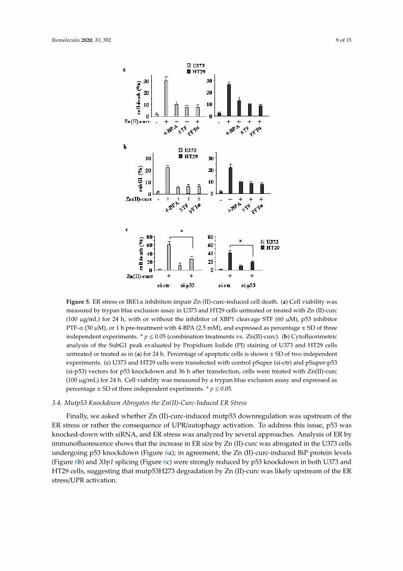
Next, we evaluated the role of ER stress/IRE1α activation on Zn (II)-curc-induced cell death.To this aim, we combined Zn (II)-curc with either ER stress or IRE1α inhibitors. The results show thatboth 4-BPA and STF efficiently reduced the Zn (II)-curc-induced cell death (Figure 5a,b), suggesting thatthe activation of ER stress balanced the cell death/survival equilibrium towards cell death. Similarly,the inhibition of wtp53 transcriptional activity with PFT-α (Figure 5a,b), as well as p53 knockdown(Figure 5c), reduced the Zn (II)-curc-triggered cell death, in agreement with the effect of Zn(II)-curc onthe reactivation of wtp53 oncosuppressor functions, as previously reported [12,14,15]. Altogether, theseresults suggest that both ER stress/UPR activation and wtp53 reactivation by Zn (II)-curc promoted celldeath in mutp53 cells.
Biomolecules 2020, 10, 392 9 of 15
Figure 5. ER stress or IRE1α inhibition impair Zn (II)-curc-induced cell death. (a) Cell viability wasmeasured by trypan blue exclusion assay in U373 and HT29 cells untreated or treated with Zn (II)-curc(100 µg/mL) for 24 h, with or without the inhibitor of XBP1 cleavage STF (60 µM), p53 inhibitorPTF-α (30 µM), or 1 h pre-treatment with 4-BPA (2.5 mM), and expressed as percentage ± SD of threeindependent experiments. * p ≤ 0.05 (combination treatments vs. Zn(II)-curc). (b) Cytofluorimetricanalysis of the SubG1 peak evaluated by Propidium Iodide (PI) staining of U373 and HT29 cellsuntreated or treated as in (a) for 24 h. Percentage of apoptotic cells is shown ± SD of two independentexperiments. (c) U373 and HT29 cells were transfected with control pSuper (si-ctr) and pSuper-p53(si-p53) vectors for p53 knockdown and 36 h after transfection, cells were treated with Zn(II)-curc(100 µg/mL) for 24 h. Cell viability was measured by a trypan blue exclusion assay and expressed aspercentage ± SD of three independent experiments. * p ≤ 0.05.
3.4. Mutp53 Knockdown Abrogates the Zn(II)-Curc-Induced ER Stress
Finally, we asked whether Zn (II)-curc-induced mutp53 downregulation was upstream of theER stress or rather the consequence of UPR/autophagy activation. To address this issue, p53 wasknocked-down with siRNA, and ER stress was analyzed by several approaches. Analysis of ER byimmunofluorescence shows that the increase in ER size by Zn (II)-curc was abrogated in the U373 cellsundergoing p53 knockdown (Figure 6a); in agreement, the Zn (II)-curc-induced BiP protein levels(Figure 6b) and Xbp1 splicing (Figure 6c) were strongly reduced by p53 knockdown in both U373 andHT29 cells, suggesting that mutp53H273 degradation by Zn (II)-curc was likely upstream of the ERstress/UPR activation.

Biomolecules 2020, 10, 392 10 of 15
Figure 6. Mutp53 knockdown abrogates the Zn (II)-curc-induced ER stress. (a) U373 cells weretransfected with control pSuper (si-ctr) and pSuper-p53 (si-p53) vectors for p53 knockdown and 36 hafter transfection, cells were treated with Zn (II)-curc (100 µg/mL) for 16 h, before undergoing ER-RedFluorescence staining. Quantization of ER content in U373 cells from ER-Red Fluorescence-stained cellsas evaluated by the mean fluorescence intensity (MFI) of each individual cell normalized to cell size andexpressed as fold-change compared with untreated cells at the same time point. Histograms representthe mean ± SD of three independent experiments. * p ≤ 0.05. (b) Western blot analysis of BiP andp53 protein levels in U373 and HT29 cells transfected for 36 h with si-ctr and si-p53, and then treatedwith Zn (II)-curc (100 µg/mL) for 24 h. Densitometric analysis was performed using Image J softwareto calculate the ratio of BiP and p53 protein levels vs. β-actin, as indicated ns: not specific signal.(c) Total mRNA was extracted from U373 cells treated as in (b), and spliced (s) and unspliced (u) Xbp1gene expression were assayed by the PCR of reverse-transcribed cDNA. (upper panel) Densitometricanalysis was performed using Image J software to calculate the Xbp1s/28S ratio. Histograms representthe mean ± SD of three independent experiments. * p ≤ 0.05.
4. Discussion
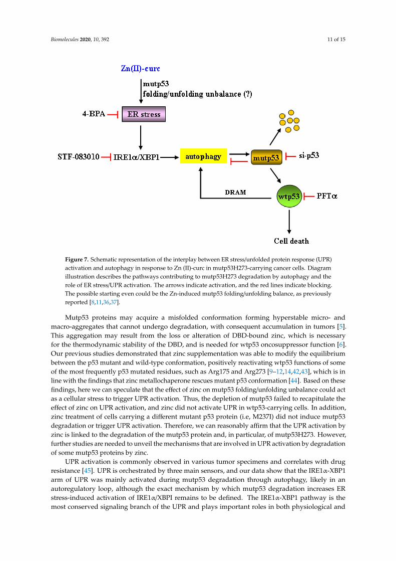
Cancer cells are often exposed to intrinsic and external factors that alter protein homeostasis.The consequence is the activation of ER stress and UPR, which is the adaptive mechanism used tocope with ER stress and to restore ER proteostasis [21]. Activation of the UPR through three differentbut interconnected signaling pathways may favor pro-death or prosurvival signaling and is strictlylinked to autophagy. Autophagy is usually a prosurvival mechanism, playing a crucial role in drugresistance, although it may also induce cell death [39,40]. The interplay between autophagy and UPRand their abnormal activation may pave the way to degenerative and chronic diseases includingcancer [28,41]. Therefore, understanding the crosstalk between UPR activities and autophagy shouldhelp in developing new treatment options for various pathologies, including cancer. Here, we foundthat autophagy-mediated mutp53H273 degradation following zinc supplementation correlated withactivation of ER stress and of the IRE1α/XBPI arm of the UPR. ER stress or IRE1α/XBPI inhibitionimpaired mutp53 degradation and reduced cell death, suggesting that both ER stress/UPR activationand the clearance of mutp53 by zinc played a pro-death role in mutp53-carrying cells, as summarizedin Figure 7.
Biomolecules 2020, 10, 392 11 of 15
Figure 7. Schematic representation of the interplay between ER stress/unfolded protein response (UPR)activation and autophagy in response to Zn (II)-curc in mutp53H273-carrying cancer cells. Diagramillustration describes the pathways contributing to mutp53H273 degradation by autophagy and therole of ER stress/UPR activation. The arrows indicate activation, and the red lines indicate blocking.The possible starting even could be the Zn-induced mutp53 folding/unfolding balance, as previouslyreported [8,11,36,37].
Mutp53 proteins may acquire a misfolded conformation forming hyperstable micro- andmacro-aggregates that cannot undergo degradation, with consequent accumulation in tumors [5].This aggregation may result from the loss or alteration of DBD-bound zinc, which is necessaryfor the thermodynamic stability of the DBD, and is needed for wtp53 oncosuppressor function [6].Our previous studies demonstrated that zinc supplementation was able to modify the equilibriumbetween the p53 mutant and wild-type conformation, positively reactivating wtp53 functions of someof the most frequently p53 mutated residues, such as Arg175 and Arg273 [9–12,14,42,43], which is inline with the findings that zinc metallochaperone rescues mutant p53 conformation [44]. Based on thesefindings, here we can speculate that the effect of zinc on mutp53 folding/unfolding unbalance could actas a cellular stress to trigger UPR activation. Thus, the depletion of mutp53 failed to recapitulate theeffect of zinc on UPR activation, and zinc did not activate UPR in wtp53-carrying cells. In addition,zinc treatment of cells carrying a different mutant p53 protein (i.e, M237I) did not induce mutp53degradation or trigger UPR activation. Therefore, we can reasonably affirm that the UPR activation byzinc is linked to the degradation of the mutp53 protein and, in particular, of mutp53H273. However,further studies are needed to unveil the mechanisms that are involved in UPR activation by degradationof some mutp53 proteins by zinc.
UPR activation is commonly observed in various tumor specimens and correlates with drugresistance [45]. UPR is orchestrated by three main sensors, and our data show that the IRE1α-XBP1arm of UPR was mainly activated during mutp53 degradation through autophagy, likely in anautoregulatory loop, although the exact mechanism by which mutp53 degradation increases ERstress-induced activation of IRE1α/XBPI remains to be defined. The IRE1α-XBP1 pathway is themost conserved signaling branch of the UPR and plays important roles in both physiological and

Biomolecules 2020, 10, 392 12 of 15
pathological settings, with its activity having profound effects on disease progression and prognosis [35].The IRE1α signaling pathway induces the expression of the transcription factor Xbp1s, which increasesthe expression of ER chaperones and ER mass, stimulates lipid biogenesis, and degrades unfoldedproteins to enhance the secretory function of ER and triggers autophagy initiation, mainly serving as apro-survival pathway in multiple human cancers [35]. XBP1 pathway activation has been shown toinduce triple-negative breast cancer progression and is correlated with poor patient survival, suggestingthat UPR inhibitors in combination with chemotherapy may improve tumor regression [46]. In addition,unspliced XBP1 is associated with longer survival of breast cancer patients treated with tamoxifen,which is opposite to XBP1 splicing that is associated with shorter survival [47]. Therefore, developingcombination therapies that target specific UPR signaling pathways will hopefully bypass anticancerdrug resistance. On the other hand, we found here that XBP1 inhibition impaired zinc-inducedautophagy and mutp53 degradation, indicating that UPR modulation may achieve different effectsdepending on cell context. To make the story more complex, mup53 has been shown to favor cancercell survival and promote cancer progression by its crosstalk with cellular stress pathways [48].For instance, the downregulation of mutp53 interferes with the downstream activation of the cross-talkbetween NRF2 and p62, restoring the cytotoxic effect of chemotherapies [49], and mutp53 inhibits thepro-apoptotic and pro-survival UPR effectors PERK and ATF6 in cancer cell lines [50], suggesting thatUPR inhibitors combined with mutp53 inhibitors may increase cell death.
5. Conclusions
In conclusion, this study demonstrates for the first time that a curcumin-based zinc complex wasable to induce UPR activation to trigger autophagy and mutp53H273 degradation, highlighting theinterplay between ER stress and autophagy. As UPR activation has both pro-survival and pro-deatheffects, caution is necessary in the design of therapies that target UPR components in combination withchemotherapies to increase the antitumor response, as they could hamper mutp53 degradation.
Supplementary Materials: The following are available online at http://www.mdpi.com/2218-273X/10/3/392/s1.
Author Contributions: G.D. and M.C. conceived and designed the experiments; A.G., G.F., M.S.G.M. carried outthe experiments; A.C. provided the Zn (II)-curc reagents; G.D. and M.C. analyzed the data; G.D. wrote the paper.All authors have read and agreed to the published version of the manuscript.
Funding: This research was funded by the Italian Association for Cancer Research (AIRC) Grant (IG 2015 Id.16742)to G.D.; by PRIN 2017 (2017K55HLC), Istituto Pasteur Italia-Fondazione Cenci Bolognetti Grant, and AIRC Grant(IG 2019 Id.23040) to M.C.
Acknowledgments: The authors wish to thank S. Soddu and A. Verdina for sharing reagents and for criticaldiscussion.
Conflicts of Interest: The authors declare no conflict of interest.
References
1. Schulz-Heddergott, R.; Moll, U.M. Gain-of-function (GOF) mutant p53 are actionable therapeutic target.Cancers 2018, 10, 188. [CrossRef] [PubMed]
2. Muller, P.A.; Vousden, K.H. Mutant p53 in cancer: New functions and therapeutic opportunities. Cancer Cell2014, 25, 304–317. [CrossRef] [PubMed]
3. Garcia, P.B.; Attardi, L.D. Illuminating p53 function in cancer with genetically engineered mouse models.Semin. Cell. Dev. Biol. 2014, 27, 74–85. [CrossRef] [PubMed]
4. Kolukula, V.K.; Sahu, G.; Wellstein, A.; Rodriguez, O.C.; Preet, A.; Iacobazzi, V.; D’Orazi, G.; Albanese, C.;Palmieri, F.; Avantaggiati, M.L. SLC25A1, or CIC, is a novel transcriptional target of mutant p53 and anegative tumor prognostic marker. Oncotarget 2014, 5, 1212–1225. [CrossRef]
5. Joenger, A.C.; Fersht, A.R. Structural biology of the tumor suppressor p53 and cancer-associated mutants.Adv. Cancer Res. 2007, 97, 1–23. [CrossRef]
6. Loh, S.N. The missing zinc: P53 misfolding and cancer. Metallomics 2010, 2, 442–449. [CrossRef]

Biomolecules 2020, 10, 392 13 of 15
7. Hoe, K.K.; Nerma, C.S.; Lane, D.P. Drugging the p53 pathway: Understanding the route to clinical efficacy.Nat. Rev. Drug Discov. 2014, 13, 217–236. [CrossRef]
8. Blandino, G.; Di Agostino, S. New therapeutic strategies to treat human cancers expressing mutant p53proteins. J. Exp. Clin. Cancer Res. 2018, 37, 30. [CrossRef]
9. Puca, R.; Nardinocchi, L.; Gal, H.; Rechavi, G.; Amariglio, N.; Domany, E.; Notterman, D.A.; Scarsella, M.;Leonetti, C.; Sacchi, A.; et al. Reversible dysfunction of wild-type p53 following homeodomain-interactingprotein kinase-2 knockdown. Cancer Res. 2008, 68, 3707–3714. [CrossRef]
10. Puca, R.; Nardinocchi, L.; Porru, M.; Simon, A.J.; Rechavi, G.; Leonetti, C.; Givol, D.; D’Orazi, G. Restoringp53 active conformation by zinc increases the response of mutant p53 tumor cells to anticancer drugs.Cell Cycle 2011, 10, 1679–1689. [CrossRef]
11. Margalit, O.; Simon, A.J.; Yakubov, E.; Puca, R.; Yosepovich, A.; Avivi, C.; Jacob-Hirsch, J.; Gelernter, I.;Harmelin, A.; Barshack, I.; et al. Zinc supplementation augments in vivo antitumor effect of chemotherapyby restoring p53 function. Int. J. Cancer 2012, 131, E562–E568. [CrossRef] [PubMed]
12. Garufi, A.; Trisciuoglio, D.; Porru, M.; Leonetti, C.; Stoppacciaro, A.; D’Orazi, V.; Avantaggiati, M.L.;Crispini, A.; Pucci, D.; D’Orazi, G. A fluorescent curcumin-based Zn(II)-complex reactivates mutant (R174Hand R273H) p53 in cancer cells. J. Exp. Clin. Cancer Res. 2013, 32, 72. [CrossRef] [PubMed]
13. Pucci, D.; Bellini, T.; Crispini, A.; D’Agnano, I.; Liguori, P.F.; Garcia-Orduña, P.; Pirillo, S.; Valentini, A.;Zanchetta, G. DNA binding and cytotoxicity of fluorescent curcumin-based Zn(II) complexes. Med. Chem.Commun. 2012, 3, 462–468. [CrossRef]
14. Garufi, A.; D’Orazi, V.; Crispini, A.; D’Orazi, G. Zn(II)-curc targets p53 in thyroid cancer. Int. J. Oncol. 2015,47, 1241–1248. [CrossRef] [PubMed]
15. Garufi, A.; Pucci, D.; D’Orazi, V.; Cirone, M.; Bossi, G.; Avantaggiati, M.L.; D’Orazi, G. Degradation ofmutant p53H175 protein by Zn(II) through autophagy. Cell Death Dis. 2014, 5, e1271. [CrossRef]
16. Garufi, A.; Pistritto, G.; Baldari, S.; Toietta, G.; Cirone, M.; D’Orazi, G. p53-Dependent PUMA to DRAMantagonistic interplay as a key molecular switch in cell-fate decision in normal/high glucose condition. J. Exp.Clin. Cancer Res. 2017, 36, 126. [CrossRef]
17. Cordani, M.; Butera, G.; Pacchiana, R.; Donadelli, M. Molecular interplay between mutant p53 proteins andautophagy in cancer cells. Biochim. Biophys. Acta Rev. Cancer 2017, 1867, 19–28. [CrossRef]
18. Klionsky, D.J.; Abdelmohsen, K.; Abe, A.; Abedin, M.U.; Abeliovich, H.; Acevedo Arozena, A.; Adams, C.M.;Adams, P.D.; Adeli, K.; Adhihetty, P.J.; et al. Guidelines for the use and interpretation of assays for monitoringautophagy. Autophagy 2016, 12, 1–222. [CrossRef]
19. Yun, C.W.; Lee, S.H. The roles of autophagy in cancer. Int. J. Mol. Sci. 2018, 19, 3466. [CrossRef]20. Yadav, R.K.; Chae, S.W.; Kim, H.R.; Char, J. Endoplasmic reticulum stress and cancer. J. Cancer Prev. 2014, 19,
75–88. [CrossRef]21. Urra, H.; Dufey, E.; Avril, T.; Chevet, E.; Hetx, C. Endoplasmic reticulum stress and the hallmarks of cancer.
Trends Cancer 2016, 2, 252–262. [CrossRef] [PubMed]22. Sano, R.; Reed, J.C. ER stress-induced cell death mechanisms. Biochim. Biophys. Acta 2013, 1833, 3460–3470.
[CrossRef] [PubMed]23. Senft, D.; Ronai, Z.A. UPR, autophagy, and mitochondria crosstalk underlies the ER stress response. Trends
Biochem. Sci. 2015, 40, 141–148. [CrossRef] [PubMed]24. Wu, R.; Zhang, Q.H.; Lu, Y.J.; Ren, K.; Yi, G.H. Involvement of the IRE1alpha-XBP1 pathway and
XBP1s-dependent transcriptional reprogramming in metabolic diseases. DNA Cell Biol. 2015, 34, 6–18.[CrossRef]
25. Cheng, X.; Liu, H.; Jiang, C.C.; Fang, L.; Chen, C.; Zhang, X.D.; Jiang, Z.W. Connecting endoplasmic reticulumstress to autophagy through IRE1/JNK/beclin-1 in breast cancer cells. Int. J. Mol. Med. 2014, 34, 772–781.[CrossRef]
26. Rozpedek, W.; Pytel, D.; Mucha, B.; Leszczynska, H.; Diehl, J.A.; Majsterek, I. The Role of thePERK/eIF2alpha/ATF4/CHOP signaling pathway in tumor progression during endoplasmic reticulumstress. Curr. Mol. Med. 2016, 16, 533–544. [CrossRef]
27. Rashid, H.O.; Yadav, R.K.; Kim, H.R.; Chae, H.J. ER stress: Autophagy induction, inhibition and selection.Autophagy 2015, 11, 1956–1977. [CrossRef]
Biomolecules 2020, 10, 392 14 of 15
28. Cirone, M.; Gilardini Montani, M.S.; Granato, M.; Garufi, A.; Faggioni, A.; D’Orazi, G. Autophagymanipulation as a strategy for efficient anticancer therapy: Possible consequences. J. Exp. Clin. Cancer Res.2019, 38, 262. [CrossRef]
29. Komarov, P.G.; Komarova, E.A.; Kondratov, R.V.; Christov-Tselkov, K.; Coon, J.S.; Chernov, M.V.; Gudkov, A.V.A chemical inhibitor of p53 that protects mice from the side effects of cancer therapy. Science 1999, 285,1733–1737. [CrossRef]
30. Ozcan, U.; Yilmaz, E.; Ozcan, L.; Furuhashi, M.; Vaillancourt, E.; Smith, R.O.; Gorgun, C.Z.; Hotamisligil, G.S.Chemical chaperones reduce ER stress and restore glucose homeostasis in a mouse model of type 2 diabetes.Science 2006, 313, 1137–1140. [CrossRef]
31. Papandreu, I.; Denko, N.C.; Olson, M.; Van Melckebeck, H.; Lust, S.; Tam, A.; Solow-Cordero, D.E.;Bouley, D.M.; Offner, F.; Niwa, M.; et al. Identification of an Ire1alpha endonuclease specific inhibitor withcytotoxic activity against human multiple myeloma. Blood 2011, 117, 1311–1314. [CrossRef] [PubMed]
32. Vokmann, K.; Lucas, J.L.; Vuga, D.; Wang, X.; Brumm, D.; Stiles, C.; Kriebel, D.; Der-Sarkissian, A.; Scheitzer, C.;Liu, Z.; et al. Potent and selective inhibitors of the inositol-requiring enzyme 1 endoribonuclease. J. Biol.Chem. 2011, 286, 12743–12755. [CrossRef] [PubMed]
33. Schneider, C.A.; Rasband, W.S.; Eliceiri, K.W. NIH Image to ImageJ: 25 years of image analysis. NatureMethods 2012, 9, 671–675. [CrossRef] [PubMed]
34. Brummelkamp, T.R.; Bernards, R.; Agami, R. A system for stable expression of short interfering RNAs inmammalian cells. Science 2002, 5567, 550–553. [CrossRef]
35. Jiang, D.; Niwa, M.; Koong, A.C. Targeting the IRE1α–XBP1 branch of the unfolded protein response inhuman diseases. Semin Cancer Biol. 2015, 33, 48–56. [CrossRef]
36. Garufi, A.; Ubertini, V.; Mancini, F.; D’Orazi, V.; Baldari, S.; Moretti, F.; Bossi, G.; D’Orazi, G. The beneficialeffect of Zn(II) on low-dose chemotherapeutic sensitivity involves p53 activation in wild-type p53-carryingcolorectal cancer cells. J. Exp. Clin. Cancer Res. 2015, 34, 87. [CrossRef]
37. Tkacz, J.S.; Lampen, O. Tunicamycin inhibition of polyioprenyl N-acetylglucosaminyl pyrophosphateformation in calf-liver microsomes. Biochem. Biophys. Res. Commun. 1975, 65, 248–257. [CrossRef]
38. Margariti, A.; Li, H.; Chen, T.; Martin, D.; Vizczy-Barrena, G.; Alam, S.; Karamiti, E.; Xiao, Q.; Zampetaki, A.;Zhang, Z.; et al. XBP1 mRNA splicing triggers an autophagic response in endothelial cells through BECLIN-1transcriptional activation. J. Biol. Chem. 2013, 288, 859–872. [CrossRef]
39. Huang, Z.; Zhou, L.; Chen, Z.; Nive, E.C.; Huang, C. Stress management by autophagy: Implications forchemoresistance. Int. J. Cancer 2016, 139, 23–32. [CrossRef]
40. Granato, M.; Santarelli, R.; Lotti, L.V.; Di Renzo, L.; Gonnella, R.; Garufi, A.; Trivedi, P.; Frati, L.; D’Orazi, G.;Faggioni, A.; et al. JNK and macroautophagy activation by bortezomib has a pro-survival effect in primaryeffusion lymphoma cells. PLoS ONE 2013, 8, e75965. [CrossRef]
41. Cirone, M. Perturbation of bulk and selective macroautophagy, abnormal UPR activation and their interplaypave the way to immune dysfunction, cancerogenesis and neurodegeneration in ageing. Ageing Res. Rev.2020, 58, 101026. [CrossRef] [PubMed]
42. D’Orazi, G.; Givol, D. p53 reactivation: The link to zinc. Cell Cycle 2012, 11, 2581–2582. [CrossRef] [PubMed]43. Cirone, M.; Garufi, A.; Di Renzo, L.; Granato, M.; Faggioni, A.; D’Orazi, G. Zinc supplementation is required
for the cytotoxic and immunogenic effects of chemotherapy in chemoresistant p53-functionally deficientcells. Oncoimmunology 2013, 2, e26198. [CrossRef] [PubMed]
44. Blanden, A.R.; Yu, X.; Loh, S.N.; Levine, A.J.; Carpizo, D.R. Reactivating mutant p53 using small moleculesas zinc metallochaperones: Awakening a sleeping giant in cancer. Drug Discov. Today 2015, 20, 1391–1397.[CrossRef] [PubMed]
45. Avril, T.; Vauleon, E.; Chevet, E. Endoplasmic reticulum stress signaling and chemotherapy resistance insolid cancers. Oncogenesis 2017, 6, e373. [CrossRef] [PubMed]
46. Chen, X.; Iliopoulos, D.; Zhang, Q.; Tang, Q.; Greenblatt, M.B.; Hatziapostolou, M.; Lim, E.; Tam, W.L.; Ni, M.;Chen, Y.; et al. XBP1 promotes triple-negative breast cancer by controlling the HIF1alpha pathway. Nature2014, 508, 103–107. [CrossRef]
47. Davies, M.P.; Barraclough, D.L.; Stewart, C.; Joyce, K.A.; Eccles, R.M.; Barraclough, R.; Rudland, P.S.;Sibson, D.R. Expression and splicing of the unfolded protein response gene XBP-1 are significantly associatedwith clinical outcome of endocrine-treated breast cancer. Int. J. Cancer 2008, 123, 85–88. [CrossRef] [PubMed]
Biomolecules 2020, 10, 392 15 of 15
48. D’Orazi, G.; Cirone, M. Mutant p53 and cellular stress pathways: A criminal alliance that promotes cancerprogression. Cancers 2019, 11, 614. [CrossRef]
49. Gilardini Montani, M.S.; Cecere, N.; Granato, M.; Romeo, M.A.; Falcinelli, L.; Ciciarelli, U.; D’Orazi, G.;Faggioni, A.; Cirone, M. Mutant p53, stabilized by its interplay with HSP90, activates a positive loop betweenNRF2 and p62 that induces chemo-resistance to apigenin in pancreatic cancer cells. Cancers 2019, 11, 703.[CrossRef]
50. Sicari, D.; Fantuz, M.; Bellazzo, A.; Valentino, E.; Apollonio, M.; Pontisso, I.; Di Cristino, F.; Dal Ferro, M.;Bicciato, S.; Del Sal, G.; et al. Mutant p53 improves cancer cells’ resistance to endoplasmic reticulum stressby sustaining activation of the UPR regulator ATF6. Oncogene 2019, 38, 6184–6195. [CrossRef]
© 2020 by the authors. Licensee MDPI, Basel, Switzerland. This article is an open accessarticle distributed under the terms and conditions of the Creative Commons Attribution(CC BY) license (http://creativecommons.org/licenses/by/4.0/).